Efficacy of Intrathecal Morphine in a Model
of Surgical Pain in Rats
Aurelie Thomas1*, Amy Miller2, Johnny Roughan1, Aneesa Malik3, Katherine Haylor4,
Charlotte Sandersen5, Paul Flecknell1, Matthew Leach2
1 Comparative Biology Centre, Institute of Neuroscience, Newcastle University, Newcastle upon Tyne, United Kingdom, 2 School of Agriculture, Food and Rural Development, Newcastle University, Newcastle upon Tyne, United Kingdom, 3 Royal (Dick) School of Veterinary Studies, Edinburgh, United Kingdom, 4 School of Biomedical Sciences, Newcastle University, Newcastle Upon Tyne, United Kingdom, 5 Clinique Ve´te´rinaire Universitaire, Faculte´ de Me´decine Ve´te´rinaire, Universite´ de Liège, Liège, Belgium
*aurelie.thomas@me.com
Abstract
Concerns over interactions between analgesics and experimental outcomes are a major reason for withholding opioids from rats undergoing surgical procedures. Only a fraction of morphine injected intravenously reaches receptors responsible for analgesia in the central nervous system. Intrathecal administration of morphine may represent a way to provide rats with analgesia while minimizing the amount of morphine injected. This study aimed to assess whether morphine injected intrathecally via direct lumbar puncture provides suffi-cient analgesia to rats exposed to acute surgical pain (caudal laparotomy).In an initial blinded, randomised study, pain-free rats received morphine subcutaneously (MSC, 3mg. kg-1, N = 6), intrathecally (MIT, 0.2mg.kg-1, N = 6); NaCl subcutaneously (NSC, N = 6) or intrathecally (NIT, N = 6). Previously validated pain behaviours, activity and Rat Grimace Scale (RGS) scores were recorded at baseline, 1, 2, 4 and 8h post-injection. Morphine-treated rats had similar behaviours to NaCl rats, but their RGS scores were significantly dif-ferent over time and between treatments. In a second blinded study, rats (N = 28) were ran-domly allocated to one of the following four treatments (N = 7): MSC, 3mg.kg-1, surgery; MIT, 0.2mg.kg-1, surgery; NIT, surgery; NSC, sham surgery. Composite Pain Behaviours (CPB) and RGS were recorded as previously. CPB in MIT and MSC groups were not signifi-cantly different to NSC group. MSC and MIT rats displayed signifisignifi-cantly lower RGS scores than NIT rats at 1 and 8h postoperatively. RGS scores for MIT and MSC rats were not sig-nificantly different at 1, 2, and 8h postoperatively. Intraclass correlation value amongst operators involved in RGS scoring (N = 9) was 0.913 for total RGS score. Intrathecal mor-phine was mostly indistinguishable from its subcutaneous counterpart, providing pain relief lasting up to 8 hours in a rat model of surgical pain. Further studies are warranted to clarify the relevance of the rat grimace scale for assessing pain in rats that have received opioid analgesics.
a11111
OPEN ACCESS
Citation: Thomas A, Miller A, Roughan J, Malik A, Haylor K, Sandersen C, et al. (2016) Efficacy of Intrathecal Morphine in a Model of Surgical Pain in Rats. PLoS ONE 11(10): e0163909. doi:10.1371/ journal.pone.0163909
Editor: Michael Bader, Max Delbruck Centrum fur Molekulare Medizin Berlin Buch, GERMANY Received: May 27, 2016
Accepted: September 17, 2016 Published: October 26, 2016
Copyright: © 2016 Thomas et al. This is an open access article distributed under the terms of the Creative Commons Attribution License, which permits unrestricted use, distribution, and reproduction in any medium, provided the original author and source are credited.
Data Availability Statement: All relevant data are within the paper and its Supporting Information files.
Funding: The authors received no specific funding for this work.
Competing Interests: The authors have declared that no competing interests exist.
Introduction
Rodents remain the most commonly used animals for fundamental science and translational medicine [1]. Public perception and acceptance of animal models for biomedical research relies on the respect of fundamental ethical rules, such as the systematic implementation of the 3Rs: replacement, reduction and refinement [2–5]. In particular, refinement, which aims to reduce to a minimum any pain or distress caused by research procedures [6] can be applied to any sur-gical procedures by providing effective analgesia. The need to adopt this approach is a require-ment of the European Directive EU 2010/63/EU (Article 14) [4]. Whilst the vast majority of rodent surgical research procedures are performed under anaesthesia, surveys indicate that the use of perioperative analgesics remains low; e.g. in less than 25% of rats undergoing surgery [7–9]. The main reported reasons for this are concerns over interactions with results of studies or potential negative side-effects from the analgesic themselves, or simply that there was no perceived need for using pain relief as a consequence of an inability to effectively recognise pain [7,8,10].
Morphine, widely used in humans since the early years of the 19thcentury, is a full agonist
of the mu-opioid receptor that is well absorbed under clinically used routes of administration [11–13]. It is commonly used for the prevention and treatment of severe acute and chronic pain in both humans and animals [14,15]. Clinically relevant side effects of morphine in pain-ful rodents are negligible when administered at appropriate doses for a short duration [14]. However, pica has been cited as a reason for withholding morphine and other opioids such as buprenorphine in rats [16,17]. The incidence of pica-type behaviour depends on several factors such as dose, frequency of opioid administration, strain of rat and type of bedding [17,18]. In rodents, respiratory depression, another commonly quoted side effect following opioid use appears to have little clinical significance in rodents [19,20]. Concerns related to the effects of opioids on various physiological systems may be relevant in some specific research areas; for example the immunomodulatory effects of morphine. Morphine use could trigger a series of effects, such as pro-inflammatory variation in the nervous system [21] and altered tumour growth profiles [22,23]. Many of these effects can be minimised by reduction of the dose of morphine administered [23]. For example, following intravenous administration of morphine in humans, only about 0.1% of the total drug administered is present in the central nervous sys-tem (CNS) at the time of peak plasma concentration [24]. Reasons for the relative poor pene-tration of morphine into the CNS include its relatively poor lipid solubility compared to other opioids and rapid conjugation (metabolism) with glucuronic acid. If the same is true in ani-mals, then use of alternative routes of administration could enable effective analgesia to be pro-vided with lower total doses of morphine. Morphine is commonly administered neuraxially (epidural or intrathecal routes) in non-rodent species [25,26]. In humans, the ratio of potencies between intrathecal (IT) and intravenous (IV) routes of administration is 1/200 [27]; i.e. equi-potent analgesic effects can be achieved using a dose that is 200 times less if the IT route is chosen.
Techniques for intrathecal injection in rats have been described and although technically more challenging than parenteral routes of injection, could provide a practical means of estab-lishing effective analgesia with a much reduced dose of morphine [28–31]. Intrathecal mor-phine was shown to alleviate pain in rats using nociceptive assays in the Brennan model of post-operative pain [32], and to restore exploratory behaviour post sub-costal laparotomy [33] and rearing as well as ambulation in a rat model of knee surgery [34]. Intrathecal injection in rats is also widely used to test therapeutic targets [35].
Refinement of research procedures involving animals partly relies on effective ways to pre-vent and alleviate pain; which requires effective and reliable methods to detect and quantify
pain. Considerable progress has been made in developing such techniques in rats, providing assessment methods that are more relevant to the assessment of post-operative pain than basic nociceptive tests. Current widely used approaches measure a range of pain specific behaviours and use these to construct scales for assessing pain (e.g. the Composite Pain Scale) [36]. These scales can be used effectively, but can be influenced by non-specific behavioural effects of opi-oids such as morphine [37,38]. Facial expressions can also be used to assess pain [39]. Grimace scales have been developed for rodents [40–42], and have been used to refine experimental models [43,44] or assess the efficacy of common analgesic drugs [45,46], However, such scales have been suggested to be influenced by non-specific behavioural effects of opioids [41] and further validation must be carried out to determine if they are a suitable method of pain assess-ment following morphine administration.
The aims of this study were to assess if intrathecal morphine could provide sufficient analge-sia to prevent pain in rats undergoing caudal laparotomy, as assessed by analysis of behavioural changes and the rat grimace scale. We hypothesised that a lower dose of morphine, adminis-tered pre-emptively by the intrathecal route would have similar analgesic properties to a rou-tine dose injected SC. We also tested the hypothesis that this lower dose of morphine would have fewer non-specific behavioural effects than morphine administered at higher doses by the SC route.
Material and Methods
Ethical statement
All procedures were carried out under Home Office approved project and personal licenses (PPL 60/4431), compliant with the Animals (Scientific Procedures) Act 1986, EU directive (2010/63) and the Animal Welfare, Ethics Review Body at Newcastle University (AWERB).
Animals and husbandry
Fifty-two female Wistar rats (Charles River Laboratories, Kent, UK) were used in this study (270±14.7g; 61±3 days old). All animals were housed in randomly allocated groups of 3 to 5 rats per cage (RC2 cages, North Kent Plastics Company), provided with environmental enrich-ment (cardboard tubes, Datesand, Manchester, UK), sawdust substrate (Aspen 4HK, UK) and nesting material (Sizzle nest, LBS technology, Surrey, UK). Food (RM3, SDS Ltd, Witham, UK) and tap water were provided ad libitum. Room temperature was 21±2°C, humidity was 55 ±10%, with a 12h light cycle (7am-7pm). All rats were allowed to acclimatize for 7 days before starting the experiment, during which time the rats were habituated to the laboratory and researchers. The animals were free from any common pathogens in accordance with the FELASA health monitoring recommendations.
Study Design
This study comprised of two phases. The 1stphase was designed to assess the effect of
mor-phine, delivered subcutaneously (SC) or intrathecally (IT), on behaviour and facial expressions in pain free rats. The second phase was designed to assess similar effects on rats following abdominal surgery. While each phase was performed and analysed separately; the study design and the method of collecting data were identical in each phase. The study design is therefore only described once. Two main operators were involved in this study: the anaesthetist injecting the rats (AT- blinded to the nature of the injection) and the surgeon (PAF, 2ndphase only,
recording of behaviour and facial expression data. All 3 operators were blinded to the nature and site of the injection received by the rats (NaCl vs. morphine).
Treatment groups
Twenty-four and twenty-eight rats were randomly allocated to one of 4 treatment groups in phase 1 and 2 of the study, respectively. All treatment groups are described in Tables1and2. Random allocated was carried out using an online random number generator (www.random. org). Treatment groups 1.1 to 1.4 (1stphase) were control groups designed to assess the
possi-ble effect of morphine (SC and IT) on behaviour and facial expression of pain-free rats. Treat-ment groups 2.1 to 2.4 (2ndphase) were designed to assess the analgesic action of morphine
(SC and IT) on acute surgical pain in rats. The morphine used (treatment groups 1.3, 1.4, 2.3, 2.4) was sterile and free of preservative (Morphine sulphate, 1mg.ml-1, South Devon
Health-care, Paignton, UK). The NaCl solution used (treatments groups 1.1, 1.2, 2.1, 2.2) was normal (0.9% w/v) and sterile (Braun, Melsungen, Germany). The dose of morphine selected for SC administration is commonly used in rats post-operative analgesia [14,47,48]. The dose for intrathecal injection (0.2 mg.kg-1) was selected from previous pilot studies (non-published
data) and represents the highest dose and volume (average 54 μg and 54 μl) of morphine that can be administered without causing significant side effects (i.e. respiratory depression).
Anaesthesia
All treatments were carried under general anaesthesia, between 08.00am and 12.00noon. Anaesthesia was induced in a 7L Plexiglas induction chamber with sevoflurane (inspired frac-tion of sevoflurane (FiSevo) = 8%, Abbott, Maidenhead, UK) delivered in 4 l.min-1O2. After
loss of consciousness (assessed by loss of the righting reflex), the rats were transferred from the chamber and anaesthesia maintained using a rodent-size facemask (VetTech Solutions Ltd, Cheshire, UK) with sevoflurane (FiSevo = 2.4% delivered in 1.5l.min-1O2). Normothermia was
maintained using a homeothermic heat pad (Harvard Apparatus, Kent, UK). Heart rate and hemoglobin saturation in oxygen were monitored using a rodent pulse oxymeter (Physiosuite, Kent Scientific Corporation, Torrington, USA). At the end of the procedure (see below), sevo-flurane was discontinued and the rats were allowed to recover for a few minutes on the heat pad (O2: 1.5 l.min-1) before transfer to an incubator (28±3 deg C) until full recovery of
coordi-nated motor function (10–15 min). The duration of anaesthesia was approximately 5 minutes (no surgery) to 15 min (surgery, see below).
Table 1. Treatment groups in phase 1 of the study.
A. 1stPhase Treatment N Drug Route Dose [mg.kg-1] Volume [ml.kg-1]
1.1 6 NaCl 0.9% SC N/A 3
1.2 6 NaCl 0.9% IT N/A 0.2
1.3 6 Morphine SC 3 3
1.4 6 Morphine IT 0.2 0.2
doi:10.1371/journal.pone.0163909.t001
Table 2. Treatment groups in phase 2 of the study.
B. 2nd Phase Treatment N Surgery Drug Route Dose [mg.kg-1] Volume [ml.kg-1]
2.1 7 Sham NaCl 0.9% SC N/A 3
2.2 7 Laparotomy NaCl 0.9% IT N/A 0.2
2.3 7 Laparotomy Morphine SC 3 3
2.4 7 Laparotomy Morphine IT 0.2 0.2
Injections
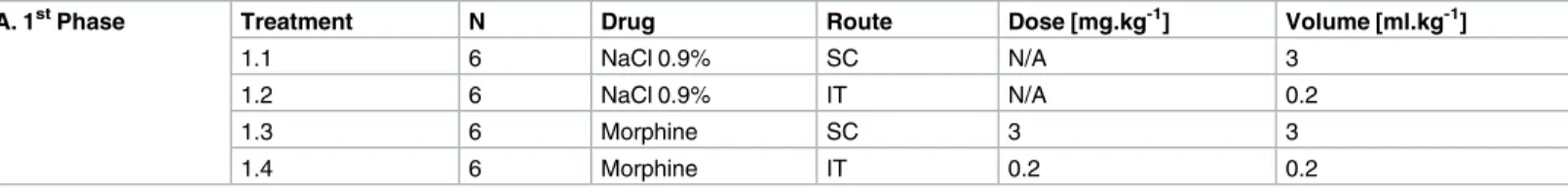
Regardless of their allocated treatment groups, all rats had the abdomen and lumbosacral area shaved at the start of the maintenance of anaesthesia. The shaved areas were cleaned with a diluted solution of chlorhexidine. Rats allocated to treatments 1.1, 1.2, 2.1, 2.2 were injected subcutaneously in the scruff with a sterile 25G needle and hypodermic syringe. Rats allocated to treatments 1.3, 1.4, 2.3, 2.4 were positioned in sternal recumbency with their pelvic limbs brought under the abdomen as cranially as possible in order to arch the lumbosacral area. The intrathecal injection was performed using aseptic techniques, using a 25G hypodermic needle and an insulin syringe (0.5ml). The injection site was between the last lumbar vertebra and the 1stsacral vertebrae (L6-S1) (Fig 1). The injection was considered successful if one of the 2
fol-lowing signs were noted: presence of CSF in the needle hub prior to injection and/or twitch of the tail during the injection. If none of these signs were noted, or if blood was visible in the nee-dle hub prior to injection, the neenee-dle was withdrawn and another sterile neenee-dle was used to repeat the procedure. All intrathecal injections were successful at the 1stor 2ndattempt.
Fig 1. Intrathecal injection in rats: anatomical landmarks and site of injection. Cx: coccygeal vertebrae, L5: fifth lumbar verterbrae, L6: sixth lumbar vertebrae, S: sacrum. $: injection site (L6-S1).
Surgery
Rats allocated to treatments 2.2, 2.3, 2.4 underwent a caudal laparotomy with bladder wall injection immediately after the injections described above. The procedure is an adaptation from a previously described technique to produce a clinically relevant model of abdominal sur-gery [36,49]. Briefly, a caudal and midline laparotomy incision (1cm) was performed. The blad-der was exposed and its wall injected with 0.1ml of sterile NaCl 0.9% (Braun, Melsungen, Germany) with an insulin needle and syringe (Terumo, Inchinnan, UK). The abdominal mus-cles were sutured with an interrupted pattern (4.0 Vicryl, Ethicon, Wokingham, UK) and the skin with a subcuticular pattern of the same suture material.
Behaviour recording
A custom made filming cage (40 x 26 x 28 cm) was used for behavioural recording. The cage did not contain any bedding, food or water. Three vertical sides, floor and ceiling were lined with a matte black film (Fablon) to minimise sensory distraction and reflections. A high defini-tion camera (Sony High Definidefini-tion HandyCam model HDR-XR155, Sony, Japan) was posi-tioned using a tripod to face the remaining clear Perspex side of the cage. The rats were placed individually in the cage, allowed to acclimatize for 5 minutes and filmed for 10 minutes without an observer being present in the filming area. The box was thoroughly cleaned after filming each animal. Each rat was filmed on the day prior to the procedure (baseline = T0) as well as at
1, 2, 4, and 8h post-injection.
Phase 1 of the study. An operator blinded to the rats’ treatment scored each 10-minute
video clip using open-source software designed to score animal behaviour from video clips (Cowlog) [50]. Behavioural scoring was performed using a validated rat ethogram [36,49,51]. The ethogram is summarised inTable 3. Duration and frequency for each of the defined behav-iours were recorded. A Composite Pain Behaviour (CPB) score was calculated by summing the frequency of the pain behaviours for each individual rat at each time-point. Pain behaviours included in the CPB in this phase were: back arching, twitch and stagger/fall.
Phase 2 of the study. One treatment-blinded operator (KH) (blinded to time point and
treatment) was responsible for all behavioural assessments. Briefly, the video was played in real time, and the operator scored the incidence of certain specific behaviours. Pain behaviours included in the CPB score in this phase were: back arching, twitch, stagger/fall, and belly press-ing. Behaviour scoring of 5 minute per rat, per time point was carried out.
Rat Grimace Scale
Following video recording for behavioural analysis, rats were placed in a photography box for a period of 5 minutes and photographed using a high definition camera (Casio EX-ZR100, Casio Computer Co., Ltd., Japan) by a treatment-blinded observer. The photography box consisted of two matte black walls and two clear Perspex walls (27cm x 19cm x 17cm). Rats were photo-graphed on every occasion they directly faced the camera, apart from when grooming in accor-dance with the method described by Sotocinal and colleagues [40]. The rats were then returned to their home cages. A treatment-blinded observer recorded any unexpected event or complica-tion related to picture acquisicomplica-tion. The box was thoroughly cleaned and dried after photogrpah-ing each animal. All out-of-focus or out-of-frame images were manually deleted. The remainphotogrpah-ing images were cropped, leaving only the face of the rat in view to prevent bias due to body posture [41]. Using a random number generator, one image per rat, per time point was selected. Using the random number generator again, the selected images were re-ordered and inserted into a cus-tom designed excel file for scoring. Nine trained observers who were blinded to the experimental details, design and purpose of the study scored each photograph for the four facial action units
(FAUs) comprising the RGS as described by Sotocinal and colleagues [40]. For each image, the 4 individual FAUS: orbit tightening, nose / cheek flattening, ear changes and whisker change were scored using a 3-point scale (0 = not present, 1 = moderately present, 2 = obviously present). The nine scorers consisted of 5 veterinarians (including 2 with experience of working with rodents) and 4 scientists with no experience of working with rodents.
All rats were euthanized using a rising concentration of inhaled C02(filling rate: 20% of the
chamber volume per minute) following recording of the last set of data, 8h post-injection, in accordance with current guidelines and legislation [4,52].
Statistical Analysis
All statistical analyses were conducted using SPSS (SPSS Inc. Chicago, USA). A sphericity test was performed to verify the parametric nature of the data after which repeated measures
Table 3. Ethogram used for behavioural observations in phase 1. Adapted from references [36,49,51]. Behaviour Description
Pain Behaviours Back arching Vertical stretch as in felines upon waking, including both partial and full arches, while inactive or walking. Twitch Transient involuntary muscular contraction of any body part. Usually occurs while inactive.
Stagger/ fall Rapid transition to crouch from high or low rear. Also, falling during grooming while crouched. Belly Pressing Rubbing the laparotomy site purposefully across the floor of the cage
Writhe One or more contractions of the abdominal muscle on either side of the stationary or moving animal, lasting until the abdomen relaxes.
Ambulatory Behaviours
High rear Bipedal stance, fully erect posture
Down to partial Downward movement from high rear to partial rear Partial rear Bipedal stance, low or half–erect posture
Down to floor Downward movement from either high rear, partial rear, jump or climb to quadrupedal contact with the cage floor Walk Quadrupedal ambulatory movement
Inactive Still, no on–going activity
Jump Transient vertical projection of entire body, usually follows high rear
Climb Hind paws not in contact with the cage floor, usually follows high rear or precedes jump Other Any behaviour not specified. See below.
Grooming Behaviours
Groom head Grooming, licking or scratching head Groom limbs Grooming, licking or scratching limbs Groom
abdomen
Grooming, licking or scratching abdomen, includes paying attention to wound site or abdominal shaved area Groom back Grooming, licking or scratching back, includes paying attention to lumbar shaved area
Groom tail Grooming, licking or scratching tail
Other Behaviours Investigate Vigorously sniff, explore, or inspect something. Recorded only when done in isolation (animal usually inactive or between ambulatory behaviours). Usually investigate a cage item but can sniff the air.
Chew Bite or gnaw part of the cage. Usually cage walls but can include masticating a cage item such as a piece of substrate
Eat Ingestion of an item
Stretch Horizontal elongation of body. Usually precedes and follows walking. Shake Instantaneous shuddering lateral whole body movements
Lift hind leg Transient upward movement of one hind paw or hind leg so that it is no longer in contact with the floor Squint Partial closure of eyes
Teeth chatter Vertical or lateral mouth/ jaw movements or teeth grinding in the absence of any obvious item to be chewed or eaten
Dig Digging on cage floor, usually with forepaws. Usually follows investigate
Miscellaneous ‘other’ behaviours On each occasion ‘other’ was scored, a note was made of this behaviour that was being performed by the rat. doi:10.1371/journal.pone.0163909.t003
ANOVA with ‘time’ as a within subjects factor and ‘treatment’ as a between subjects factor were performed. Pairwise comparisons were examined with post hoc analysis (Bonferroni). Data from the 1stand 2ndphases of the study were analysed separately. Differences were
con-sidered significant if p<0.05.
The RGS (0–8) score was obtained by averaging the scores (0–2) obtained for each of the 4 action units (AU) for each analysed image. Time effects within treatments were assessed with a Friedman’s test. Treatment effect at each time points were identified using a Kruskall-Wallis test and further investigated with Mann-Whitney U Tests and Bonferroni corrections for mul-tiple comparisons. Comparisons of equivalent treatments across study phases (i.e. treatments 1.1 vs. 2.1; 1.2 vs. 2.2; 1.3 vs. 2.3; 1.4 vs. 2.4) were also performed using Mann-Whitney U Tests with adjusted Bonferroni corrections for multiple comparisons. Finally, the level of consistency (reliability) between scorers was illustrated with Intraclass Correlation Coefficients(ICC) for each action unit as well as for the total RGS score. In all cases, differences were considered sig-nificant if p<0.05.
Power of the study
Retrospective assessment of the power of the study (2ndphase only) was performed using
G⇤Power (Softnews NET SRL, Romania).
For the behavioural analysis (CPB), the power exceeded 0.8 with respect to differences between time points and with respect to differences between the saline surgical group and both morphine groups. The power with which to detect differences between the morphine groups was approximately 0.4. Similar results were obtained for the RGS (within subjects, power >0.8; within morphine groups: 0.33).
Results
Phase 1: treatments 1.1 to 1.4
Effect of morphine (SC and IT) on the behaviour of pain-free rats. There was no
signifi-cant treatment effect on the behaviour of rats. Neither morphine administered SC or IT, or NaCl significantly affected the frequency and duration of behaviours identified in the rat ethogram
Time had a significant effect on some of the behaviours across all treatments (1.1 to 1.4), as illustrated inFig 2. As such, the frequency of high rears significantly decreases (p<0.001), the duration of walking decreased (p<0.001), the duration of climbing behaviours were signifi-cantly higher at baseline compared to other time points (p = 0.03). Lastly, the duration of inac-tivity increased over time with duration of inacinac-tivity being significantly lower at baseline compared to other time points (p<0.001). There was no significant effect of time on any of the other analysed behaviours. No pain specific behaviours were detected.
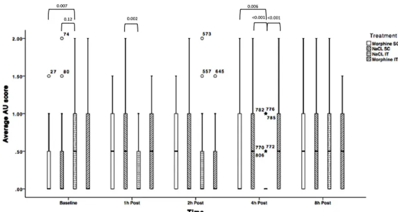
Effect of morphine (SC and IT) on Rat Grimace Scale (RGS) score in pain-free rats.
Average AU RGS results are documented inFig 3andTable 4.
Average AU RGS scores were significantly different across treatments at baseline, 1 and 4h post injection. At 1h post-injection, rats receiving intrathecal NaCl had a significantly lower RGS score than rats having received NaCl subcutaneously (p = 0.002). At 4h post-injection, total RGS was significantly lower in rats allocated to the NaCl IT group compared to: NaCl SC (p<0.001) and the morphine groups (morphine SC: p = 0.006; morphine IT: p<0.001) com-pared to their respective control groups.
Average AU RGS score also significantly increased over time in pain-free rats for all treat-ments except morphine intrathecally (Fig 3andTable 4).
Phase 2: treatments 2.1 to 2.4
Analgesic properties of intrathecal morphine in rats subjected to acute surgical pain.
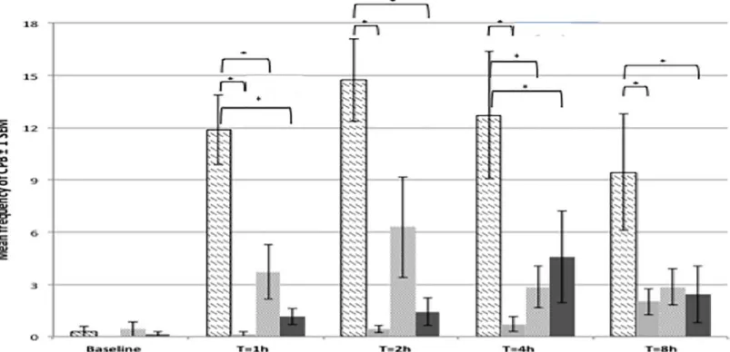
The surgical model used in the present study (caudal laparotomy with bladder wall injection) resulted in the presence of previously validated, specific pain behaviours such as writhing, fall/ stagger, twitches, and belly pressing. The incidence of back arching was very low and was there-fore removed from the analysis. The composite pain behaviour (CPB) score was therethere-fore obtained using the mean frequencies of the following behaviours: writhing, fall/stagger, belly pressing and twitches (Fig 4). CPB score was very low and not significantly different between treatment groups at baseline. Rats allocated to undergo surgery and receive an intrathecal injec-tion of NaCl showed had a significantly higher CPB score than other rats allocated to the sham group at all postoperative time points (Table 5). The peak of CPB was observed at 2h post surgery.
Morphine, administered subcutaneously or intrathecally, resulted in significantly lower CPB scores compared to control rats (NaCl IT, surgery) at all time points. CPB scores in mor-phine-treated rats (either SC or IT) were not significantly different to CPB scores in the sham surgery group. There was no significant difference in the CPB score of rats undergoing surgery
Fig 2. Representation of mean duration or frequency of the most commonly observed behaviours in pain-free rats (1stphase study) ± 2 standard error to the mean. N = 24, pooled data for treatment groups 1.1 to 1.4. There was no significant treatment effect between groups. A: significant decrease of rearing frequency over time (p<0.001); B: significant decrease of time spent walking over time (p<0.001); C: Time spent climbing is significantly higher at baseline than at any further time point (P = 0.036); D: significant increase of the time spent inactive (p = 0.001). doi:10.1371/journal.pone.0163909.g002
and receiving morphine either subcutaneously or intrathecally, at any time point. SeeTable 5 for the full list of significant differences between treatments at all time points.
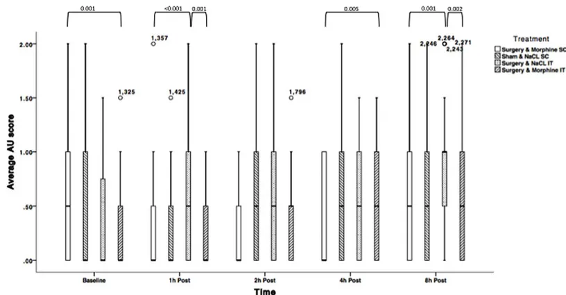
Average AU RGS scores for rats undergoing surgery and receiving NaCl IT significantly changed over time: average AU scores were significantly lower at baseline compared to 8h post laparotomy (p<0.01), but not at other time points. Similar results were obtained for rats under-going sham surgery. Average AU scores for rats underunder-going surgery and receiving morphine remained similar over time if the drug was administered subcutaneously, but were higher 8h post surgery if morphine was injected IT (p<0.001). Detailed results are displayed inFig 5and Table 6.
Fig 3. Average Action Unit RGS scores for treatments 1.1 (NaCl SC); 1.2 (NaCl IT); 1.3 (Morphine SC) and 1.4 (morphine IT) over time. N = 24 (Solid line = median, box = 1stand 3rdquartiles, whiskers = minimum and maximum, $, = Outliers). AU: Action Unit. P-values are indicated where differences are significant.
doi:10.1371/journal.pone.0163909.g003
Table 4. Within-subjects comparison between post treatment time-points with baseline with associ-ated p-values for differences where significance was found (phase 1 of the study). Differences are sig-nificant if p<0.05. AU: Action Unit; SC: Subcutaneous; IT: Intrathecal; ND: No sigsig-nificant difference.
Average AU Scores- NaCl SC
Time point 1h 2h 4h 8h
Baseline <0.001 0.002 0.003 0.001
Average AU Scores–NaCl IT
Time point 1h 2h 4h 8h
Baseline ND ND 0.001 ND
Average AU Scores- Morphine SC
Time point 1h 2h 4h 8h
Baseline 0.007 0.007 ND 0.007
Morphine-treated rats (SC and IT) undergoing surgery, displayed significantly lower total RGS scores than control rats (NaCl IT) 1h post-surgery (p<0.001, p = 0.001 respectively). Sim-ilar pattern of significant differences were identified 8h post-surgery (p = 0.001–0.002). RGS scores were not significantly different between the morphine groups at 1, 2, and 8h postopera-tively. RGS scores were not significantly different across treatments 2h post laparotomy. At 4h post laparotomy; RGS scores were not significantly different between rats allocated to the sham surgery groups and those allocated to the surgical groups and intrathecal NaCl.
Average AU RGS scores across treatments were significantly different at baseline between rats allocated to receive morphine SC and (p = 0.01). No other significant differences were noted between groups at baseline.
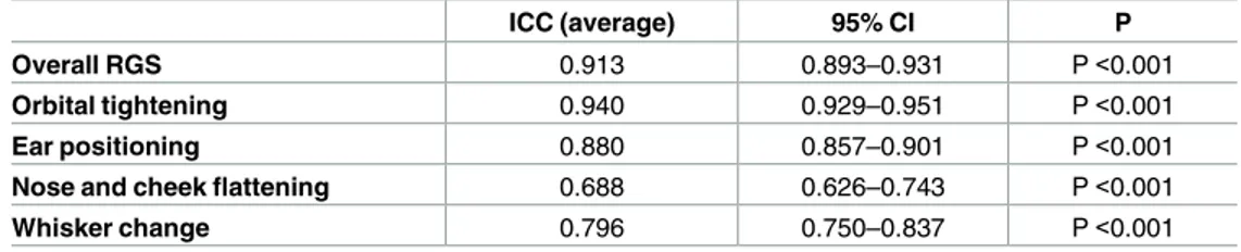
Reliability of RGS scoring
The reliability of both the individual AUs and the overall RGS scoring across all 9 scorers was high with intraclass correlation (ICC) values for the individual AUs ranging from 0.69 (nose/ cheek flattening) to 0.94 (orbital tightening) and a value of 0.91 for the overall RGS score (P<0.0.001 for all comparisons) (Table 7).
Discussion
This novel study demonstrates the effects of morphine injected intrathecally via direct lumbar puncture in pain-free rats and in a model of acute surgical pain. In addition, this study repre-sents the first use of the Rat Grimace Scale to assess the analgesic properties of IT morphine.
Fig 4. Mean frequencies for Composite Pain Behaviour (CPB) scores across treatments and time points. Data presented ± 2 standard error of the mean. N = 28. Significant differences (*) are explained in the text andTable 5.
Table 5. Composi te Pain Behaviour scores and associ ated p -values for phase 2 of the study. Time Baseline 1h post surgery 2h post surgery 4h post surgery 8h post surger y Treatmen t NaCl IT, Sx NaCl SC, Sh Mor IT, Sx Mor SC, Sx NaCl IT, Sx NaCl SC, Sh Mor IT,Sx Mor SC, Sx NaCl IT, Sx NaCl SC, Sh Mor IT,Sx Mor SC, Sx NaCl IT, Sx NaCl SC, Sh Mor IT,Sx Mor SC, Sx NaCl IT, Sx NaCl SC, Sh Mor IT,Sx Mor SC, Sx NaCl IT, Sx / ND ND ND / < 0.001 0.006 < 0.001 / < 0.001 ND 0.039 / 0.001 0.011 0.045 / 0.013 ND 0.035 NaCl SC, Sh ND / ND ND < 0.001 / ND ND < 0.001 / ND ND 0.001 / ND ND 0.013 / ND ND Mor IT, Sx ND ND / ND 0.006 ND / ND ND ND / ND 0.011 ND / ND ND ND / ND Mor SC, Sx ND ND ND / < 0.001 ND ND / 0.039 ND ND / 0.045 ND ND / 0.035 ND ND / P values below 0.05 indicated a significa nt difference . ND: No significa nt difference (p > 0.05). IT: Intratheca l, Mor: Morphin e, NaCl: Saline, SC: Subcut aneous, Sh: Sham, Sx: Surgery. See Table 2 for full information on treatments doi: 10.137 1/journal.pone .0163909.t005
Effects of morphine SC and IT on the behaviour and facial expression of
pain-free rats
The 1stphase of the study was designed to determine whether morphine, administered SC or
IT, influenced behaviour or rat grimace score (RGS) of pain-free rats. At the doses chosen in this study morphine, injected subcutaneously or intrathecally, did not affect the frequency and duration of behaviours used in a validated rat ethogram [36]. All changes in behavioural scope and pattern in the second phase of the study, in particular behaviours included in the CPB score, can therefore be attributed to the acute surgical pain caused by caudal laparotomy and
Fig 5. Average Action Unit RGS scores for treatments 2.1 (NaCl SC + sham surgery); 2.2 (NaCl IT + surgery); 2.3 (morphine SC + surgery) and 2.4 (morphine IT + surgery) over time (Solid line = median, box = 1stand 3rdquartiles, whiskers = minimum and maximum, $, = Outliers). N = 28. P-values are indicated where differences are significant (<0.05).
doi:10.1371/journal.pone.0163909.g005
Table 6. Within-subjects comparison between post treatment time-points with baseline with associ-ated p-values for differences where significance was found (Phase 2 of the study). Differences are sig-nificant if p<0.05. AU: Action Unit; SC: Subcutaneous; IT: Intrathecal; ND: No sigsig-nificant difference.
Average AU Scores- NaCl SC, Sham
Time Point 1h 2h 4h 8h
Baseline ND ND ND 0.010
Average AU Scores–NaCl IT, Surgery
Time Point 1h 2h 4h 8h
Baseline ND ND ND <0.001
Average AU Scores- Morphine IT, Surgery
Time Point 1h 2h 4h 8h
Baseline ND ND <0.001 <0.001
bladder injection. This finding is contrary to the common belief that opioids alter the behav-iour of pain-free rodents [38]. Morphine as well as other opioids do influence the behaviours of pain-free rats, but this has generally been reported when the doses used are much greater than those recommended for clinical use in rats [14,38], or when opioids are administered repeat-edly for example in models of opioid tolerance [53,54]. The activity of all rats decreased over time, regardless of the drug received (NaCl or morphine). This reflects either insufficient habit-uation to the recording box (5 min) prior to the recording (novelty effect) during the early time points; or habituation of the rats, and possible boredom after several recordings in quick suc-cession (a total of 5, 10 min long, behaviour recording sessions) [55,56].
The effect of morphine and the route of administration on pain-free rats were not easily interpreted using RGS. Average AU scores increased over time for all pain-free treatment groups, with the exception of intrathecal morphine. Each of the behavioural recording session was followed by another 10 min long session in a smaller Plexiglas box for RGS scoring. Whilst activity was not recorded in the RGS box, it was observed that the rats were also more inactive over time during the RGS sessions. Several rats were seen to remain immobile for a large part of the RGS session, and appeared disinterested by their surroundings. Such observations seem to correlate with the overall activity patterns (i.e. inactivity and walk). In the authors’ experi-ence, it would be reasonable to assume that inactive rats placed in a familiar environment could intermittently show signs of drowsiness. Given that orbital tightening is a natural conse-quence of drowsiness, then an increase in RGS score over time within all treatment groups could have been expected. One might argue that the sedative properties of morphine [14,57,58] could have further accentuated this expected effect, as sedation has already been shown to increase grimace scale scores [59]. While pain-free rats having received NaCl SC had a signifi-cantly lower RGS at baseline than at other time points; this pattern was not repeated in the NaCl IT group.
Interestingly, RGS scores for pain-free rats receiving morphine IT did not change signifi-cantly over time, despite the same decrease in activity shown by control rats. Intrathecal mor-phine may therefore have an effect on facial expression of pain-free rats. Typically, pain-free rats receiving IT morphine displayed seemingly wider and slightly more protuberant eyes, than in control rats. This facial appearance of pain-free rats receiving morphine was documented as exophthalmos by some of the RGS treatment-blinded scorers. Morphine is known to cause mydriasis and exophthalmos in pain-free rats [60–62], but this effect was not analysed for sig-nificance in our study. Exophthalmos, which is not taken into account by the RGS, might have counteracted any possible orbital tightening from somnolence in the later time points.
Lastly, pain-free rats receiving morphine SC had a significantly higher RGS score 1, 2 and 8h post injection. This could be explained by the sedative properties of morphine [14,57,58] causing some additional degree of orbital tightening. However, if any sedation occurred, this was not reflected by behaviour changes 1h post-injection.
Table 7. Inter-rater reliability for the Rat Grimace Scale (2ndphase of the study).
ICC (average) 95% CI P
Overall RGS 0.913 0.893–0.931 P <0.001
Orbital tightening 0.940 0.929–0.951 P <0.001
Ear positioning 0.880 0.857–0.901 P <0.001
Nose and cheek flattening 0.688 0.626–0.743 P <0.001
Whisker change 0.796 0.750–0.837 P <0.001
Intraclass correlation coefficient (ICC) calculated for multiple (average) raters (n = 9). doi:10.1371/journal.pone.0163909.t007
Analgesic properties of intrathecal morphine for acute surgical pain
The pain caused by the model chosen for this study (caudal laparotomy with bladder wall injec-tion) has been well documented using composite pain behaviour scoring [36,63]. This surgical procedure remains widely used in orthotopic models of bladder cancers in our institution and elsewhere [64,65]. The presence of detectable pain was confirmed by the significant increase in composite pain behaviours in the positive control group up to the last recorded time point, 8h post surgery. This is in line with previously reported pain mediated behavioural alteration, where laparotomy was associated with an increase of pain specific pain behaviour for up to 6.5h [51]; as well as non-specific behavioural alteration for up to 24h [66].Morphine administered intrathecally via direct lumbosacral puncture significantly attenu-ated composite pain behaviours (1 and 4h postoperatively) and rat grimace scores (1 and 8h post surgery), and therefore seems to alleviate acute postsurgical pain caused by caudal laparot-omy. Morphine IT was mostly indistinguishable from subcutaneous administration based on RGS scoring, but analysis of the composite pain behavior score suggested that SC morphine might provide uninterrupted analgesia for up to 8h.
The duration of action of morphine administered subcutaneously (8h) was longer than expected since morphine SC is usually considered to provide effective post-operative analgesia for no more than 2-4h [14,67,68], with a peak of analgesic and anti-hyperalgesic activity 45–60 min post administration [67]. Morphine administered intrathecally is expected to provide long lasting analgesia in people [69] and animals [70–72]. While neuraxial morphine was shown to have a duration of action of 21h in primates [70], and up to 24h in dogs and cats [71,72], intra-thecal morphine has been associated with markedly shorter duration of action in rats. Most studies undertaken in rats reported a duration of action of approximately 120 min
[32,33,73,74]. Results obtained in the present study suggest that intrathecal morphine in rats, might have a longer duration of action. Three key elements of our study design could explain this difference from previous studies. Firstly, the present study documents analgesic properties of morphine in the context of acute surgical pain, using specific pain behaviours validated for post-laparotomy pain in rats. Most other studies assessed the anti-nociceptive properties of morphine using various nociceptive tests (e.g. Von Frey, Hargreaves). Secondly, in our study, in order to assess the practicality of intrathecal administration of post-operative pain manage-ment, morphine was administered without prior surgical implantation of a spinal catheter. A spinal catheter is widely used in pharmacological or toxicological studies to facilitate multiple injections of a substance. The presence of the catheter may be associated with chronic inflam-matory pain, alteration of rats’ behaviours, and intrinsic variation in the pharmacological prop-erties of some molecules, including morphine [33,75]. Thirdly, the dose used in the present study (0.2 mg.kg-1, IT, i.e. average 54μg per rat) was higher than doses used in most
anti-noci-ceptive studies (e.g. 0.16 to 10 μg per rat) [28,29,32,33]. When selected morphine doses were higher, anti-hyperalgesic properties of intrathecal morphine was anecdotally documented to last for up to 4h [76].
The dose chosen was based on pilot studies conducted in our laboratory (unpublished data) and represented the highest dose that could be administered intrathecally without producing clinical and behavioural side effects. We ensured that the total amount remained below 150μg per rat, the intrathecal dose reported to trigger hyperalgesia in rats [77]. A total dose of 0.2 mg. kg-1in rats is also higher than intrathecal doses commonly used in human analgesia
[25,27,78,79], even after applying allometric scaling [80]. Further studies would be required to identify the lowest intrathecal dose required to inhibit composite pain behaviours in rats undergoing laparotomies and other surgical procedures involving the abdomen and the pelvic limbs. Lastly, the effects of a low morphine dose (54μg or below) administered intrathecally on
the immune system and cancer models is unknown to date, but would be expected to be lower than systemic doses previously investigated [22,23].
Relevance of the rat grimace scale for assessing pain in opioid
medicated rats
A range of different approaches have been developed to assess the degree of pain experienced by rats undergoing surgical and other traumatic pain, with the view of refining the procedures and/or improving the relevance of translational studies. Beyond nonspecific and retrospective methods (such as weight loss, biochemical stress markers etc.), two prospective pain assess-ment methods are currently most relied upon. Behaviour-based assessassess-ments of pain have been developed for both rats and mice following surgery and other traumatic procedures, and use either the appearance of abnormal behaviours, or the change in the frequency of normal behav-iour patterns to score pain [11,49,51,81]. There remain a number of limitations to using behav-iour to assess pain in animals, such as possible confounding factor of opioid analgesic on the behaviour of pain-free animals [51] and the specific behavioural responses to painful stimuli varying markedly depending on the nature of the surgical procedure (abdominal-based or other) [11,49,51,81]. The use of facial expressions to assess pain [40] was suggested to over-come some of the above difficulties.Variation of facial expression during painful events was codified as the Rat Grimace Scale (RGS), validated for the assessment of acute surgical and nociceptive pain [40,43,82], and used to assess the efficacy of commonly used analgesics in rats [45]. Amongst the proposed advantages supporting the use of RGS was the lack of a confound-ing effect of opioids on the facial expression of pain-free animals. The present study suggests that opioids may have an effect on facial expression of pain-free rats (overall decrease of RGS scores over time) caused by a degree of opioid-mediated exophthalmos, but no behavioural effect over time. These findings would benefit from further investigation.
In rats subjected to acute surgical pain, unexpected findings have weakened our interpreta-tion from RGS results. RGS scores in pain free and opioid free rats were expected to be lower than were observed in this study [40] and similar across treatment groups. Importantly, signifi-cant differences were detected amongst treatment groups at baseline during both phases of the study. This major inconsistency existed despite overall excellent inter-scorer reliability of the scoring method (ICC 0.913). Three factors either in isolation or combination may have con-tributed to such findings. Firstly, in mice, baseline mouse grimace scale scores were shown to differ depending on the strain, sex, and methods used for the scoring of facial action units (ret-rospective vs. p(ret-rospective scoring) [83]. While variation of baseline scores across strains in Wistar rats, or the impact of retrospective RGS scoring on still images are yet to be documented in a specific study, significant differences amongst baseline RGS scores between treatment groups were unexpected. Rats (strain, age, sex, age), operators, time of the days and methodol-ogy used was similar between both study phases. However, this requires further study as the above finding is based on a single study in 6 mice. A second reason that potentially contributed to the differences in RGS scores at baseline between rats could, more simply, be a cohort effect. All rats were bred by the same supplier, were of a similar age, strain, and received equivalent husbandry in our laboratory. In addition, group housing and random allocation of each rat to his treatment group would be expected to minimize the likelihood of such cohort effect. None-theless, the statistical analysis used for this study compensated for any possible cohort effect since using a within-subjects design accounts for the variation between individuals when com-paring across time points. Thirdly, a relatively short yet consistent habitation period was used in these studies (5 min), and therefore it is possible that the novel nature of the box used con-tributed to the variability of facial expressions of the rats, possibly causing inadvertent false
positive scoring at baseline that were reduced with repeated exposures. This effect has however not been noted in other studies using grimace scales when similar habitation times [59] or dif-ferent [82] habituation times were used.
While the overall power of our studies was acceptable (>0.8), the lower power observed between the morphine comparisons should be taken into account when interpreting the results of this pilot study. These comparisons bear further investigation in a future study. This pilot study will provide more accurate information for the sample size determination of future stud-ies than was available when this pilot study was undertaken.
Only female rats were used in this study. Pain perception and analgesic properties of opioid are influenced by gender in rats. Briefly, female rodents are more sensitive than males to nox-ious stimuli and have lower levels of stress-induced analgesia, whereas male rodents generally have stronger analgesic responses to mu-opioid receptor agonists than females [84,85]. In spite of this, gender was not found to significantly influence specific pain behaviours in similar surgi-cal models of pain in rats [49], and no sex-differences were reported in the original RGS paper [40]. We chose to use single sex female groups and minimise the number of rats involved in this study. Further studies would be required to characterise gender-based difference in the analgesic properties of intrathecal morphine.
Conclusion
Findings from the present study suggest that administration of intrathecal morphine by percu-taneous injection may represent an effective way of providing long lasting pain relief in rodents subjected to caudal laparotomy and bladder wall injection. Intrathecal morphine (0.2 mg.kg-1)
may provide comparable analgesia to the subcutaneous route (3 mg.kg-1) using less that a 10th
of the dose required for subcutaneous administration. As a result, the intrathecal route of administration may reduce concerns related to the non-specific effects of opioids when these agents are used to alleviate pain in rodents used in a range of different areas of biomedical research. Further studies would be required to better characterize the effects of such reduced morphine dose on individual experimental outcomes.
While the RGS may have advantages for the assessment of pain in rats compared to scoring of composite pain behaviours, morphine may impact facial expression of pain-free rats and influence use of the RGS in opioid medicated rats. The variation in detection of analgesic effects between the RGS and the CPB score support the use of both techniques, as complemen-tary measures of the behavioural changes induced both by analgesics and post-surgical pain.
Supporting Information
S1 File. Rat Grimace Scale scores and behavioural dataset.
(XLSX)
Acknowledgments
The authors would like to thank Peter Nicholson, Ashleigh Brown, Kimberley Wells, Keila Ida, and Alexandru Tuturanu for their assistance with scoring the Rat Grimace Scale. The animals were donated by Charles River UK.
Author Contributions
Conceptualization: AT PF ML. Data curation: ML.
Formal analysis: ML JR A. Miller AT. Investigation: AT A. Miller A. Malik KH. Methodology:AT PF A. Miller ML. Project administration: AT A. Miller. Supervision: ML PF JR.
Validation: ML JR AT.
Visualization: AT ML A. Miller.
Writing – original draft: AT A. Miller CS. Writing – review & editing: AT ML A. Miller CS.
References
1. Home Office Science. Annual Statistics of Scientific Procedures on Living Animals Great Britain 2014. 2015 Jan pp. 1–62.
2. Burden N, Chapman K, Sewell F, Robinson V. Pioneering better science through the 3Rs: an introduction to the national centre for the replacement, refinement, and reduction of animals in research (NC3Rs). J Am Assoc Lab Anim Sci. 2015 Mar; 54(2):198–208. PMID:25836967
3. Graham ML, Prescott MJ. European Journal of Pharmacology. European Journal of Pharmacology. Elsevier; 2015 Jul 15;759(C):19–29.
4. European Parliament. Directive 2010/63/EU of the European Parliament and of the Council of 22 September 2010 on the protection of animals used for scientific purposes [Internet]. Official Journal of the European Union Oct 19, 2010 pp. 1–47. Available from:http://eur-lex.europa.eu/legal-content/EN/TXT/?uri= CELEX:32010L0063
5. Bratcher NA, Reinhard GR. Creative implementation of 3Rs principles within industry programs: beyond reg-ulations and guidelines. J Am Assoc Lab Anim Sci. 2015 Mar; 54(2):133–8. PMID:25836958
6. Russell WMS, Burch RL. The principles of humane experimental technique. 1959. 1 p.
7. Coulter CA, Flecknell PA, Richardson CA. Reported analgesic administration to rabbits, pigs, sheep, dogs and non-human primates undergoing experimental surgical procedures. Laboratory Animals. 2009 Jul 1; 43 (3):232–8. doi:10.1258/la.2008.008021PMID:19116294
8. Stokes EL, Flecknell PA, Richardson CA. Reported analgesic and anaesthetic administration to rodents undergoing experimental surgical procedures. Laboratory Animals. SAGE Publications; 2009 Apr; 43 (2):149–54. doi:10.1258/la.2008.008020PMID:19116297
9. Bara M, Joffe AR. The ethical dimension in published animal research in critical care: the public face of sci-ence. Crit Care. 2014; 18(1):R15. doi:10.1186/cc13694PMID:24423201
10. Flecknell PA. Refinement of animal use-assessment and alleviation of pain and distress. 2007 May 3;:1–10. 11. Leach MC, Bailey HE, Dickinson AL, Roughan JV, Flecknell PA. A preliminary investigation into the
practical-ity of use and duration of action of slow-release preparations of morphine and hydromorphone in laboratory rats. Laboratory Animals. 2010 Jan 1; 44(1):59–65. doi:10.1258/la.2009.007160PMID:19858166 12. Iwamoto K, Klaassen CD. First-pass effect of morphine in rats. J Pharmacol Exp Ther. 1977 Jan; 200
(1):236–44. PMID:833759
13. Papathanasiou T, Juul RV, Gabel-Jensen C, Kreilgaard M, Lund TM. Population Pharmacokinetic Modelling of Morphine, Gabapentin and their Combination in the Rat. Pharm Res. Pharmaceutical Research; 2016 Jul 5;1–14. doi:10.1007/s11095-015-1770-7PMID:26334501
14. Flecknell P. Laboratory Animal Anaesthesia. Academic Press; 2015. 1 p.
15. Stoelting RK, Hillier SC. Pharmacology and Physiology in Anesthetic Practice. Lippincott Williams & Wilkins; 2012. 1 p.
16. Clark JA, Myers PH, Goelz MF, Thigpen JE, Forsythe DB. Pica behavior associated with buprenorphine administration in the rat. Lab Anim Sci. 1997 Jun; 47(3):300–3. PMID:9241634
17. Roughan JV, Flecknell PA. Buprenorphine: a reappraisal of its antinociceptive effects and therapeutic use in alleviating post-operative pain in animals. Laboratory Animals. SAGE Publications; 2002 Jul; 36(3):322–43. PMID:12144743
18. Schaap MWH, Uilenreef JJ, Mitsogiannis MD, van ’t Klooster JG, Arndt SS, Hellebrekers LJ. Optimizing the dosing interval of buprenorphine in a multimodal postoperative analgesic strategy in the rat: minimizing
side-effects without affecting weight gain and food intake. Laboratory Animals. SAGE Publications; 2012 Oct; 46 (4):287–92. doi:10.1258/la.2012.012058PMID:23097561
19. Ohtani M, Kotaki H, Nishitateno K, Sawada Y, Iga T. Kinetics of respiratory depression in rats induced by buprenorphine and its metabolite, norbuprenorphine. J Pharmacol Exp Ther. 1997 Apr; 281(1):428–33. PMID:9103526
20. Gueye PN, Borron SW, Risède P, Monier C, Buneaux F, Debray M, et al. Buprenorphine and midazolam act in combination to depress respiration in rats. Toxicol Sci. 2002 Jan; 65(1):107–14. PMID:11752690 21. Grace PM, Strand KA, Galer EL, Urban DJ, Wang X, Baratta MV, et al. Morphine paradoxically prolongs
neu-ropathic pain in rats by amplifying spinal NLRP3 inflammasome activation. Proc Natl Acad Sci USA. 2016 Jun 14; 113(24):E3441–50. doi:10.1073/pnas.1602070113PMID:27247388
22. Al-Hashimi M, Scott SWM, Thompson JP, Lambert DG. Opioids and immune modulation: more questions than answers. British Journal of Anaesthesia. Oxford University Press; 2013 Jul; 111(1):80–8. doi:10.1093/ bja/aet153PMID:23794649
23. Afsharimani B, Doornebal CW, Cabot PJ, Hollmann MW, Parat M-O. Comparison and analysis of the animal models used to study the effect of morphine on tumour growth and metastasis. Br J Pharmacol. 2015 Jan; 172(2):251–9. doi:10.1111/bph.12589PMID:24467261
24. Stoelting PK, Miller RD. Basics of anesthesia. 5th. Philadelphia: Churchill Living stone; 2007.
25. Barash PG, Cullen BF, Stoelting RK, Cahalan M, Stock MC. Clinical Anesthesia. Lippincott Williams & Wil-kins; 2011. 1 p.
26. Tranquilli WJ, Thurmon JC, Grimm KA. Lumb and Jones’ Veterinary Anesthesia and Analgesia. John Wiley & Sons; 2013.
27. Rathmell JP, Lair TR, Nauman B. The role of intrathecal drugs in the treatment of acute pain. Anesthesiology. 2005 Nov; 101(5 Suppl):S30–43.
28. Yaksh TL, Rudy TA. Analgesia mediated by a direct spinal action of narcotics. Science. American Associa-tion for the Advancement of Science; 1976 Jun 25; 192(4246):1357–8. PMID:1273597
29. Yaksh TL, Rudy TA. ANALGESIA MEDIATED BY A DIRECT SPINAL ACTION OF NARCOTICS. Survey of Anesthesiology. 1977 Apr 1; 21(2):122.
30. Mestre C, Pe´lissier T, Fialip J, Wilcox G, Eschalier A. A method to perform direct transcutaneous intrathecal injection in rats. Journal of Pharmacological and Toxicological Methods. 1994 Dec; 32(4):197–200. PMID: 7881133
31. Fairbanks CA. Spinal delivery of analgesics in experimental models of pain and analgesia. Advanced Drug Delivery Reviews. 2003 Aug 28; 55(8):1007–41. PMID:12935942
32. Zahn PK, Gysbers D, Brennan TJ. Effect of systemic and intrathecal morphine in a rat model of postoperative pain. Anesthesiology. The American Society of Anesthesiologists; 1997 May; 86(5):1066–77. PMID: 9158356
33. Martin TJ, Zhang Y, Buechler N, Conklin DR, Eisenach JC. Intrathecal morphine and ketorolac analgesia after surgery: comparison of spontaneous and elicited responses in rats. PAIN. 2005 Feb; 113(3):376–85. doi:10.1016/j.pain.2004.11.017PMID:15661447
34. Buvanendran A, Kroin JS, Kari MR, Tuman KJ. A New Knee Surgery Model in Rats to Evaluate Functional Measures of Postoperative Pain. Anesthesiology. 2008 Jul; 107(1):300–8.
35. Mamet J, Klukinov M, Yaksh TL, Malkmus SA, Williams S, Harris S, et al. Single intrathecal administration of the transcription factor decoy AYX1 prevents acute and chronic pain after incisional, inflammatory, or neuro-pathic injury. PAIN. International Association for the Study of Pain; 2014 Feb 1; 155(2):322–33. doi:10.1016/ j.pain.2013.10.015PMID:24145208
36. Roughan JV, Flecknell PA. Evaluation of a short duration behaviour-based post-operative pain scoring sys-tem in rats. Eur J Pain. 2003; 7(5):397–406. doi:10.1016/S1090-3801(02)00140-4PMID:12935791 37. Roughan JV, Flecknell PA. Effects of surgery and analgesic administration on spontaneous behaviour in
sin-gly housed rats. Research in Veterinary Science. 2000 Dec; 69(3):283–8. doi:10.1053/rvsc.2000.0430 PMID:11124101
38. Flecknell PA. The relief of pain in laboratory animals. Laboratory Animals. 1984 Apr; 18(2):147–60. PMID: 6146743
39. Williams AC de C. Facial expression of pain: an evolutionary account. Behav Brain Sci. 2002 Aug; 25 (4):439–55–discussion455–88. PMID:12879700
40. Sotocinal SG, Sorge RE, Zaloum A, Tuttle AH, Martin LJ, Wieskopf JS, et al. The Rat Grimace Scale: A par-tially automated method for quantifying pain in the laboratory rat via facial expressions. Mol Pain. BioMed Central Ltd; 2011 Jul 29; 7(1):55.
41. Langford DJ, Bailey AL, Chanda ML, Clarke SE, Drummond TE, Echols S, et al. Coding of facial expressions of pain in the laboratory mouse. Nature Publishing Group. Nature Publishing Group; 2010 May 9; 7(6):447–9. 42. Oliver V, De Rantere D, Ritchie R, Chisholm J, Hecker KG, Pang DSJ. Psychometric Assessment of the Rat
Grimace Scale and Development of an Analgesic Intervention Score. McCormick C, editor. PLoS ONE. 2014 May 16; 9(5):e97882–7. doi:10.1371/journal.pone.0097882PMID:24838111
43. Liao LA, Long HA, Zhang LB, Chen HB, Zhou YA, Ye NA, et al. Evaluation of pain in rats through facial expression following experimental tooth movement. European Journal of Oral Sciences. 2014; 122(2):121– 4. doi:10.1111/eos.12110PMID:24428464
44. Lilley EA, Armstrong RB, Clark NC, Gray PD, Hawkins PA, Mason KD, et al. Refinement of animal models of sepsis and septic shock. Shock. 2015; 43(4):304–16. doi:10.1097/SHK.0000000000000318PMID: 25565638
45. Waite ME, Tomkovich A, Quinn TL. Efficacy of Common Analgesics for Postsurgical Pain in Rats. Journal of the American Association for Laboratory Animal Science. 2015; 54:420–5. PMID:26224443
46. Roughan JV, Bertrand HGMJ, Isles HM. Meloxicam prevents COX-2-mediated post-surgical inflammation but not pain following laparotomy in mice. European Journal of Pain (United Kingdom). 2016; 20(2):231–40. 47. Thomas AA, Flecknell PA. Comparative Anesthesia and Analgesia of Laboratory Animals. In: Lumb Jones
Veterinary Anaesthesia and Analgesia. 2015. pp. 1–10.
48. Flecknell P, Lofgren JLS, Dyson MC, Marini RR, Swindle MM, Wilson RP. Chapter 24. Preanesthesia, Anes-thesia, Analgesia, and Euthanasia. Third Edition. Laboratory Animal Medicine. Elsevier Inc; 2015. 66 p. 49. Roughan JV, Flecknell PA. Behavioural effects of laparotomy and analgesic effects of ketoprofen and
carpro-fen in rats. PAIN. 2001 Feb 1; 90(1–2):65–74. PMID:11166971
50. Ha¨nninen L, Pastell M. CowLog: Open-source software for coding behaviors from digital video. Behavior Research Methods. Springer-Verlag; 2009; 41(2):472–6. doi:10.3758/BRM.41.2.472PMID:19363187 51. Roughan JV, Flecknell PA. Behaviour-based assessment of the duration of laparotomy-induced abdominal
pain and the analgesic effects of carprofen and buprenorphine in rats. Behav Pharmacol. 2004 Nov; 15 (7):461–72. PMID:15472568
52. Hawkins P, Playle L, Golledge H, Leach M. Newcastle concensus meeting on carbon dioxide euthanasia of laboratory animals. Animal Technology and . . .. 2006.
53. Babbini M, Davis WM. Time-dose relationships for locomotor activity effects of morphine after acute or repeated treatment. Br J Pharmacol. Blackwell Publishing Ltd; 1972 Oct 1; 46(2):213–24. PMID:4651770 54. Craig MM, Bajic D. Long-term behavioral effects in a rat model of prolonged postnatal morphine exposure.
Behavioral Neuroscience. 2015; 129(5):643–55. doi:10.1037/bne0000081PMID:26214209
55. Groves PM, Thompson RF. Habituation: a dual-process theory. Psychol Rev. 1970 Sep; 77(5):419–50. PMID:4319167
56. Veloso AWN, Filgueiras GB, Lorenzo P, Estanislau C. Modulation of Grooming Behavior in Rats by Different Test Situations. Psychology & Neuroscience. 2016;:1–15.
57. Massa H, Lacoh CM, Vutskits L. Effects of Morphine on the Differentiation and Survival of Developing Pyra-midal Neurons During the Brain Growth Spurt. Toxicol Sci. 2012 Oct 17; 130(1):168–79. doi:10.1093/toxsci/ kfs234PMID:22843570
58. Emery MJ, Groves CC, Kruse TN, Shi C, Terman GW. Ventilation and the Response to Hypercapnia after Morphine in Opioid-naive and Opioid-tolerant Rats. Anesthesiology. 2016 Jan 6;: 1.
59. Miller A, Kitson G, Skalkoyannis B, Leach M. The effect of isoflurane anaesthesia and buprenorphine on the mouse grimace scale and behaviour in CBA and DBA/2 mice. Applied Animal Behaviour Science. Elsevier B. V; 2015 Nov 1; 172(C):58–62.
60. Wallenstein MC. Role of sympathetic system in morphine-induced mydriasis in rat. Am J Physiol. 1981 Sep; 241(3):R130–5. PMID:6269441
61. Wallenstein MC. Effect of prostaglandin synthetase inhibitors on non-analgesic actions of morphine. Euro-pean Journal of Pharmacology. 1983 May 20; 90(1):65–73. PMID:6409656
62. Suzuki T, Futakata A, Shimada M, Yoshii T, Yanaura S. [Comparison of three methods of inducing physical dependence to morphine in rats using short-term medication]. Yakubutsu Seishin Kodo. 1984 Sep; 4(2):149– 56. PMID:6543077
63. Roughan JV, Flecknell PA. Behavioural effects of laparotomy and analgesic effects of ketoprofen and carpro-fen in rats. PAIN. 2001 Feb 1; 90(1–2):65–74. PMID:11166971
64. Abaza R, Keck RW, Selman SH. Intraperitoneal chemotherapy for the prevention of transitional cell carci-noma implantation. J Urol. Elsevier; 2006 Jun; 175(6):2317–22. doi:10.1016/S0022-5347(06)00257-6 PMID:16697866
65. Dornelas CA, Almeida PRC de, Nascimento GLD, Lima EB, Moraes MO de. [Experimental model of Walker 256 carcinosarcoma in rats bladder]. Acta Cirurgica Brasileira. 2006 Jan; 21(1):38–42. PMID:16491221 66. Martin TJ, Buechler NL, Kahn W, Crews JC, Eisenach JC. Effects of laparotomy on spontaneous exploratory
activity and conditioned operant responding in the rat: a model for postoperative pain. Anesthesiology. 2004 Jul; 101(1):191–203. PMID:15220791
67. Papathanasiou T, Juul RV, Heegaard A-M, Kreilgaard M, Lund TM. Co-administration of morphine and gaba-pentin leads to dose dependent synergistic effects in a rat model of postoperative pain. PHASCI. Elsevier B. V; 2016 Jan 20; 82(C):97–105.
68. Whiteside GT, Harrison J, Boulet J, Mark L, Pearson M, Gottshall S, et al. Pharmacological characterisation of a rat model of incisional pain. Br J Pharmacol. 2004 Jan; 141(1):85–91. doi:10.1038/sj.bjp.0705568PMID: 14597606
69. Jacobson L, Chabal C, Brody MC, Ward RJ, Wasse L. Intrathecal methadone: a dose-response study and comparison with intrathecal morphine 0.5 mg. PAIN. 1990; 43(2):141–8. PMID:2087326
70. Yaksh TL. Spinal opiate analgesia: Characteristics and principles of action. PAIN. 1981; 11(3):293–333– 336–346.
71. Tung AS, Yaksh TL. In vivo evidence for multiple opiate receptors mediating analgesia in the rat spinal cord. Brain Research. 1982 Sep 9; 247(1):75–83. PMID:6127147
72. Saleh AS, Bonath KH. Long term pain treatment in the dog by peridural morphines. 1985.
73. Buerkle H, Yaksh TL. Comparison of the spinal actions of the mu-opioid remifentanil with alfentanil and mor-phine in the rat. Anesthesiology. 1996 Jan; 84(1):94–102. PMID:8572360
74. Pettersen VLA, Zapata-Sudo G, Raimundo JM, Trachez MM, Sudo RT. The Synergistic Interaction Between Morphine and Maprotiline After Intrathecal Injection in Rats. Anesthesiology. 2009 Oct; 109(4):1312–7. 75. Prado WA. Antinociceptive potency of intrathecal morphine in the rat tail flick test: a comparative study using
acute lumbar catheter in rats with or without a chronic atlanto-occipital catheter. Journal of Neuroscience Methods. 2003 Oct; 129(1):33–9. PMID:12951230
76. Brennan TJ, Umali EF, Zahn PK. Comparison of Pre- versus Post-incision Administration of Intrathecal Bupi-vacaine and Intrathecal Morphine in a Rat Model of Postoperative Pain. Anesthesiology. The American Soci-ety of Anesthesiologists; 1997 Dec 1; 87(6):1517–28. PMID:9416737
77. Woolf CJ. Intrathecal high dose morphine produces hyperalgesia in the rat. Brain Research. 1981 Mar 30; 209(2):491–5. PMID:6261872
78. Girgin NK, Gurbet A, Turker G, Aksu H, Gulhan N. Intrathecal morphine in anesthesia for cesarean delivery: dose-response relationship for combinations of low-dose intrathecal morphine and spinal bupivacaine. Jour-nal of Clinical Anesthesia. Elsevier; 2008 May; 20(3):180–5. doi:10.1016/j.jclinane.2007.07.010PMID: 18502360
79. Ganesh A, Kim A, Casale P, Cucchiaro G. Low-dose intrathecal morphine for postoperative analgesia in chil-dren. Anesthesiology. 2007 Feb; 104(2):271–6.
80. Mager DE, Woo S, Jusko WJ. Scaling pharmacodynamics from in vitro and preclinical animal studies to humans. Drug Metab Pharmacokinet. 2009; 24(1):16–24. PMID:19252333
81. Wright-Williams SL, Courade J-P, Richardson CA, Roughan JV, Flecknell PA. Effects of vasectomy surgery and meloxicam treatment on faecal corticosterone levels and behaviour in two strains of laboratory mouse. PAIN. 2007 Jul; 130(1–2):108–18. doi:10.1016/j.pain.2006.11.003PMID:17196337
82. De Rantere DA, Schuster CJA, Reimer JNA, Pang DSJAB. The relationship between the Rat Grimace Scale and mechanical hypersensitivity testing in three experimental pain models. European Journal of Pain (United Kingdom). 2016; 20(3):417–26.
83. Miller AL, Leach MC. The Mouse Grimace Scale: A Clinically Useful Tool? Samal SK, editor. PLoS ONE. 2015 Sep 25; 10(9):e0136000–10. doi:10.1371/journal.pone.0136000PMID:26406227
84. Cicero TJ, Nock B, Meyer ER. Gender-related Differences in the Anticociceptive Properties of Morphine. J Pharmacol Exp Ther. 2006 Jan 17; 279(2):767–73.
85. Wiesenfeld-Hallin Z. Sex differences in pain perception. Gender Medicine. 2005; 2(3):137–45. PMID: 16290886
![Table 3. Ethogram used for behavioural observations in phase 1. Adapted from references [36,49,51].](https://thumb-eu.123doks.com/thumbv2/123doknet/6889097.193471/7.918.51.868.134.808/table-ethogram-used-behavioural-observations-phase-adapted-references.webp)