Availableonlineat
ScienceDirect
www.sciencedirect.com
CLINICAL
RESEARCH
Prediction
of
new
onset
of
resting
pulmonary
arterial
hypertension
in
systemic
sclerosis
Facteurs
prédictifs
d’apparition
d’hypertension
artérielle
pulmonaire
chez
les
patients
atteints
de
sclérodermie
systémique
Damien
Voilliot
a,
Julien
Magne
a,
Raluca
Dulgheru
a,
Seisyou
Kou
a,
Christine
Henri
a,
Luis
Caballero
a,
Carla
De
Sousa
a,
Muriel
Sprynger
a,
Béatrice
Andre
a,
Luc
A.
Pierard
a,∗,
Patrizio
Lancellotti
b,∗aUniversityofLiègeHospital,GIGACardiovascularSciences,DepartmentofCardiology,
HeartValveClinic,UniversityHospitalSartTilman,Liège,Belgium
bGruppoVillaMariaCareandResearch,AntheaHospital,Bari,Italy
Received25April2015;receivedinrevisedform13 November2015; accepted18November 2015
Availableonline15February2016
KEYWORDS Scleroderma; Exercise echocardiography; Pulmonaryarterial hypertension; Nailfold videocapillaroscopy Summary
Background.—Earlydetectionofpulmonaryarterialhypertension(PH)iscrucialinsystemic scleroderma.However, predictorsofnew onsetofrestingPH duringfollow-up (FUPH)have beenpoorlyexplored.
Aim.—Todeterminewhethernailfoldvideocapillaroscopy(NVC)gradeandexercise echocar-diographicvariablesarepredictorsofFUPH.
Methods.—Weprospectivelyenrolled40patientswithsystemicsclerosis(age54±13years; 68% women). All patients underwent graded semisupine exercise echocardiography and NVC. Baseline resting PH and FUPH were defined as systolic pulmonary arterial pressure (sPAP)>35mmHg,andexercise-inducedPH(EIPH)asexercisesPAP>50mmHg.
Results.—SeventeenpatientsdevelopedEIPH(43%).Duringfollow-up(FU)(25±15months),11 patientswithoutbaselinePHdevelopedFUPH(28%),allfromtheEIPHgroup.PatientswithFUPH
Abbreviations: BNP,B-typenatriureticpeptide;CI,confidenceinterval;EIPH,exercise-inducedpulmonaryarterialhypertension;FUPH, newonsetofrestingpulmonaryarterialhypertensionduringfollow-up;HR,hazardratio;LV,leftventricular;NVC,nailfold videocapil-laroscopy;OR,oddsratio;PH,pulmonaryarterialhypertension;sPAP,systolicpulmonaryarterialpressure.
∗Correspondingauthors.
E-mailaddresses:lpierard@chu.ulg.ac.be(L.A.Pierard),plancellotti@chu.ulg.ac.be(P.Lancellotti).
http://dx.doi.org/10.1016/j.acvd.2015.11.014
weresignificantlyolder(60±14vs50±12years;P=0.04),hadhigherrestingandexercisesPAP (30±4vs22±5and60±12vs40±11mmHg,respectively;P<0.0001)andahigherexercise E/e’ratio(9.4±0.7vs5.8±0.4;P=0.0003)andpresentedmorefrequentlyNVCgrade>2(90% vs35%;P=0.0009).Afteradjustmentforage,restingsPAP,exercisesPAPandNVCgrade>2were associatedwithmaximalrestingsPAPduringfollow-upandFUPH(P<0.05).Patientswithboth EIPHandNVCgrade>2hadaveryhighincidenceofFUPH(82%),andbothvariablesremained strongly associated withFUPH after adjustmentfor age(hazard ratio11.6, 95%confidence interval2.4—55.3;P=0.002).
Conclusion.—Exercise echocardiographyand NVC can identify a subgroup of patients with systemicsclerosiswhoareatriskofdevelopingFUPH.
©2016ElsevierMassonSAS.Allrightsreserved.
MOTSCLÉS Sclérodermie; Échocardiographie d’effort; Hypertension artérielle pulmonaire; Capillaroscopie Résumé
Contexte.—Lesfacteursprédictifsd’apparitiond’HTAPpendantlesuivi(HTAPsuivi),dansla sclérodermiesystémique,demeurentmalconnus.
Objectif.—Étudiersilestadedelacapillaroscopiedigitale(CD)etl’échocardiographied’effort peuventprédirel’HTAPSuivi.
Méthodes.—Quarantepatientsavecsclérodermiesystémiqueontétéinclus(âge54±13ans ;68 %femme).Tousbénéficiaientd’uneéchocardiographied’effort,etd’uneCD.L’HTAPde reposétaitdéfinieparunepressionartériellepulmonairesystolique(PAPs)>35mmHg,l’HTAP d’effort(HTAPeffort)parunePAPsd’effort>50mmHg.
Résultats.—Dix-septpatients(43 %)développaientuneHTAPeffort.Pendantlesuivi(25±15 mois),11 patientssansd’HTAPàl’inclusion,développaientuneHTAPsuivi(28 %),tousavaient uneHTAPEffort.Les patientsavec uneHTAPsuiviétaient plusâgés(60±14 vs50±12ans ;
p=0,04)avaientunePAPsdereposetd’effortplushautes(respectivement,30±4 vs22±5 and60±12 vs40±11mmHg;p<0,0001),unE/e’d’effortplusélevé(9,4±0,7 vs5,8±0,4;
p=0,0003)etprésentaientplusfréquemmentunstadeCD>2(90 %vs35 %;p=0,0009).Après ajustementàl’âge,lesPAPsdereposetd’effortainsiquelestadeCD>2étaientassociéesàla PAPsmaximalederepospendantlesuivietàl’apparitiond’HTPSuivi(p<0,05).Lespatientsavec uneHTAPEffortetunstadeCD>2 avaientuneincidenced’HTPsuivitrèsélevée(82 %).Après ajustementàl’âge,l’associationdecesparamètresétaientfortementassociéeàl’HTPsuivi (HR11,6,IC2,4—55,3 ;p=0,002).
Conclusion.— Danslasclérodermiesystémique,l’échocardiographied’effortetlaCDsontutiles pouridentifierungroupedepatientsàrisquededévelopperuneHTAPSuivi.
©2016ElsevierMassonSAS.Tousdroitsréservés.
Background
Systemicsclerosisisarareandcomplexautoimmunedisease characterized by widespread vascular lesions and fibrosis leadingtomultiple organimpairmentfor themostsevere presentationofthedisease[1].Lung,pulmonarycirculation andmyocardiumimpairmentcan leadtothedevelopment ofpulmonaryarterialhypertension(PH)causedby precapil-laryand/orpost-capillarymechanisms.Earlydetectionand treatmentofPHiscrucialinsystemicsclerosis,asitisamain causeofdeathandpooroutcome[2].Insystemicsclerosis, nailfoldvideocapillaroscopy(NVC)isaninterestingtoolfor identifyingmicrovascularimpairment.Althoughsome stud-ies have shown the association of NVCwith severe organ involvement[3],ithasnotbeenshowntobelinkedtothe newonsetofrestingPHduringfollow-up(FUPH). Exercise-inducedPH(EIPH)hasrecentlybeensuggestedasapotential highlysensitive toolfortheearlyidentificationofpatients
withsystemicsclerosiswhoareatriskofdevelopingresting
PH [4,5]. However, probablydue to lack of specificity, it
seemsthattheincidenceofEIPHmayoverestimatethe per-centageofFUPH[6].Althoughanexaminationofthe haemo-dynamicsofpulmonarycirculationisofvalue[6,7],exercise echocardiography was not included in the last European SocietyofCardiologyrecommendations[8],becauseofthe ongoingdebateaboutitsusefulnessindaily-lifepractice.
The presentstudy soughttoevaluate theusefulnessof exerciseechocardiography,B-typenatriureticpeptide(BNP) blood concentration and NVC in an integrated screening approachforpatientsatriskofFUPHinsystemicsclerosis.
Methods
FromJanuary2008toNovember2012weprospectively stud-ied 68 consecutive patients with a diagnosis of systemic
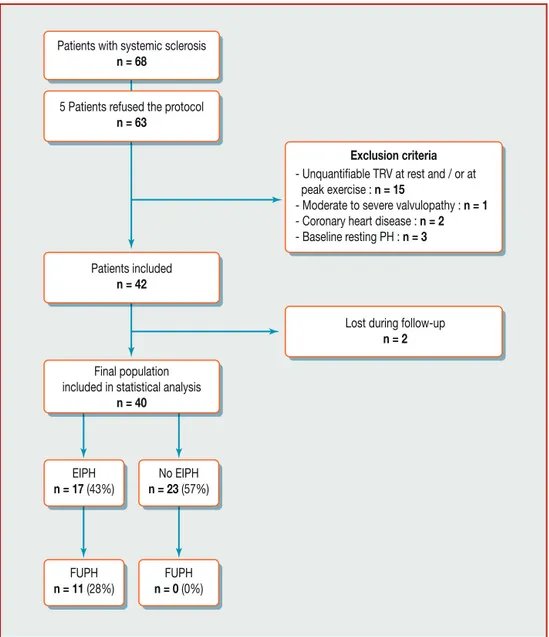
Figure1. Studyflow chart. EIPH:exercise-inducedpulmonary hypertension;FUPH:onsetofpulmonary arterialhypertension during follow-up;PH:pulmonaryhypertension;TRV:tricuspidregurgitationvelocity.
sclerosisfollowedintherheumatologycentreofCHU Sart-TilmaninLiège.Fivepatientsrefusedtoparticipateinthe study.Exclusioncriteriawereinabilitytoprovideinformed consent; previous ischaemic heart or valvular heart dis-ease;inabilitytoperformanexercisetest;baselineresting PH;andunquantifiablesystolicpulmonaryarterialpressure (sPAP).Fifteenpatientswereexcludedfromthepopulation (n=63) because of unquantifiable sPAP (24% of the popu-lation), one for ≥moderate mitral regurgitation, two for coronaryarterydiseaseandthreeforbaselinerestingPH(7% ofpatientswithregistrabletricuspidregurgitation).Finally, twopatientswerelostduringfollow-up,sothefinal analy-sisincluded 40patients(Fig.1).Therelevantinstitutional reviewboardapprovedtheprotocol,andallpatientsgave writteninformedconsent.
Echocardiographic
examination
All patients underwent comprehensive resting echocardi-ography, using a conventional method, with a Vivid 9
ultrasoundsystem(GeneralElectricHealthcare,Little Chal-font,UK),atbaselineandduringfollow-up(twiceayear), blindedforNVCresultsatbaseline.Offlineanalysiswas per-formedretrospectivelyusingacustomizedsoftwarepackage (EchoPac). Left ventricular (LV) stroke volume was cal-culated as the difference between LV end-diastolic and end-systolicvolumesassessedbythebiapicalSimpson’sdisc method, and LV ejection fraction was derived from the stroke volume/LV end-diastolicvolumeratio.Cardiac out-putwasobtainedbymultiplyingLV,strokevolumeandheart rate.PeakEwaveandAwavevelocitiesofthemitralinflow were measured with pulsed-waveDoppler. Tissue Doppler imagingwasappliedforthemeasurementofe’waveatthe lateralmitralannulusaspect.BothmeasurementsofEandA wavesandtissueDopplerimagingatthemitralannuluslevel wereperformedduringexercise,justbeforefusionofEand Awaves.Rightventricularfractionalareachange,tricuspid annularplanesystolicexcursionandmaximalsystolic veloc-ityofthetricuspidannulus(s’)wereassessedinallpatients. Thesystolicpulmonaryarterialpressure(sPAP)wasderived
fromthemaximalvelocityofthetricuspidregurgitantjet, according to the simplified Bernoulli equation, with the additionofrightatrialpressure,estimatedfromthe dimen-sionandcollapsibilityoftheinferiorvenacava[9].Apeak value>35mmHg wasconsidered todefine resting PH [10]
atbaselineandFUPH. Atpeakexercise,sPAPwasderived fromthe tricuspidregurgitantjetvelocity,withthe addi-tionof10mmHgfor theestimationofrightatrial pressure
[11]. EIPH wasdefinedas sPAP>50mmHg [12]. mPAPwas estimatedusingtheChemlaformula:mPAP=0.61×sPAP+2. TheslopeofthemPAP/LVCOrelationshipwasestimatedas theratiobetweenchanges(peak—restvalue)inmPAPand changes in LVCO [13,14]. All echocardiographic variables were acquired at peak exercise, except for mitral inflow velocitiesandtissueDopplerimagingatthemitralannulus. Asymptom-limitedgradedbicycleexercisetestwas per-formedinasemisupinepositiononatiltedtable.Afteran initialworkloadof25Wmaintainedfor2minutes,the work-load was gradually increased by 25W every 2minutes. A 12-leadelectrocardiogramwasmonitoredcontinuously,and blood pressurewasmeasured atrest andat each levelof exercise.Allpatientspresentednormaltests,definedasthe absenceof the occurrenceof: angina; ≥2mm ST-segment depressioncomparedwithbaselinelevel;or complex ven-triculararrhythmias.
NVC
and
lung
function
assessment
Using an optical probe videocapillaroscope, the nailfold of the second, third, fourth and fifth fingers was exam-inedbilaterallyineachpatientasdescribedpreviously[3]. NVC grades were qualitatively assessed as normal (grade 1:normalcapillarymorphology,regulardistributionandno capillary loss), early (grade 2: few capillary microhaem-orrhages and giant capillaries, no loss of capillaries and preserveddistribution),active(grade3:frequentcapillary microhaemorrhagesandgiantcapillaries,moderatelossof capillaries,milddisorganizationofthemicrovascular archi-tecture and absent or mild ramified capillaries) and late (grade4:irregularenlargementofcapillaries,feworabsent giant capillaries and microhaemorrhages, severe loss of capillariesandlargeavascularareas,disorganizationof cap-illaryandramifiedcapillary).
All patients underwent standard pulmonary function tests,withassessmentoftotallungcapacity,vitalcapacity, forcedvitalcapacity,forcedexpiratoryvolumein1second, ratioofforcedexpiratoryvolumein1secondtovital capac-ityanddiffusingcapacityofthelungforcarbonmonoxide.
BNP
blood
concentration
assessment
BNPbloodconcentrationwasassessedatrest, justbefore echocardiographyexamination,inasubset of30 patients. Venousbloodsamplesweredrawnbeforeechocardiography, after 10minutes of supine rest. Chilled ethylenediamine-tetraacetic acid tubes were centrifuged immediately at 4000rpm(4—8◦C)for15minutes.
Statistical
analysis
Continuous variables are expressed as means±standard deviations; categorical variables are presented as
numbers and percentages. Data comparisons were per-formedaccordingtothepresenceorabsenceofFUPHusing Student’sunpairedandpairedttests,the2testorFisher’s
exacttest,asappropriate.Therelationshipsbetween max-imalsPAPduringfollow-up andother continuousvariables (i.e.demographicdata,resting andexercise echocardiog-raphic data) were evaluated by simple linear regression. PredictorsofhighestsPAPduringfollow-upwereidentified withtheuseofunivariateandmultivariablelinear regres-sions.Univariate and multivariable logisticregression and Coxproportional-hazardsmodelsanalyseswereperformed todefinepredictorsofFUPH.Inallmultivariableanalyses, because of the sample size and the limited number of outcomedata,adjustmentswereperformed withonlytwo variablesin ordertolimitstatistical power reductionand to avoid type II error. The value of 2 for each variable
definedits prognosticvalue. Sequential Cox modelswere performedtodeterminetheincrementalprognosticbenefit of different variables over age. A statistically significant increaseinthegloballog-likelihood2ofthemodeldefined
incremental prognostic value. Probabilities of FUPH-free survivalwereobtainedbyKaplan-Meierestimates,andthen comparedusingatwo-sidedlog-ranktest.ValuesofP<0.05 were considered statistically significant. All statistical analyseswereperformedwithSPSSsoftware,version16.0.
Results
Population
characteristics
The mean age of the population was 54±13 years; 68% werewomen. sPAP increased significantly during exercise (from24±6to46±14mmHg;P<0.0001).Seventeen(43%) patientsdevelopedEIPH.Afterameanfollow-upof25±15 months(median28months),11patients(28%)hadFUPH,all intheEIPHgroup.PatientswithEIPHandFUPHwere signifi-cantlyolder(62±12vs48±11yearsand60±14vs50±12 years,respectively;P<0.05inbothcases).Femalesexwas morefrequentintheEIPHgroup(90%vs62%;P=0.03),but notin the FUPH group (64% vs 69%; P=0.69. There were nosignificantdifferencesbetweenthetwogroupsregarding medicationandlungfunctiontestvariables(Table1).There werenosignificantdifferencesbetweenthetwogroupsin termsofRaynaud’sphenomenon(82%vs87%;P=1.00orthe presenceofScl-70antibodies(36%vs31%;P=0.70.Patients withFUPHhadahigherBNPbloodconcentration(79±86vs 23±21pg/mL;P=0.01).
NVC
grades
NVCgrade 1wasfound in8% ofpatients, grade2 in38%, grade3in28%andgrade4in20%(eightpatients).Therewas asignificantrelationshipbetweenthedifferentNVCgrades and the onset of PH during follow-up (P=0.01) (Fig. 2). PatientswithFUPHpresented more frequentlyNVCgrade 4 (45% vs 11%; P=0.03) and less frequently NVC grade 2 (54%vs9%;P=0.006).Therewerenosignificantdifferences betweentheFUPHandnoFUPHgroupsregardingNVCgrade 1(0vs12%;P=0.13)andgrade3(45vs23%;P=0.18).There wasasignificantlyhigherproportionofNVC>grade2inthe FUPHgroup(90%vs35%;P=0.0009).
Table1 Demographic,clinicalandexercisedata.
Variables Wholecohort
(n=40) NoFUPH (n=29,72%) FUPH (n=11,28%) P Demographic,clinicalandbiologicaldata
Age(years) 54±13 50±12 60±14 0.04
Femalesex 27(68) 20(69) 7(64) 0.69
Bodymassindex(kg/m2) 24±5 24±5 25±6 0.72
Heartrate(bpm) 74±14 73±13 74±16 0.72
Systolicarterialpressure(mmHg) 128±19 126±19 134±16 0.36
Diastolicarterialpressure(mmHg) 73±9 73±10 74±7 0.90
Raynaud’sphenomenon 34(85) 25(87) 9(82) 1.00
DelaybetweendiagnosisandTTE(months) 36±36 37±39 31±28 0.62
NYHAclass>II 3(8) 3(10) 0(0) 0.26 PresenceofScl-70antibodies 13(33) 9(31) 4(36) 0.70 BNP(pg/mL) 41±61 23±21 79±86 0.01 Riskfactors Systemichypertension 4(10) 2(7) 2(11) 0.33 Hypercholesterolaemia 9(23) 7(25) 2(18) 0.64 Smoker 10(24) 9(31) 1(9) 0.08
FamilyhistoryofCVdisease 2(5) 2(7) 0(0) 0.24
Pulmonaryfunction
Totallungcapacity(%predicted) 93±21 95±21 82±16 0.15
Vitalcapacity(%predicted) 104±24 105±26 96±17 0.32
Forcevitalcapacity(%predicted) 101±25 102±21 93±17 0.30
FEV1(%predicted) 95±20 96±20 87±22 0.27
FEV1/vitalcapacity(%predicted) 99±12 100±12 96±13 0.38
DLCO(%predicted) 65±14 66±15 60±12 0.33 Medication ACEinhibitors 2(5) 1(4) 1(10) 0.51 Beta-blockers 3(8) 1(4) 2(9) 0.15 Diuretics 2(5) 1(4) 1(10) 0.51 Calciuminhibitors 20(50) 13(52) 7(70) 0.32 Corticoids 9(23) 5(20) 4(40) 0.23 Immunosuppressors 6(16) 4(16) 2(20) 0.77 Exercisedata Workload(W) 75±31 81±30 65±36 0.22
Durationofexercise(minutes) 4.9±1.3 4.7±1.3 5.1±1.6 0.55
Heartrate(bpm) 119±17 119±16 122±21 0.65
Systolicarterialpressure(mmHg) 168±23 163±23 176±21 0.19
Diastolicarterialpressure(mmHg) 82±12 81±11 84±13 0.55
Dataareexpressedasmean±standarddeviationornumber(%).ACE:angiotensin-convertingenzyme;BNP:B-typenatriureticpeptide; bpm:beats per minute; CV: cardiovascular; DLCO: diffusing capacity of the lung for carbon monoxide; FEV1: forced expiratory volumein1second;FUPH:onsetofpulmonaryhypertensionduringfollow-up;NYHA:NewYorkHeartAssociation;TTE:transthoracic echocardiography.
Resting
and
exercise
echocardiography
Patients with FUPH had higher baseline sPAP (30±4 vs 22±5mmHg; P< 0.0001, baseline mPAP (20±2 vs 15±3mmHg;P<0.0001),indexedleftatrialarea(10±3vs 8±1cm2;P=0.01),exercisesPAP(60±12vs40±11mmHg; P<0.0001; Fig.3), exercisemPAP(38±7 vs26±7mmHg;
P<0.0001)andexercise-inducedchangesinsPAP(+25±10 vs+14±9mmHg;P=0.001)(Tables2and3).Patientswith EIPHhadahigherexerciseE/e’ratio(9.1±1.2vs5.7±1.4;
P=0.0002). During exercise (Table 3), LV ejection frac-tion (67±5 vs 71±5%; P=0.001) and its rate of change (+4±3vs+8±4%;P=0.008)werelowerintheFUPHgroup.
Conversely, the E/e’ ratio, resulting from lower e’ wave velocity,washigherintheFUPHgroup(9.4±0.7vs5.8±0.4 [P=0.0003]and17±0.1vs12±0.2cm/s[P=0.03], respec-tively).TheslopeofthemPAP-LVCOrelationship(6.7±2.7 vs3.4±2.1mmHg/L/min;P=0.003)washigherintheFUPH group.
Finally,40%ofpatientspresentedneitherEIPHnorNVC grade>2,32% presentedeither EIPH or NVCgrade>2 and 18%hadbothEIPHandNVCgrade>2.Therewasasignificant differenceintheincidenceofFUPHbetweenpatientswith noEIPH and NVCgrade<2 andpatients withEIPH or NVC grade>2,andthosewhohad bothEIPH andNVCgrade>2 (0%vs17%vs82%,respectively;P<0.0001).
Figure 2. Comparison of theincidence of nailfold videocapil-laroscopy(NVC)gradesaccordingtothetwogroups.FUPH:onset ofpulmonaryarterialhypertensionduringfollow-up.
Figure3. Comparisonofrestingandexercisesystolicpulmonary arterialpressure(sPAP)accordingtothetwogroups.FUPH:onset ofpulmonaryarterialhypertensionduringfollow-up.
Determinants
of
the
maximal
value
of
sPAP
during
follow-up
Asignificant correlationwasfoundbetween maximalsPAP duringfollow-upandexerciseleftatrialpressure(r2=0.43; P<0.004), as well as resting indexed left atrial area (r2=0.43; P<0.0001), resting sPAP (r2=0.36; P<0.0001),
exercisesPAP(r2=0.36;P<0.0001),theslopeofthe
mPAP-LVCOrelationship(r2=0.22;P=0.01)andexerciseindexed
right atrial area (r2=0.31; P=0.002). A correlation was
found with age (r2=0.18; P=0.009) and exercise-induced
changesinLVejectionfraction(r2=0.15;P=0.01).
After adjustment for age, resting sPAP and exercise sPAP remained independently associated with maximal resting sPAP during follow-up (=0.9±0.3 [P=0.02] and =0.4±0.1 [P=0.02], respectively), asdid NVCgrade>2 (=9.1±2.9;P=0.004).
Inthe subgroupof 30patients withBNPblood concen-tration measurements available, BNP was related to the maximal value of sPAP during follow-up (r2=0.41; P<0.001)andremainedassociatedafteradjustmentforage (=0.1±0.03;P=0.001).
Determinants
of
FUPH
Logistic
regression
analyses
Age was associated with the onset of PH during follow-up (odds ratio [OR] 1.06; 95% confidence interval [CI] 1.01—1.13;P=0.047),aswererestingsPAPandexercisesPAP (OR1.71,95%CI1.19—2.45[P=0.003]andOR1.18,95%CI 1.06—1.32[P=0.003],respectively).Similarly,NVCgrade>2 wasrelatedtoFUPH(hazardratio[HR]18.9,95%CI2.1—172;
P=0.009).
After adjustment for age, resting sPAP and exercise sPAPremainedassociatedwithFUPH(oddsratio1.68,95% CI 1.17—2.4 [P=0.005] and OR 1.17, 95% CI 1.05—1.32 [P=0.006],respectively),asdidNVCgrade>2(OR16.9,95% CI1.7—168;P=0.02).
Beyondage (2=4.5%),therewasanincrementalvalue
for NVC grade>2 (2=14.1%; P<0.0001), resting sPAP
(2=22.1%)andEIPH(2=25.2%;P<0.0001)topredictFUPH
(Fig.4).
In thesubgroup of 30 patients withBNPconcentration measurementsavailable,BNPwasnotassociatedwithFUPH (OR1.04,95%CI0.99—1.08;P=0.054).
Time-dependency
statistical
analyses
Afterafollow-upof24months,patientswithNVCgrade>2 andEIPHhadlowerFUPH-freesurvivalrates(61.8±11.4%vs 100%[P<0.05]and55.4±12.8%vs100%[P<0.05], respec-tively).Inaddition,patientswithnoEIPHandNVCgrade<2 had a greater FUPH-free survival rate than patients with either EIPH or NVC grade>2 and patients with both EIPH and NVC grade>2 (100% vs 75.8±15.6% vs 41.6±15.6%, respectively;P=0.0001).
InCoxregressionanalyses,agewasassociatedwithFUPH (HR1.06,95%CI1.02—1.11;P=0.007),aswererestingsPAP andexercisesPAP(HR1.41,95% CI1.18—1.69[P=0.0001] andHR1.16, 95%CI1.08—1.25[P=0.0001],respectively). NVCgrade>2 wasassociatedwith theonsetof PH during follow-up(HR8.9,95%CI1.13—69.4;P=0.04).Finally,the combinationofEIPHandNVCgrade>2wasrelatedtoFUPH (HR15.1,95%CI3.2—70.3;P=0.001).
Afteradjustmentforage,restingsPAPandexercisesPAP were associated with FUPH (HR 1.41, 95% CI 1.08—1.70 [P=0.0001] and HR 1.2, 95% CI 1.08—1.25 [P=0.0001], respectively),aswasNVCgrade>2(HR9.1,95%CI1.1—74.8;
P=0.04).Finally,EIPHandNVCgrade>2alsoremained asso-ciatedwithFUPH(HR11.6,95%CI2.4—55.3;P=0.002).
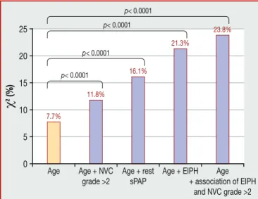
Beyondage (2=7.7%),therewasanincrementalvalue
for NVC grade>2 (2=11.8%; P<0.0001), resting sPAP
(2=16.1%; P<0.0001), EIPH (2=21.3%; P<0.0001) and
the combination of EIPH and NVC grade>2 (2=23.8%; P<0.0001)topredictFUPH(Fig.4).
Inthesubsetof30patientswithBNPconcentration mea-surements available, BNP was associated with FUPH (HR 1.02, 95% CI 1.01—1.03; P=0.004), and this association
Table2 Restingechocardiographicdata.
Variables Wholecohort
(n=40) NoFUPH (n=29,72%) FUPH (n=11,28%) P RestingLVechocardiographicdata
LVindexedend-diastolicvolume(mL) 45±11 47±13 42±7 0.30
LVindexedend-systolicvolume(mL) 16±5 17±7 15±3 0.48
LVindexedstrokevolume(mL) 29±6 30±7 28±5 0.40
Simpson’sLVEF(%) 63±4 64±4 62±5 0.41
Cardiacoutput(L/min) 3.7±0.9 3.7±0.2 3.5±0.3 0.65
E(cm/s) 6.9±1.3 6.9±0.2 7.1±0.5 0.75 Edecelerationtime(ms) 179±37 179±7 172±13 0.75 A(cm/s) 6.7±1.8 6.7±0.3 7.2±0.6 0.63 E/Aratio 1.1±0.5 1.1±0.1 1.2±0.2 0.59 e’(cm/s) 1.1±0.5 1.3±0.1 1.1±0.1 0.10 E/e’ratio 5.9±1.7 5.6±0.3 6.8±0.6 0.08
RestingRVechocardiographicdata
RV/LVratio 0.75±0.1 0.7±0.1 0.8±0.1 0.30 RVend-diastolicarea(cm2) 13.9±3.7 14.1±4.2 13.3±2.4 0.53 RVend-systolicarea(cm2) 7.2±2.3 7.5±2.4 7.0±2.1 0.59 RVFAC(%) 47.8±8.8 47.3±7.7 47.4±10.2 0.98 TAPSE(mm) 23±5 23±4 24±6 0.77 s’(cm/s) 12.3±2.7 12.5±2.6 12.6±2 0.89 ICVmax(cm/s) 12±4 12±4 13±3 0.33 IVRT(ms) 46±23 49±22 41±29 0.39 MPI 0.36±0.14 0.35±0.12 0.38±0.19 0.63
Pulmonaryaccelerationtime(ms) 138±35 144±35 119±31 0.06
sPAP(mmHg) 24±6 22±5 30±4 <0.0001
mPAP(mmHg) 16±4 15±3 20±2 <0.0001
Restingatrialarea
LAindexedarea(cm2) 8±2 8±1 10±3 0.01
RAindexedarea(cm2) 7±3 7±1 9±4 0.03
Dataareexpressedasmean±standarddeviation.FUPH:onsetofpulmonaryhypertensionduringfollow-up;ICV:isovolumiccontraction velocity;IRVT:isovolumicrelaxation time;LA:left atrial;LV:leftventricular;LVEF: leftventricularejectionfraction;mPAP: mean pulmonary arterial pressure; MPI: myocardial performance index; RA: right atrial; RV: right ventricular; RVFAC: right ventricular fractionalareachange;sPAP:systolicpulmonaryarterialpressure;TAPSE:tricuspidannularplanesystolicexcursion.
remainedsignificantafteradjustmentforage(HR1.02,95% CI1.01—1.03;P=0.01).
Discussion
The present study shows that: the developmentof PH at follow-upisnotarareconditionin patientswithsystemic sclerosis(28%);NVCgrade>2isapowerfulpredictorofthe onsetofFUPHinpatientswithsystemicsclerosis;and exer-cisesPAP canpredict the evolutionof resting sPAPduring FU.Ourdataalsosuggestthatanintegratedapproachbased upon both comprehensive exercise echocardiography and NVCcouldbe useful in identifyinga subgroup of patients withsystemicsclerosisathigherriskofdevelopingPH.
NVC
grade
and
onset
of
resting
PH
NVCis aninterestingtoolinsystemicsclerosisfor investi-gatingtheunderlyingdiseaselesion.However,ithasbeen poorlyexplored,anditspotential roleinpredicting FUPH
has, to the best of our knowledge, hardly been studied. A few studies have reported a correlation between wors-ening NVC grade and severe organ involvement. Smith etal.[3]showedthepotentialclinicalsignificanceofNVC in 66 patients with systemic sclerosis; the authors clas-sified microvascular lesions within four different stages, dependingonthepresenceandimportanceofgiant capillar-ies, microhaemorrhages and loss of capillaries (i.e. from ‘‘normal’’to‘‘late’’ NVCgrade).Patientswere followed-upclinicallyat18—24months,andsevereorganinvolvement ofthenineorgansystemswasstudied.Theauthors demon-stratedacloserelationship betweenworseningNVCgrade andfuturesevereperipheralvascularinvolvement,aswell as future severe lung involvement. Similarly, we demon-strated a close relationship between NVC grade>2 and FUPH, strengthening the potential clinical value of NVC in risk stratification of patients with systemic sclerosis. Theseresultssuggestthatlocalmicrovascularstatus prob-ably represents global microvascular impairment, and is linkedto myocardialand pulmonary microvascular status, assuggestedbyrestingandexerciseechocardiographydata
Table3 Exerciseechocardiographicdata.
Variables Wholecohort
(n=40) NoFUPH (n=29,72%) FUPH (n=11,28%) P ExerciseLVechocardiographicdata
LVindexedend-diastolicvolume(mL) 50±11 51±13 47±6 0.36
LVindexedend-systolicvolume(mL) 15±5 15±5 16±3 0.79
LVindexedstrokevolume(mL) 35±8 36±9 31±3 0.15
Simpson’sLVEF(%) 0±5 71±5 67±5 0.001
Cardiacoutput(L/min) 7.2±2.1 7.6±0.4 6.2±0.8 0.15
E(cm/s) 9.8±1.9 9.6±1.9 10.0±2.2 0.21 Edecelerationtime(ms) 118±37 114±8 118±19 0.86 A(cm/s) 9.4±1.7 9.2±0.3 10.1±0.8 0.34 E/Aratio 1.1±0.2 1.1±0.1 1.0±0.1 0.76 e’(cm/s) 1.6±0.4 1.7±0.1 1.2±0.2 0.03 E/e’ratio 6.5±1.4 5.8±0.4 9.4±0.7 0.0003
ExerciseRVechocardiographicdata
RVend-diastolicarea(cm2) 12.1±2.9 12.0±3.0 12.5±3.3 0.71 RVend-systolicarea(cm2) 5.7±1.6 5.8±1.8 5.5±1.1 0.67 RVFAC(%) 52.6±10 51.9±9.6 54.2±11.8 0.59 LVend-diastolicdiameter,(mm) 39.6±4.8 40.6±4.9 36.6±4.1 0.05 RV/LVratio 71.0±12 69.3±12.2 79.4±10.7 0.04 TAPSE(mm) 27.1±5.4 26.5±3.5 29.3±8.4 0.15 s’(cm/s) 16.3±3.7 16.9±3.8 14.7±3.3 0.18 ICVmax(cm/s) 15.7±4.8 15.5±5.4 15.7±2.4 0.95 IVRT(ms) 37.4±15.7 35±12 44±26 0.32 sPAP(mmHg) 46±14 40±11 60±12 <0.0001 mPAP(mmHg) 29±8 26±7 38±7 <0.0001
SlopeofmPAPa(mmHg/L/min) 4.2±2.7 3.4±2.1 6.7±2.7 0.003
EIPH 17(43) 6(21) 11(100) <0.0001
Exerciseatrialarea
LAindexedarea(cm2) 9±3 8±2 10±5 0.09
RAindexedarea(cm2) 8±3 7±1 10±5 0.03
Dataare expressed as mean±standarddeviation or number(%). EIPH: exercise-inducedpulmonary hypertension;FUPH: onsetof pulmonaryhypertensionduringfollow-up;ICV:isovolumiccontractionvelocity;IRVT:isovolumicrelaxationtime;LA:leftatrial;LV:left ventricular;LVEF:leftventricularejectionfraction;mPAP:meanpulmonaryarterialpressure;RA:rightatrial;RV:rightventricular; RVFAC:right ventricularfractional areachange;sPAP: systolicpulmonary arterialpressure;TAPSE: tricuspidannular planesystolic excursion.
a SlopeofmPAPindicatestheratiobetweenchangesinmPAPandchangesinLVCO.
(i.e. higher exercise E/e’ and lower exercise-induced changeinthesevariablesintheFUPHgroup).
Exercise
echocardiography
and
evolution
of
sPAP
The accuracy of exercise echocardiography in detecting the development of PH during exercise in systemic scle-rosis has been reported by several authors. Steen et al.
[5] reported an EIPH incidence of 44% (defined by a post-exercise≥20mmHg increase in sPAP) in 54 patients. Using right heart catheterization, the authors confirmed the involvement of a precapillary mechanism in the vast majorityofcasesand,lessfrequently,apost-capillary aeti-ology. Ofnote, fourpatients hadresting PH notdetected by echocardiography, and only one patient had normal catheterization.Thepotentialclinical significanceofEIPH wasdemonstratedbyAlkotobetal.[4]in65patients;they foundasimilarrateofEIPH(46%),withaslightlydifferent
definitionof EIPH (exercisesPAP>40mmHg). Finally, they showedarelationshipbetweenEIPHanddecreasedexercise capacity,withaweakinverserelationshipbetweenexercise sPAPandmaximalworkload achieved(r=—0.34;P=0.006) or exercise duration (r=—0.31; P=0.01). Nevertheless, despitethesefindings,thepotentialroleofEIPHand exer-ciseechocardiographyfortheriskstratificationofpatients withPHwaslittlehighlighted inthelastEuropean recom-mendations[8]. Infact, onlyone recentstudy reporteda significantrelationshipbetweenexercisesPAPandFUPHin systemicsclerosis[15].Usingexerciseechocardiographyina cohortof170patientswithsystemicsclerosis,Codulloetal.
[15]foundthatpatientswhodevelopedPHduringfollow-up (3.5±0.2 years) had markedly significant higher exercise sPAP, exercise-induced change in sPAP and change in PAP indexedtochangeincardiacoutput.Inamultivariable anal-ysis,theoccurrenceofFUPH(definedasmPAP≥25mmHgin rightheartcatheterization)wasbestpredictedby exercise-induced changes in sPAP. Although the definitions of EIPH
Figure 4. Incremental value over age of nailfold videocapil-laroscopy(NVC)grade,restingsystolicpulmonaryarterialpressure (sPAP),exercise-induced pulmonary hypertension (EIPH) and the combinationofEIPHandNVCgrade.
(exercisesPAP>50mmHg)andFUPH(restingPH>35mmHg on echocardiography) were different from previous stud-ies, our data confirmed that EIPH assessed by exercise echocardiographycanpredict the evolutionof future res-tingsPAP.Resting sPAPaswellasexercisesPAParestrong predictors of FUPH. In addition, exercise sPAP provides incrementalvalueinpredictingtheonsetofFUPH,and exer-ciseechocardiographybringsvaluableinformationaboutthe pathophysiologyofthedisease.Exerciseechocardiography showstheconsequencesforthemyocardiumandpulmonary microvascularfunction.Thus,increasedrestingandexercise pulmonaryvascularresistanceintheFUPHgroupmay sug-gestapulmonaryvascularimpairment.Reducedmyocardial microvascularfunctionissuggestedbyincreasedestimated exercise LV filling pressure and lower exercise-induced changeinLVejectionfraction,whichprobablycorresponds tomicrovascularischaemiaduringexercise.Thishypothesis issupportedbypreviousstudieswithsinglephotonemission computedtomographyassessmentofmyocardialperfusion, whichshowedischaemia insystemicsclerosiswithout any coronaryarterylesions[16].Myocardialscintigraphy demon-strated evidence of reversible ischaemia together with irreversible lesions, and showed inducibility of coronary vasospasm by cold pressor provocation, suggesting both myocardialischaemiaandfibrosis[17].Vignauxetal.[18]
showedan increase in LVfunction andin myocardial per-fusion after administration of nifedipine in patients with systemicsclerosis,suggestingalinkbetweenmicrovascular dynamicreserveandLVfunction.
Study
limitations
The main limitation of our study was the low incidence ofright heartcatheterizationtoconfirm restingPH inthe FUPH group. Two patients had confirmed resting PH dur-ingfollow-up(mPAP26mmHgandmPAP33mmHg),andone hadresting mPAPduringfollow-upin thegreyzone (mPAP 21mmHg).However,thisdidnotaffect themainresultof ourstudy, which was that exercise echocardiographyand NVCcanscreenpatientsatriskofdevelopingFUPH.Inthis
group, patientshad ahigherincrease in restingsPAP dur-ingfollow-up(3±4.5vs0.4±0.7mmHg/month;P=0.005), suggestingadynamicandrapidincreaseinsPAPovertime, correspondingtoahighriskofdevelopingrestingPH.
Thesecondlimitationofourstudywastherelativesmall sizeofthepopulation,butthisreflectsthelowincidenceof systemicsclerosis.Consequently, regardingthesizeofthe populationandthenumberofeventsduringFU,itwasnot possibletoperformcomplexmultivariableanalyses (includ-ingmorethantwovariables).Asageisawell-knownvariable influencing the level of sPAP, we decided to perform an adjustmentforageinthemultivariableanalysis.
Inaddition,thetimingoffollow-upwasdetermined arbi-trarily, and could be modified depending on the clinical statusofthepatients.Therefore,therealtimeofonsetof FUPHwasnotknownprecisely(itoccurredbetweenthetime of diagnosis of FUPH and the previous echocardiographic control)andconstitutesabiasfortime-dependency analy-ses.Thus,CoxregressionandKaplan-Meieranalysesshould be interpreted withcaution, and either linear or logistic regression may be more informative in the present case. However,evenwiththislimitation,theresultsoflinearand logistic regression were quite similarto time-dependency analysisresults.
ThesmallLVvolumesreportedcouldberelatedto fore-shortening views, and may also explain the low cardiac output reported. However, this underestimation affected thewholepopulationand,consequently,didnotinfluence thereliabilityofthemainresultofthisstudy,whichwasthe demonstrationthatexercisesPAPandNVCgradeare inde-pendentdeterminantsofFUPH.Becauseofthehighresting heart ratein some patientsand theearly fusion ofE and Awaves, the E/e’ratio wasonly available in50% ofboth groups ofpatients.Although useful,E/e’ratioassessment duringexerciseseemsmoderatelyfeasibleandapplicablein routineassessmentinthisparticularpopulation.Itisknown that pulmonary function status influences sPAP; however, therewerenosignificantdifferencesbetweenpatientswith andwithoutPHduringfollow-upforeachofthelung func-tion variables. This could be a consequence of the small population and a type II error. Patients with FUPH were significantly older, andsPAP increaseswith age.However, afteradjustmentforageandrestingsPAP,bothexercisesPAP andNVCgrade>2remainedsignificantlyassociatedwiththe occurrenceofPHduringFU,suggestingthatthesetwo varia-blesareindependentofageandthebaselinevalueofresting sPAP.
Finally, 15/63 (24%) patients had unquantifiable sPAP due to the absenceof registrable tricuspidregurgitation, whichcouldbealimitationtothelargeapplicationof exer-cise echocardiographytotherisk stratificationof patients with systemicsclerosis. However,contrast echocardiogra-phy couldbe performed in thesepatients toincrease the feasibilityoftheregistrationofthetricuspidregurgitation, andfurtherstudiesshouldassessthispoint.
Conclusion
Exerciseechocardiographyand NVCcanhelp usto under-standthepathophysiologicalmechanismsleadingtoresting PH in patients withsystemic sclerosis,andcould beused
for individual risk stratification and decisionmaking. NVC revealstheprimarylesionofthedisease(i.e.microvascular impairment) and echocardiographyallows the assessment of the consequences of this lesion for myocardial and pulmonary vascular function. Patients with no abnormal increase in sPAP during exercise and presenting an NVC grade<2 are at very low risk of FUPH, contrasting with patientswithbothEIPHandNVCgrade>2,inwhomtherisk ofdevelopingPHduringfollow-upisparticularlyhigh(82%).
Funding
DamienVoilliot is supported bya researchgrantfromthe EuropeanAssociationofCardiovascularImaging.
Acknowledgements
WeespeciallythankCarmineCelentanoforhisvaluable sup-portandhelpwiththeinvestigationsinthisstudy.
Disclosure
of
interest
Theauthorsdeclarethattheyhavenocompetinginterest.
References
[1]GabrielliA,AvvedimentoEV,KriegT.Scleroderma.NEnglJMed 2009;360:1989—2003.
[2]KawutSM,TaichmanDB,Archer-ChickoCL,PalevskyHI, Kim-melSE.Hemodynamicsandsurvivalinpatientswithpulmonary arterial hypertension related to systemic sclerosis. Chest 2003;123:344—50.
[3]SmithV,DecumanS,SulliA,etal.Doworseningscleroderma capillaroscopicpatternspredictfuture severeorgan involve-ment?Apilotstudy.AnnRheumDis2012;71:1636—9. [4]AlkotobML,SoltaniP,SheattMA,etal.Reducedexercise
capac-ityandstress-inducedpulmonaryhypertensioninpatientswith scleroderma.Chest2006;130:176—81.
[5]SteenV,ChouM,ShanmugamV,MathiasM,KuruT,MorrisseyR. Exercise-inducedpulmonaryarterialhypertensioninpatients withsystemicsclerosis.Chest2008;134:146—51.
[6]NaeijeR,VanderpoolR,DhakalBP,etal.Exercise-induced pul-monaryhypertension:physiologicalbasisandmethodological concerns.AmJRespirCritCareMed2013;187:576—83.
[7]LewisGD,BossoneE,NaeijeR,etal.Pulmonaryvascular hemo-dynamic response to exercise in cardiopulmonary diseases. Circulation2013;128:1470—9.
[8]GalieN,HoeperMM,HumbertM,etal.Guidelinesforthe diag-nosisandtreatmentofpulmonaryhypertension:theTaskForce fortheDiagnosisandTreatmentofPulmonaryHypertensionof theEuropean SocietyofCardiology (ESC) and theEuropean RespiratorySociety(ERS),endorsedbytheInternational Soci-ety of Heart and Lung Transplantation (ISHLT).Eur HeartJ 2009;30:2493—537.
[9]RudskiLG,LaiWW,AfilaloJ,etal.Guidelinesforthe echocardi-ographicassessmentoftherightheartinadults:areportfrom the American Society of Echocardiographyendorsed bythe EuropeanAssociationofEchocardiography,aregisteredbranch oftheEuropeanSocietyofCardiology,andtheCanadianSociety ofEchocardiography.JAmSocEchocardiogr2010;23:685—713 [quiz86—8].
[10]Mukerjee D,St George D,Coleiro B, et al. Prevalence and outcome in systemicsclerosis associatedpulmonary arterial hypertension:applicationofaregistryapproach.AnnRheum Dis2003;62:1088—93.
[11]MagneJ,LancellottiP,PierardLA.Exercisepulmonary hyper-tension in asymptomatic degenerative mitral regurgitation. Circulation2010;122:33—41.
[12]Gargani L, PignoneA, AgostonG, et al. Clinical and echo-cardiographic correlations of exercise-induced pulmonary hypertension in systemic sclerosis: a multicenter study.Am HeartJ2013;165:200—7.
[13]ArgientoP,CheslerN,MuleM,etal.Exercisestress echocardi-ographyforthestudyofthepulmonarycirculation.EurRespir J2010;35:1273—8.
[14]ArgientoP,VanderpoolRR,MuleM,etal.Exercisestress echo-cardiographyofthepulmonarycirculation:limitsofnormaland sexdifferences.Chest2012;142:1158—65.
[15]CodulloV,CaporaliR, CuomoG, etal.Stress Doppler echo-cardiography in systemic sclerosis: evidence for a role in the prediction of pulmonary hypertension. Arthritis Rheum 2013;65:2403—11.
[16]Steen VD, Follansbee WP, Conte CG, Medsger Jr TA. Thallium perfusion defects predict subsequent cardiac dys-function inpatientswithsystemicsclerosis. ArthritisRheum 1996;39:677—81.
[17]GustafssonR,ManntingF,KazzamE,WaldenstromA,Hallgren R.Cold-induced reversiblemyocardialischaemiainsystemic sclerosis.Lancet1989;2:475—9.
[18]Vignaux O, Allanore Y, Meune C, et al. Evaluation of the effectofnifedipineuponmyocardial perfusionand contrac-tility using cardiac magnetic resonance imaging and tissue Dopplerechocardiographyinsystemicsclerosis.AnnRheumDis 2005;64:1268—73.