Discovery and Characterization of R/S‑N‑3-Cyanophenyl‑N
′‑(6-tert-
butoxycarbonylamino-3,4-dihydro-2,2-dimethyl‑2H‑1-benzopyran-4-yl)urea, a New Histone Deacetylase Class III Inhibitor Exerting
Antiproliferative Activity against Cancer Cell Lines
Michael Schnekenburger,
†,◆Eric Goffin,
‡,◆Jin-Young Lee,
§Jun Young Jang,
§Aloran Mazumder,
§Seungwon Ji,
§Bernard Rogister,
∥Nafila Bouider,
‡Florence Lefranc,
⊥Walter Miklos,
#Véronique Mathieu,
○Pascal de Tullio,
‡Kyu-Won Kim,
∇Mario Dicato,
†Walter Berger,
#Byung Woo Han,
§Robert Kiss,
○Bernard Pirotte,
*
,‡,¶and Marc Diederich
*
,§,¶†
Laboratoire de Biologie Moléculaire et Cellulaire du Cancer, Hôpital Kirchberg, 9, Rue Edward Steichen, L-2540 Luxembourg,
Luxembourg
‡
Laboratory of Medicinal Chemistry, Center for Interdisciplinary Research on Medicines (CIRM), University of Liège, 4000 Liège,
Belgium
§
Department of Pharmacy, Research Institute of Pharmaceutical Sciences, College of Pharmacy, Seoul National University,
1 Gwanak-ro, Gwanak-gu, Seoul 151-742, Korea
∥
Nervous System Diseases and Treatment, GIGA-Neurosciences, University of Liège, 4000 Liège, Belgium
⊥Service de Neurochirurgie, Hôpital Erasme, Université Libre de Bruxelles, 1070 Brussels, Belgium
#
Department of Medicine I, Comprehensive Cancer Center and Institute of Cancer Research, Medical University of Vienna,
1090 Vienna, Austria
∇
SNU-Harvard Neurovascular Protection Center, College of Pharmacy and Research Institute of Pharmaceutical Sciences,
Seoul National University, Seoul 151-742, Korea
○
Laboratoire de Cancérologie et de Toxicologie Expérimentale, Faculté de Pharmacie, Université Libre de Bruxelles, 1050 Brussels,
Belgium
*
S Supporting InformationABSTRACT:
A new series of
N-aryl-N′-3,4-dihydro-2,2-dimethyl-2H-1-benzopyran-4-yl)ureas bearing an alkoxycarbonylamino group at the
6-position were synthesized and examined as putative anticancer agents
targeting sirtuins in glioma cells. On the basis of computational docking
combined to in vitro sirtuin 1/2 inhibition assays, we selected compound
18
[R/S-N-3-cyanophenyl-N′-(6-tert-butoxycarbonylamino-3,4-dihydro-2,2-dimethyl-2H-1-benzopyran-4-yl)urea] which displays a potent
anti-proliferative activity on various glioma cell types, assessed by quantitative
videomicroscopy, eventually triggering senescence. The impact on
normal glial cells was lower with a selectivity index of >10. Furthermore,
human U373 and Hs683 glioblastoma cell lines served to demonstrate
the inhibitory activity of 18 against histone deacetylase (HDAC) class III
sirtuins 1 and 2 (SIRT1/2) by quantifying acetylation levels of histone
and non-histone proteins. The translational potential of 18 was validated by an NCI-60 cell line screen and validation of growth
inhibition of drug resistant cancer cell models. Eventually, the anticancer potential of 18 was validated in 3D glioblastoma
spheroids and in vivo by zebrafish xenografts. In summary, compound 18 is the first representative of a new class of SIRT
inhibitors opening new perspectives in the medicinal chemistry of HDAC inhibitors.
■
INTRODUCTION
Glioblastomas are associated with dismal prognoses due to their
intrinsic resistance to proapoptotic stimuli,
1and temozolomide,
a generic compound, remains today the most efficient
chemo-therapeutic agent to fight glioblastoma once maximal surgery
and radiotherapy have already been performed.
2Consider-ing the absence of chemotherapeutically active drugs against
glioblastoma, the reduced life expectancy of the patients, and
the specific molecular characteristics of this type of cancer, it
becomes increasingly clear that new compound groups and
molecular targets need to be investigated. Recently, histone
Received: April 6, 2017 Published: May 5, 2017
deacetylase (HDAC) and sirtuin (SIRT) inhibitors (SIRTi) were
shown to trigger metabolic effects in glioblastoma independently
or combined with temozolomide,
3allowing speculation about
the potential impact of SIRTi on this cancer type.
NAD
+-dependent deacetylase class III of HDACs comprises a
family of seven members, namely, SIRT1−7, known to
dea-cetylate histones and other target proteins. Intracellular
local-ization characterizes the different isoforms (SIRT1/2, nucleus or
cytoplasm; SIRT3−5, mitochondria; SIRT6, nucleus; SIRT7,
nucleolus) as well as substrate specificity: besides histones, many
transcriptional regulators and mitochondrial proteins are
deacetylated by SIRTs.
4,5SIRTs are emerging targets for the treatment of various
chronic diseases including cancer.
6,7Especially, both SIRT1 and
-2 have been reported to play a key role in the development of
cancer.
8,9Accordingly, first inhibitors comprised compounds
resembling the physiological substrates such as NAD
+analogs
that were described as competitive SIRTi compounds that
com-pete with NAD
+for the NAD
+-binding domain, thus inhibiting
enzymatic activity of multiple SIRTs. Such compounds showed
anticancer activity in B-cell chronic lymphoblastic leukemia or
prostate cancer.
10Suramin (1) (
Figure 1
), an adenosine receptor
antagonist, acts as a SIRTi by binding the C pocket, B pocket,
and partially the enzyme’s substrate-binding site.
111 was shown
to possess anticancer potential in combination with cytotoxic
agents in xenografted prostate, lung, and breast cancer cells.
12Similarly, sirtinol (2) (
Figure 1
), a well described SIRT1 and -2
selective inhibitor, displays anticancer properties against various
cancer models.
9,10Although, most SIRTi compounds lack
selectivity against a specific isoform, EX-527 (3) (
Figure 1
)
was reported to inhibit SIRT1 but is less active against SIRT2 and
SIRT3.
13A combination treatment using 3 and other HDAC
inhibitors was found to induce apoptosis in acute myeloid
leukemia cells.
10The selective inhibition of SIRT2 by AGK2 (4)
(
Figure 1
)
14has been reported to induce anticancer effects by
suppressing tumor cell growth and migration.
15Recently, some of our structural analogs of the ATP-sensitive
potassium (K
ATP) channel opener cromakalim (5), 6-halo-substituted
3,4-dihydro-2,2-dimethyl-2H-1-benzopyrans (or
2,2-dimethylchro-mans), bearing an arylurea or arylthiourea moiety at the 4-position
(6: X = F, Cl, Br), were described as potent inhibitors of glioma.
Here we synthesized a series of
N-aryl-N′-3,4-dihydro-2,2-dimethyl-2H-1-benzopyran-4-yl)ureas and thioureas bearing an
alkoxycarbonylamino group (“carbamate” group) at the 6-position
(6: X = NHCOOR) and investigated their anticancer effects on
glial tumor cells (
Figure 2
). Our results show that compound 18
[R/S-N-3-cyanophenyl-N′-(6-tert-butoxycarbonylamino-3,4-dihy-dro-2,2-dimethyl-2H-1-benzopyran-4-yl)urea] inhibits SIRT1/2
activity associated with a potent antiproliferative activity on
glioblastoma cells while having a lower impact on normal glial
cells, thus demonstrating an excellent differential toxicity. For the
fi
rst time, we document here the in vitro characterization of 18 as a
novel SIRT inhibitor and its in cellulo epigenetic effect combined
with its anticancer potential in 2D and 3D cell culture. Our
results are validated in vivo by zebrafish glioblastoma xenografts.
■
RESULTS AND DISCUSSION
Chemistry. The synthesis of the key intermediate 9 has been
previously described.
16The target compounds 10−28 were
Figure 1. Reference HDAC inhibitors.
Figure 2. Chemical structure of cromakalim (5) and general formula of 4-arylurea/arylthiourea-substituted 2,2-dimethylchromans (6) previ-ously reported as KATP channel openers. Compounds 7 and 8 are
examples of 2,2-dimethylchromans exerting antiproliferative activity against glioma cell lines.
prepared in good yields by reaction of 9 with the appropriate
monosubstituted phenyl isocyanate in methylene chloride at
room temperature (
Scheme 1
).
Starting from the N-3-cyanophenyl-substituted urea 18,
the corresponding N-3-aminomethyl-substituted analog 22 was
obtained after reduction of the cyano group using hydrogen gas
in the presence of Raney nickel
0(
Scheme 2
). On the other hand,
acid-catalyzed deprotection of the tert-butyl carbamate group of
18 provided the corresponding 6-amino-substituted chroman 33,
which by formylation gave access to the 6-formamido-substituted
analog 34 (
Scheme 2
).
Reduction of the nitro group of compound 19 into the
amino-substituted compound 23 was performed with iron powder in the
presence of ammonium chloride in ethanol/water 3:1 (
Scheme 3
).
The target compound 32 was obtained by reaction of the
pre-viously described compound 31
17with 3-cyanophenyl isocyanate
(
Scheme 4
).
All the compounds were crystallized from appropriate solvents
and characterized by
1H and
13C NMR. Their purity was assessed
by elemental analysis to obtain the final materials with the
chemical purity required before pharmacological evaluations.
Growth Inhibitory Activity against Glioma Cells. The
in vitro effects of the 2,2-dimethylchromans 10−28 and 32−37
on the growth of three human high-grade glioma cell lines,
i.e., U373 and T98G from astroglial origin and Hs683 from
oligodendroglial origin, were examined (
Tables 1
and
2
; see
Experimental Section
for details). Moreover, the selective
tox-icity between normal astrocytes and glioma cells regarding in
vitro growth inhibition was also investigated for several
com-pounds listed in
Table 1
.
The 2,2-dimethylchroman compounds tested on glioma cells
were characterized by the presence of a variety of substituents
at the ortho, meta, or para position of the phenyl ring in the
arylurea or arylthiourea moiety located at the 4-position of the
benzopyran ring. The monosubstitution was investigated in these
three positions with moderate to strong electron withdrawing
(halogen, CN, NO
2, CF
3) or electron donating (OCH
3, NH
2)
groups (
Table 1
). With a second set of compounds, the nature of
the substituent in the 6-position was also examined (
Table 2
).
Particular attention was paid to the progressive increase of the
steric hindrance of the “carbamate” moiety in this position.
From the data reported in
Table 1
, it was observed that for the
compounds bearing the same substituent on the phenyl ring, the
ortho position was generally found to be less favorable for growth
inhibitory activity on glioma cells than the meta and the para
positions (compare 10 versus 16 and 24; compare 11 versus 18
and 25; compare 13 versus 20 and 27). However, no general
trend can be formally deduced. The nitro group, for example, was
better tolerated in the ortho position than in the para position,
while the meta position remains the best choice for improved
activity against all tumor cell lines (see 12, 19, and 26).
It also appears that for growth inhibitory activity on glioma
cells, an electron withdrawing group was preferred to an electron
donating group (i.e., positive mesomeric effect of OCH
3and
NH
2), in particular in the meta and the para positions (compare
21 and 23 versus 15−20; compare 28 versus 24−27).
Taken as a whole, the two best compounds from
Table 1
were
the 3-cyano and the 3-nitro-substituted arylurea compounds
18 and 19, with an equal potency compared to cisplatin in
these three tumor cell lines models, and 4-cyano-substituted
arylthiourea compound 30, previously described as an inhibitor
of insulin release.
16Interestingly, the reduction of the cyano
Scheme 1
aaReagents: (i) X-C
6H4-NCO, CH2Cl2, rt, 75−85%.
Scheme 2
aaReagents: (i) 5 N HCl in EtOH, rt, 80% ; (ii) HCOOH, Ac
group of 18, which provides the corresponding
aminomethyl-substituted derivative 22, gave rise to a complete loss of biological
activity.
Results reported in
Table 2
clearly indicated that the presence
of a bulky tert-butoxycarbonylamino group (see 18) was preferred
to less bulky “carbamate” moieties (see 35, 36, 37) or the absence
of a substituent (see 32). The introduction of an amino (33) or a
formamido group (34) was responsible for a complete loss of
growth inhibitory activity against the three tumor cell lines.
Finally, looking at the selectivity index defined as the ratio
between the GI
50values on normal and tumor astrocytes, 18
Scheme 4
aaReagents: (i) 3-cyanophenyl isocyanate, CH
2Cl2, rt, 90−95%.
Table 1. In Vitro Growth Inhibitory Activity against Three Human Glioma Cell Lines and Determination of Cell Selectivity Using
Murine Normal Astrocytes (Modulation of the 4-Position)
in vitro IC50(μM)a,d
compd Y R U373 T98G Hs683 meand Selb,d AclogPc
10 O 2-Cl 59 ± 11 88 ± 24 34 ± 3 60 ± 27 - 4.65 11 O 2-CN 45 ± 8 67 ± 7 54 ± 5 55 ± 11 - 3.99 12 O 2-NO2 12 ± 1 22 ± 2 13 ± 1 16 ± 6 >10 4.20 13 O 2-CF3 >100 - - - - 4.86 14 O 2-OCH3 30 ± 1 39 ± 1 59 ± 4 43 ± 15 - 3.96 15 O 3-F 20 ± 1 26 ± 1 19 ± 2 22 ± 4 - 4.23 16e O 3-Cl 18 ± 2 20 ± 3 12 ± 3 17 ± 4 >12 4.65 17 O 3-Br 22 ± 1 26 ± 3 18 ± 1 22 ± 4 - 4.79 18e O 3-CN 6 ± 1 14 ± 1 4 ± 1 8 ± 5 >10 3.85 19 O 3-NO2 1 ± 1 7 ± 4 16 ± 2 8 ± 8 <5 4.04 20 O 3-CF3 9 ± 1 21 ± 2 9 ± 1 13 ± 7 <2 4.86 21 O 3-OCH3 30 ± 1 >100 37 ± 2 >56 - 3.97 22 O 3-CH2NH2 >100 >100 95 ± 4 >98 - 3.10 23 O 3-NH2 97 ± 2 >100 91 ± 5 >96 - 3.24 24e O 4-Cl 22 ± 1 26 ± 3 26 ± 3 25 ± 2 - 4.65 25e O 4-CN 10 ± 1 26 ± 1 18 ± 2 18 ± 8 >10 3.85 26 O 4-NO2 24 ± 1 26 ± 1 20 ± 1 23 ± 3 - 4.05 27 O 4-CF3 10 ± 1 15 ± 2 11 ± 1 12 ± 3 >10 4.86 28 O 4-OCH3 33 ± 1 49 ± 2 62 ± 9 48 ± 15 - 3.24 29e S 3-CN 19 ± 3 10 ± 2 26 ± 2 18 ± 8 <2 4.28 30e S 4-CN 7 ± 1 7 ± 1 7 ± 1 7 ± 1 - 4.28 cisplatin NAf NAf 4 ± 1 12 ± 4 4 ± 1 7 ± 3 NAf NAf aIC
50values: concentration of drug responsible for the inhibition of 50% of the growth of the specified cell line after 72 h [mean ± SEM (n = 6
replicates)].b“Sel” means the level of selectivity, i.e., IC
50values obtained on murine normal astrocytes (n = 3)/IC50values obtained on human
tumor astrocytes (n = 3).cAclogP: calculated log P values according to the ALOGPS 2.1 software [VCCLAB, Virtual Computational Chemistry
(with a selectivity index of >10) was identified as one of the most
promising compounds.
Biology. Therapeutic Potential of SIRT1 and -2
Target-ing in Glioblastoma. Since the simultaneous inhibition of
SIRT1 and -2 has been shown to display promising anticancer
properties,
8,18we aimed to assess the therapeutic potential of the
pharmacological targeting of SIRT1 and -2 in glioblastoma. First,
we investigated basal SIRT1 and -2 protein expression levels in
U373 and Hs683 (
Figure 3
A). Results showed that Hs683 cells
expressed a high constitutive level of SIRT1 compared to U373
cell, whereas SIRT2 was expressed at higher levels in U373 cells.
Accordingly, we then knocked down SIRT1 in HS683 and
SIRT1 and SIRT2 in U373 using siRNAs (
Figure 3
B) and
followed their effects on cell growth (
Figure 3
C) by
videomicros-copy over 72 h. Results suggested that a depletion of SIRT1 or -2
strongly reduced the proliferation of glioblastoma cells, thus
further validating SIRT1 and -2 as interesting pharmacological
targets.
Predicting SIRT1/2 Binding Affinity of
2,2-Dimethyl-chroman Derivatives. To combine the differential
cytotox-icity of our 2,2-dimethylchromans to their potential to inhibit
SIRT1/2 activities, we compared the binding affinity of selected
2,2-dimethylchroman derivatives to known SIRTi including 2 as
a dual SIRT1/2 inhibitor, 3 (SIRT1 inhibitor), and 4 (SIRT2
inhibitor). First, derivatives were analyzed against the human
SIRT1 complex [Protein Data Bank (PDB) code 4I5I] and
SIRT2 complex (PDB code 4RMG) using AutoDock Vina
19as
described in “
Experimental Section
”. Briefly, the SIRT1 and -2
complexes were divided into ligand and protein and then the
ligands were docked into the protein complexes. In silico docking
results are represented in
Table 3
and
Figure SI-1 in Supporting
Information
.
Since compound 18 with the N-alkyl-N′-arylurea group in its
cis, trans low-energy conformation demonstrated best docking
affinity against SIRT1/2 (
Figure 4
), a qualitative molecular
docking was then performed with compound 18 in comparison
to the SIRTi 4 and 2 against seven available experimental
cocry-stallized human SIRT2 complexes. The resulting lowest binding
affinities were compared (
Table 4
A).
20−23Overall, compound 18
exhibited a similar affinity to 2 but less than 4. In
Figure 4
A, the
lowest affinity pose of compound 18 was selected as the likely
binding modes in the binding pocket of SIRT2 (PDB code
4RMG) where compound 18 formed a hydrogen bond with
the backbone carbonyl oxygen of Val233. In addition, similar
docking experiments were performed using compound 18 in
comparison with the SIRTi 3 and 2 against six available
experi-mental cocrystallized human SIRT1 complexes (
Table 4
B).
24−27Compound 18 exhibited better binding affinities than 3 and 2.
The lowest binding affinity pose of compound 18 was selected as
the likely binding mode in the binding pocket of SIRT1 protein
(PDB code 4I5I) where compound 18 formed two hydrogen
bonds with the backbone nitrogen of Ala262 and the Nδ atom of
His363 (
Figure 4
B).
Compound 18 Acts as a SIRT1/2 Inhibitor. Considering
the proficiency of various HDAC inhibitors to impair
glioblas-toma cell proliferation
3,28and the strong effect of the depletion of
SIRT1 and -2 on glioblastoma cell growth, we then validated the
potential of various 2,2-dimethylchromans selected based on our
computational docking results to inhibit SIRT activities in vitro
(
Table 5
). In agreement with our docking data, compound 18
markedly inhibited both SIRT1 (IC
50= 6.2 ± 1.7 μM) and
SIRT2 (IC
50= 4.2 ± 1.6 μM) activities within a concentration
range comparable to 1 used as a positive control, even though 1
showed higher selectivity for SIRT1 (2.8 ± 0.3 μM) versus
SIRT2 (13 ± 1 μM), whereas other derivatives performed less
well or not at all.
Collectively, these data pointed out compound 18 as a good
drug candidate for further investigation. First, we evaluated the
selectivity of 18 by testing its activity against total in vitro HDAC
activities (
Figure 5
A) as well as against the in vitro activity of
various nonsirtuin HDAC isoenzymes (
Figure 5
B). Results
showed only residual inhibition of total HDAC activity, whereas
increasing concentrations (0.1−100 μM) of 18 did not
sig-nificantly impact HDAC 1, 2, 3, 8, 6, 10, and 11 activities.
There-fore, compound 18 appeared essentially as a potent inhibitor
of SIRT1/2 but not of SIRT3. Presently, we cannot exclude
that 18 also targets other SIRT isoforms insofar expressed by
glioblastoma.
To validate in vitro results, 18 was then investigated on human
U373 and Hs683 glioblastoma to assess in cellulo effects of SIRT
inhibition on acetylation levels of histone and non-histone
pro-teins. Cells were treated for 24 h at concentrations of 1−10 μM
Table 2. In Vitro Growth Inhibitory Activity against Three Human Glioma Cell Lines (Modulation of the 6-Position)
in vitro IC50(μM)a
compd R U373 T98G Hs683 mean AclogPb
32 H 38 ± 2 32 ± 5 31 ± 5 34 ± 3 2.52 33 NH2 >100 >100 >100 >100 2.52 34 NHCHO >100 >100 >100 >100 2.41 35c NHCOOCH 3 45 ± 10 56 ± 7 84 ± 8 60 ± 20 2.82 36c NHCOOC 2H5 38 ± 2 46 ± 4 15 ± 2 33 ± 16 3.19 37c NHCOOCH(CH 3)2 8 ± 1 40 ± 5 45 ± 6 31 ± 20 3.53 18c NHCOOC(CH 3)3 6 ± 1 14 ± 1 4 ± 1 8 ± 5 3.85 aIC
50values: concentration of drug responsible for the inhibition of 50% of the growth of the specified cell line after 72 h [mean ± SEM (n = 6
replicates)].bAclogP: calculated log P values according to the ALOGPS 2.1 software [VCCLAB, Virtual Computational Chemistry Laboratory,
18 compared to the pan-HDAC inhibitor suberoylanilide
hydroxamic acid (SAHA, 38; 2 μM) used as a positive control
(
Figures 1
and
6
). We observed a concentration-dependent
increase of α-tubulin, a well-known protein substrate for
SIRT2,
29and histone H4 acetylation starting at 5 μM in U373
cells and 1−2.5 μM in Hs683 cells, respectively. The differential
dose−response of protein acetylation observed in those two cell
lines after 18 exposure depending on the analyzed sirtuin
substrate could be explained by the constitutive SIRT1 and -2
protein expression levels observed in glioblastoma cell lines
(
Figure 3
A). Furthermore, 18 increased acetylation levels in
both lines for acetylated H4 and H3K56, reported to serve as
substrates for SIRT1 (
Figure SI-2
).
Furthermore, we compared the growth inhibitory activity
of 18 to reference SIRTi against U373 and Hs683 cell lines
(
Table 6
). MTT results highlighted 18 as a promising anticancer
drug as it had the most pronounced growth inhibitory potency
against glioblastoma cells, about 6 times compared to the SIRT1/
2 inhibitor 2 and about 20 and 6−14 times for the SIRT1 and
SIRT2 inhibitors 3 and 4, respectively.
Finally, we investigated the effect of the depletion of SIRT1
or SIRT2 on the anticancer effects of compound 18 in
glio-blastoma cells. Results demonstrated that the downregulation
of either one of these two HDAC enhanced the growth
inhib-itory properties of compound 18 on U373 and Hs683 cells
(
Figure 7
).
Figure 3. SIRT1/2 silencing decreases glioblastoma cell proliferation. (A) Constitutive SIRT1 and SIRT2 protein expression levels in U373 and Hs683 cell lines analyzed by Western blot. α-Tubulin was used as a loading control. (B, C) U373 and Hs683 cell lines were transfected with the indicated siRNA. (B) SIRT1 and SIRT2 protein expression levels were analyzed 24 h transfection. β-Actin was used as loading control. (C) 24 h post-transfection cell proliferation was monitored by time-lapse videomicroscopy. Graphs are representative of three independent experiments and correspond to the mean ± SD of 16 pictures per well acquired in triplicate. Blots are representative of three independent experiments, and quantifications are indicated under the blots.
Compound 18 Does Not Exert Its Growth Inhibitory
Activity through Proapoptotic Effects in Human
Glio-blastoma Cells. We then assessed the biological effect of
com-pound 18 by using computer-assisted phase contrast microscopy
(quantitative videomicroscopy) to grossly analyze the way by
which compound 18 inhibits the growth of human U373 and
Hs683 glioma cell lines (
Figure 8
).
Figure 8
A shows that 10 μM
(i.e., approximately the GI
50value revealed by the MTT
colorimetric assay,
Table 1
) compound 18 decreased by
∼70%
the growth of the Hs683 cell line after 72 h of culture. While the
growth inhibitory effects of 18 were pronounced on Hs683 cell
line growth, the 18-related growth inhibitory activity rather
corresponded to cytostatic than to cytotoxic effects on these
Hs683 tumor cells as morphologically illustrated in
Figure 8
.
The definitions of the global growth and the global growth ratio
are provided in the legend of
Figure 8
. The same features were
observed on the U373 glioblastoma cells (
Figure 8
B).
Remark-ably, while U373 glioblastoma cells display certain levels of
resistance to proapoptotic stimuli,
30these U373 cells appear as
sensitive as Hs683 oligodendroglioma cells to the growth
inhibitory effects of compound 18 (
Table 1
,
Figure 8
) with the
fact that Hs683 cells are sensitive to proapoptotic stimuli.
30,31The quantitative videomicroscopy-related analyses suggested
that compound 18 is cytostatic rather than cytotoxic in both
Hs683 and U373 glioma cell lines. We thus made use of flow
cytometry analyses to investigate whether compound 18
induced, or not, significant proapoptotic effects in these two
glioma cell lines. The data obtained with 10 μM compound 18
indicated modest proapoptotic effects in Hs683 and weak, if any,
effects in U373 glioma cells (
Figure SI-3
). It is thus unlikely that
the growth inhibitory effects observed at 10 μM for compound
18 (
Table 1
,
Figure 8
) could relate to cytotoxic proapoptotic
effects, even if, as expected,
30,31Hs683 oligodendroglioma cells
are more prone to enter apoptosis than U373 glioblastoma cells.
Indeed, while both the MTT colorimetric assay and the
quan-titative videomicroscopy approach revealed that 10 μM
com-pound 18 inhibited by
≥50% the growth of both Hs683 and
U373 glioma cell lines (
Table 1
,
Figure 8
), flow cytometry
analyses revealed that 10 μM compound 14 induced
∼25% and
<10% apoptosis in Hs683 and U373 glioma cells, respectively
(
Figure SI-3
). These results were confirmed and generalized by
the absence of a significant accumulation of cells in the sub G1
population (
Figure SI-4
) when cells were treated with various
SIRTi at the IC
50calculated from MTT assays (
Table 6
). Only 4
induced a modest increase of sub G1 population in both cell lines
as observed with the pan-non-sirtuin HDAC inhibitor 38 in
U373 cells.
Effect of Compound 18 on Cell Cycle and Senescence.
Considering the strong effect of 18 on glioblastoma cell growth,
we then investigated the effect of various SIRTi compounds on
cell cycle distribution. As shown in
Figure 9
A, compound 18
induced an accumulation in G1 in both cell lines comparable to
the one observed with the SIRT1 inhibitor 3 as well as with the
SIRT1/2 inhibitor 2 and the pan-non-sirtuin inhibitor 38 in
Hs683 cells. In contrast, the SIRT2 inhibitor 4 triggered G2/M
accumulation in both cell lines as observed with 2 and 38 in U373
cells.
Irreversible cell cycle arrest may result in senescence
induc-tion.
32Therefore, we investigated whether sustained exposure
to subtoxic concentrations of compound 18 may promote
senescence-associated β-galactosidase (SA-β-gal) activity. As
shown in
Figure 9
B, the number of senescent cells, identified as
blue-stained cells, was markedly increased in 18-treated U373
cells (≅20%), whereas no blue-stained cells were detected in
untreated cells.
Compound 18 Inhibits Glioblastoma Cell Growth
in Vivo. Considering the drastic antiproliferative properties of
18 in vitro, we next investigated whether this compound could
also hamper the spheroid-forming capacity of glioblastoma
cells. Our results show that 5 and 10 μM compound 18
sig-nificantly reduced the surface area and volume of multicellular
tumor spheroids from glioma Hs683 (
Figure 10
A) and U373
(
Figure SI-5
) cell lines. The propensity of compound 18 to
reduce the 3D tumor-like growth of glioblastoma cells was also
confirmed by decreased ATP levels in 18-treated tumor spheroid
glioblastoma cultures (
Figures 10
A and
SI-5
).
To further extend our evaluation of the growth inhibitory
capacities of 18 toward in vivo settings, we examined the ability of
18 to abrogate tumor development in a zebrafish
xenotrans-plantation model. Fluorescently labeled tumor cells were
pre-treated for 30 (
Figure SI-5
) or 36 h (
Figure 10
B) with compound
18 and then injected into the yolk sacs of zebrafish embryos.
Results revealed that the tumor-associated fluorescence intensity
signal was drastically lowered in the 18-treated zebrafish group
compared to the untreated control group for both glioblastoma
Hs683 and U373 cell lines. The capacity of 18 to impede tumor
formation in vivo validated our in vitro results and sustained the
potential anticancer activity of 18.
Compound 18 Is Active against a Broad Range of
Tumor Cell Lines Including Drug-Resistant Cancer Cells
Harboring Different Resistance Mechanisms. Next, to
extend the anticancer potential of 18,
Figure 11
(revealed with
the permission of the NCI) shows that the mean GI
50value of
compound 18 in the NCI-60 cell line panel is log
10GI50=
−5.52,
i.e.,
∼3 μM, a concentration very close to the one we obtained on
the three Hs683, U373, and T98G glioma cell lines, i.e., 8 ±5 μM
(
Table 1
).
Figure 11
further shows that compound 18 displays
similar growth inhibitory activity against a broad majority of the
NCI cell lines.
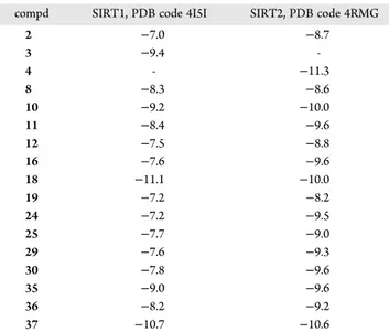
Table 3. In Silico Docking Scores of Selected Compounds
against Human SIRT1 and SIRT2
acompd SIRT1, PDB code 4I5I SIRT2, PDB code 4RMG
2 −7.0 −8.7 3 −9.4 -4 - −11.3 8 −8.3 −8.6 10 −9.2 −10.0 11 −8.4 −9.6 12 −7.5 −8.8 16 −7.6 −9.6 18 −11.1 −10.0 19 −7.2 −8.2 24 −7.2 −9.5 25 −7.7 −9.0 29 −7.6 −9.3 30 −7.8 −9.6 35 −9.0 −9.6 36 −8.2 −9.2 37 −10.7 −10.6
aBinding affinity energy values (kcal/mol) for the indicated PDB
codes were calculated using AutoDock Vina (for more details see Experimental Section). 2, 3, and 4 were used as reference inhibitors of SIRT1/2, SIRT1, and SIRT2, respectively. “-” = not determined.
Successful cancer therapy by chemotherapeutic agents but also
targeted small molecule inhibitors is frequently limited by drug
resistance mechanisms leading to treatment failure and
recur-rence. Resistance might be mediated by mutations in sensor
genes of DNA damage, like the tumor suppressor p53
33or
overexpression of pleiotropic efflux pumps for multiple drugs
causing a broad-scale multidrug resistance (MDR) phenotype.
34Hence we tested whether mutation of p53 or overexpression of
the major MDR proteins ABCB1 (P-glycoprotein) and ABCC1
(multidrug resistance protein 1, MRP1) significantly reduces the
anticancer activity of 18 (
Figure 12
). Neither loss of p53 nor
overexpression of these ABC transporters resulted in a significant
loss of activity against the investigated drug resistance models
(
Table SI-1
). In contrast, ABCB1 overexpression in SW1573
2R160 and KB-C-1 lines even resulted in a slight hypersensitivity
to 18 (
Figure 12
B and
Figure 12
D, respectively, and
Table SI-1
).
Additionally, an ovarian cancer cell model with acquired cisplatin
resistance (A2780cis) and a hydroxyurea-resistant HeLa
deriva-tive overexpressing ribonucleotide reductase model (KB-HU)
35were compared to their respective parental cell lines (
Figure 12
C
and
Figure 12
D, respectively). Also in these two cases, no
sig-nificant impact of the resistance phenotype on the cytotoxic
activity of 18 was detectable. Collectively these data demonstrate
that 18 is not susceptible to several important chemotherapy
resistance mechanisms.
Finally, the effect of 18 was tested after 24 and 48 h of
treatment on the viability of peripheral blood mononuclear cells
(PBMCs) from healthy donors (
Figure 13
). Compound 18 does
not exhibit any major acute toxicity on this normal cell model.
■
CONCLUSIONS
We prepared new series of
N-aryl-N′-3,4-dihydro-2,2-dimethyl-2H-1-benzopyran-4-yl)ureas and thioureas bearing an
alkoxycar-bonylamino group (“carbamate” group) at the 6-position of the
Figure 4. Compound 18 docked into human SIRT1 and SIRT2. (A) Docking pose of compound 18 on the crystal structure of SIRT1 (light pink; PDB code 4I5I). Close-up view on the right shows that compound 18 (lime) binds to the active site of SIRT1 structure. Hydrogen bonds are shown as dashed black lines. The residues forming hydrophobic interactions are represented as gray colored sticks. (B) Docking pose of compound 18 on the crystal structure of SIRT2 (wheat; PDB code 4RMG). Close-up view on the right shows that compound 18 (lime) binds to the active site of SIRT2 structure. The hydrogen bond is shown as dashed black lines. The residues forming hydrophobic interactions are represented as gray colored sticks.
chroman nucleus. These compounds, structurally related to
previously described 2,2-dimethylchroman derivatives acting on
glioma cells, were examined as putative anticancer agents.
Growth inhibitory activity was measured in three glioma cell lines
(U373, T98G, and Hs683), as well as on normal astrocytes.
Those data revealed that the compound bearing a cyano group at
the meta position of the phenyl ring of the 4-arylurea moiety, as
well as a bulky tert-butoxycarbonylamino group at the 6-position
(compound 18), exhibited a potent antiproliferative activity
against the tumor glial cells while having lower impact on normal
glial cells (selectivity index of >10). From the computational
docking analysis, compound 18 also showed a potent binding
affinity when compared with other derivatives as observed with
our antiproliferative activity assay. This compound was therefore
selected for further investigations to better understand its
mechanism of action. Compound 18 was assayed in the NCI-60
cell line panel screening demonstrating interesting inhibitory
potential in various cell types. 18 was also submitted to a
COMPARE analysis (results not presented) to further decipher
the mode of action of compound 18 as successfully done
pre-viously with respect to other compounds but without providing
further clues. The screening confirmed the antiproliferative
Table 4. Qualitative Molecular Docking of Compound 18
against Human SIRT1 (A) and SIRT2 (B)
a(A) SIRT1 PDB code 18 2 3 4I5I −11.1 −7.0 −9.4 4IG9 −8.5 −8.8 −7.3 4ZZH −8.5 −8.4 −7.5 4ZZI −9.9 −9.9 −8.7 4ZZJ −7.4 −9.8 −8.8 5BTR −8.4 −7.9 −6.8 average −9.0 −8.6 −8.1 (B) SIRT2 PDB code 18 2 4 4RMG −10.0 −8.7 −11.3 4RMH −10.6 −8.8 −11.5 1J8F −7.5 −9.8 −9.3 3ZGO −8.1 −10.3 −9.1 3ZGV −9.1 −9.8 −8.7 5DY4 −9.4 −11.0 −10.6 5DY5 −9.5 −7.5 −9.8 average −9.2 −9.4 −10.0
aBinding affinity energy values (kcal/mol) for the indicated PDB
codes were calculated using AutoDock Vina program (for more details seeExperimental Section). 2, 3, and 4 were used as reference inhib-itors of SIRT1/2, SIRT1, and SIRT2, respectively.
Table 5. Inhibitory Activity of Selected SIRT Inhibitors and
Compound 18 on SIRT1, -2, and -3 Activities
IC50(μM)a
compd SIRT1 SIRT2 SIRT3
1 2.8 ± 0.3 13 ± 1 >200 2 82.5 ± 7.1 47.1 ± 4.0 -3 0.10 ± 0.06 20.1 ± 4.2 -4 98.1 ± 2.4 2.8 ± 1.0 -8 >200 >200 -16 32.1 ± 6.5 18.5 ± 3.6 -18 6.2 ± 1.7 4.2 ± 1.6 >200 19 >200 99.8 ± 11.0 -25 121.2 ± 18.3 110.2 ± 18.9 -35 109.9 ± 6.1 49.4 ± 7.6 -36 108.7 ± 5.9 51.1 ± 11.1 -37 54.8 ± 8.3 22.7 ± 3.5 -aIC
50values: concentration of drug required for the inhibition of 50%
of the specified sirtuin activity [mean ± SD (n = 3 independent experiments)]. 1 and 2, 3, and 4 were used as in vitro reference inhib-itors of SIRT1/2, SIRT1, and SIRT2, respectively. “-”: not deter-mined.
Figure 5. In vitro HDAC inhibition potential of compound 18. (A) Residual inhibition of total HDAC activity. (B) Effect of 18 on activity of selected HDAC isoenzymes. Data are the mean ± SD of three independent experiments.
Figure 6. In cellulo assessment of HDAC inhibition by compound 18 through the study of histone and α-tubulin acetylation levels. Cells were treated for 8 h at the indicated concentrations of compound 18. The acetylation (Ac) of histone H4 (H4) and α-tubulin was analyzed by Western blot. Histone H1 (H1) and β-actin were used as loading controls for the analysis of acid (top panel) and total (bottom panel) extracts, respectively. 38 (2 μM) was used as a reference HDAC inhibitor.
Table 6. Growth Inhibitory Activities of Selected SIRT
Inhibitors and Compound 18 in Glioma Cell Lines
IC50(μM)a compd U373 Hs683 2 39.3 ± 5.4 33.9 ± 4.3 3 157.4 ± 23.0 115.9 ± 23.3 4 47.6 ± 4.4 80.2 ± 9.3 18 7.5 ± 1.1 5.8 ± 0.9 aIC
50 values: concentration of drug responsible for the inhibition of
50% of the growth of the specified cell line after 72 h [mean ± SEM of three independent experiments (n = 4 replicates)].
activity of 18 on a variety of cancer cell lines (mean GI
50value of
3 μM), while the COMPARE tool did not allow obtaining a clear
hypothesis about its potential mechanism of action. Quantitative
videomicroscopy analysis revealed that 18 was a cytostatic rather
than a cytotoxic agent.
Furthermore, neither mutation of p53 nor overexpression of
MDR efflux pumps including ABCB1 and ABCC1 significantly
reduced the anticancer activity of 18. The same holds true for
acquired cisplatin and hydroxyurea resistance. These data
suggest that several major drug resistance mechanisms limiting
the success of current systemic cancer therapy are not affecting
the activity of 18. Hence this substance might be especially
attractive for targeting treatment-resistant recurrences.
Then, 18 was tested against a panel of HDAC activities and
a potent inhibitory activity was observed in vitro on HDAC
SIRT1/2 and IC
50values in the low micromolar range (
Table 3
).
Presently, we cannot exclude that inhibition of other SIRT
isoforms led to growth arrest if expressed by the targeted cancer
type. This observation was validated in cellulo when 18 was
investigated on human U373 and Hs683 glioblastoma cell lines
to assess effects of deacetylase inhibition on acetylation levels of
histone and non-histone proteins. Interestingly, compound 18
acted as an in vitro SIRTi with activities commonly reported in
the literature. Importantly we demonstrated associated biological
activities (i.e., increased protein acetylation levels and important
antiproliferative properties) at concentrations in the low
micro-molar range, which is usually not the case of SIRTi published
over the past years (
Table SI-4
).
Finally, the anticancer potential of 18 was further highlighted
through the growth inhibition of multicellular glioblastoma
tumor spheroids as well as the reduction of tumor formation in
vivo using zebrafish xenografts.
In conclusion, compound 18 is new anticancer agent active on
glioma cells and expressing a cytostatic rather than a cytotoxic
activity. Moreover, this compound is a potent inhibitor of SIRT1
and -2 and constitutes a model of chemical structure opening
new perspectives in the medicinal chemistry of HDAC inhibitors
against class III.
■
EXPERIMENTAL SECTION
Chemistry. Melting points were determined on a Stuart SMP3 capillary apparatus and are uncorrected. The1H NMR spectra were
recorded on a Bruker Avance (500 MHz) instrument using DMSO-d6as
the solvent with TMS as an internal standard; chemical shifts are reported in δ values (ppm) relative to that of internal TMS. The abbreviations s = singlet, d = doublet, t = triplet, q = quadruplet,
m = multiplet, b = broad are used throughout. Elemental analyses (C, H, N, S) were realized on a Thermo Scientific FlashEA 1112 elemental analyzer and were within ±0.4% of the theoretical values. This analytical method certified a purity of ≥95% for each tested com-pound. All reactions were routinely checked by TLC on silica gel Merck 60 F254.
R/S-N-2-Chlorophenyl-N ′-(6-tert-butoxycarbonylamino-3,4-dihydro-2,2-dimethyl-2H-1-benzopyran-4-yl)urea (10). 3-Chlor-ophenyl isocyanate (0.13 g, 0.82 mmol) was added to a solution of 9 (0.2 g, 0.68 mmol) in methylene chloride (5 mL). After 1 h, the solvent was removed under vacuum; the title product was obtained from the crude product by DCVC purification: mp 192−194 °C; 1H NMR
(DMSO-d6) δ 1.25 (s, 3H, CH3), 1.36 (s, 3H, CH3), 1.43 (s, 9H, NHCOOC(CH3)3), 1.66 (dd, J = 13 Hz/11 Hz, 1H, 3-H), 2.13 (dd, J = 13 Hz/6 Hz, 3-H), 4.91 (dd, J = 16 Hz/9 Hz, 1H, 4-H), 6,65 (d, J = 8.8 Hz, 1H, 8-H), 6.97 (td, J = 7.9 Hz/1.5 Hz, 1H, 4′-H), 7.19 (d, J = 7.1 Hz, 1H, 7-H), 7.27 (td, J = 7.9 Hz/1.5 Hz, 1H, 5′-H), 7.38 (d, J = 8.4 Hz, 1H, CHNHCONHAr), 7.41−7.42 (m, 2H, 5-H/3′-H), 8.07 (s, 1H, CHNHCONHAr), 8.26 (dd, J = 8.3 Hz/1.2 Hz, 1H, 6′-H), 9.10 (s, 1H, NHCOOC(CH3)3).13C NMR (DMSO-d6) δ 24.6 (CH3), 28.1 (C(CH3)3), 28.9 (CH3), 40.0 (C-3), 42.4 (C-4), 74.7 (C-2), 78.6 (C(CH3)3), 116.7 (C-8), 117.7 (C-5), 119.7 (C-7), 120.7 (C-6′), 121.1 (C-2′), 122.5 (C-4′), 122.6 (C-4a), 127.5 (C-5′), 129.1 (C-3′), 132.1 (C-6), 136.6 (C-1′), 148.6 (C-8a), 153.0 (NHCOOC(CH3)3), 154.7
(CHNHCONHAr). Anal. (C23H28ClN3O4) theoretical: C, 61.95; H,
6.33; N, 9.42. Found: C, 61.71; H, 6.33; N, 9.44.
R/S-N-2-Cyanophenyl-N ′-(6-tert-butoxycarbonylamino-3,4-dihydro-2,2-dimethyl-2H-1-benzopyran-4-yl)urea (11). The title compound was obtained as described for 10, starting from 9 (0.2 g, 0.68 mmol) and 2-cyanophenyl isocyanate (0.12 g, 0.82 mmol): mp 206−208 °C;1H NMR (DMSO-d 6) δ 1.25 (s, 3H, CH3), 1.36 (s, 3H, CH3), 1.43 (s, 9H, NHCOOC(CH3)3), 1.69 (dd, J = 13 Hz/11 Hz, 1H, 3-H), 2.14 (dd, J = 13 Hz/6 Hz, 3-H), 4.91 (dd, J = 16 Hz/9 Hz, 1H, 4-H), 6,65 (d, J = 8.8 Hz, 1H, 8-H), 7.12 (t, J = 8 Hz, 1H, 4′-H), 7.20 (d, J = 7.3 Hz, 1H, 7-H), 7.42 (d, J = 8.8 Hz, 1H, CHNHCONHAr), 7.43 (s, 1H, 5-H), 7.61 (m, 1H, 5′-H), 7.71 (dd, J = 7.8 Hz/1.4 Hz, 3′-H), 8.22 (d, J = 8.5 Hz, 1H, 6′-H), 8.54 (s, 1H, CHNHCONHAr), 9.11 (s, 1H, NHCOOC(CH3)3). 13C NMR (DMSO-d6) δ 24.6 (CH3), 28.1 (C(CH3)3), 28.8 (CH3), 40.0 (C-3), 42.6 (C-4), 74.7 (C-2), 78.6 (C(CH3)3), 100.5 (C-2′), 116.8 (C-8), 117.0 (CN), 117.6 (C-5), 119.7 (C-7), 120.4 (C-6′), 122.2 (C-4′), 122.4 (C-4a), 132.1 (C-6), 133.0 (C-3′), 133.9 (C-5′), 142.7 (C-1′), 148.5 (C-8a), 153.0 (NHCOOC-(CH3)3), 154.4 (CHNHCONHAr). Anal. (C24H28N4O4) theoretical:
C, 66.04; H, 6.47; N, 12.84. Found: C, 66.12; H, 6.11; N, 13.26. R/S-N-2-Nitrophenyl-N ′-(6-tert-butoxycarbonylamino-3,4-di-hydro-2,2-dimethyl-2H-1-benzopyran-4-yl)urea (12). The title compound was obtained as described for 10, starting from 9 (0.2 g, 0.68 mmol) and 2-nitrophenyl isocyanate (0.13 g, 0.82 mmol): mp 218−219 °C;1H NMR (DMSO-d
6) δ 1.25 (s, 3H, CH3), 1.37 (s, 3H,
CH3), 1.42 (s, 9H, NHCOOC(CH3)3), 1.69 (dd, J = 13 Hz/11 Hz, 1H,
3-H), 2.11 (dd, J = 13 Hz/6 Hz, 3-H), 4.92 (dd, J = 16 Hz/9 Hz, 1H, Figure 7. SIRT1 or SIRT2 knock-down enhances the growth inhibitory effects of 18 in glioblastoma cells. U373 and Hs683 cell lines were transfected with the indicated siRNA. 24 h post-transfection cells were treated with compound 18 at the respective IC50values calculated from MTT assays
(Table 6), and proliferation was monitored by time-lapse videomicroscopy. Graphs are representative of three independent experiments and correspond to the mean ± SD of 16 pictures per well acquired in triplicate.
4-H), 6,65 (d, J = 8.8 Hz, 1H, 8-H), 7.16 (m, 1H, 4′-H), 7.20 (d, J = 6.4 Hz, 1H, 7-H), 7.40 (s, 1H, 5-H), 7.68 (m, 1H, 5′-H), 7.98 (d, J = 8.1 Hz, 1H, CHNHCONHAr), 8.09 (dd, J = 8.4 Hz/1.5 Hz, 1H, 6′-H), 8.45 (d, J = 8.5 Hz, 1H, 3′-H), 9.09 (s, 1H, NHCOOC(CH3)3), 9.45 (s, 1H, CHNHCONHAr). 13C NMR (DMSO-d 6) δ 24.4 (CH3), 28.1 (C(CH3)3), 29.0 (CH3), 40.0 (C-3), 42.7 (C-4), 74.7 (C-2), 78.6 (C(CH3)3), 116.7 (C-8), 117.7 (C-5), 119.6 (C-7), 121.5 (C-4′), 122.1 (C-3′), 122.4 (C-4a), 125.4 (C-6′), 132.0 (C-6), 135.0 (C-5′), 135.8 (C-1′), 136.7 (C-2′), 148.5 (C-8a), 152.9 (NHCOOC(CH3)3), 154.2
(CHNHCONHAr). Anal. (C23H28N4O6) theoretical: C, 60.52; H, 6.18;
N, 12.27. Found: C, 60.18; H, 6.17; N, 11.86.
R/S-N-2-Trifluoromethylphenyl-N′-(6-tert-butoxycarbonyl-amino-3,4-dihydro-2,2-dimethyl-2H-1-benzopyran-4-yl)urea (13). The title compound was obtained as described for 10, starting from 9 (0.2 g, 0.68 mmol) and 2-trifluoromethylphenyl isocyanate (0.15 g, 0.82 mmol): mp 239−239.5 °C;1H NMR (DMSO-d
6) δ 1.25 (s, 3H,
CH3), 1.36 (s, 3H, CH3), 1.44 (s, 9H, NHCOOC(CH3)3), 1.67
(dd, J = 13 Hz/11 Hz, 1H, 3-H), 2.12 (dd, J = 13 Hz/6 Hz, 3-H), 4.90 Figure 8. Digitized images of Hs683 oligodendroglial (A) and U373 glioblastoma (B) cells were obtained using computer-assisted phase-contrast microscopy (quantitative videomicroscopy). A global growth ratio (the GGR index) was calculated, resulting in a value that can be directly compared to the IC50value determined by the MTT colorimetric assay (the hatched horizontal line in the bottom chart, i.e.,∼10 μM (seeTable 1)). First, the global
growth (GG) is calculated for each control and each treated condition at 24, 48, and 72 h by dividing the number of cells on the last image by the number of cells on the first image. The GGR index is obtained by dividing the GG values calculated for Hs683 tumor cells treated with 18 by the GG values calculated for the control. The experiment was performed once in triplicate, and the data represent the mean ± SEM values. White, gray, and black bars represent the data obtained at 24, 48 and 72 h, respectively.
(dd, J = 16 Hz/9 Hz, 1H, 4-H), 6.64 (d, J = 8.8 Hz, 8-H), 7.15 (d, J = 7.6 Hz, 1H, 7-H), 7.21 (t, J = 7.6 Hz, 1H, 4′-H), 7.38 (d, J = 8.3 Hz, 1H, CHNHCONHAr), 7.48 (s, 1H, 5-H), 7.60 (t, J = 7.8 Hz, 1H, 5′-H), 7.63 (d, J = 7.9 Hz, 1H, 3′-H), 7.83 (s, 1H, CHNHCONHAr), 8.09 (d, J = 8.3 Hz, 1H, 6′-H), 9.12 (s, 1H, NHCOOC(CH3)3).13C NMR (DMSO-d6) δ 24.6 (CH3), 28.1 (C(CH3)3), 28.9 (CH3), 40.0 (C-3), 42.6 (C-4), 74.7 (C-2), 78.6 (C(CH3)3), 116.7 (C-8), 117.8 (C-5), 119.6 (C-7), 122.7 (C-4a), 122.8 (C-4′), 123.0−125.1 (d, J = 273 Hz, CF3), 124.8 (C-6′), 125.7 (C-3′), 127.3 (C-2′), 132.0 (C-6), 132.7 (C-5′), 137.2 (C-1′), 148.5 (C-8a), 152.9 (NHCOOC(CH3)3), 154.9 (CHNHCONHAr).
Anal. (C24H28F3N3O4) theoretical: C, 60.12; H, 5.89; N, 8.76. Found: C,
60.12; H, 5.95; N, 8.73.
R/S-N-2-Methoxyphenyl-N′-(6-tert-butoxycarbonylamino-3,4-dihydro-2,2-dimethyl-2H-1-benzopyran-4-yl)urea (14). The title compound was obtained as described for 10, starting from 9
(0.2 g, 0.68 mmol) and 2-methoxyphenyl isocyanate (0.12 g, 0.82 mmol): mp 204−205 °C;1H NMR (DMSO-d 6) δ 1.25 (s, 3H, CH3), 1.35 (s, 3H, CH3), 1.42 (s, 9H, NHCOOC(CH3)3), 1.62 (dd, J = 13 Hz/11 Hz, 1H, 3-H), 2.10 (dd, J = 13 Hz/6 Hz, 3-H), 3.83 (s, 3H, OCH3), 4.90 (dd, J = 16 Hz/9 Hz, 1H, 4-H), 6,63 (d, J = 8.8 Hz, 1H, 8-H), 6.88 (pd, J = 7.4 Hz/1.6 Hz, 2H, 4′-H/5′-H), 6.98 (dd, J = 7.7 Hz/1.6 Hz, 1H, 3′-H), 7.19 (bs, 1H, CHNHCONHAr), 7.20 (d, J = 8.5 Hz, 1H, 7-H), 7.40 (s, 1H, 5-H), 8.01 (s, 1H, CHNHCONHAr), 8.18 (dd, J = 7.5 Hz/1.9 Hz, 1H, 6′-H), 9.10 (s, 1H, NHCOOC(CH3)3).13C NMR (DMSO-d6) δ 24.6 (CH3), 28.1 (C(CH3)3), 28.9 (CH3), 40.0 (C-3), 42.2 (C-4), 55.6 (OCH3), 74.7 (C-2), 78.5 (C(CH3)3), 110.5 (C-3′), 116.6 (C-8), 117.8 (C-5/C-6′), 119.6 (C-7), 120.5 (C-5′), 121.0 (C-4′), 122.9 (C-4a), 129.4 (C-1′), 132.0 (C-6), 147.2 (C-2′), 148.6 (C-8a), 153.0 (NHCOOC-(CH3)3), 155.0 (CHNHCONHAr). Anal. (C24H31N3O5) theoretical: C,
65.29; H, 7.08; N, 9.52. Found: C, 65.20; H, 7.09; N, 9.51.
Figure 9. Compound 18 induces G1 cell cycle arrest in glioblastoma cell lines and promotes the accumulation of senescent U373 cells. (A) Cells were treated, or not, with 18, 2, 3, 4 (at the respective IC50values for each cell lines reported inTable 6) and 2 μM 38. After 24 and 48 h of exposure, cell cycle
distribution was analyzed. Histograms correspond to the mean ± SD of the quantification of three independent experiments.∗, ∗∗ indicate p < 0.05, p < 0.01 compared to control cells, respectively. (B) Cells were incubated in a medium containing DMSO as a vehicle control or 5 μM 18 for 6 days and were stained for β-galactosidase activity. Representative pictures of two independent experiments are depicted where SA-β-gal-positive cells are revealed by blue staining. The percentage of senescent cells, expressed as a percentage of the total number of cells counted, is indicated for each picture. Data represent the mean ± SD of two independent experiments.
R/S-N-3-Fluorophenyl-N ′-(6-tert-butoxycarbonylamino-3,4-dihydro-2,2-dimethyl-2H-1-benzopyran-4-yl)urea (15). The title compound was obtained as described for 10, starting from 9 (0.2 g, 0.68 mmol) and 3-fluorophenyl isocyanate (0.11 g, 0.82 mmol): mp 115−125 °C;1H NMR (DMSO-d 6) δ 1.24 (s, 3H, CH3), 1.35 (s, 3H, CH3), 1.42 (s, 9H, NHCOOC(CH3)3), 1.70 (dd, J = 13 Hz/11 Hz, 1H, 3-H), 2.08 (dd, J = 13 Hz/6 Hz, 3-H), 4.92 (dd, J = 16 Hz/10 Hz, 1H, 4-H), 6.55 (d, J = 8.6 Hz, 1H, CHNHCONHAr), 6,64 (d, J = 8.7 Hz, 1H, 8-H), 6.73 (t, J = 7.6 Hz, 1H, 4′-H), 7.06 (d, J = 7.8 Hz, 1H, 6′-H), 7.17 (d, J = 7.1 Hz, 1H, 7-H), 7.26 (dd, J = 15.2 Hz/7.7 Hz, 1H, 5′-H), 7.41 (s, 1H, 5-H), 7.53 (d, J = 12 Hz, 1H, 2′-H), 8.73 (s, 1H, CHNHCONHAr), 9.11 (s, 1H, NHCOOC(CH3)3).13C NMR (DMSO-d6) δ 24.4 (CH3), 28.1 (C(CH3)3), 29.0 (CH3), 40.0 (C-3), 42.3 (C-4), 74.8 (C-2), 78.5 (C(CH3)3), 104.4 (d, J = 27 Hz, C-2′), 107.4 (d, J = 21 Hz, C-4′), 113.5 (d, J = 2 Hz, C-6′), 116.7 (C-8), 117.6 (C-5), 119.6 (C-7), 122.9 (C-4a), 130.1 (d, J = 10 Hz, C-5′), 132.0 (C-6), 142.2 (d, J = 12 Hz, C-1′), 148.5 (C-8a), 152.9 (NHCOOC(CH3)3), 154.9 (CHNHCONHAr), 161.5−
163.4 (d, J = 240 Hz, C-3′). Anal. (C23H28FN3O4) theoretical: C, 64.32;
H, 6.57; N, 9.78. Found: C, 64.68; H, 6.26; N, 10.09.
R/S-N-3-Bromophenyl-N ′-(6-tert-butoxycarbonylamino-3,4-dihydro-2,2-dimethyl-2H-1-benzopyran-4-yl)urea (17). The title compound was obtained as described for 10, starting from 9 (0.2 g, 0.68 mmol) and 3-bromophenyl isocyanate (0.16 g, 0.82 mmol): mp 209.5−210.5 °C;1H NMR (DMSO-d 6) δ 1.24 (s, 3H, CH3), 1.35 (s, 3H, CH3), 1.42 (s, 9H, NHCOOC(CH3)3), 1.71 (dd, J = 13 Hz/11 Hz, 1H, 3-H), 2.08 (dd, J = 13 Hz/6 Hz, 3-H), 4.92 (dd, J = 16 Hz/10 Hz, 1H, 4-H), 6.57 (d, J = 8.6 Hz, 1H, CHNHCONHAr), 6,63 (d, J = 8.7 Hz, 1H, 8-H), 7.09 (d, J = 8.5 Hz, 1H, 4′-H), 7.16 (d, J = 7.1 Hz, 1H, 7-H), 7.20 (t, J = 8 Hz, 1H, 5′-H), 7.26 (d, J = 8.7 Hz, 1H, 6′-H), 7.40 (s, 1H, 5-H), 7.90 (t, J = 1.9 Hz, 1H, 2′-H), 8.71 (s, 1H, CHNHCONHAr), 9.09 (s, 1H, NHCOOC(CH3)3).13C NMR (DMSO-d6) δ 24.4 (CH3), 28.1 (C(CH3)3), 29.0 (CH3), 40.0 (C-3), 42.4 (C-4), 74.8 (C-2), 78.5 (C(CH3)3), 116.6 (C-8/C-6′), 117.5 (C-5), 119.6 (C-7), 120.1 (C-2′), 121.7 (C-3′), 122.9 (C-4a), 123.7 (C-4′), 130.6 (C-5′), 132.0 (C-6), 142.0 (C-1′), 148.5 (C-8a), 152.9 (NHCOOC(CH3)3), 154.9
(CHNHCONHAr). Anal. (C23H28BrN3O4) theoretical: C, 56.33; H,
5.76; N, 8.57. Found: C, 56.40; H, 5.83; N, 8.44.
R/S-N-3-Nitrophenyl-N′-(6-tert-butoxycarbonylamino-3,4-di-hydro-2,2-dimethyl-2H-1-benzopyran-4-yl)urea (19). The title compound was obtained as described for 10, starting from 9 (0.2 g, 0.68 mmol) and 3-nitrophenyl isocyanate (0.13 g, 0.82 mmol): mp 120−155 °C;1H NMR (DMSO-d 6) δ 1.25 (s, 3H, CH3), 1.36 (s, 3H, CH3), 1.41 (s, 9H, NHCOOC(CH3)3), 1.76 (dd, J = 13 Hz/11 Hz, 1H, 3-H), 2.09 (dd, J = 13 Hz/6 Hz, 3-H), 4.95 (dd, J = 16 Hz/10 Hz, 1H, 4-H), 6.64 (d, J = 8.8 Hz, 1H, 8-H), 6.71 (d, J = 8.7 Hz, 1H, CHNHCONHAr), 7.17 (bs, 1H, 7-H), 7.41 (s, 1H, 5-H), 7.53 (t, J = 8.2 Hz, 1H, 5′-H), 7.69 (d, J = 7.8 Hz, 1H, 4′-H), 7.78 (d, J = 8.1 Hz, 1H, 6′-H), 8.61 (t, J = 2 Hz, 1H, 2′-H), 9.09 (s, 2H, CHNHCONHAr/ NHCOOC(CH3)3). 13C NMR (DMSO-d6) δ 24.4 (CH3), 28.1 (C(CH3)3), 29.1 (CH3), 40.0 (C-3), 42.5 (C-4), 74.8 (C-2), 78.5 (C(CH3)3), 111.7 (C-2′), 115.7 (C-6′), 116.7 (C-8), 117.5 (C-5), 119.6 (C-7), 122.8 (C-4a), 123.9 (C-4′), 129.9 (C-5′), 132.0 (C-6), 141.7 (C-1′), 148.1 (C-3′), 148.5 (C-8a), 152.9 (NHCOOC(CH3)3), 154.9
(CHNHCONHAr). Anal. (C23H28N4O6) theoretical: C, 60.52; H, 6.18;
N, 12.27. Found: C, 61.01; H, 6.13; N, 12.18.
R/S-N-3-Trifluoromethylphenyl-N′-(6-tert-butoxycarbonyl-amino-3,4-dihydro-2,2-dimethyl-2H-1-benzopyran-4-yl)urea (20). The title compound was obtained as described for 10, starting from 9 (0.2 g, 0.68 mmol) and 3-trifluoromethylphenyl isocyanate (0.15 g, 0.82 mmol): mp 140−155 °C;1H NMR (DMSO-d
6) δ 1.25 (s, 3H,
CH3), 1.36 (s, 3H, CH3), 1.42 (s, 9H, NHCOOC(CH3)3), 1.74 (dd,
J = 13 Hz/11 Hz, 1H, 3-H), 2.09 (dd, J = 13 Hz/6 Hz, 3-H), 4.94 (dd, Figure 10. continued
J = 16 Hz/10 Hz, 1H, 4-H), 6.62 (d, J = 8.6 Hz, 1H, CHNHCONHAr), 6.64 (d, J = 8.8 Hz, 1H, 8-H), 7.17 (bd, J = 6.7 Hz, 1H, 7-H), 7.26 (d, J = 7.5 Hz, 1H, 4′-H), 7.41 (s, 1H, 5-H), 7.47 (t, J = 7.9 Hz, 1H, 5′-H), 7.53 (d, J = 8.2 Hz, 1H, 6′-H), 8.07 (s, 1H, 2′-H), 8.89 (s, 1H, CHNHCONHAr), 9.08 (s, 1H, NHCOOC(CH3)3). 13C NMR (DMSO-d6) δ 24.4 (CH3), 28.1 (C(CH3)3), 29.1 (CH3), 40.0 (C-3), 42.4 (C-4), 74.8 (C-2), 78.5 (C(CH3)3), 113.8 (C-2′), 116.6 (C-8), 117.4 (C-5/C-4′), 119.6 (C-7), 121.3 (C-6′), 122.9 (C-4a), 123.2− 125.4 (d, J = 272 Hz, CF3), 129.3−129.6 (d, J = 31 Hz, C-3′), 129.7 (C-5′), 132.0 (C-6), 141.2 (C-1′), 148.5 (C-8a), 153.0 (NHCOOC-(CH3)3), 155.0 (CHNHCONHAr). Anal. (C24H28F3N3O4) theoretical:
C, 60.12; H, 5.89; N, 8.76. Found: C, 60.19; H, 6.03; N, 8.59. R/S-N-3-Methoxyphenyl-N ′-(6-tert-butoxycarbonylamino-3,4-dihydro-2,2-dimethyl-2H-1-benzopyran-4-yl)urea (21). The title compound was obtained as described for 10, starting from 9 (0.2 g,
0.68 mmol) and 3-methoxyphenyl isocyanate (0.12 g, 0.82 mmol): mp 189−191 °C;1H NMR (DMSO-d 6) δ 1.24 (s, 3H, CH3), 1.35 (s, 3H, CH3), 1.42 (s, 9H, NHCOOC(CH3)3), 1.68 (dd, J = 13 Hz/11 Hz, 1H, 3-H), 2.08 (dd, J = 13 Hz/6 Hz, 3-H), 3.72 (s, 1H, OCH3), 4.91 (dd, J = 16 Hz/10 Hz, 1H, 4-H), 6.46 (d, J = 8.7 Hz, 1H, CHNHCONHAr), 6.50 (dd, J = 8.2 Hz/1.9 Hz, 1H, 4′-H), 6.63 (d, J = 8.8 Hz, 1H, 8-H), 6.89 (dd, J = 8.1 Hz/1 Hz, 1H, 6′-H), 7.13 (t, J = 8 Hz, 1H, 5′-H), 7.17 (d, J = 7.8 Hz, 1H, 7-H), 7.22 (t, J = 2 Hz, 1H, 2′-H), 7.40 (s, 1H, 5-H), 8.52 (s, 1H, CHNHCONHAr), 9.08 (s, 1H, NHCOOC(CH3)3).13C NMR (DMSO-d6) δ 24.5 (CH3), 28.1 (C(CH3)3), 29.0 (CH3), 40.0 (C-3), 42.3 (C-4), 54.8 (OCH3), 74.8 (C-2), 78.5 (C(CH3)3), 103.4 (C-2′), 106.7 (C-4′), 110.1 (C-6′), 116.6 (C-8), 117.6 (C-5), 119.6 (C-7), 123.0 (C-4a), 129.4 (C-5′), 132.0 (C-6), 141.6 (C-1′), 148.6 (C-8a), 153.0 (NHCOOC(CH3)3), 155.0 (CHNHCONHAr), 159.7 (C-3′).
Figure 10. Antitumor potential of compound 18. (A) Representative bright field images and 3D plots of spheroids generated from Hs683 cells (top panels). Cells were pretreated for 24 h with the indicated concentrations of 18 and then left untreated for 96 h (4-day incubation, upper left panel), or untreated cells were cultured for 96 h in the presence of the indicated concentrations of 18 (24 h pretreated cells, upper right panel). The surface, volume, and ATP levels of spheroids were measured as described in theExperimental Section(bottom panels). Data are the mean ± SD of three independent experiments. (B) Fluorescent Hs683 and U373 cells were treated, or not, in vitro at 5 or 10 μM compound 18 for 30 or 36 h and then injected in the zebrafish yolk sac. The figure represents results obtained with a pretreatment of 36 h at 10 μM; for the other conditions of treatment we refer toFigure SI-5. After 72 h, fluorescence was quantified and representative images from a total of eight fish per condition are shown. For each fish, the upper panel represents the bright field, the middle panel the red fluorescence, and the bottom panel the merged picture. Fluorescence intensity quantification graphs are shown. PBS injection was used as a control for injection side effects.∗, ∗∗, ∗∗∗ indicate p < 0.05, p < 0.01, p < 0.05 compared to control cells, respectively.
Anal. (C24H31N3O5) theoretical: C, 65.29; H, 7.08; N, 9.52. Found: C,
65.25; H, 7.13; N, 9.43.
R/S-N-3-Aminomethylphenyl-N ′-(6-tert-butoxycarbonyl-amino-3,4-dihydro-2,2-dimethyl-2H-1-benzopyran-4-yl)urea (22). Raney nickel was added to a solution of 18 (0.3 g, 0.69 mmol) and ammonia (0.3 mL) in ethanol (10 mL). The solution was stirred in a sealed hydrogenator under a hydrogen pressure of 7 bar. When the reaction was ended (3 h), the catalyst was filtered off and the filtrate was evaporated under pressure. The title compound was obtained after purification on a DCVC system: mp 120−170 °C;1H
NMR (DMSO-d6) δ 1.24 (s, 3H, CH3), 1.35 (s, 3H, CH3), 1.42 (s, 9H, NHCOOC(CH3)3), 1.68 (dd, J = 13 Hz/11 Hz, 1H, 3-H), 2.09 (dd, J = 13 Hz/6 Hz, 3-H), 3.3 (bs, 2H, CH2NH2), 3.67 (s, 2H, CH2NH2), 4.91 (dd, J = 16 Hz/10 Hz, 1H, 4-H), 6.47 (d, J = 8.7 Hz, 1H, CHNHCONHAr), 6.63 (d, J = 8.8 Hz, 1H, 8-H), 6.89 (d, J = 7.5 Hz, 1H, 4′-H), 7.17 (m, 2H, 7-H/5′-H), 7.31 (d, J = 8.2 Hz, 1H, 6′-H), 7.37 (s, 1H, 2′-H), 7.41 (s, 1H, 5-H), 8.46 (s, 1H, CHNHCONHAr), 9.08 (s, 1H, NHCOOC(CH3)3).13C NMR (DMSO-d6) δ 24.5 (CH3), 28.1 (C(CH3)3), 29.0 (CH3), 40.0 (C-3), 42.3 (C-4), 45.7 (CH2NH2), 74.8 (C-2), 78.5 (C(CH3)3), 115.8 (C-6′), 116.5 (C-2′), 116.7 (C-8), 117.5 (C-5), 119.6 (C-7), 120.0 (C-4′), 123.0 (C-4a), 128.4 (C-5′), 132.0 (C-6), 140.2 (C-1′), 144.5 (C-3′), 148.6 (C-8a), 153.0
(NHCOOC(CH3)3), 155.1 (CHNHCONHAr). Anal. (C24H32N4O4)
theoretical: C, 65.43; H, 7.32; N, 12.72. Found: C, 64.96; H, 7.31; N, 12.70.
R/S-N-3-Aminophenyl-N ′-(6-tert-butoxycarbonylamino-3,4-dihydro-2,2-dimethyl-2H-1-benzopyran-4-yl)urea (23). Ammo-nium chloride (0.15 g, 2.83 mmol) and iron (0.6 g, 10,7 mmol) were added to a solution of 19 (0.3 g, 0.66 mmol) in ethanol/water 3:1 (10 mL). The solution was heated at 80 °C for 15 min. After filtration (without cooling), the filtrate was treated with charcoal, filtered, and evaporated under vacuum. The title compound was obtained after puri-fication on a DCVC system: mp 130−140 °C;1H NMR (DMSO-d
6) δ 1.24 (s, 3H, CH3), 1.35 (s, 3H, CH3), 1.43 (s, 9H, NHCOOC(CH3)3), 1.65 (dd, J = 13 Hz/11 Hz, 1H, 3-H), 2.08 (dd, J = 13 Hz/6 Hz, 3-H), 4.89 (dd, J = 16 Hz/10 Hz, 1H, 4-H), 4.95 (s, 1H, NH2), 6.14 (dd, J = 7.9 Hz/1.3 Hz, 1H, 4′-H), 6.35 (d, J = 8.7 Hz, 1H, CHNHCONHAr), 6.57 (dd, J = 8.1 Hz/1 Hz, 1H, 6′-H), 6.63 (d, J = 8.8 Hz, 1H, 8-H), 6.72 (t, J = 1.9 Hz, 1H, 2′-H), 6.85 (t, J = 8 Hz, 1H, 5′-H), 7.17 (d, J = 6.6 Hz, 1H, 7-H), 7.39 (s, 1H, 5-H), 8.17 (s, 1H, CHNHCONHAr), 9.10 (s, 1H, NHCOOC(CH3)3). 13C NMR (DMSO-d6) δ 24.6 (CH3), 28.1 (C(CH3)3), 29.0 (CH3), 40.0 (C-3), 42.2 (C-4), 74.8 (C-2), 78.5 (C(CH3)3), 103.4 (C-2′), 105.8 (C-6′), 107.6 (C-4′), 116.6 (C-8), 117.7 (C-5), 119.6 (C-7), 123.1 (C-4a), 128.9 (C-5′), 132.0 (C-6), 140.9 (C-1′), 148.6 (C-8a), 149.0 (C-3′), 153.0 (NHCOOC(CH3)3),
154.9 (CHNHCONHAr). Anal. (C23H30N4O4) theoretical: C, 64.77;
H, 7.09; N, 13.14. Found: C, 64.32; H, 7.06; N, 12.95.
R/S-N-4-Nitrophenyl-N ′-(6-tert-butoxycarbonylamino-3,4-di-hydro-2,2-dimethyl-2H-1-benzopyran-4-yl)urea (26). The title compound was obtained as described for 10, starting from 9 (0.2 g, 0.68 mmol) and 4-nitrophenyl isocyanate (0.13 g, 0.82 mmol): mp 233−233.5 °C;1H NMR (DMSO-d 6) δ 1.25 (s, 3H, CH3), 1.36 (s, 3H, CH3), 1.42 (s, 9H, NHCOOC(CH3)3), 1.75 (dd, J = 13 Hz/11 Hz, 1H, 3-H), 2.10 (dd, J = 13 Hz/6 Hz, 3-H), 4.94 (dd, J = 16 Hz/9 Hz, 1H, 4-H), 6.64 (d, J = 8.8 Hz, 1H, 8-H), 6.82 (d, J = 8.6 Hz, 1H, CHNHCONHAr), 7.18 (d, J = 6.5 Hz, 1H, 7-H), 7.40 (s, 1H, 5-H), 7.69 (d, J = 7.7 Hz, 2H, 2′-H/6′-H), 8.17 (d, J = 9.3 Hz, 2H, 3′-H/5′-H), 9.09 (s, 1H, NHCOOC(CH3)3), 9.32 (s, 1H, CHNHCONHAr).13C NMR (DMSO-d6) δ 24.4 (CH3), 28.1 (C(CH3)3), 29.0 (CH3), 40.4 (C-3), 42.5 (C-4), 74.8 (C-2), 78.6 (C(CH3)3), 116.7 (C-8), 117.0 (C-2′/C-6′), 117.5 (C-5), 119.6 (C-7), 122.5 (C-4a), 125.1 (C-3′/C-5′), 132.1 (C-6), 140.5 (C-1′), 147.0 (C-4′), 148.5 (C-8a), 152.9 (NHCOOC(CH3)3),
154.4 (CHNHCONHAr). Anal. (C23H28N4O6) theoretical: C, 60.52;
H, 6.18; N, 12.27. Found: C, 59.95; H, 6.16; N, 12.09.
R/S-N-4-Trifluoromethylphenyl-N′-(6-tert-butoxycarbonyl-amino-3,4-dihydro-2,2-dimethyl-2H-1-benzopyran-4-yl)urea (27). The title compound was obtained as described for 10, starting from 9 (0.2 g, 0.68 mmol) and 4-trifluoromethylphenyl isocyanate (0.15 g, 0.82 mmol): mp 186−188 °C;1H NMR (DMSO-d 6) δ 1.25 (s, 3H, CH3), 1.36 (s, 3H, CH3), 1.42 (s, 9H, NHCOOC(CH3)3), 1.72 (dd, J = 13 Hz/11 Hz, 1H, 3-H), 2.09 (dd, J = 13 Hz/6 Hz, 3-H), 4.94 (dd, J = 16 Hz/9 Hz, 1H, 4-H), 6.63−6.66 (m, 2H, CHNHCONHAr/8-H), 7.18 (d, J = 7 Hz, 1H, 7-H), 7.40 (s, 1H, 5-H), 7.59 (d, J = 8.8 Hz, 2H, 3′-H/5′-H), 7.65 (d, J = 8.8 Hz, 1H, 2′-H/6′-H), 8.95 (s, 1H, CHNHCONHAr), 9.09 (s, 1H, NHCOOC(CH3)3). 13C NMR (DMSO-d6) δ 24.4 (CH3), 28.1 (C(CH3)3), 29.0 (CH3), 40.4 (C-3), 42.4 (C-4), 74.8 (C-2), 78.5 (C(CH3)3), 116.7 (C-8), 117.4 (C-5/C-2′/ C-6′), 119.6 (C-7), 121.0−121.2 (d, J = 32 Hz, C-4′), 122.8 (C-4a), 123.5−125.7 (d, J = 271 Hz, CF3), 126.0 (C-3′/C-5′), 132.0 (C-6), 144.1 (C-1′), 148.6 (C-8a), 153.0 (NHCOOC(CH3)3), 154.8
(CHNHCONHAr). Anal. (C24H28F3N3O4) theoretical: C, 60.12; H,
5.89; N, 8.76. Found: C, 60.08; H, 5.97; N, 8.72.
R/S-N-4-Methoxyphenyl-N ′-(6-tert-butoxycarbonylamino-3,4-dihydro-2,2-dimethyl-2H-1-benzopyran-4-yl)urea (28). The title compound was obtained as described for 10, starting from 9 (0.2 g, 0.68 mmol) and 4-methoxyphenyl isocyanate (0.12 g, 0.82 mmol): mp 204−205 °C;1H NMR (DMSO-d 6) δ 1.24 (s, 3H, CH3), 1.35 (s, 3H, CH3), 1.43 (s, 9H, NHCOOC(CH3)3), 1.68 (dd, J = 13 Hz/11 Hz, 1H, 3-H), 2.06 (dd, J = 13 Hz/6 Hz, 3-H), 3.70 (s, 3H, OCH3), 4.91 (dd, J = 16 Hz/9 Hz, 1H, 4-H), 6.47 (d, J = 8.7 Hz, 1H, CHNHCONHAr), 6,62 (d, J = 8.8 Hz, 1H, 8-H), 6.83 (d, J = 9 Hz, 2H, 3′-H/5′-H), 7.16 (d, J = 7.2 Hz, 7-H), 7.35 (d, J = 9 Hz, 2H, 2′-H/6′-H), 7.40 (s, 1H, 5-H), 8.41 Figure 11. Characterization of the growth inhibitory effects exerted by
compound 18 in the NCI 60-cell-line panel (National Cancer Institute Developmental Therapeutics Program, mean graphs).
(s, 1H, CHNHCONHAr), 9.08 (s, 1H, NHCOOC(CH3)3).13C NMR
(DMSO-d6) δ 24.4 (CH3), 28.1 (C(CH3)3), 29.1 (CH3), 40.0 (C-3),
42.3 (C-4), 55.1 (OCH3), 74.8 (C-2), 78.5 (C(CH3)3), 113.8 (C-3′/
C-5′), 116.6 (C-8), 117.7 (C-5), 119.5 (C-2′/C-6′), 119.6 (C-7), 123.3 (C-4a), 132.0 (C-6), 133.6 (C-1′), 148.6 (C-8a), 153.0 (NHCOOC-(CH3)3), 154.0 (CHNHCONHAr), 155.4 (C-4′). Anal. (C24H31N3O5)
theoretical: C, 65.29; H, 7.08; N, 9.52. Found: C, 64.88; H, 7.20; N, 9.13. R/S-N-3-Cyanophenyl-N ′-(3,4-dihydro-2,2-dimethyl-2H-1-benzopyran-4-yl)urea (32). The title compound was obtained as described for 10, starting from 31 (0.2 g, 1.13 mmol) and 3-cyanophenyl isocyanate (0.18 g, 1.25 mmol): mp 217−219 °C;1H NMR (DMSO-d
6) δ 1.28 (s, 3H, CH3), 1.39 (s, 3H, CH3), 1.77 (t, J = 12 Hz, 1H, 3-H), 2.12 (dd, J = 13 Hz/6.2 Hz, 3-H), 4.98 (dd, J = 16.3 Hz/9.2 Hz, 1H, 4-H), 6.74 (d, J = 8.7 Hz, 1H, 8-H), 6.76 (d, J = 10 Hz, 1H, CHNHCONHAr), 6.88 (t, J = 7.4 Hz, 1H, 6-H), 7.14 (t, J = 7.6 Hz, 1H, 7-H), 7.26 (d, J = 7.6 Hz, 1H, 5-H), 7.37 (d, J = 7.5 Hz, 1H, 4′-H), 7.46 (t, J = 8 Hz, 1H, 5′-H), 7.64 (d, J = 8.3 Hz, 1H, 6′-H), 8.00 (s, 1H, 2′-H), 8.92 (s, 1H, CHNHCONHAr).13C NMR (DMSO-d 6) δ 25.0 (CH3), 29.6 (CH3), 40.0 (C-3), 42.8 (C-4), 75.6 (C-2), 117.2 (C-8), 119.4 (CN), 120.4 (C-6), 120.8 (C-2′), 122.9 (C-6′), 123.7 (C-4a), 125.1 (C-4′), 128.0 (C-5), 129.0 (C-7), 130.6 (C-5′), 141.8 (C-1′), 153.8 (C-8a), 155.5 (CHNHCONHAr). Anal. (C19H19N3O2) theoretical: C, 71.01; H, 5.96;
N, 13.08. Found: C, 70.90; H, 5.97; N, 12.83.
R/S-N-3-Cyanophenyl-N ′-(6-amino-3,4-dihydro-2,2-dimeth-yl-2H-1-benzopyran-4-yl)urea (33). A suspension of 18 (2.85 g, 6.5 mmol) in a 5 N hydrochloric acid ethanolic solution (100 mL) was heated for 5 min. The mixture was then poured into water (200 mL). The title compound was precipitated by addition of a 20% aqueous solution of sodium hydroxide until pH = 10, filtered, washed with water, and dried: mp 176−178 °C;1H NMR (DMSO-d
6) δ 1.21 (s, 3H, CH3), 1.32 (s, 3H, CH3), 1.67 (dd, J = 13 Hz/11 Hz, 1H, 3-H), 2.03 (dd, J = 13 Hz/6.3 Hz, 3-H), 4.57 (s, 2H, NH2), 4.87 (dd, J = 15.9 Hz/9.7 Hz, 1H, 4-H), 6.40 (dd, J = 8.5 Hz/2.5 Hz, 1H, 7-H), 6.45 (d, J = 8.5 Hz, 1H, 8-H), 6.51 (d, J = 2.2 Hz, 1H, 5-H), 6.63 (d, J = 8.7 Hz, 1H, CHNHCONHAr), 7.36 (d, J = 7.7 Hz, 1H, 4′-H), 7.45 (t, J = 8 Hz, 1H, 5′-H), 7.63 (d, J = 8.3 Hz, 1H, 6′-H), 8.00 (s, 1H, 2′-H), 8.86 (s, 1H, CHNHCONHAr).13C NMR (DMSO-d 6) δ 24.8 (CH3), 29.6 (CH3), 40.8 (C-3), 42.9 (C-4), 74.4 (C-2), 112.0 (C-3′), 112.7 (C-5), 115.7 (C-7), 117.4 (C-8), 119.5 (CN), 120.7 (C-2′), 122.8 (C-6′), 123.4 (C-4a), 125.1 (C-4′), 130.6 (C-5′), 141.8 (C-6), 142.2 (C-1′), 144.8 (C-8a), 155.4 (CHNHCONHAr). Anal. (C19H20N4O2) theoretical: C,
67.84; H, 5.99; N, 16.66. Found: C, 67.47; H, 6.03; N, 16.53. R/S-N-3-Cyanophenyl-N ′-(6-formamido-3,4-dihydro-2,2-di-methyl-2H-1-benzopyran-4-yl)urea (34). A mixture of acetic anhydride (2 mL, 21.2 mmol) and formic acid (1 mL, 26.5 mmol) was heated at 55 °C for 2 h. The mixture was then cooled to 0 °C, and anhydrous THF (2 mL) was added. A solution of 33 (0.3 g, 0.89 mmol) in anhydrous THF was added dropwise. When the reaction was complete, water was added and the resulting precipitate was filtered, washed with water, and dried. The title product was obtained from the crude product by DCVC purification: mp 200−202 °C; 1H NMR
(DMSO-d6) δ 1.26 (s, 3H, CH3), 1.37 (s, 3H, CH3), 1.76 (t, J = 12 Hz, 1H, 3-H), 2.09 (dd, J = 13 Hz/6.2 Hz, 3-H), 4.96 (dd, J = 15.3 Hz/ 10.3 Hz, 1H, 4-H), 6.70 (d, J = 8.7 Hz, 1H, 8-H), 6.80 (d, J = 8.7 Hz, 1H, CHNHCONHAr), 7.37 (d, J = 7.7 Hz, 1H, 4′-H), 7.42−7.48 (m, 3H, 5-H/7-H/5′-H), 7.66 (m, 1H, 6′-H), 8.01 (s, 1H, 2′-H), 8.15 (d, J = 2 Hz, 1H, NHCHO), 8.96 (s, 1H, CHNHCONHAr), 10.00 (s, 1H, NHCHO). 13C NMR (DMSO-d 6) δ 24.4 (CH3), 29.1 (CH3), 40.0 (C-3), 42.3 (C-4), 75.1 (C-2), 111.5 (C-3′), 116.8 (C-8), 118.3 (C-5), 119.0 (CN), 120.2 (C-7), 120.3 (C-2′), 122.4 (C-6′), 123.2 (C-4a), 124.0 (), 124.7 (C-4′), 130.1 (C-5′), 131.0 (C-6), 141.2 (C-1′), 149.5 Figure 12. Impact of clinically significant drug resistance mechanisms on the anticancer activity of 18. (A) HCT-116 p53wt and HCT-116 p53ko (p53 knockout), (B) SW1573, SW1573 2R120 (ABCC1- and LRP-overexpressing) and 2R160 (ABCB1-overexpressing), (C) A2780 and A2780cis (cisplatin resistant), and (D) KB-3-1, KB-C-1 (ABCB1-overexpressing) and KB-HU (ribonucleotide reductase subunit 2-overexpressing) were treated with the indicated concentrations of compound 18. After 72 h of continuous drug exposure, cell viability was determined by MTT assay. The experiments were performed in quintuplicate, and results are shown as the mean ± SD.
Figure 13. Effect of compound 18 on the viability of healthy peripheral blood mononuclear cells. Histograms correspond to the mean ± SD of three independent experiments. ∗, ∗∗ indicate p < 0.05, p < 0.01 compared to control cells, respectively.