Electrocardiographic changes during dobutamine stress testing in patients with recent myocardial infarction: Relation with residual infarct artery stenosis and functional recovery
6
0
0
Texte intégral
(2) 12. P. Lancellotti et al.. artery and 2) for predicting the occurrence of contractile recovery.. Methods STUDY. PATIENTS. A total of 121 consecutive patients admitted for a first AMI were prospectively recruited for the study. AMI was diagnosed by an increase in creatine kinase and creatine kinase MB myocardial enzymes to at least twice the normal values in the clinical setting of typical chest pain lasting more than 30 minutes and acute ST-segment elevation. No contraindications to graded dobutamine infusion were present: post-infarction angina, heart failure, uncontrolled systemic hypertension or ventricular arrhythmias. All patients underwent resting echocardiography, dobutamine-stress testing and coronary angiography. Twenty-seven patients were excluded because of an uninterpretable dobutamine test: the target heart rate was not reached in 17, severe arrhythmias occurred in 3 and 7 patients had a noninterpretable ECG. The final study population consisted of the 94 remaining patients (82 men and 12 women), with a mean age of 59 ± 12 years. The infarct ECG location was anterior in 35 patients, lateral in 6, and inferior in 53. The mean peak level of serum creatine kinase was 2,107±976 IU/l. Fifty-nine patients (63%) were treated with thrombolytic therapy but none underwent primary angioplasty of the infarct-related vessel. The study was approved by the Research Ethics Committee of our institution; informed consent was obtained in all patients.. STRESS. TEST. Stress dobutamine-ECG was performed 4±2 days after admission. Beta-adrenergic-blocking agents were withdrawn 24 hours before the test. A 12-lead ECG was continuously monitored and recorded every minute. Blood pressure was measured at each stage by an arm-cuff sphygmomanometer. Dobutamine was administered in doses of 5 and 10 µg/kg/min for 3 minutes each, followed by increments of 10 µg/kg/min every 2 minutes, up to a maximal dose of 40 µg/kg/min. Atropine (0.25mg to a maximum of 1mg) was added if the target heart rate (85% of maximum predicted heart rate) was not reached with dobutamine alone. End points of the test were: target heart rate, limiting chest pain and significant (≥2 mm) ST-segment depression. A follow-up resting echocardiogram was recorded 1 month later in all patients and contractile recovery was defined to be present when wall thickening increased by at least 1 score in 2 contiguous segments of the affected area.. ELECTROCARDIOGRAPHIC. ANALYSIS. ST-segment, T-waves and QT characteristics were analysed in the infarct-related ECG leads, as previously described7,9. During stress test, ST-segment elevation was defined as new or worsening ≥ 1 mm, 80 ms after J point, in ≥ 2 contiguous infarct-related leads. T-wave normalization was defined as present when inverted T-waves at rest became upright during stress in ≥2 infarct-related leads. QT interval was measured from the onset of QRS to the end of T-wave. QT dispersion was defined as the difference between the maximum and minimum QT intervals. QT dispersion was calculated at baseline, at low-dose dobutamine and at peak stress. The QT dispersion differences between low-dose dobutamine and rest and between high and low-dose dobutamine were also calculated. Since QT dispersion does not change with heart rate, heart ratecorrected QT dispersion was not calculated10.. CORONARY. ANGIOGRAPHY. Quantitative coronary angiography and left ventriculography were performed in all patients within 1 week after the dobutamine test. Quantitative measurements of coronary stenoses were performed with the use of the CAAS system (Cardiovascular Angiography Analysis System, Philips Integris-Medical System, Eindhoven, The Netherlands). Mean global left ventricular ejection fraction was 47±15%. End-diastolic volume and end-systolic volume were 102±39 ml/m2 and 61±42 ml/m2, respectively. Significant stenosis of the infarct-related artery (≥ 50%) was observed in 76 patients. Persistent occlusion of the infarct-related artery was observed in 14 patients. Mean residual stenosis of a patent infarct-related artery was 70±19%. Elective angioplasty of the infarct-related artery was performed in 56 (60%) patients who had ≥ 60% stenosis. The results of stress testing were not used for the decision to perform the procedure. Fifteen patients had multivessel disease (double-vessel disease in 9 and triple-vessel disease in 6).. STATISTICAL. ANALYSIS. Data are expressed as mean ±SD. Student’s t test was used to assess differences between mean values and categorical variables were compared with chisquare test and Fisher’s exact test when appropriate. To detect independent variables associated with functional recovery, a multivariate logistic regression procedure was performed according to the unmodified forward selection stepwise analysis (STATISTICA version 5). In this case, the variable that had the most significant relation with dependent outcome was selected first for.
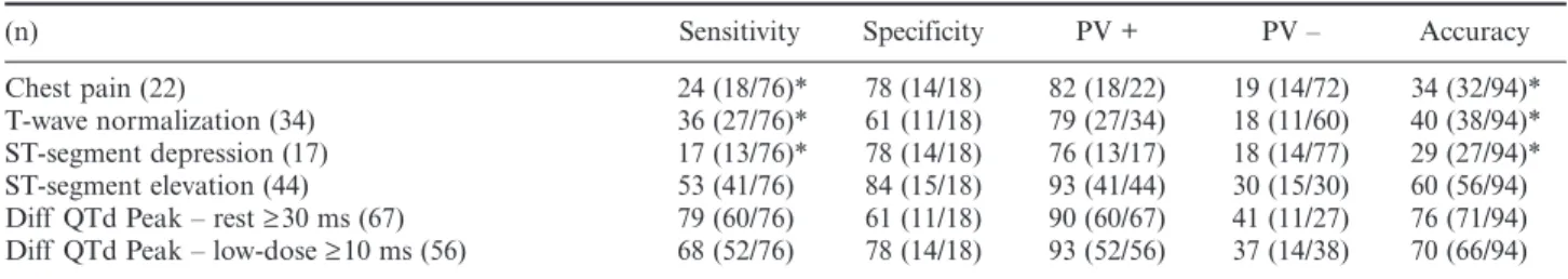
(3) Stress ECG after AMI. inclusion in the model. At the second and subsequent steps, the set of variables (clinical, angiographic and ECG variables) remaining at each point was evaluated, and the most significant was included if it significantly improved the prediction of outcome. The algorithm ceases to select variables when there was no further significant improvement in the prediction. Statistical significance was defined as p≤0.05. Reproducibility of QT dispersion measurements has been published previously 7.. Results STRESS. ELECTROCARDIOGRAPHIC CHARACTERISTICS. On the ECG recorded at rest, pathologic Q waves were present in 75 patients, negative T-waves in 45 and ST-segment elevation in 19. Heart rate increased from baseline to low and high-dose dobutamine (71±12 vs. 93 ± 9 vs. 131 ± 11 beats/min, respectively; p < 0.001). Dobutamine infusion significantly increased QT dispersion from baseline to low-dose and high-dose dobutamine (69±19 vs. 84±16 vs. 104±24 ms; p<0.0001). Significant ST-segment elevation occurred during stress in 44 patients (47%): 31 patients had new ST-segment elevation and the 13 remaining patients showed increased ST-segment elevation as compared with baseline. ST-segment depression was observed in 17 patients and T-wave normalization in 34.. PREDICTION. 13. between high-dose and low-dose dobutamine (26±28 vs. 3±20 ms; p = 0.0018) were higher in patients with significant infarct stenosis. Table 1 shows the sensitivity, specificity, predictive value and accuracy of stressECG findings for the prediction of significant stenosis of the infarct-related artery. Both an increase in QT dispersion (≥ 30 ms from baseline to high-dose dobutamine and ≥ 10 ms from low-dose to high-dose dobutamine) and dobutamine ST-segment elevation were more sensitive than the occurrence of dobutamineinduced chest pain and ST-segment depression (79%, 68% and 53% vs. 24% and 17%, respectively; p<0.05). Specificity and positive predictive values were not significantly different for all stress-ECG features. The increase in QT dispersion from baseline to high-dose dobutamine was greater in patients with multivessel disease than in those with single-vessel disease (69±26 vs. 29 ± 28 ms; p < 0.0001). A ≥ 50 ms increase in QT dispersion from baseline to high-dose dobutamine had a sensitivity of 73%, a specificity of 75%, a positive predictive value of 35%, a negative predictive value of 94% and an accuracy of 75% for predicting the presence of multivessel disease (figure 1).. OF SIGNIFICANT INFARCT-RELATED. ARTERY STENOSIS. There were no significant differences between QT dispersion on the baseline ECG in patients with or without significant stenosis of the infarct-related artery (67±19 vs. 74±17 ms). The increase in QT dispersion was significantly greater in patients with significant infarct artery stenosis. Both the difference in QT dispersion between high-dose dobutamine and rest (40±31 vs. 14±24 ms; p = 0.002) and the difference. Fig. 1. – Bar graph showing sensitivity, specificity and accuracy of an increase in QT dispersion from baseline ECG to highdose dobutamine by ≥ 30 ms for detecting significant infarct artery stenosis (white bar) and by ≥50 ms for multivessel decease (black bar).. Table 1. – Value of dobutamine-induced ECG changes for predicting significant stenosis of the infarct-related artery (n). Sensitivity. Specificity. PV +. PV –. Accuracy. Chest pain (22) T-wave normalization (34) ST-segment depression (17) ST-segment elevation (44) Diff QTd Peak – rest ≥ 30 ms (67) Diff QTd Peak – low-dose ≥ 10 ms (56). 24 (18/76)* 36 (27/76)* 17 (13/76)* 53 (41/76) 79 (60/76) 68 (52/76). 78 (14/18) 61 (11/18) 78 (14/18) 84 (15/18) 61 (11/18) 78 (14/18). 82 (18/22) 79 (27/34) 76 (13/17) 93 (41/44) 90 (60/67) 93 (52/56). 19 (14/72) 18 (11/60) 18 (14/77) 30 (15/30) 41 (11/27) 37 (14/38). 34 (32/94)* 40 (38/94)* 29 (27/94)* 60 (56/94) 76 (71/94) 70 (66/94). d = dispersion, Diff = difference, n = number of patients, PV = predictive value, * p < 0.05 vs. Diff QTd and ST-segment elevation..
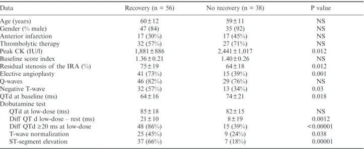
(4) 14. P. Lancellotti et al.. PREDICTORS. OF CONTRACTILE RECOVERY. Table 2 presents clinical, ECG and angiographic findings in patients with and without contractile recovery. Recovery of contraction was observed in 56 (60%) patients. There were no significant differences between groups in age, gender, site of infarction, proportion of Q wave infarction, the use of thrombolytic therapy and baseline wall motion score index. Peak level of creatine kinase was lower in patients who recovered (1,881±886 vs. 2,441 ± 1,017 IU/l; p = 0.012). Elective coronary angioplasty was more frequently performed in patients who showed contractile recovery (41 of 56 vs. 15 of 38; p = 0.001). QT dispersion on the baseline ECG was shorter in patients with contractile recovery. QT dispersion increased by 21±10 ms from baseline to low-dose dobutamine in patients with contractile recovery and only by 8 ± 19 ms in patients without recovery (p = 0.0012). An increase in QT dispersion by ≥ 20 ms allowed the best distinction between groups (p<0.00001). Contractile improvement was also more frequent in patients with stress-induced T-wave normalization, and ST-segment elevation. Using multivariate analysis, 4 independent variables were selected stepwise: an increase in QT dispersion by ≥ 20 ms from baseline to low-dose dobutamine (χ2 = 15.3; p = 0.00016), dobutamine-induced ST-segment elevation (χ2 = 11.8; p = 0.0009), elective angioplasty of the infarct-related artery (χ2 = 11.4; p = 0.001), and T-wave normalization (χ2 = 8.4; p = 0.005). A ≥ 20 ms increase in QT dispersion from baseline to low-dose dobutamine had a higher sensitivity for predicting contractile recovery than dobutamine-induced ST-segment elevation or T-wave normalization (86% vs. 66% vs. 45%, respectively; p<0.05) (figure 2). Specificity was not significantly different for dobutamine-induced. Fig. 2. – Bar graph showing sensitivity, specificity and accuracy of dobutamine-induced T-waves normalization (left bar), ST-segment elevation (middle bar) and an increase of QT dispersion from baseline ECG to low-dose dobutamine by ≥ 20 ms (right bar) for predicting the occurrence of functional recovery. * p < 0.05. ST-segment elevation, T-wave normalization and QT dispersion changes (82% vs. 76% vs. 61%; respectively). The accuracy of QT dispersion changes and of ST elevation was greater than that of T-wave normalization (76% vs. 72% vs. 57%, respectively; p<0.05).. Discussion This study shows that after AMI, the analysis of predischarge stress ECG is useful for predicting residual stenosis of the infarct-related artery and contractile recovery in the affected area. Changes in QT dispersion during graded infusion of dobutamine were the most accurate parameter for this purpose. The clinical value of dobutamine-induced chest pain or ST-segment depression was modest. Stress-induced ST-segment. Table 2. – Comparison between patients with and without functional recovery Data Age (years) Gender (% male) Anterior infarction Thrombolytic therapy Peak CK (IU/l) Baseline score index Residual stenosis of the IRA (%) Elective angioplasty Q-waves Negative T-wave QTd at baseline (ms) Dobutamine test QTd at low-dose (ms) Diff QT d low-dose – rest (ms) Diff QTd ≥ 20 ms at low-dose T-wave normalization ST-segment elevation. Recovery (n = 56). No recovery (n = 38). P value. 60±12 47 (84) 17 (30%) 32 (57%) 1,881±886 1.36±0.21 75±19 41 (73%) 46 (82%) 32 (57%) 64±16. 59 ± 11 35 (92) 17 (45%) 27 (71%) 2,441 ± 1,017 1.40 ± 0.26 64 ± 18 15 (39%) 29 (76%) 13 (34%) 74 ± 21. NS NS NS NS 0.012 NS 0.012 0.001 NS 0.03 0.018. 85±18 21±10 48 (86%) 25 (45%) 37 (66%). 82 ± 15 8 ± 19 15 (39%) 9 (24%) 7 (18%). NS 0.0012 < 0.00001 0.038 0.00001. CK = creatine kinase, d = dispersion, Diff = difference, IRA = infarct-related artery..
(5) Stress ECG after AMI. elevation was a sensitive marker of viable myocardium but its sensitivity to predict significant residual stenosis was moderate.. SIGNIFICANT. STENOSIS OF THE INFARCT-RELATED. ARTERY AND MULTIVESSEL DISEASE. After AMI, ischaemic myocardium usually reflects the presence of significant residual stenosis of the infarct-related artery2. Several studies have assessed the relation between dobutamine-induced ECG changes and the presence of jeopardized myocardium in the affected area. T-wave normalization in the infarctrelated leads during dobutamine infusion has been identified as an accurate marker of ischaemia in patients with non-Q wave AMI11. In Q wave AMI, Lombardo et al. have demonstrated that T-wave normalization was specifically associated with ischaemic myocardium when developed at high-dose dobutamine4. In our study, the positive predictive value of T-wave normalization for detecting significant infarct stenosis was good (79%), but its negative predictive value was very low (18%). We have recently found a good relationship between ST-segment elevation and the observation of a biphasic response during dobutamine administration9. In the present study, the sensitivity and the specificity of dobutamine ST-segment elevation for predicting significant infarct artery stenosis were similar to those reported by Smart et al12. No study has assessed the accuracy of QT dispersion changes during dobutamine stress testing for the identification of infarct artery stenosis. In patients with significant infarct artery stenosis, QT dispersion increased significantly during graded infusion of dobutamine. This increase was more sensitive and accurate than dobutamine-induced chest pain or ST-segment depression. Our results confirm and extend recent observations indicating that an increase in QT dispersion immediately after exercise was more sensitive for detecting ischaemic myocardium than ST-segment depression13. The increase in QT dispersion was even higher in patients with multivessel disease. These data suggest that the degree of heterogeneity of ventricular repolarization is determined by the extent of stressinduced myocardial ischaemia.. CONTRACTILE. RECOVERY. In the present era of thrombolysis, the incidence of transmural infarction and left ventricular aneurysm has largely decreased. Most patients develop an incomplete infarction, with an admixture of subendocardial necrosis and salvaged subepicardium. Several studies have recently found an association between stressinduced T-wave normalization14, ST-segment eleva-. 15. tion15 or an increase in QT dispersion7 and the presence of myocardial viability. Few studies have shown the usefulness of stress-induced ECG changes for predicting improvement of function after AMI. Recently, we16 and others17 have found that recovery in regional function was more frequent in patients with than in patients without dobutamine-induced ST-T segment changes. However, no investigation has assessed the accuracy of QT dispersion changes during dobutamine infusion for the prediction of contractile recovery. In our study, stepwise multivariate analysis selected an increase in QT dispersion of ≥ 20 ms from baseline to low-dose dobutamine as the best predictor of functional recovery. The sensitivity of dobutamine ST-segment elevation (66%) tended to be lower but was not significantly different to that observed in our previous investigation (74%)16. T-wave normalization was also a less sensitive marker of myocardial viability as previously demonstrated4. It should be emphasized that these results are not necessarily applicable to patient populations infrequently treated by revascularization after AMI. In this study, elective angioplasty of the infarct-related artery also emerged as an independent predictor of functional recovery. Indeed, persistence of a significant infarct artery stenosis may preclude functional recovery18.. LIMITATIONS Our observations pertain only to patients with small or moderate infarct size. Wall motion analysis was done by a semi-quantitative method rather than by quantitative techniques, but this method remains the standard. Coronary angiography was not repeated at follow-up. Thus, restenosis or reocclusion cannot be excluded: this could have resulted in lack of recovery in some patients. However, repeated catheterization is not indicated in an asymptomatic patient. It is also possible that the measurement of QT interval cannot always be assessed in every lead and the terminal portion of the T-wave may be difficult to determine. In the present study, QT intervals were assessable in ≥ 8 leads in all patients. The duration of the dynamic changes in QT dispersion during recovery was not examined. Correction of QT interval is plagued by controversy due to the weakness of the classical Bazett’s formula19. Since QT dispersion does not change with heart rate, heart rate-corrected QT dispersion was not calculated.. Conclusions In patients with uncomplicated AMI, the identification of viable but jeopardized myocardium is of great importance for selecting patients who could benefit.
(6) 16. P. Lancellotti et al.. from a revascularization procedure. Dobutamine stress echocardiography has been widely validated in this clinical setting. However, the interpretation of the test remains difficult in some patients with low echogenicity or with subtle changes in wall thickening. Our study shows that careful ECG observation, especially dynamic changes in QT dispersion during graded infusion of dobutamine provides an accurate and cost-effective strategy for detecting the presence of significant infarct artery stenosis and for predicting functional recovery after AMI.. References 1. Carlos ME, Smart SC, Wynsen JC, Sagar KB. Dobutamine stress echocardiography for risk stratification after myocardial infarction. Circulation 1997; 95: 1402-10. 2. Piérard LA, De Landsheere CM, Berthe C, Rigo P, Kulbertus H. Identification of viable myocardium by echocardiography during dobutamine infusion in patients with myocardial infarction after thrombolytic therapy: comparison with positron emission tomography. J Am Coll Cardiol 1990; 15: 1021-31. 3. Marwick T, D’Hondt AM, Baudhuin T, Willemart B, Wijns W, Detry JM, Melin J. Optimal use of dobutamine stress for the detection and evaluation of coronary artery disease: combination with echocardiography or scintigraphy, or both. J Am Coll Cardiol 1993; 22: 159-67. 4. Lombardo A, Loperfido F, Pennestri F. Significance of transient ST-T segment changes during dobutamine testing in Q wave myocardial infarction. J Am Coll Cardiol 1996; 27: 599-605. 5. Stiles GL, Rosati RA, Wallace AG. Clinical relevance of exercise-induced ST-segment elevation. Am J Cardiol 1980; 46: 931-6. 6. Yamagishi H, Akioka K, Takagi M. Exercise four hour redistribution thallium-201 single photon emission computed tomography and exercise induced ST-segment elevation in detecting the viable myocardium in patients with acute myocardial infarction. Heart 1999; 81: 17-24. 7. Lancellotti P, Bilge AR, Mipinda JB, Piérard LA. Significance of dobutamine-induced changes in QT dispersion early after acute myocardial infarction. Am J Cardiol 2001; 88: 939-43. 8. Bodi V, Sanchis J, Llacer A. ST-segment elevation on Q leads at rest and during exercise: relation with myocardial viability and left ventricular remodeling within the first 6 months after infarction. Am Heart J 1999; 137: 1107-15.. 9. Piérard LA, Lancellotti P, Kulbertus HE. ST-segment elevation during dobutamine stress testing predicts functional recovery after acute myocardial infarction. Am Heart J 1999; 137: 500-11. 10. Malik M, Batchvarov VN. Measurement, interpretation, and clinical potential of QT dispersion. J Am Coll Cardiol 2000; 36: 1749-66. 11. Elhendy A, Geleijnse ML, Salustri A, van Domberg RT, Cornel JH, Arnese M, Roelandt JR. T-wave normalization during dobutamine stress testing in patients with non-Q wave myocardial infarction: a marker of myocardial ischemia. Eur Heart J 1996; 17: 526-31. 12. Smart SC, Knickelbine T, Stoiber TR, Carlos M, Wijnsen JC, Sagar B. Safety and accuracy of dobutamine-atropine stress echocardiography for the detection of residual stenosis of the infarct-related artery and multivessel disease during the first week after myocardial infarction. Circulation 1997; 95: 1394401. 13. Naka M, Shotani I, Koretsune Y, Imai K, Akamatsu Y, Hishida E, Kinoshita N, Katsube Y, Sato H, Hori M. Occurrence of sustained increase in QT dispersion following exercise in patients with residual myocardial ischemia after healing of anterior wall myocardial infarction. Am J Cardiol 1997; 80: 1528-31. 14. Schneider CA, Helmig AK, Baer FM. Significance of exercise-induced ST-segment elevation and T-wave pseudonormalization for improvement of function in healed Q-wave myocardial infarction. Am J Cardiol 1998; 82: 148-53. 15. Margonato A, Chierchia SL, Xuereb RG, Xuereb M, Fragasso G, Cappelletti A, Landoni C, Lucignani G, Fazio F. Specificity and sensitivity of exercise-induced ST-segment elevation for detection of residual viability: comparison with fluorodeoxyglucose and positron emission tomography. J Am Coll Cardiol 1995; 25: 1032-8. 16. Lancellotti P, Seidel L, Hoffer E, Kulbertus HE, Piérard LA. Exercise versus dobutamine-induced ST elevation in the infarct-related electrocardiographic leads: clinical significance and correlation with functional recovery. Am Heart J 2001; 141: 772-9. 17. Elhendy A, Cornel JH, Roelandt JRTC. Relation between ST-segment elevation during dobutamine stress test and myocardial viability after a recent myocardial infarction. Heart 1997; 77: 115-21. 18. Barilla F, Gheorghiade M, Alam M. Low-dose dobutamine in patients with acute myocardial infarction identifies viable but not contractile myocardium and predicts the magnitude of improvement in wall motion abnormalities in response to coronary revascularization. Am Heart J 1991; 122: 1522-31. 19. Malik M, Camm AJ. Mystery of QTc interval dispersion. Am J Cardiol 1997; 79: 785-7..
(7)
Figure
Documents relatifs
(2013) Global and regional longitudinal strain assessed by two-dimensional speckle tracking echocardiography identifies early myocardial dysfunction and transmural extent of
However, simulating a full cardiac cycle of the ventri- cle looped with vascular system under varying physiological conditions (e.g. during stress) using a 3D approach would lead
Je n’veux plus rentrer… Elles sont fatiguées… Je dois les forcer à me supporter,.. Elles gémissent, se mettent
with a complex vector space complex vector space with inner product with inner product called the. called the State Space State Space of
Aymar est accompagné d’un très vieil homme avec une grande barbe et un vêtement très long.. -Allez, disparaissez vous deux, hurle alors Aymar en
Sur le site d’André Boileau, dont l’adresse est donnée ci-dessous, on peut déplacer le point P dans un Applet Java et refaire l’exploration réalisée par
Daptomycin plasma concentrations in our ICU patients undergoing CRRT and receiving daptomycin q48 h showed low peak and very low trough concentrations (data not shown) compared
Calculez le p´ erim` etre et l’aire de chaque trap` eze.. Aire et P´erim`etre des Trap`ezes
