Diagnostic value of interleukine-6, transforming growth factor-beta 1 and
vascular endothelial growth factor in malignant pleural effusions
Bernard C. Duysinxa,b, Jean-Louis Corhaya,b, Laurent Hubina,b, Delphine Nguyena,b, Monique Henketa,b, Renaud Louisa,b
aDivision of Pulmonary Medicine, University of Liège, CHU Sart-Tilman B35, B-4000 Liège, Belgium
bGIGA Infection, Immunity and Inflammation Research Group, University of Liège, CHU Sart-Tilman B35, B-4000 Liège, Belgium
SUMMARY:
Study objectives: We evaluate the accuracy of pleural interleukine-6 (IL-6), transforming growth factor-beta 1
(TGF-β1 ), and vascular endothelial growth factor (VEGF) levels for differentiating benign from malignant pleural exudates.
Patients and methods: Levels of IL-6, TGF-β1, and VEGF were measured by ELISA in 103 patients with non
neutrophilic (<50%) exudative pleurisy including both benign and malignant effusions. Pleurisies were splitted into benign and malignant according to the pathological diagnosis.
Results: Thirty-nine benign (seven infections; 32 inflammatory diseases) and 64 malignant (34 extrathoracic
tumors; 25 lung cancers; five mesotheliomas) pleural exudates were diagnosed by thoracoscopy. Pleural reticulo-monocyte count, protein Light's ratio and lactic dehydrogenase Light's ratio were significantly higher in
malignant than in benign effusions (p < 0.05, ρ<0.001 and ρ<0.001, respectively). The median (range) level of VEGF was significantly higher in malignant than in benign effusions (664.50 pg/ml [10-40,143] vs 349pg/ml [10-8888]) (p<0.05). VEGF levels correlated with pleural LDH (r = 0.41, ρ<0.0001), glucose (r = -0.30, p < 0.01 ) and red cell count (r = 0.57, p < 0.0001 ).
No significant difference was found between malignant and benign effusions with respect to IL-6 (26.8 ng/ml [1.8-421] vs 18.4ng/ml [0.45-400], respectively) and TGF-β1 (1079 pg/ml [18-6206] vs 1123 pg/ml [34-5447]) levels.
ROC analysis between benign and malignant pleurisies for VEGF showed an area under the curve of 619 (p = 0.03) with a value of 382 pg/ml as the best threshold for distinguishing benign from malignant effusions.
Conclusions: Malignant effusions may enhance the release of VEGF in pleural space and its measurement may
help in the diagnosis of malignant effusion.
Keywords : Pleural cytokines; Pleural effusion; Cancer; Lymphocyte effusion; Exudate; VEGF
INTRODUCTION
Pleural effusion occurs as a complication of many different diseases and accurate diagnosis is difficult without resorting to invasive procedures. Particularly, the diagnosis of malignant pleural effusion is sometimes a challenging medical problem because differentiation from benign effusion is often difficult with the currently available parameters derived from thoracocentesis. Although being essential for distinguishing between
transudate and exudate,1 biochemical, microbiological and cytological pleural fluid analyses have poor value for identifying the cause of a pleural lymphocytic exudate.2 The sensitivity of cytological examination of pleural fluid and blind needle biopsy, even when combined together, is generally less than 75%.3-7 Moreover there is a lack of accepted and reliable diagnosis criteria particularly for malignancy based on morphological imaging (CT and MR imaging).3,8-10 Metabolic imaging has shown its usefulness11-14 but the technique is not yet largely applied because of the financial burden of the material needed.
The development of inflammation in the pleura results in an increased vascular permeability and pleural liquid accumulation is the result of increased fluid production and/or reduced lymph drainage. This pleural fluid is enriched in proteins, inflammatory cells and mediators.3,15 Cytokine-producing cells and cytokines have been reported in pleural effusions from patients with malignant as well as benign diseases. 15-21
Interleukine-6 (IL-6) is a multifunctional cytokine secreted by lymphoid and non lymphoid cells that regulates B cell and T cell function and is a potent inducer of the acute-phase protein response.22-24 Conflicting results have been reported regarding the ability of IL-6 in distinguishing malignant from benign pleural effusion.19,20,25 Transforming growth factor-beta 1 (TGF-β1) is a multifunctional cytokine that stimulates cell proliferation and angiogenesis in areas of inflammation26 and increases the permeability of mesothelial cells.27 TGF-β1 is produced by and acts on mesothelial cells in an autocrine loop. Therefore this mediator may be central in the pathogenesis of pleural diseases. High levels of TGF-β1 have been demonstrated in both malignant and infectious pleural effusions.28,29 Furthermore, TGF-β1 is a potent fibrogenic cytokine that contributes to fibrin deposition and tissue fibrosis in loculated pleural effusion.30
Finally, vascular endothelial growth factor (VEGF) is a key cytokine in the control of vascular permeability and is thought to be important in pleural fluid formation.31 VEGF is able to increase angiogenesis and enhance the permeability of vascular endothelial cells.32,33 High VEGF concentrations in pleural effusions were found in malignancies including metastasis of the lung or other primary tumors.34
In this study we have wondered whether the determination of IL-6, TGF-β1, or VEGF pleural fluid levels might help in identifying the etiology of non neutrophilic pleural effusions and in particular in differentiating malignant from benign effusions.
MATERIALS AND METHODS Patient selection
Two hundred and seventeen consecutive patients presenting with pleural exudative effusion after thoracocentesis were diagnosed in our CHU pneumology unit between 2003 and 2006. In the chemical analysis, pleural effusion was considered as an exudate according to Light's criteria,1 that is if pleural effusion met at least one of the following criteria: a ratio of pleural fluid protein to serum protein greater than 0.5; a ratio of pleural fluid lactic dehydrogenase to serum lactic dehydrogenase greater than 0.6; pleural fluid lactic dehydrogenase level greater than two-thirds of the upper limit of the normal value for serum lactic dehydrogenase. Neutrophilic pleurisy (>50% neutrophils)2 and empyema were not included in this study. The neutrophilic pleural effusions accounted for 53 out of the 217 cases (24%). Additionally among the non neutrophilic exudates, 61 patients (28%) had received a diagnosis based on radiological evaluation and thoracocentesis. The patients selected for this study were those 103 patients (mean age of 66 ±13.9 years; range 18-96 years; 62 males and 41 females) presenting with a non neutrophilic exudative pleural effusion in whom the combination of chest X-ray, thoracic CT
scanning (PQ 2000 4th generation, Picker, Cleveland, Ohio, USA) and thoracocentesis failed to give an etiologic diagnosis, thus justifying the realization of a thoracoscopy with pleural biopsies. This group represented 48% of all pleural effusions. In our series the thoracoscopy procedure allowed to establish a diagnosis in each case. Pleural fluid analysis
A successful thoracocentesis of pleural fluid was performed on each subject before thoracoscopy was carried out. A first sample was subjected to routine biochemical analysis including tests for pleural protein, glucose, lactic dehydrogenase (LDH), and amylase levels. A second sample was added to a tube containing ethylenediamino-tetraic-potassium anticoagulant for differential cell counting.
For cytokine measurement, the pleural fluid was centrifuged at 400 g for 10min, at 4°C. The supernatant was discarded and kept at -70°C until ELISA was performed. IL-6, TGF-β1 and VEGF cytokines were measured according to the following commercially available enzyme-linked immunosorbent assay (ELISA) kits (Cytoset; Bio-source, Camarillo, USA for IL-6; Duoset; R and D Systems Europe, Abingdon, UK for TGF-β1 and VEGF). The limit of detection was 10 pg/ml for each of the three cytokines.
Etiologic diagnosis of pleural exudate
The final diagnosis of the pleural effusion was obtained by invasive pleural biopsy during a thoracoscopy. When a diagnosis of benign disease was established, the patients were followed for at least 18 months to ensure the absence of malignant pleural process. Benign pleural effusions were divided into infectious (parapneumonic and tuberculosis) vs inflammatory pleurisy. Malignant pleural effusions were divided into three groups: (1) pleural metastasis of extra-thoracic cancer, (2) pleural metastasis of primary lung cancer, (3) mesothelioma.
The size of the pleural effusion was estimated in each patient by the total pleural fluid volume aspirated when starting the thoracoscopy procedure. The protocol was approved by the ethics committee, and informed consent was obtained from each subject before the study.
Statistical analysis
Characteristics of the pleural fluid and pleural cell counts in malignant vs benign pleural effusions were compared using the non parametric Mann-Whitney test. For correlations between variables, we used the Spearman rank coefficient of correlation. The accuracy of each pleural cytokine to distinguish malignant from benign pleural lesions was calculated with receiver operating characteristics (ROC) analyses. Fisher's exact test was performed to evaluate the use of two or three Light's criteria as cut-off for distinguishing neoplasic from benign pleural effusion. A p value <0.05 was considered as statistically significant.
RESULTS
Thoracoscopic biopsies showed benign lesions in 39 patients and malignant pleural effusions in 64. The mean age was 65 ± 15 years in the benign group and 66 ± 13 years in the malignant group. The sex ratio was different with females accounting for 50% of the malignant group while only representing 23% in the benign group (ρ<0.01). In the malignant group, 34 were metastases of extrathoracic tumors: 11 breast cancers, four ovarian cancers, four kidney cancers, four pancreatic cancers, four colic tumors, two prostatic neoplasms, one skin cancer, one genital carcinoma, one acute leukemia, one laryngeal cancer and one unknown primary carcinoma. Twenty-five pleural effusions were secondary to a pleural invasion of a lung cancer (five squamous non small cell carcinomas, 18 adenocarcinomas, one large cell carcinoma and one small cell cancer) and mesothelioma was diagnosed in five patients. Among the benign pleural diseases, the clinical context, bacteriological and
histological data led to a final diagnosis of infection in seven patients (five parapneumonic pleurisies, two tuberculosis) and inflammatory effusion in 32 patients (one Dressler's syndrome, one chronic pancreatitis, two heart failures, one post-radic pleurisy, five benign asbestos pleurisies, one uremic pleurisy and 21 non specific chronic inflammatory changes). So far the follow up of all these patients with benign pleural disease has been at least 18 months with no disease recurrence or change to pleural malignancy diagnosis.
There was no significant difference in pleural protein, LDH, glucose and amylase levels between malignant and benign pleural effusions. However, protein Light's ratio and LDH Light's ratio were significantly higher in malignant effusions (p<0.001 for both). Malignant effusions exhibited a greater proportion of reticulo-monocyte cell counts (p<0.05) (Table 1).
None of the pleural samples showed a positive bacterial growth during the culture even when the pleural effusion was deemed to be of infectious origin.
The median level of pleural VEGF was 664.5 pg/ml [10-40,143] in patients with malignant pleural effusions and significantly higher compared to the value of 349 pg/ml [10-8888] found in benign effusions (Table 2 and Fig. 1). By contrast no significant difference was found in IL-6 and TGF-β1 pleural levels between malignant and benign effusions (Table 2).
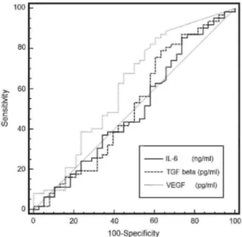
Receiver operating characteristic curves are presented in Fig. 2. Only the measurement of VEGF showed a significant performance in distinguishing between malignant and benign effusions with an area under the curve (AUC) of 619 (p<0.05). Derived from this curve, the best cut-point was found to be 382 pg/ml that gave a sensitivity, specificity and accuracy of 69%, 54% and 63% respectively. It was possible to use Light's criteria to distinguish neoplasic from benign pleural effusions as long as we used the presence of the three criteria as cut-off (Table 3). Combining the three Light criteria gave a relative risk (RR) of 1.97 (95% CI -1.16-3.34, p<0.05) for having malignant effusion and sensitivity, specificity and accuracy of 62%, 64% and 63% respectively.
VEGF and IL-6 levels did not differ according to the three sub-groups of malignant effusions. However there was a strong trend of a raised level of TGF-β1 among the patients suffering from a mesothelioma (1698 pg/ml [699-4415]) as compared to those suffering from lung cancer {767 pg/ml (130-2149)} (p = 0.06). We found no significant difference in the three cytokines levels according to the etiology of benign effusions.
Table 1: Pleural cell count and biochemical parameters in malignant and benign pleural effusions Benign pleural effusions Malignant pleural effusions
RBC/mm3 7200 [40-340,000] 5220 [10-940,000] WBC/mm3 640 [117-14,920] 710 [4-1000] Neutrophils (%) 6 [0-47] 11 [0-48] Lymphocytes (%) 67 [4-98] 50 [0-98] Reticulo-monocytes (%) 12.5 [0-78] 23 [0-87]* Eosinophils (%) 0 [0-40] 0.5 [0-43] Proteins (g/l) 39 [24-59] 42 [10-61] LDH <UI/l) 443 [105-9987] 551 [155-3859] Amylase <UI/l) 38 [12-22,540] 43 [5-903] Glucose (g/l) 0.86 [0.02-1.94] 0.84 [0.18-1.69] Protein Pl/Sg ratio 0.57 [0.33-0.82] 0.63 [0.41-0.88]*** LDH Pl/Sg ratio 0.97 [0.15-21.6] 1.53 [0.47-7.57]*** Values are expressed as median (range).
*p < 0.05; **p< 0.01 ; ***p < 0.001 ; ****p < 0.0001.
Table 2: Comparison of pleural cytokine levels between benign and malignant pleural effusions Pleural cytokines Benign pleurisy (N = 39) Malignant pleurisy (N = 64) IL-6 (ng/ml) 18.41 [0.45-400] 26.83 [1.82-421] TGF-β (pg/ml) 1123 [34-5447] 1079 [18-6206] VEGF (pg/ml) 349 [10-8888] 664.50 [10-40,143]* Values are expressed as median (range).
*ρ < 0.05; **ρ < 0.01 ; ***ρ < 0.001 ; ****ρ < 0.0001.
Figure 1: Comparison of pleural VEGF levels in malignant vs benign pleural effusions. The dashed line
represents the limit of detection of our assay. The solid line represents the median value in benign and malignant pleurisy.
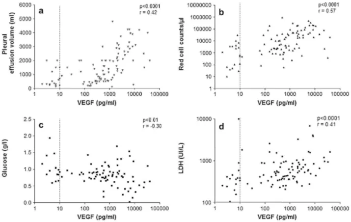
Pleural VEGF levels were positively correlated with LDH (r = 0.41, p<0.0001) but negatively correlated with glucose (r = -0.30, p<0.01) (Fig. 3). No correlation was found with the total pleural proteins levels (r =-0.12,
not with pleural leukocytes (r = -0.114, p > 0.05). Finally, pleural VEGF levels strongly correlated with the pleural effusion volume (r =0.42, p<0.0001) (Fig. 3). These correlations may reflect the aggressiveness and clinical presentation of the effusion.
Figure 2: Receiver operating characteristics (ROC) curves for differential diagnosis of pleural effusion
(malignant vs benign) by IL-6, TGF-β1 and VEGF.
Table 3: Fisher's exact test for the use of three Light's criteria as cut-off for distinguishing neoplasic from
benign pleural effusion
Number of Light's criteria Benign pleurisy (N = 39) Malignant pleurisy (N = 64) Total (N= 103) <2 24% 23% 47% 3 14% 39% 53% 38% 62% 100% p = 0.0142. DISCUSSION
Diagnosing the etiology of pleural lymphocytic exudate is sometimes a challenging medical problem because of a lack accuracy of non invasive investigations.2-10 Identifying new biochemical fluid markers is therefore suitable for differentiating benign from malignant lesions. Our study shows, in a large and well characterized population of patients with lymphocytic exudate, that although greater in malignant pleural effusions, pleural VEGF level is of a rather limited clinical interest in distinguishing between malignant from benign pleural effusion.
VEGF is a multifunctional cytokine that increases vascular permeability and it is an important angiogenic and lymphogenic factor.35,36 Literature has produced conflicting results about pleural VEGF levels in malignancies so
far.31,34,36-41 Part of these discrepancies may be explained by the selection criteria of pleural effusion and the
nature of the comparisons performed by the authors to single out the malignant cases. In our study we took care to exclude transudate and highly neutrophilic effusions thereby excluding acute bacterial pleural infection reported to be associated with high VEGF levels.42 In contrast Daniil et al.42 compared neoplasic effusions to effusion occurring in an infectious setting (tuberculosis and parapneumonic effusions). On the other hand Cheng et al.39 as well as Kaya et al.41 compared a limited number of malignant effusions to various miscellaneous pleural diseases including infection. The goal of our study was to assess the value of cytokines in pleural
effusion where etiology is not obvious. In these patients we found that malignant effusions were characterized by raised VEGF levels. The raised VEGF levels found here in malignant effusions confirm some of the previous
studies.31,34,37-39 ROC curve analysis indicates that 382 pg/ml yields the best accuracy (63%), although we recognize it to be fairly modest as a diagnostic tool. When targeting a specificity of 80%, our ROC curve yields a sensitivity greater than 22%. This level of accuracy however can be compared to that of other techniques of investigation such as chest CT scan, the sensitivity of which may range from 22% to 35% according to the morphologic chosen criterion43 when specificity is around 80%. In contrast to previous data, no difference was observed between the different sub-groups of malignant pleural effusions split according to histology31 or primary tumor localization.37 We are confident about our results as our series is larger than the ones which previously reported differences according to lung histology and primary localization.
Figure 3: Correlation between pleural VEGF levels and pleural effusion volume (a), pleural red cell count (b),
pleural glucose (c) and LDH (d). r is the Spearman coefficient of correlation. The dashed line represents the limit of detection of our assay.
The reason why VEGF is increased in malignant effusions is not clear. Although we cannot exclude that raised VEGF levels may partly be related to raised pleural endothelial permeability, the lack of a correlation between pleural VEGF and protein levels suggests that there may be additional mechanisms. Local production of VEGF by mesothelial cells, recruited inflammatory cells and malignant cells is likely to contribute to the raised levels found in malignant effusions.44 In our study we found no correlation between inflammatory cell counts and VEGF suggesting that recruited leucocytes may not be central in local VEGF production. By contrast the positive correlation between pleural LDH, a marker of tumor activity, and VEGF supports the hypothesis that malignant cells may contribute to the production of VEGF and is in line with Cheng et al.37 Interestingly we found an inverse relationship between pleural glucose and VEGF levels. This supports the idea that VEGF is raised when metabolic activity of the pleura is intense as in cancer involvement. 11-14 Additionally we found a strong correlation between pleural red cell counts and VEGF levels which is in line with the role of VEGF in pleural neoangiogenesis, a phenomenon present in tumor proliferation.
In contrast to VEGF, neither TGF-β1 nor IL-6, although readily detectable, was not found to be increased in the pleural effusion of malignancies. TGF-β1 was previously associated with loculated effusions,45 tuberculosis and mesothelioma.28 In our series most of the effusions were not loculated, as proved by thoracoscopy, and
mesothelioma only represented 8% of the malignant cases. As far as IL-6 is concerned, this cytokine has traditionally been associated with acute46 or chronic pleural infection.47 Our results indicate that the cytokine is not specifically associated with a tumoral process.
In conclusion our results show that malignant pleural effusions display raised VEGF levels which might contribute to the local growth of the tumor. Because of the modest accuracy, this finding may have, on its own, limited diagnostic value, but may open the way to a new treatment strategy using anti-VEGF in malignant pleural diseases.
REFERENCES
1. Light RW, Macgregor Ml, Luchsinger PC, Ball Jr WC. Pleural effusions: the diagnostic separation of transudates and exudates. Ann Intern Med 1972;77:507-13.
2. Light RW, Erozan YS, Ball WC. Cells in pleural fluid. Their value in differential diagnosis. Arch Intern Med 1973;132:854-60. 3. Sahn S. State of the art: the pleura. Am Rev Respir Dis 1988; 138:184-234.
4. Prakash U, Reiman H. Comparison of needle biopsy with cytologic analysis for the evaluation of pleural effusion: analysis of 414 cases. Mayo Clin Proc 1985;60:158-64.
5. Von Hoff D, Di Volsi V. Diagnostic reliability of needle biopsy of the parietal pleura: a review of 272 biopsies. Am J Clin Pathol 1979;72:48-51.
6. Schönfeld N, Loddenkemper R. Pleural biopsy and thoracoscopy. Eur Respir Mon 1998;9:135-52.
7. Poe RH, Israel RH, Utell MJ, Hall WJ, Greenblatt DW, KallayMC. Sensitivity, specificity and predictive values of closed pleural biopsy. Arch Intern Med 1984;144:325-8.
8. Dedrick CG, Mc Loud TC, Shepard JA, Shipley RT. Computed tomography of localized pleural mesothelioma. Am J Roentgenol 1985;144:275-80.
9. Leung A, Muller N, Miller R. CT in differential diagnosis of diffuse pleural disease. Am J Roentgenol 1990; 154:487-92.
10. Sahn S. Malignant pleural effusions. In: Fischman AP, editor. Pulmonary diseases and disorders. 2nd ed. New York: Mc Graw-Hill; 1988. p. 2159-70.
11. Gupta NC, Rogers JS, Graeber GM, Gregory JL, Waheed U, Mullet D, et al. Clinical role of F-18 fluorodeoxyglucose positron emission tomography imaging in patients with lung cancer and suspected malignant pleural effusion. Chest 2002; 122(6): 1918-24.
12. Schaffler GJ, Wolf G, Schoellnast H, Groell R, Maier A, Smolle-Juttner FM, et al. Non-small cell lung cancer: evaluation of pleural abnormalities on CT scans with 18F-FDG PET. Radiology 2004;231(3):858-65.
13. Duysinx B, Nguyen D, Louis R, Cataldo D, Belhocine T, Bartsch P, et al. Evaluation of pleural disease with 18-fluo-rodeoxyglucose positron emission tomography imaging. Chest 2004;125(2):489-93.
14. Duysinx BC, Larock MP, Nguyen D, Corhay JL, Bury T, Hustinx R, et al. 18F-FDG PET imaging in assessing exudative pleural effusions. Nucl Med Commun 2006;27:971-6.
15. Antony VB, Godbey SW, Kunkel SL, Hott JW, Hartman DL, Burdick MD, et al. Recruitment of inflammatory cells to the pleural space: chemotactic cytokines, IL-8, and monocyte chemotactic peptide-1 in human pleural fluids. J Immunol 1993;151:7216-23.
16. Alexandrakis MG, Coulocheri SA, Bouros D, Mandalaki K, Karkavitsas N, Eliopoulos GD. Evaluation of inflammatory cytokines in malignant and benign pleural effusions. Oncol Rep 2000;7:1327-32.
17. Hoheisel G, Izbicki G, Roth M, Chan CH, Reichenberger F, Schauer J, et al. Proinflammatory cytokine levels in patients with lung cancer and carcinomatous pleurisy. Respiration 1998;65:183-6.
18. Shimokata K, Saka H, Murate T, Hasegawa Y, Hasegawa T. Cytokine content in pleural effusion: comparison between tuberculous and carcinomatous pleurisy. Chest 1991;99: 1103-7.
19. Yokoyama A, Kohno N, Fujino S, Abe M, Ishida O, Hiwada K. Soluble interleukine-6 receptor levels in pleural effusions. Respir Med 1996;90:329-32.
20. Alexandrakis MG, Coulocheri SA, Bouros D, Eliopoulos GD. Evaluation of ferritin, interleukin-6, interleukin-8 and tumor necrosis factor-α in the differentiation of exudates and transudates in pleural effusions. Anticancer Res 1999;19:3607-12.
21. Xirouchaki N, Tzanakis N, Bouros D, Kyriakou D, Karkavitsas N, Alexandrakis M, et al. Diagnostic value of 1α, interleukin-6, and tumor necrosis factor in pleural effusions. Chest 2002;121:815-20.
22. Opal SM, DePalo VA. Anti-inflammatory cytokines. Chest 2000; 117:1162-72. 23. Nicod LP. Cytokines: l. overview. Thorax 1993;48:660-7.
24. Elias JA, Freundlich B, Kern JA, Rosenbloom J. Cytokine networks in the regulation of inflammation and fibrosis in the lung. Chest 1990;97:1439-45.
25. Lin CC, Liu CC, Lin CY. Changes in cell population and tumor necrosis factor, interleukin-6, and interleukin-8 in malignant pleural effusions after treatment with intrapleural tetracycline. Am Rev Respir Dis 1993;147:1503-6.
26. Sporn MB, Robert AB, Wakefield LM, de Crombrugghe B. Some recent advances in the chemistry and biology of transforming growth factor-beta. J Cell Biol 1987;105:1039-45.
27. Ikubo A, Morisaki T, Katano M, Kitsuki H, Anan K, Uchiyama A, et al. A possible role of TGF-β in the formation of malignant effusions. Clin Immunol Immunopathol 1995;77:27-32.
28. Maeda J, Ueki N, Ohkawa T, Iwahashi N, Nakano T, Hada T, et al. Transforming growth factor-beta 1 (TGF-β1)- and β2-like activities in malignant pleural effusions caused by malignant mesothelioma or primary lung cancer. Clin Exp Immunol 1994; 98:319-22.
29. Marie C, Losser M-R, Fitting C, Kermarrec N, Payen D, Cavaillon JM. Cytokines and soluble cytokine receptors in pleural effusions from septic and non septic patients. Am J Respir Crit Care Med 1997;156:1515-22.
30. Border WA, Noble NA. Transforming growth factor-β in tissue fibrosis. N Engl J Med 1994;331:1286-92.
31. Thickett DR, Armstrong L, Millar AB. Vascular endothelial growth factor (VEGF) in inflammatory and malignant pleural effusions. Thorax 1999;54:707-10.
32. Senger DR, Galli SJ, Dvorak AM, Perruzzi CA, Harvey VS, Dvorak HF. Tumor cells secrete a vascular permeability factor that promotes accumulation of ascites fluid. Science 1983;219: 983-5.
33. Ferrara N, Henzel WJ. Pituitary follicular cells secrete a novel heparin-binding growth factor specific for vascular endothelial cells. Biochem Biophys Res Commun 1989;161:851-8.
34. Sack U, Hoffmann M, Zhao XJ, Chan KS, Hui DS, Gosse H, et al. Vascular endothelial growth factor in pleural effusions of different origin. Eur Respir J 2005;25:600-4.
35. Grove CS, Lee YC. Vascular endothelial growth factor: the key mediator in pleural effusion formation. Curr Opin Pulm Med 2002;8:294-301.
36. Light RW, Hamm H. Malignant pleural effusion: would the real cause please stand up? Eur Respir J 1997;10:1701-2.
37. Cheng D, Rodriguez RM, Perkett EA, Rogers J, Bienvenu G, Lappalainen U, et al. Vascular endothelial growth factor in pleural fluid. Chest 1999; 116:760-5.
38. Lim SC, Jung SI, Kim YC, Park KO. Vascular endothelial growth factor in malignant and tuberculous pleural effusions. J Korean Med Sci 2000;15:279-83.
39. Cheng D, Lee YC, Rogers JT, Perkett EA, Moyers JP, Rodriguez RM, et al. Vascular endothelial growth factor level correlates with transforming growth factor-β isoform levels in pleural effusions. Chest 2000;118:1747-53.
40. Mommi H, Matsuyama W, Inoue K, et al. Vascular endothelial growth factor and proinflammatory cytokines in pleural effusions. Respir Med 2002;96:817-22.
41. Kaya A, Poyraz B, Celik G, Ciledag A, Gulbay BE, Savas H, et al. Vascular endothelial growth factor in benign and malignant pleural effusions. Arch Bronconeumol 2005;41: 376-9.
42. Daniil ZD, Zintzaras E, Kiropoulos T, Papaioannou AI, Koutsokera A, Kastanis A, et al. Discrimination of exudative pleural effusions based on multiple biological parameters. Eur Respir J 2007;30:957-64.
43. Yilmaz U, Polat G, Sahin N, Soy O, Gulay U. CT in differential diagnosis of benign and malignant pleural disease. Monaldi Arch Chest Dis 2005;63:17-22.
44. Lee YCG. Cytokines in pleural diseases. Chapter 6. In: Light RW, Lee YCG, editors. Textbook of pleural disease. London: Arnold Publishers; 2003. p. 63-89.
45. Chung C-L, Chen C-H, Sheu J-R, et al. Proinflammatory cytokines, transforming growth factor-β1, and fibrinolytic enzymes in loculated and free-flowing pleural exudates. Chest 2005; 128:690-7.
46. Akarsu S, Kurt AN, Dogan Y, et al. The differential diagnostic values of cytokine levels in pleural effusions. Mediators Inflamm 2005;2005(1):2-8.