Salivary Gland www.proteomics-journal.com
Proteins Identified from Saliva and Salivary Glands of the
Chinese Gall Aphid
Schlechtendalia chinensis
Zixiang Yang, Lin Ma, Frederic Francis, Ying Yang, Hang Chen, Haixia Wu,
and Xiaoming Chen*
Aphid saliva plays an essential role in the interaction between aphids and their host plants. Several aphid salivary proteins have been identified but none from galling aphids. Here the salivary proteins from the Chinese gall aphid are analyzed,Schlechtendalia chinensis, via an LC-MS/MS analysis.
A total of 31 proteins are identified directly from saliva collected via an artificial diet, and 141 proteins are identified from extracts derived from dissected salivary glands. Among these identified proteins, 17 are found in both collected saliva and dissected salivary glands. In comparison with salivary proteins from ten other free-living Hemipterans, the most striking feature of the salivary protein fromS. chinensis is the existence of high
proportion of proteins with binding activity, including DNA-, protein-, ATP-, and iron-binding proteins. These proteins maybe involved in gall formation. These results provide a framework for future research to elucidate the molecular basis for gall induction by galling aphids.
The Chinese gall aphid Schlechtendalia chinensis (Hemiptera, Aphididae, Eriosomatinae) induces galls on the Chinese sumac Rhus chinensis that are valuable for both industry and medicine due to the high level of gallotannins, which have been used in traditional Chinese medicine for more than 2000 years.[1]Usually
each gall is induced by a single aphid fundatrix and her offspring. Both gall initiation and growth manipulation in host plants are likely achieved through aphid salivary secretions, which are crit-ical in an active interplay between host plants and gall aphids.[2]
Nevertheless, the functions of these aphid salivary proteins in the host plants remain unknown. Several lines of evidence suggested that aphid saliva is rich in secretory proteins that play roles anal-ogous to effectors in plant–pathogen interactions.[3]Effector
pro-teins can elicit various physiological changes within host plants to create a favorable environment for insects and pathogens to
Dr. Z. Yang, L. Ma, Y. Yang, Dr. H. Cheng, Dr. H. Wu, Prof. X. Chen Research Institute of Resource Insects
Chinese Academy of Forestry
Key Laboratory of Breeding and Utilization of Resource Insects of State Forestry Administration
Kunming, Yunnan, P. R. China E-mail: cafcxm@139.com Dr. F. Francis
Functional and Evolutionary Entomology Gembloux Agro-Bio Tech
Liege University Gembloux, Belgium
DOI: 10.1002/pmic.201700378
feed on host plants.[4] At present,
lit-tle is known about the precise compo-nents in aphid saliva that play effector roles, especially for galling aphids. Here, the protein composition of S. chinensis saliva collected though a Parafilm diet sa-chet was analyzed and proteins from dis-sected salivary glands were also investi-gated based on LC-MS/MS analyses.
An S. chinensis aphid colony was maintained on R. chinensis and Plagiom-nium maximoviczii hosts under natu-ral conditions and wingless fundatri-geniae were obtained from fresh galls on R. chinensis trees. Saliva from wing-less fundatrigeniae was collected fol-lowing a modified protocol of Carolan et al.[5] while salivary glands were
ob-tained by dissecting wingless fundatrige-niae collected from fresh galls following a modified protocol of Mutti et al.[6]One thousand wingless
fun-datrigeniae were collected from 24 replicates for saliva collec-tion as presented in Figure 1. Multiple saliva colleccollec-tions were combined with equal volume of PBS, and then centrifuged twice through a 3000 molecular weight cutoff polyethersulfone membrane at 4°C. The extract was resuspended in 100 μL of 9.5 m urea, 2% CHAPS, 0.8% Pharmalyte pH 3–10, and 1% DTT. Salivary glands from wingless fundatrigeniae were dissected on sterile glass slides in ice-cold PBS using sterile fine needles. The glands were dissected and separated from the aphid bodies fol-lowing the methods of Rao et al.[7]Extracts of collected saliva and
dissected salivary glands were combined with Laemmli buffer, separately. Proteins were separated on 13% resolving gels with a 4% stacking gel prior to gel picture analysis using MagicScan 5.1 software (Suzhou, China).
After electrophoresis, visible protein bands were excised from preparative 1D gels destained, reduced, alkylated, and trypsin-digested as described by Ben Amira et al.[8]Generated peptides
were analyzed by LC-MS/MS on a Q Exactive Mass Spectrome-ter (Thermo Fisher Scientific, UK). Peptides were separated us-ing a BioBasic C18 at a flow rate of 200 nL min−1 and gradi-ent of 5–95% ACN over 45 min. All data were acquired with the mass spectrometer operating in automatic data-dependent switching mode. A zoom scan was performed on the five most intense ions to determine charge state prior to an MS/MS analy-sis. Protein identification from the MS/MS data was performed using the TurboSEQUEST algorithm in BioWorks v. 3.2 to corre-late the data against the pea aphid ACYPI proteins and UniProt databases. Precursor ion’s mass tolerance was set at ±0.1 Da,
Figure 1. Experimental setup and protein functional distribution from saliva and salivary gland proteomes of the Chinese gall aphid.
the fragment ion’s tolerance at ±0.2 Da with a maximum of two missed cleavage sites allowed. A transcriptomic database from the fundatrigenia of S. chinensis was also used for the pro-tein searching and identification. Comparison between salivary proteins from S. chinensis and those from other insects was per-formed according to Huang et al.[9]The mass spectrometry
pro-teomics data have been deposited to the ProteomeXchange Con-sortium via the PRIDE[10] partner repository with the dataset
identifier PXD008928 and PXD008929.
A total of 31 saliva and 141 salivary gland proteins were identified after MASCOT searches of MS/MS spec-tra against UniProt (http://www.uniprot.org/) and an aphid database (https://bipaa.genouest.org/is/aphidbase/iagc/;Tables S1 and S2, Supporting Information). Among the 31 saliva and 141 salivary gland proteins, 20 and 64, respectively, were only detected in S. chinensis at present, and the remaining 11 and 77, respectively, have been reported in other free-living insects (Figure 2 and Table S3, Supporting Information). There were 17 proteins present in both saliva and dissected salivary glands of S. chinensis (Figure 2, Table 1 and Table S2, Supporting Informa-tion). Six of the these 17 proteins were novel, including a zinc finger protein, a cadherin, a piccolo isoform, a charybde-like iso-form, and two unknown proteins (Table S3, Supporting Infor-mation). The remaining 11 proteins were also found in other free-living aphids. The same proteomic data were also searched against a S. chinensis transcriptome. However, no hits were found, which may be due to the low coverage of the transcriptome. Al-ternatively, no transcript hits were due to the fact that these two
Figure 2. The Venn diagram of identified salivary proteins in the Chinese
gall aphid and ten free-living insect species.
datasets were obtained from two different stages of the insect. Saliva and salivary glands were obtained from aphids collected from galls on the sumac, R. chinensis, whereas the transcriptome was generated from free-living aphids on the alternative host moss, P. maximoviczii.
One of the interesting characteristics of S. chinensis saliva and salivary gland proteins was the high proportion of pro-teins with various binding activities. For the 21 annotated proteins from saliva, 17 were with various binding functions
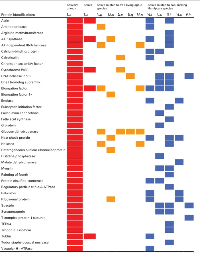
Table 1. Comparative analysis of Chinese gall aphid salivary proteins with other insect species. Salivary
glands
Saliva Saliva related to free-living aphid species
Saliva related to sap-sucking Hemiptera species
-Protein identifications S.c S.c A.p M.e D.n S.g M.p N.l L.s S.f N.c H.h
Actin
Aminopeptidase
Arginine methyltransferase ATP synthase
ATP-dependent RNA helicase Calcium-binding protein Calreticulin
Chromatin assembly factor Cytochrome P450
DNA helicase Ino80 DnaJ homolog subfamily Elongation factor Elongation factor 1γ Enolase
Eukaryotic initiation factor Failed axon connections Fatty acid synthase G protein
Glucose dehydrogenase Heat shock protein Helicase
Heterogeneous nuclear ribonucleoprotein Histidine phosphatase
Malate dehydrogenase Myosin
Painting of fourth
Protein disulfide-isomerase Regulatory particle triple-A ATPase Reticulon
Ribosomal protein Spectrin
Synaptotagmin
T-complex protein 1 subunit TER94
Troponin T isoform Tublin
Tudor staphylococcal nuclease Vacuolar H+ ATPase
Red, orange, and blue areas are related to gall aphid, free-living aphids, and other free-living sucking Hemiptera species, respectively.;S.c, Schlechtendalia chinensis; A.p, Acyrthosiphon pisum; M.e, Macrosiphum euphorbiae; D.n, Diuraphis noxia; S.g, Schizaphis graminum; M.p, Myzus persicae; N.l, Nilaparvata lugens; L.s, Laodelphax striatellus; S.f, Sogatella furcifera; N.c, Nephotettix cincticeps; H.h, Halyomorpha halys.
(Table S1, Supporting Information). For the 117 annotated pro-teins from dissected salivary glands, 75 were with various bind-ing activities. The proteins with bindbind-ing activities included ATP-, GTP-ATP-, mRNA-ATP-, protein-ATP-, actin-ATP-, magnesium ion-ATP-, calcium ion-ATP-, NADH-, DNA-, chromatin-, tropomyosin-, and zinc ion-binding proteins. The second most abundant group of proteins was those with various enzymatic activities. Annotated enzymes included five dehydrogenases, four ATPases, three disulfide isomerases, three ubiquitin-protein transferases, two oxidoreductases, two endopeptidases, and one of each of the following: citrate syn-thase, aminobutyrate transaminase, and acid phosphatase. There were also three structural proteins and a translation elongation factor.
Aphid saliva plays multiple roles once injected into host tis-sues, including partial digestion of food prior to ingestion, detox-ification, suppression of host defense, and serving as effectors for various host manipulations.[2,11,12] Unlike other free-living
aphids, the feeding of a single S. chinensis aphid can induce the formation of a gall. It is very likely that some of aphid saliva pro-teins play a role in gall induction in addition to other functions common to free-living aphids. In gall midges, a huge number of putative effector proteins are likely delivered into host tissues for host manipulation and gall induction.[13–16]The most striking
characteristic for putative gall midge effectors is a large number of highly diversified, small peptides. The most striking charac-teristic of S. chinensis saliva proteins is the high proportions of proteins with binding activities including protein-, DNA-, ATP-, and ion-binding proteins.[17,18]No similarity has been found
be-tween S. chinensis saliva proteins identified here and reported gall midge salivary proteins, suggesting that each insect–plant system has a unique mechanism for gall induction.
Another interesting feature was that many of the proteins de-tected in the aphid saliva lacked a typical secretion signal peptide. The way for these proteins to be transported out of insect’s sali-vary glands without a secretion signal peptide was not clear. How-ever, similar phenomena have also been observed in other aphid– plant systems, suggesting that there are likely other mechanisms to deliver proteins from salivary glands into host tissues from aphids in addition to the traditional signal peptide pathway.[9]
Here, we wanted to emphasize that this study on both collected saliva and dissected salivary glands is very preliminary due to the small amounts of samples obtained even though huge amounts of time and effort have been spent on this project. There are major challenges in both obtaining saliva and dissecting salivary glands from small insects.[9]In this study, more than 24 000 aphid
individuals were used to collect saliva, and the amount of protein samples was perhaps still insufficient for a comprehensive analy-sis. It can be seen that much more protein species were identified from dissected salivary glands than from the collected saliva sam-ple. This analysis may have only identified the most abundant proteins in the aphid saliva. A more effective method for saliva collection or a more sensitive technique for protein detection is needed for comprehensive analyses of tiny insects. Variation in salivary secretion has been observed on different artificial diets and at different feeding stages of aphid.[11,12]It is possible to
es-tablish a more efficient protocol to collect saliva from this aphid in the future. It was also very difficult to obtain large amounts of dissected salivary glands from S. chinensis individuals with our current dissection tools. The major challenge for gland dissection
is to transfer salivary glands into RNA extraction solution after dissection. We are currently in the process to improve procedures to collect larger amounts of saliva samples and obtain more dis-sected salivary glands for more comprehensive analyses. Finally, S. chinensis has different developmental stages on two different hosts,[19]and this study analyzed only one of the stages. Further
studies on salivary proteins from other developmental stages on alternative hosts may provide more useful information on the functions of specific salivary proteins.
Supporting Information
Supporting Information is available from the Wiley Online Library or from the author.
Acknowledgements
X.C. and Z.Y. conceived experiments. Z.Y. conducted the experiments. L.M. performed the experiments. Z.Y. and L.M. analyzed data and wrote the manuscript. X.C., F.F., Y.Y., H.W., and H.C. analyzed the results. All au-thors reviewed the manuscript. The auau-thors would like to acknowledge Prof. Ming-Shun Chen, Kansas State University, for his helpful comments. The LC-MS/MS analyses were processed by Mr. Shuo Chen in Beijing Pro-teome Research Center, China. This research is funded by the National Natural Science Foundation of China (Grant No. U1402263; 31372266; 31370651), and the Grant for Essential Scientific Research of Chinese Na-tional Non-profit Institute (Grant No. CAFYBB2014ZD005).
Conflict of Interest
The authors declare no conflict of interest.
Keywords
gall aphid, proteomics, saliva, salivary gland,Schlechtendalia chinensis
Received: October 11, 2017 Revised: March 14, 2018 Published online:
[1] G. X. Zhang, T. S. Zhong,Economic Insect Fauna of China, Science
Press, Beijing 1983, p. 77.
[2] R. L. Blackman, V. F. Eastop,Aphids on the World’s Crops: An Identifi-cation and Information Guide, Wiley & Sons, England 2000.
[3] J. C. Carolan, D. Caragea, K. T. Reardon, N. S. Mutti, N. Dittmer, K. Pappan, F. Cui, M. Castaneto, J. Poulain, C. Dossat, D. Tagu, J. C. Reese, G. R. Reeck, T. L. Wilkinson, O. R. Edwards,J. Proteome. Res.
2011,10, 1505.
[4] X. Tang, R. D. Frederick, J. Zhou, D. A. Halterman, Y. Jia, G. B. Martin,
Science 1996, 274, 2060.
[5] J. C. Carolan, C. I. J. Fitzroy, P. D. Ashton, A. E. Douglas, T. L. Wilkin-son,Proteomics 2009, 9, 2457.
[6] N. Mutti, Y. Park, J. Reese, G. Reeck,J. Insect Sci. 2006, 6, 38.
[7] S. A. K. Rao, J. C. Carolan, T. L. Wilkinson,PLoS One 2013, 8, e57413.
[8] A. B. Amira, J. Bauwens, E. De Pauw, S. Besbes, H. Attia, F. Francis, C. Blecker,J. Chem. Biol. 2017, 10, 25.
[9] H. J. Huang, J. B. Lu, Q. Li, Y. Y. Bao, C. X. Zhang.J. Proteomics 2018, 172, 25.
[10] J. A. Vizcaino, R. G. Cote, A. Csordas, J. A. Dianes, A. Fabregat, J. M. Foster, J. Griss, E. Alpi, M. Birim, J. Contell, G. O’Kelly, A. Schoeneg-ger, D. Ovelleiro, Y. Perez-Riverol, F. ReisinSchoeneg-ger, D. Rios, R. Wang, H. Hermjakob,Nucleic Acids Res. 2013, 41, 1063.
[11] P. W. Miles,Biol. Rev. 1999, 74, 41.
[12] J. I. B. Bos, D. Prince, M. Pitino, M. E. Maffei, J. Win, S. A. Hogenhout,
PLoS Genet. 2010, 6, e1001216.
[13] M. S. Chen, X. M. Liu, Z. Yang, H. Zhao, R. H. Shukle, J. J. Stuart, S. Hulbert,BMC Evol. Biol. 2010, 10, 296.
[14] C. Zhao, L. N. Escalante, H. Chen, T. R. Benatti, J. Qu, S. Chellapilla, R. M. Waterhouse, D. Wheeler, M. N. Andersson, R. Bao, M. Batterton,
S. K. Behura, K. P. Blankenburg, M. S. Chen, S. J. Brown, J. J. Stuart, S. Richards,Curr. Biol. 2015, 25, 613.
[15] C. Zhao, R. Shukle, L. Navarro, M. S. Chen, S. Richards, J. J. Stuart,J. Insect Physiol. 2016, 84, 22.
[16] R. Aggarval, S. Subramanyam, C. Zhao, M. S. Chen, M. O. Harris, J. J. Stuart,PLoS One 2014, 9, e100958.
[17] S. Vandermoten, N. Harmel, G. Mazzucchelli, E. D. Pauw, E. Haubruge, F. Francis,Insect Mol. Biol. 2014, 23, 67.
[18] H. J. Huang, C. W. Liu, X. H. Huang, X. Zhou, J. C. Zhuo, C. X. Zhang, Y. Y. Bao, J. Proteome Res. 2016, 15,
1883.
[19] P. Liu, Z. X. Yang, X. M. Chen, R. G. Foottit,Ann. Entomol. Soc. Am.