HAL Id: hal-02978110
https://hal.archives-ouvertes.fr/hal-02978110
Submitted on 26 Oct 2020HAL is a multi-disciplinary open access
archive for the deposit and dissemination of sci-entific research documents, whether they are pub-lished or not. The documents may come from teaching and research institutions in France or abroad, or from public or private research centers.
L’archive ouverte pluridisciplinaire HAL, est destinée au dépôt et à la diffusion de documents scientifiques de niveau recherche, publiés ou non, émanant des établissements d’enseignement et de recherche français ou étrangers, des laboratoires publics ou privés.
A new numerical scheme for the measurement of strain
fields in cellular microstructures
Ali Rouwane, Robin Bouclier, Jean-Charles Passieux, Jean-Noël Périé
To cite this version:
Ali Rouwane, Robin Bouclier, Jean-Charles Passieux, Jean-Noël Périé. A new numerical scheme for the measurement of strain fields in cellular microstructures. International Digital Image Correlation Society Virtual Conference 2020, Oct 2020, Virtual Conference, France. �hal-02978110�
A new numerical scheme for the measurement
of strain fields in cellular microstructures.
Ali Rouwane1,2, Robin Bouclier1,2, Jean-Charles Passieux 1,
Jean-Noël Périé1
1ICA, Université de Toulouse, UPS, INSA, ISAE-SUPAERO, MINES-ALBI, CNRS, 3 rue Caroline Aigle, F-31055 31400 Toulouse, France 2IMT, Université de Toulouse, UPS, UT1, UT2, INSA, CNRS, 135 avenue de Rangueil, F-31077 Toulouse Cedex 04, France
Abstract — Full field measurements provided by digital volume correlation (DVC) would undoubtedly contribute to a better understanding of the mechanical behaviour of cellular materials. Nevertheless, when dealing with micro-structures that do not exhibit sufficient micro gray level gradients, traditional digital image correlation (DIC) algorithms lead to a rough spatial resolution that does not allow capturing local strains. In this critical situation, one effective method to increase spatial resolution while master-ing measurement uncertainties is the use of mechanical regularization of the underlymaster-ing optimization problem.
Keywords — Digital Volume Correlation, elastic registration, B-splines, fictitious domain methods, image based models, cellular materials.
Introduction With the recent advances in imaging techniques such as Computed Micro-Tomography (µ-CT), it is possible to acquire volumetric images of the micro-structure of complex heterogeneous materials in an non-invasive way. In the following, the focus is on full-field measurement in cellular ma-terials such as foams. DVC in complex micro-structures presents multiple changes and challenges when compared to 2D DIC. For instance, in most two-dimensional cases, a speckle pattern can be deposited onto the sample in order to introduce sufficient gray-level gradients in the images. Conversely, for DVC, the only information available is the intrinsic gray-level distribution related to the microstructure. That is why it seems that conventional DVC algorithms can only be applied when materials exhibit a natural speckle pattern (for instance graphite nodules [1]). Our objective is to develop a correlation algorithm that can capture local deformations at the micro-structural scale using only the natural texture of the studied material. To do so, we suggest constructing an automated digital image-based mechanical model and using it to regularize the ill-posed problem of DIC.
Methods Given two images of a cellular specimen captured during a mechanical test, we are consider-ing regularizconsider-ing the DIC problem usconsider-ing a penalization term based on the equilibrium gap [2]:
argmin u 1 2R(u) + λ 2 ||Ku||22 (1)
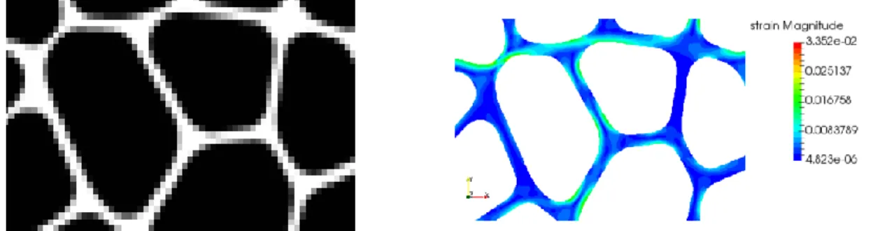
where R is the discrete sum of squared differences between the initial and the advected images,u is the displacement vector at the control points of a cubic structured B-spline grid defined over the region of interest andK is an elastic rigidity operator that simulates the mechanical behavior of the texture being observed. Our contribution concerns the automated construction of a fairly priced image-based model that allows to obtainK. To do so, we use a high order fictitious domain method using a level-set repre-sentation of the materials boundary [3, 4]. We mean by fairly priced the fact that our model’s parameters (mesh size and degree of approximation) are set such that the error made on the mechanical solution is insignificant compared to the geometric errors related to the image-based geometry approximation. In contrary to boundary-fitted meshing techniques, the degrees of freedom of the fictitious domain method are defined over all the specimen’s region (including the empty black region in Fig. 1a). We emphasize thatK is defined only at the degrees of freedoms on which the elastic forces are known.
Results In order to validate our method, we consider measuring the strain field on a planar specimen representing a two-dimensional cellular material (see Fig. 1a). Fig. 1b shows that the suggested mechan-ical regularization allows to measure accurate strain fields compared to traditional low order interpolation schemes and this using only the natural texture of the specimen.
(a) Poorly resolved image of a cellular specimen (4 pixels in the micro-structure’s thickness (of length equal
to 0.6mm). The image gradient is either small (black region) or has a dominant direction (white regions).
(b) Local bending observed at the microscopic scale when using the mechanical regularization without any
speckle pattern.
Figure 1: Strain measurement at the microscopic scale in a cellular specimen.
Discussion and Conclusion High order basis functions such as B-splines, allow to capture non-rigid and smooth displacements in the contrary of low order registration techniques. This method was vali-dated in two dimensions for synthetic images. Its generalization to real foam specimens will be done in the future and we assume that the three-dimensional generalization is only related to the computational complexity and efficiency. However, multiple challenges are still open concerning the elaboration of an in-situ mechanical test using micro-tomography. In fact, in order to acquire sufficiently resolved images, we need to consider small volume samples. This constraint obliges us to resort to adapted mechanical testing machines so as not to disturb the tomography acquisition system [5].
References
[1] Hugo Leclerc, Jean-Noël Périé, Stéphane Roux and François Hild. Voxel-scale digital volume correlation. Experimental Mechanics, 2011.
[2] Julien Réthoré, Stéphane Roux and François Hild. An extended and integrated digital image correlation technique applied to the analysis of fractured samples. European Journal of Computational Mechanics, 2009. [3] Dominik Schillinger and Martin Ruess. The finite cell method: A review in the context of higher-order struc-tural analysis of cad and image-based geometric models. Archives of Computational Methods in Engineering, 2015.
[4] Clemens V Verhoosel, GJ Van Zwieten, B Van Rietbergen, and René de Borst. Image-based goal-oriented adaptive isogeometric analysis with application to the micro-mechanical modeling of trabecular bone. Com-puter Methods in Applied Mechanics and Engineering, 2015.
[5] Buffiere, J-Y and Maire, E and Adrien, J and Masse, J-P and Boller, E. In situ experiments with X ray tomography: an attractive tool for experimental mechanics. Experimental mechanics, 2010.