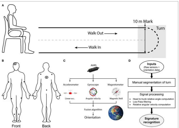
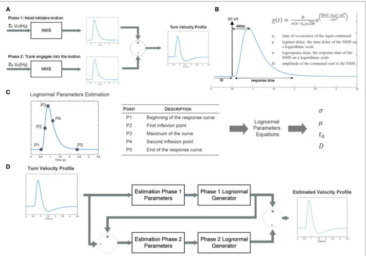
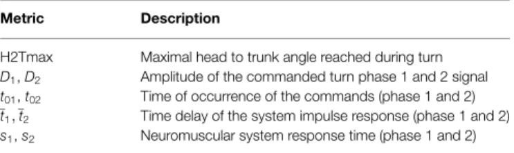
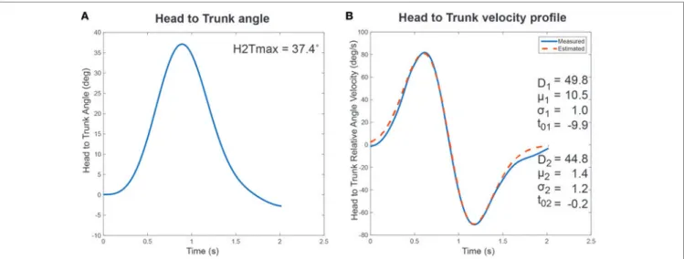
Capturing the Cranio-Caudal Signature of a Turn with Inertial Measurement Systems: Methods, Parameters Robustness and Reliability
Texte intégral
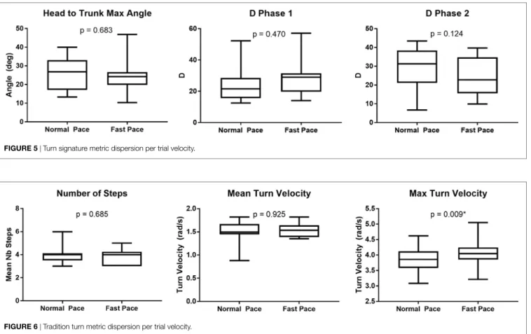
Figure
Documents relatifs
O livro estrutura-se em prefácio, prólogo e nove capítulos, ao longo dos quais são desenvolvidos os três eixos temáticos que Bernstein se pro- põe a criticamente analisar:
The proposed closed-loop control circuit does not slow down the switching speed of the current because the V Lsmax obtained during the di/dt is lower than the minimum
“translation” of Bortkiewicz’s article is very free, summing up in three lines what was developped over three pages in the original, and, inversely, dedicating a lot of print to
The epilogue to the book offers concluding remarks on how abstract concepts such as regionalism and regional identity construction materialised, or not, in works of
In this paper I argue that recent technological transformations in the life-cycle of information have brought about a fourth revolution, in the long process of reassessing humanity’s
The right panel depicts a boundary pinching scenario, where around the bound- ary pinching point, a near-boundary three-arm event (with alternating pattern) occurs. We show in the
In order to show the specific intellectual and scientific context in which we place our research activities on digital (audiovisual) archives in the sense of an integrated set
Thanks to technological advances, we can consider how a bioinspired model of information communication could be implemented into smart human environments: through wearable
