THESE
Pour l’obtention du Grade de
DOCTEUR DE L’UNIVERSITE DE POITIERS Faculté de médecine et de pharmacie (Diplôme National – arrêté du 7 août 2006)
Ecole doctorale : Ingénierie Chimique, Biologique et Géologique
Secteur de Recherche : Recherche clinique, innovation technologique, santé publique
Présentée par :
Elise MOK
************************
EFFET DE LA PRISE ORALE DE GLUTAMINE SUR LA FONCTION ET LA MASSE MUSCULAIRE DANS LA DYSTROPHIE DE DUCHENNE DE BOULOGNE
************************
Directeur de thèse : Pr Régis HANKARD
************************
Soutenue le 9 Décembre 2008, à Poitiers Devant la commission d’examen
************************
JURY
Dominique DARMAUN PU-PH Université de Nantes Rapporteur Frédéric GOTTRAND PU-PH CHRU de Lille Rapporteur Evelyne JACQZ-AIGRAIN PU-PH Université Paris VII Examinateur François GUILHOT PU-PH CHU de Poitiers Examinateur Benoit DUGUE Professeur Université de Poitiers Président du jury
RESUME EN FRANCAIS :
La prise de glutamine (Gln) sur 5 h inhibe la dégradation protéique dans la dystrophie de Duchenne de Boulogne (DDB) mais le bénéfice clinique restait à évaluer. Nous avons testé dans la DDB, si l’inhibition de la dégradation protéique persistait après 10 j, et étudié les mécanismes de cette inhibition, et l’impact clinique de la prise orale de Gln au long cours. Nous avons aussi évalué la pertinence de la mesure de la composition corporelle par impédancemétrie comme index d’effet thérapeutique. L’inhibition de la dégradation protéique persistait après 10 j de prise de Gln mais était aussi retrouvée avec un apport iso-azoté d’acides aminés. Pour la première fois cet effet était observé à distance de la prise de Gln. Nous n’avons observé aucune détérioration de la marche avec la prise orale de Gln sur 4 mois mais la Gln n’apportait aucun bénéfice par rapport à un placebo. Chez la souris mdx, l’administration de Gln était associée à une diminution du rapport glutathion oxydé au glutathion total (un index du stress oxydatif) ce qui peut contribuer à l’effet antiprotéolytique observé chez l’humain. La mesure de la composition corporelle par impédancemétrie était plus proche d’une méthode de référence et plus sensible que la mesure des plis cutanés pour détecter de faibles variations de composition corporelle dans la DDB. En synthèse, la Gln ne présente pas d’indication chez les enfants atteints de DDB avec conservation de la marche. L’impédancemétrie est une méthode simple et sensible permettant d’évaluer l’effet de thérapeutiques dans la DDB. L’intérêt d’une prise de Gln dans la DDB reste à tester en situation de stress avec hyper catabolisme protéique.
TITRE EN ANGLAIS : Oral glutamine supplementation on muscle mass and function in
Duchenne muscular dystrophy
RESUME EN ANGLAIS :
Glutamine (Gln) administration for 5 h decreased whole body protein degradation in Duchenne muscular dystrophy (DMD). However the clinical benefit remained to be tested. In DMD, we tested whether the inhibition of whole body protein degradation persisted after 10 d and studied the mechanisms of Gln’s antiproteolytic effect as well as the clinical benefit of long term Gln supplementation. We also evaluated the use of bioelectrical impedance analysis method for estimating body composition in DMD, as an indicator of treatment efficacy. The inhibition of whole body protein degradation persisted after 10 d oral Gln supplementation, however the effect was also reproduced using a mixture of isonitrogenous amino acids. For the first time, this effect was observed 24 h after supplementation ceased. Although we did not observe functional deterioration with Gln throughout the trial, 4 months oral Gln supplementation did not provide additional benefit over placebo. Using the mdx mouse model, we observed a decrease in the ratio of oxidized to total glutathione (an indicator of oxidative stress) after Gln administration; which could explain in part Gln’s antiproteolytic effect observed in humans. In DMD, the bioelectrical impedance method provided more accurate estimates of body composition and its evolution over time than skinfold thickness measurement. In summary, Gln supplementation is not indicated in ambulatory DMD boys. Bioelectrical impedance analysis is a simple and sensitive technique appropriate for use in DMD children to evaluate the effect of new treatments. Further research is needed to test the therapeutic role of Gln in DMD under conditions of increased catabolic stress.
MOTS CLES EN FRANCAIS :
Pédiatrie, Maladie, Glutamine, Etudes cliniques, Nutrition, Enfant, Supplémentation, Aliments fonctionnels
MOTS CLES EN ANGLAIS :
Pediatrics, Disease, Glutamine, Clinical trials, Nutrition, Child, Supplementation, Functional foods
LABORATOIRE D’ACCUEIL :
Laboratoire des Adaptations Physiologiques aux Activités Physiques (EA 3813) Faculté des Sciences du Sport – Université de Poitiers
REMERCIEMENTS
A mon mari et ma fille, pour leur soutien pendant la réalisation de ce travail.
A mes parents, la famille et les amies à Montréal, pour leur soutien de loin.
A Pr Régis Hankard, pour son intelligence, ses idées originales et sa confiance.
A Guy Letellier, pour son aide indispensable dans le protocole clinique.
A Pr Frédéric Gottrand, Pr Dominique Darmaun, Pr Evelyne Jacqz-Aigrain et Pr François Guilhot, votre acceptation d’être membre de jury de ma thèse est pour moi un grand honneur.
A Pr Benoit Dugué et l’équipe EA3813, pour leur accueil chaleureux.
A Fanny Thourer, Christelle Guimber, Véronique Berruer, Christine Samy, Maria Ferraz, Marie Lise Tixier, Pr André Denjean, Dr Ying Wang et l’équipe du CIC de Robert Debré-Paris.
A Dr Laurent Béghin, Fernande Noté, Charlotte Delcroix, Dr Catalina Iliescu et l’équipe du CIC de Lille.
A Elodie Rogeon, Anne-Sophie Gourgues, Brigitte Lafaille, Marie-France Bourin et l’équipe du CIC de Poitiers.
A Dr Jean-Marie Cuisset et Dr Louis Viollet, pour leur aide dans le recrutement des patients.
A Dr. Odile Rigal en biochimie à l’hôpital Robert Debré-Paris, pour sa gentillesse.
A l’AGEPS pour les produits thérapeutiques et à Florence Barat, pour sa disponibilité et son aide durant le protocole.
Debré-A Dr Bruno Constantin et l’équipe muscle de l’UMR CNRS 6187, pour leur aide dans l’étude fondamentale. A Pr Christian Cognard et Pr Guy Raymond pour m’avoir accueilli au laboratoire. A Ludivine Mondin, pour son aide avec les souris. A Dr Nicolas Bourmeyster, Dr Jean-Claude Lecron, Dr Frédéric Favreau, Nathalie Neveux, Christophe Magaud, Adriana Delwail, pour leur aide technique.
Aux enfants (DA, YB, GB, LB, AB, FC, FC, KC, MC, LD, HF, HG, RG, FH, AL, AL, SL, TL, KL, LL, BL, VM, KP, BP, SR, MT, GV, HV, NV, DW) et leurs familles qui participaient dans le protocole clinique, pour leur courage.
Aux Fonds de la Recherche en Santé Québec (Bourse FRSQ de 2006 à 2008), à la Fédération Association Nationale pour les Traitements A Domicile, les Innovations et la Recherche et la Société Francophone de Nutrition Entérale et Parentérale (Prix ANTADIR en 2005), pour financer ma thèse.
TABLE DES MATIERES
SOMMAIRE DES FIGURES ... 9
SOMMAIRE DES TABLEAUX ... 10
Chapitre 1... 11
INTRODUCTION... 11
Chapitre 2... 13
REVUE DE LA LITERATURE ... 13
Supplémentation en Gln chez l’enfant malade ... 14
Chapitre 3... 120
OBJECTIFS... 120
METHODES GENERALES ... 121
1. Recherche clinique chez l’enfant ... 121
1.1 Métabolisme protéique ... 121
1.1.1 Mesures isotopiques ... 121
1.1.1.1 Principe ... 121
1.1.1.2 Détermination du flux total de leucine et de glutamine... 121
1.1.2 Evaluation du catabolisme musculaire... 124
1.1.2.1 Concentration plasmatique de créatine phospho kinase ... 124
1.1.2.2 Rapport 3-méthyl-histidine urinaire / créatininurie ... 125
1.1.3 Déterminants du métabolisme protéique ... 127
1.1.3.1 Estimation de la composition corporelle ... 127
1.1.3.1.1 Impédancemétrie... 127
1.1.3.1.2 Anthropométrie (mesure des plis cutanés)... 131
1.1.3.1.3 Absorptiométrie biphotonique (DXA)... 134
1.1.3.2 Apports nutritionnels (enquête diététique) ... 135
1.2 Masse et fonction musculaires... 136
1.2.1 Test de marche... 136
1.2.2 Estimation de la masse musculaire par créatininurie des 24 h ... 137
2. Recherche fondamentale sur l’animal... 138
2.1 Voies de signalisation (expressions des protéines)... 138
2.1.1 Western Blot ... 138
2.1.2 Enzyme-Linked Immuno Sorbent Assay (ELISA)... 140
2.1.2.1 Principe ... 140
2.1.2.2 Préparation des échantillons ... 141
2.2 Métabolisme du glutathion... 141
2.2.1 Dosage du glutathion total et du glutathion oxydé... 141
2.2.1.1 Principe ... 141
2.2.1.2 Préparation des échantillons ... 142
Chapitre 4... 144
TRAVAUX PUBLIES OU EN COURS DE SOUMISSION ... 144
1. La prise orale de supplémentation azotée diminue la dégradation protéique corporelle totale chez l’enfant myopathe ... 145
2. Glutamine diminue le glutathion oxydé et la voie de MAP Kinase dans le muscle dystrophique de la souris mdx... 152
3. Absence de dégradation de la marche avec glutamine ou placebo chez l’enfant atteint de dystrophie de Duchenne de Boulogne (DDB) : Etude randomisée en « crossover »... 159
4. Estimation de la composition corporelle chez l’enfant myopathe : comparaison impédancemétrie/anthropométrie ... 184
5. Evolution de la composition corporelle chez les enfants atteints la dystrophie de Duchenne de Boulogne : comparaison impédancemétrie / anthropométrie / absorptiométrie biphotonique... 190
Chapitre 5... 215
CONCLUSIONS - PERSPECTIVES ... 215
BIBLIOGRAPHIE ... 218
ANNEXES... 224
1. Détermination de la concentration de Tumour necrosis factor-alpha (TNF-Į) par ELISA... 224
2. Dosage du glutathion... 226
2.1 Dosage du glutathion (GSH)... 227
2.2 Dosage du glutathion oxydé (GSSG) ... 227
SOMMAIRE DES FIGURES
Figure 1 : Estimation du métabolisme protéique par la perfusion de [1-13C]leucine
Figure 2 : La concentration plasmatique de créatine phospho kinase (CPK) en fonction de
l’âge dans la dystrophie de Duchenne de Boulogne
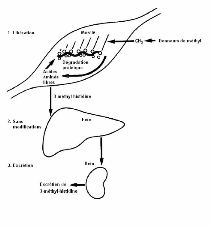
Figure 3 : La 3-méthyl-histidine (3-MH) : un index de la dégradation des protéines
myofibrillaires du muscle chez l’humain
Figure 4 : L’impédancemétrie (BIA) : placement des électrodes Figure 5 : La mesure anthropométrique des plis cutanés
Figure 6 : La pince anthropométrique
Figure 7 : L’absorptiométrie biphotonique (DXA) : le modèle à 3 compartiments
Figure 8 : La créatininurie des 24 h en fonction de l’âge dans la dystrophie de Duchenne de
Boulogne
Figure 9 : Recyclage du glutathion
SOMMAIRE DES TABLEAUX
Tableau 1 : L’impédancemétrie (BIA) : conditions de mesure Tableau 2 : Préparation de la gamme glutathion total (GSH) Tableau 3 : Préparation de la gamme glutathion oxydé (GSSG)
Chapitre 1 INTRODUCTION
La prise orale de glutamine (Gln) permet une amélioration de l’évolution clinique chez l’humain en situation de stress (1). Cependant, la place de la Gln dans la prise en charge nutritionnelle des enfants atteints de pathologie reste encore débattue.
La Gln est l’acide aminé libre le plus abondant de l’organisme humain (2). Elle est synthétisée principalement dans le muscle et utilisée par les cellules à renouvellement rapide, comme les cellules intestinales (3, 4), les lymphocytes (5) et les fibroblastes (6). La Gln participe à la synthèse des protéines, des acides nucléiques et des bases puriques et pyrimidiques (7). Elle est un précurseur pour la néoglucogenèse hépatique (8), rénale (9) et intestinale (10). La Gln assure un rôle central dans la régulation de l’homéostasie azotée, par le transport inter organe d’azote, afin de permettre la synthèse d’autres acides aminés (11). Enfin, elle contribue à la régulation du métabolisme acido-basique (12).
Chez l’humain, en situation de stress (chirurgie, cancer, infections) la Gln est exportée des muscles squelettiques et la concentration intramusculaire de Gln est effondrée (13). Pour cette raison, la Gln, acide aminé non indispensable est considérée « conditionnellement indispensable » lorsque l’organisme ne peut la synthétiser en quantité suffisante (7, 14).
En thérapeutique humaine, la Gln stimule l’anabolisme protéique. Chez le sujet sain, elle stimule la synthèse protéique (15, 16). En pathologie pédiatrique, elle inhibe la dégradation protéique. Chez l’enfant atteint la dystrophie de Duchenne de Boulogne (DDB), maladie de transmission récessive liée au chromosome X qui se caractérise par une perte progressive et sévère de la masse musculaire, la prise orale de la Gln sur une courte période (5 h) inhibait la dégradation protéique et la synthèse endogène de Gln (17). Mais rien n’indiquait que cet effet persistait avec le temps et les mécanismes d’actions restaient encore méconnus. Chez la souris
mdx, un modèle animal de la DDB (18), la Gln a été identifiée comme présentant un intérêt
thérapeutique, car elle améliorait les performances des animaux (19).
Ce travail comprend deux parties, la première partie comprend une revue de la littérature, sous forme d’article de synthèse qui porte sur la supplémentation en Gln en pathologie
expliquer les bénéfices thérapeutiques de la Gln sont abordés, ainsi qu’une synthèse et évaluation critique des essais cliniques chez l’enfant. Nous présentons aussi de différents modèles animaux de pathologie pédiatrique, afin de mieux comprendre les mécanismes mis en jeu lors d’une supplémentation en Gln (article de synthèse).
La deuxième partie de ce travail est consacrée à notre contribution personnelle avec l’exposé de la méthodologie, des études publiées ou en cours de soumission et une discussion résumée des différentes études avec perspectives thérapeutiques.
La première étude concerne l’effet d’une administration orale de Gln sur le métabolisme protéique d’enfants atteints de DDB. La prise de Gln sur une période de 5 h a montré un effet d’épargne protéique, dû à une inhibition de la dégradation protéique corporelle totale (17). La courte période utilisée incitait à déterminer si cet effet persistait sur une période plus longue (10 jours) d’administration et s’il était spécifique (article 1).
La deuxième étude est une approche des mécanismes physiopathologiques mis en jeu au niveau du muscle dystrophique de la souris mdx lors d’une supplémentation en Gln. Cette connaissance permettra de mieux comprendre le mécanisme de l’effet inhibiteur de la Gln sur la dégradation protéique observé chez l’enfant myopathe (article 2).
La démonstration que l’effet d’épargne protéique de la Gln persistait après 10 jours, nous a incité à évaluer dans une troisième étude le bénéfice clinique associé à la prise de Gln au long cours (4 mois) pour freiner la progression de la maladie en évaluant notamment l’effet sur la masse et la fonction musculaire (article 3).
Enfin, l’évaluation de l’effet anabolique d’une supplémentation en Gln nécessite une estimation fiable et précise de la composition corporelle. Cependant, les techniques pour évaluer la composition corporelle chez l’enfant en pathologie (notamment dans la DDB) sont peu testées. Dans les deux dernières études, nous avons évalué la pertinence (article 4) et la sensibilité (article 5) de l’impédancemétrie (BIA) par rapport à une technique de référence (eau marquée et absorptiométrie biphotonique) et la mesures des plis cutanés.
Chapitre 2
REVUE DE LA LITERATURE
Supplémentation en Gln chez l’enfant malade
TRAVAIL EN COURS DE SOUMISSION A PUBLICATION
Le travail est présenté sous forme d’article de synthèse. L’article (en anglais) est précédé d’un résumé en français.
E. Mok, R. Hankard. Glutamine supplementation in sick children: Is it beneficial? Soumis à
Supplémentation en Gln chez l’enfant malade
Glutamine supplementation in sick children: Is it beneficial? E. Mok, R. Hankard
Soumis à publication dès acceptation de l’article 3
Résumé
La Glutamine (Gln), acide aminé non indispensable, possède plusieurs fonctions cellulaires, et peut devenir « conditionnellement indispensable » dans les situations de stress où la production de Gln ne suffit plus à couvrir les besoins de l’organisme. Le but de cet article de synthèse est de préciser la place d’une supplémentation en Gln en pédiatrie. Pour cela, nous présentons une évaluation critique de la littérature concernant l’intérêt thérapeutique d’une supplémentation en Gln de la période néonatale jusqu’à l’adolescence. Les différents mécanismes d’actions sont discutés ainsi que les modèles animaux en pathologie pédiatrique. En synthèse, les données de la littérature concernant le bénéfice clinique d’une supplémentation en Gln dans la pathologie pédiatrique restent à ce jour contradictoires et insuffisantes, notamment chez l’enfant né prématurément ou en pathologie pédiatrique (maladie de Crohn, syndrome de grêle court, dénutrition, diarrhée, cancer, les grands brûlés ou polytraumatisés, la dystrophie de Duchenne de Boulogne, la drépanocytaire ou la mucoviscidose). De problèmes méthodologiques ont aussi été notés dans certaines études. En revanche, aucun effet biologique défavorable n’a pu être mis en rapport avec la Gln. Une meilleure compréhension des mécanismes mis en jeu est nécessaire, afin d’identifier les populations pédiatriques pour lesquelles le bénéfice clinique reste à tester.
Glutamine supplementation in sick children: Is it beneficial?
Elise Mok, PDt, MSc1,2,3 and Régis Hankard, MD, PhD1,2,3
Author affiliation:
1Pédiatrie multidisciplinaire - Nutrition de l’enfant, Centre Hospitalier Universitaire de Poitiers, Poitiers, France (EM, RH)
2EA 3813, Laboratoire Adaptation Physiologique aux Activités Physiques, Université de Poitiers, Poitiers, France (EM, RH)
3INSERM Centre d’Investigation Clinique 802, Centre Hospitalier Universitaire de Poitiers, Poitiers, France (EM, RH)
Correspondence should be addressed to: Professor Régis Hankard, MD, PhD, Pédiatrie
Multidisciplinaire-Nutrition de l’Enfant, Centre Hospitalier Universitaire de Poitiers, 2 rue de la Milétrie, 86021 Poitiers Cedex, France, telephone: 33 5 49 44 49 18, fax: 33 5 49 44 40 16, e-mail: r.hankard@chu-poitiers.fr
Sources of support: Supported by Les Fonds de la Recherche en Santé Québec PhD
Fellowship (to EM) and Le Prix de Nutrition de la Fédération Association Nationale pour les Traitements A Domicile, les Innovations et la Recherche awarded by La Société Francophone de Nutrition Entérale et Parentérale (to EM).
Running head: Glutamine in children
TABLE OF CONTENTS
ABSTRACT ... 20
INTRODUCTION... 21
GLUTAMINE MECHANISMS OF ACTION ... 22
Antioxidant capacity ... 23
Tissue protection ... 25
Immune function ... 26
Tissue metabolic function ... 27
Protein synthesis and degradation... 27
GLUTAMINE IN THE NEONATAL PERIOD... 29
Glutamine in breast milk... 30
Glutamine supplementation in premature neonates... 32
Parenteral glutamine in premature neonates ... 33
Clinical outcomes... 33
Protein metabolism ... 36
Enteral glutamine in premature neonates ... 38
Clinical outcomes... 38
Protein metabolism ... 43
Discussion... 44
GLUTAMINE IN PEDIATRIC PATIENTS WITH GASTROINTESTINAL DISEASE ... 46
Glutamine supplementation in infants with surgical gastrointestinal disease... 46
Glutamine supplementation in short bowel syndrome ... 49
Enteral glutamine in children with Crohn’s Disease ... 51
Glutamine supplementation in infants and children with diarrhea ... 54
Glutamine supplementation in malnourished infants and children... 55
Discussion... 58
GLUTAMINE IN PEDIATRIC ONCOLOGY PATIENTS ... 59
Glutamine supplementation on oral mucositis in children undergoing chemotherapy or bone marrow transplant/hematopoietic stem cell transplant... 60
Glutamine on immune function: implications for children with solid tumors and acute lymphoblastic leukaemia ... 62
Discussion... 64
GLUTAMINE IN PEDIATRIC PATIENTS WITH SEVERE BURNS / TRAUMA... 65
GLUTAMINE IN INHERITED DISEASES OF CHILDHOOD ... 68
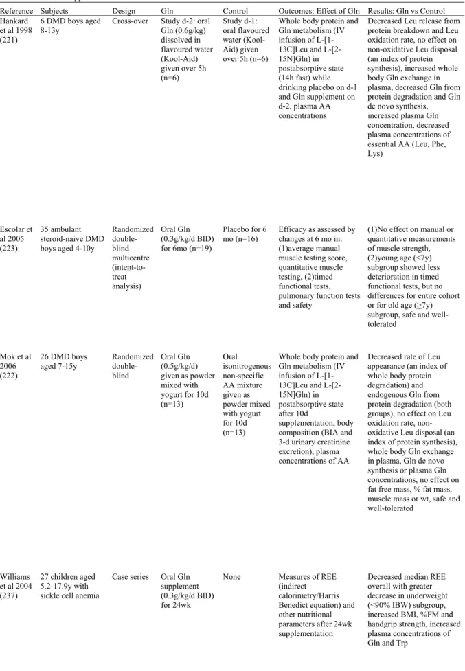
Glutamine supplementation in children with Duchenne muscular dystrophy... 68
Glutamine supplementation in children with sickle cell anemia ... 71
Glutamine supplementation in children with cystic fibrosis ... 73
SUMMARY/CONCLUSIONS ... 74
REFERENCES ... 77
TABLES ... 104
Table 1. Glutamine-supplemented parenteral nutrition in premature neonates... 104
Table 2. Glutamine-supplemented enteral nutrition in premature neonates... 106
Table 3. Glutamine supplementation in pediatric patients with gastrointestinal disease .. 110
Table 4. Glutamine supplementation in children with diarrheal disease / malnutrition.... 113
Table 5. Glutamine supplementation in pediatric oncology patients ... 115
Table 6. Glutamine in pediatric patients with severe burns / trauma ... 117
ABBREVIATIONS
Ala : alanine
ALL : acute lymphoblastic leukaemia Arg : arginine
ASK-1 : apoptosis signal-regulated kinase-1 BMT : bone marrow transplant
BSA : body surface area CNS : central nervous system
DMD : Duchenne muscular dystrophy
4E-BP 1 : eukaryotic initiation factor 4E-binding protein 1 ELBW : extremely low birth weight
EN : enteral nutrition
ERK 1/2 : extracellular signal-regulated kinase 1/2 Gln : glutamine
Glu : glutamate Gly : glycine GSH : glutathione
GSSG : oxidized glutathione
HSCT : hematopoietic stem cell transplant HSP : heat shock protein
IBD : inflammatory bowel disease ICU : intensive care unit
Ig : immunoglobulin
IGF1: insulin like growth factor 1 IV : intravenous
JNK : c-Jun N-terminal kinase LBW : low birth weight Leu: leucine
MAPK : mitogen-activated protein kinase mTOR : mammalian target of rapamycin NEC : necrotizing enterocolitis
NF-țB : nuclear factor-kappaB NICU: neonatal intensive care unit NOS : nitric oxide synthase
ORS : oral rehydration solution
PCDAI : Pediatric Crohn's Disease Activity Index Phe : phenylalanine
PN : parenteral nutrition
p70S6K : p70 ribosomal S6 kinase
rhGH : human recombinant growth hormone SAPK : stress-activated protein kinase SBS : short bowel syndrome
Ser : serine Thr : threonine
TNF-Į : tumour necrosis factor-alpha TPN : total parenteral nutrition VLBW : very low birth weight WHO : World Health Organization
ABSTRACT
Glutamine (Gln), a non essential amino acid, has several important cell specific functions and may become conditionally essential under certain stressful conditions. While pediatric patients are sick and highly stressed, they also undergo growth and development. Hence specific research on the role of Gln in children is essential. The purpose of this review is to provide a critical appraisal of the literature on Gln supplementation in various conditions or illnesses that affect children, from neonates to adolescents. First, a general overview of the proposed mechanisms for the beneficial effects of Gln is provided and subsequently clinical trials are discussed. Animal models on Gln administration in the context of these childhood conditions are also included. Despite safety, studies are conflicting and insufficient evidence is available on the benefits of Gln supplementation in pediatric patients. This includes premature infants, infants with gastrointestinal disease, children with Crohn’s disease, short bowel syndrome, malnutrition/diarrhea, cancer, severe burns/trauma, Duchenne muscular dystrophy, sickle cell anemia and cystic fibrosis. Moreover, methodological issues have been noted in some studies. Further mechanistic data is needed along with large randomized controlled trials in select populations of sick children, who may eventually benefit from supplemental Gln.
KEY WORDS :
Nutrition, Nutrition support, Therapy, Enteral, Parenteral, Oral, Feeding, Breast milk, Pediatrics, Neonate, Infant, Child, Adolescent, Growth, Childhood disease, Catabolic
INTRODUCTION
Glutamine (Gln) is the most abundant amino acid in the muscle and plasma of humans (1). Although Gln is a non-essential neutral amino acid, it is necessary for optimal growth of mammalian cells in tissue culture (2) and has important physiological functions. Apart from providing nitrogen for protein synthesis, Gln is a precursor for nucleic acids, nucleotides (3), hexosamines (4), the nitric oxide precursor-arginine (Arg) (5) and the major antioxidant-glutathione (4, 6). Gln is also an important oxidative fuel for rapidly proliferating cells such as those of the gastrointestinal tract (7) and immune system (3), reticulocytes (8), fibroblasts (9) and so on. It plays a central role in nitrogen transport between tissues (10), specifically from muscle to gut, kidney and liver. In addition to its role as a gluconeogenic substrate in the liver, kidney (11) and intestine (12), Gln is involved in the renal handling of ammonia, serving as a regulator of acid-base homeostasis (13). Present data also indicate that Gln functions as a signalling molecule (14), particularly under catabolic conditions.
Traditionally Gln is considered a non-essential amino acid, because it is synthesized by most tissue (skeletal muscle being the main producer and storage site) (15). Gln synthetase catalyzes the terminal step in Gln de novo synthesis and is a key enzyme in Gln metabolism (16, 17). In mammals, Gln synthetase expression is regulated by transcriptional and post-transcriptional mechanisms, i.e. increasing Gln synthetase mRNA in response to stress (e.g. glucorticoids) and regulation of Gln synthetase protein turnover in response to its product (Gln concentrations) (18). The importance of Gln at the whole body level is highlighted by the report of severe brain malformation resulting in multi-organ failure and neonatal death in 2 unrelated newborns with congenital Gln synthetase deficiency, in whom Gln was largely absent in plasma, urine and cerebral spinal fluid (19).
Under normal conditions Gln is released into circulation for consumption by other tissue, whereas during catabolic stress the production of Gln may be insufficient to meet the increased requirements by the gut, immune system/inflammatory cells, liver and kidneys. Demands are partly met by skeletal muscle proteolysis and release of large amounts of Gln to maintain normal concentrations in the plasma, resulting in depletion of Gln stores. Based on this abundant evidence, Lacey and Wilmore (10) suggested that Gln may become a conditionally essential amino acid for the critically ill.
In pediatrics several researchers have studied the efficacy of supplemental Gln in premature infants of low birth weight (LBW), who are highly stressed and have low energy and protein reserves (20). Similar to premature neonates, Gln supplementation may also be beneficial for other childhood conditions including; gastrointestinal disease, malnutrition, cancer, severe burns/trauma as well as genetic diseases of childhood. However, less data is available.
In addition to being sick and highly stressed, children are also in the process of growth and development. Hence specific research on the role of Gln in pediatric patients is necessary. The main purpose of this manuscript is to provide a critical review of the literature on Gln supplementation in various conditions/illnesses that affect children (from neonates to adolescents). First the proposed mechanisms of Gln action are reviewed in a general context, followed by a detailed description and critique of the clinical research on Gln supplementation in children. Fundamental research using neonatal/young animal models on Gln administration in relation to these specific childhood conditions is also described.
While it is well-established that Gln is a protein precursor as well as a major fuel and nucleotide substrate for rapidly proliferating cells (e.g. gut and immune system) (3, 7), additional mechanistic data has emerged to explain the apparent benefits of Gln. Gln can regulate the expression of many genes related to metabolism, signal transduction, cell defense and repair and can activate intracellular signaling pathways (14). In brief, Gln seems to affect antioxidant capacity, tissue protection, immune and metabolic function (21) as well as protein synthesis and degradation (14). The postulated mechanisms remain speculative and are by no means mutually exclusive, since Gln can provoke a number of different effects that interact with one another.
Antioxidant capacity
Glutathione. Gln is a precursor of the glutamate (Glu), for glutathione
(L-Ȗ-glutamyl-L-cysteinyl-glycine) synthesis, an important antioxidant in many cell types (22). Glutathione is present in the cell in reduced (GSH) and oxidized (GSSG) forms. The ratio of reduced to oxidized glutathione is the major regulator of the cellular redox potential that determines the antioxdant capacity of the cell (14, 22, 23). The effectiveness of glutathione protection in individual tissue depends on the tissue concentration of glutathione as well as the capacity of the tissue to import GSH and to export GSSG (24).
In vivo experiments in rats demonstrate that administration of Gln before ischemia/reperfusion
injury or surgical manipulation can enhance GSH concentrations and provide protection against oxidative stress in various tissues (e.g. cardiac, intestinal, lung) (25, 26). In humans,
Gln supplementation can attenuate GSH depletion in skeletal muscle following surgical trauma (27).
During critical illness, muscle concentrations of GSH decrease and a change in the redox status occurs, indicative of an elevated GSSG (24). Moreover, there is a correlation between the concentrations of Gln and GSH (24). Shifting the GSH/GSSG redox toward oxidizing conditions activates several signaling pathways, such as c-Jun N-terminal kinase (JNK), apoptosis signal-regulated kinase-1 (ASK-1), mitogen-activated protein kinase (MAPK) and the transcription factor nuclear factor-kappaB (NF-țB; a stimulator of the synthesis of pro-inflammatory cytokines and adhesion molecules) (22, 23, 28). Evidence also implicates oxidative stress as a potential regulator of NF-țB transactivation by MAPKs (in particular extracellular signal-regulated kinase 1/2 (ERK1/2) and p38) (29) which could lead to increased proteolysis in muscle (30). Moreover the cellular redox status seems to be related to the degree of muscle protein degradation (31, 32). Likewise there is significant literature on the role of inflammatory cytokines (interleukin-1,-6, tumour necrosis factor-alpha (TNF-Į)) and muscle-wasting (33).
Gln metabolism via entry into the citric acid cycle may allow activation of malic enzyme which will result in an increase in NADPH (15) and subsequently increase the GSH/GSSG ratio (14). Administration of Gln leads to an increased ratio of GSH/GSSG and reduces the activity of redox sensitive kinases subsequently preventing NF-țB activation and thus inhibiting the inflammatory response (23).
Experiments from our group in the mdx mouse model of muscular dystrophy (a condition associated with severe muscle-wasting) showed that in vivo Gln administration can reduce
GSSG in dystrophic skeletal muscle, hence increasing the ratio of GSH/GSSG (34). This was associated with decreased activation of MAPK (ERK1/2) pathway (34). Similar effects were observed in muscle of control mice, however, the magnitude was less (34). Thus in muscle tissue, Gln might affect the cellular redox state involving MAPK pathway.
Tissue protection
Heat shock protein (HSP). The HSPs serve as molecular chaperones that appear to repair
denatured/injured proteins or promote their degradation following irreparable injury. Gln has cell protective effects, as a potent enhancer of the expression of HSP25, HSP70, HSP72 and heme-oxygenase-1 in cell culture (35), in mutiple organs of both stressed and unstressed animal models (36), as well as in humans (37, 38). However, Gln depletion during the stress response can impair the expression of the major inducible HSP (HSP70), as shown in human lymphocytes (39). And recent evidence suggests that HSP70 expression is required for Gln’s protection against tissue injury and for attenuation of NF-țB activation and pro-inflammatory cytokine release (40).
In rats, pre-operative administration of Gln induces HSP70 expression and attenuates the inflammatory response by regulating nitric oxide synthase (NOS) activity in heart, lung and liver (41). Gln is a well-known precursor for Arg (5), which can increase nitric oxide formation as a result of enhanced NOS activity (6). However, in various models of human intestinal cells, Gln does not further increase nitric oxide production or inducible NOS mRNA following pro-inflammatory cytokine stimulation (42). Thus Gln’s effects on NOS activity might be tissue- or condition-specific.
The survival-promoting effects of HSP70 can also be attributed in part to the suppression of apoptosis, since reduced HSP expression in Gln-deprived cells together with their impaired antioxidant capacity may make them more susceptible to apoptosis (28).
Apoptosis. Gln starvation has been shown to induce apoptosis in intestinal epithelial cells (43)
and also renders human monocytic cells more susceptible to apoptosis induced by Fas ligand, heat shock or TNF-Į stimulation (44). In HeLa cells, Gln might also suppress ASK-1 and JNK/stress-activated protein kinase (SAPK) activation by Fas ligand (45). The effect of Gln in delaying spontaneous apoptosis in neutrophils may be mediated by the anti-oxidant effects of glutathione (46). Furthermore Gln may protect activated T cells from apoptosis, partially by up-regulating glutathione and Bcl-2 expression and inhibiting Fas (47).
Intestinal barrier function. As an important fuel for intestinal tissue and gut-associated
lymphoid tissue, Gln may contribute to gut barrier function (48). Gln is also involved in the biosynthesis of hexosamines which are important for maintaining gut wall integrity via surface mucin and glycoprotein-forming intracellular tight junctions, and thus may protect against bacterial translocation (4, 49).
Immune function
As a major fuel for immune cells, Gln is known to modulate immune function. More recently Gln has also been shown to have anti-inflammatory effects, modulating cytokine production, both in vitro (35, 50-52) and in vivo (40, 53, 54), possibly through decreased NF-țB activation (40, 51), a major transcription factor regulating immune and inflammatory responses.
Gln’s anti-inflammatory effects may also be related to enhanced HSP expression (35, 40). The induction of HSP response can attenuate pro-inflammatory cytokine release, which in turn depends on the cellular redox potential and consequently is regulated by the intracellular GSH/GSSG ratio (14) (as previously described).
Tissue metabolic function
Gln can preserve tissue metabolic function in stress states. For instance, Gln enhances myocardial tissue metabolic function after ischemia/reperfusion injury in rats (26). Gln can also enhance ATP levels in oxidant stressed endothelial cells (55).
Glucose metabolism. While Gln plays an important role in gluconeogenesis (11, 12, 56),
evidence also suggests that Gln can improve insulin sensitivity and glucose disposal in patients suffering from critical illness (21, 57), a condition frequently associated with insulin resistance and subsequent hyperglycemia. Gln also plays a role as a signaling molecule in amino acid- and glucose-stimulated insulin secretion (58). Interestingly in rats with diet induced obesity, Gln supplementation induces insulin resistance in adipose tissue and reduces adipose mass, consequently attenuating insulin resistance and activation of JNK and inhibitory kappaB kinase subunit beta in liver and muscle, thus improving insulin signaling (59). These data suggest that Gln can beneficially influence insulin-dependent glucose metabolism.
Gln appears to regulate protein turnover in cultured rat skeletal myotubes, stimulating protein synthesis in stressed myotubes while inhibiting protein degradation in long-lived proteins. This may be related to the increase in HSP70 (60). There is also abundant literature to suggest that amino acids affect protein turnover via the mammalian target of rapamycin (mTOR) pathway (61, 62).
Protein synthesis. Amino acids, particularly branched chain amino acids, e.g. leucine (Leu)
stimulate skeletal muscle protein synthesis via activation of mTOR which in turn activates p70 ribosomal S6 kinase (p70S6K) and dephosphorylates eukaryotic initiation factor 4E-binding protein 1 (4E-BP 1), stimulating translation and protein synthesis (61, 62). Gln can induce growth and maturation of neonatal rat cardiomyocytes, which is associated with an increase in the mRNAs encoding contractile proteins and metabolic enzymes via activation of protein kinase A and mTOR (63). However, the action of Gln seems to be cell-type specific. For instance, in C2C12 myogenic cells, Gln and Leu have opposite effects on the mTOR pathway (64). Whereas Leu activates this pathway, Gln inhibits it by decreasing the phosphorylation states of mTOR (on serine (Ser)2448), p70S6K and 4E-BP1, with no effect on protein synthesis (64).
Protein degradation. The major proteolytic pathways in organs such as the liver, muscle and
intestine include: the autophagic/lysosomal (cathepsins), the calcium activated (calpains) and the ATP-ubiquitin-proteosome pathway (14, 33). The proteosome system (26S) is a highly selective proteolytic pathway (61). In visceral tissues (e.g. liver) autophagy is however the major proteolytic pathway and the only pathway known to be regulated by plasma amino acids (in liver and skeletal muscle) (61, 62). In autophagic proteolysis, several amino acids
have direct regulatory potential, possibly via a plasma membrane amino acid receptor/sensor and subsequent intracellular signaling (61).
Another line of evidence suggests that amino acids activate mTOR pathway which in turn suppresses protein breakdown by the autophagy/lysosomal pathway (61, 62). Amino acids can also control autophagic lysosomal proteolysis by inhibiting MAPK (ERK1/2) phosphorylation (65). Gln may cause its antiproteolytic effect through osmotic swelling (66), involving p38 MAPK pathway (67). An increase in cellular hydration acts as an anabolic signal, whereas cell shrinkage is catabolic and there is a close relation in the regulation of cell volume, Gln and protein catabolism (23).
On the other hand in the human gut, enteral Gln may attenuate ubiquitin-dependent proteolysis as demonstrated by decreased ubiquitin mRNA, whereas mRNAs encoding for cathepsins or calpains were not affected (68). Furthermore, in lung and muscle, Gln can also regulate its own production through a post-transcriptional mechanism in which Gln regulates Gln synthetase protein degradation (16, 17, 69), by facilitating its degradation by the 26S proteosome (18). Thus the presence of Gln could have a protein-sparing effect; sparing amino acids for protein accretion (70-72).
GLUTAMINE IN THE NEONATAL PERIOD
Gln is the predominant amino acid supplied to the fetus through the placenta and is specifically suited for its rapid development (73, 74). While normally present in the enteral diet, Gln has been excluded from parenteral nutrition (PN) because of low solubility and instability in solution. In the first weeks of life however, premature infants receive most of
their nutrients from PN which is Gln-free (75). The sudden cessation of Gln supply from the mother to premature infants, who are already stressed and undergoing rapid growth may be detrimental (76). Whereas plasma Gln concentrations normally increase during the first days of life in newborn infants breastfed ad libitum (77), selective amino acid deficiencies have been reported in neonates suffering from acute illness; including reductions in serum Gln and Arg in infants who have necrotizing enterocolitis (NEC) that may predispose them to the illness (78). It has been suggested that in catabolic conditions premature infants are not able to synthesize sufficient Gln to meet demands and in these conditions Gln may become a conditionally essential amino acid (10).
Glutamine in breast milk
In addition to providing an ideal nutritional composition for the neonate, breast milk contains specific nutrients such as Gln that may influence gastrointestinal development and can modulate immune, metabolic and inflammatory responses (79). Animal models suggest that early weaning in conjunction with the lack of Gln intake impairs immune (80-82) and intestinal function (83, 84). Deprivation of dietary or endogenously synthesized Gln results in a breakdown in the intestinal epithelium of artificially reared neonatal rats, whereas Gln supplementation may help to maintain intestinal integrity (85) and reduce intestinal inflammation induced by lipopolysaccharide (53). In young mice, Gln supplementation reverses impaired macrophage function resulting from early weaning (82, 86) and reverses the lower concentrations of protein and DNA in muscle and liver (81). Similarly, in the early weaned piglet, dietary Gln supplementation influences immune cell function in a beneficial manner (87) and maintains intramuscular Gln concentrations (80). Supplying Gln to the weaning diet of piglets also prevents intestinal atrophy (83, 84, 88) and improves barrier
function (88) as well as gut adaptation (83, 84, 88), leading to increased body weight gain (83, 84). Both newborn and weaning piglets also show improved intestinal mucosa metabolic activity with Gln treatment (89) which could lead to increased barrier function. In addition to enhanced intestinal oxidative-defense capacity (as indicated by increased intestinal glutathione concentrations) the beneficial effects of Gln on intestinal development could be mediated by the increased expression of genes required for cell growth and removal of oxidants, while down-regulating those that promote oxidative stress and immune activation (84).
In extremely low birth weight (ELBW) infants, the beneficial effects of breast milk ingested in the neonatal intensive care unit (NICU) on developmental outcomes at 18 months of age (90) persist at 30 months (91). While PN is Gln-free, infant formulas also have lower Gln concentrations than in breast milk (92), and heat sterilization of infant formulas can further lower the concentrations of Gln by more than 60% (93). Agostoni et al (92) reported lower free Gln concentrations in standard infant formulas compared to breast milk collected from 40 healthy lactating mothers after delivery of term infants at age 1 month. And similar to previous reports (94), they observed that glutamic acid and Gln accounted for most of the free amino acids in breast milk (92). Thus suggesting that enrichment of infant formulas with non protein nitrogen components (particularly Gln and glutamic acid) could be beneficial. The same group followed 16 healthy exclusively breastfeeding mothers after delivery of term infants and showed that the concentrations of free glutamic acid and Gln increased by 2.5 and 20 fold, respectively with progressing lactation, representing >50% of total free amino acids by 3 months (95). To assess the potential influence of gestational age and duration of lactation, Jochum et al (96) measured the content of free and protein-bound Gln in transitional and mature breast milk of 40 healthy mothers after term and preterm delivery. While the time
of delivery had no influence on the free Gln or total Gln concentration, free Gln concentrations increased during maturation of lactogenesis, similar to previous reports (95, 97, 98). In contrast, total Gln and protein-bound Gln concentrations decreased with the duration of lactation and this correlated with the decrease in total protein concentration in mature breast milk (96). These data highlight the need to better define the role of Gln in the neonatal period and the possible benefit of supplemental Gln.
Glutamine supplementation in premature neonates
Experimental data from in vitro and neonatal animal models suggest that Gln may provide protective effects against complications associated with prematurity (99-106). For instance, Gln may be beneficial in the prevention of retinopathy of prematurity via inhibition of retinal neovascularisation, as demonstrated in the oxygen-induced retinopathy mouse model (99). Gln may also be beneficial for nutrition in neonatal sepsis. Whereas hepatic oxidative metabolism is impaired during neonatal sepsis (100-102), Gln has been shown to reverse this impairment in neonatal rat hepatocytes (101-103) and provides protective effects on metabolism in suckling rat endotoxemia, improving clinical status (104). The effectiveness of Gln in preventing liver damage in neonatal sepsis appears to be mediated via glutathione synthesis (102). Finally, in neonatal mice and rats with experimental NEC, Gln reduces intestinal injury (105, 106), via mechanisms inhibiting inflammatory cytokine release (105).
Gln is a principal fuel source in the jejunum during the suckling period (107). The supply of Gln from the luminal side is of critical importance for maintaining optimal mucosal structure and function as demonstrated in a Caco2 cell culture system and a weaning rabbit ileal loop model (108), which may have implications for premature neonates receiving PN. Gln
supplementation in infant rabbits receiving PN attenuates PN-associated liver injury (109). Neonatal piglets fed by PN show less mucosal atrophy and improved villus morphology when supplemented with Gln (110), although no effect on small intestinal growth or disaccharidase activity was observed (111) and disturbances in water balance have also been reported (112). Moreover, intravenous (IV) Gln does not acutely stimulate mucosa protein synthesis in healthy growing dogs (113).
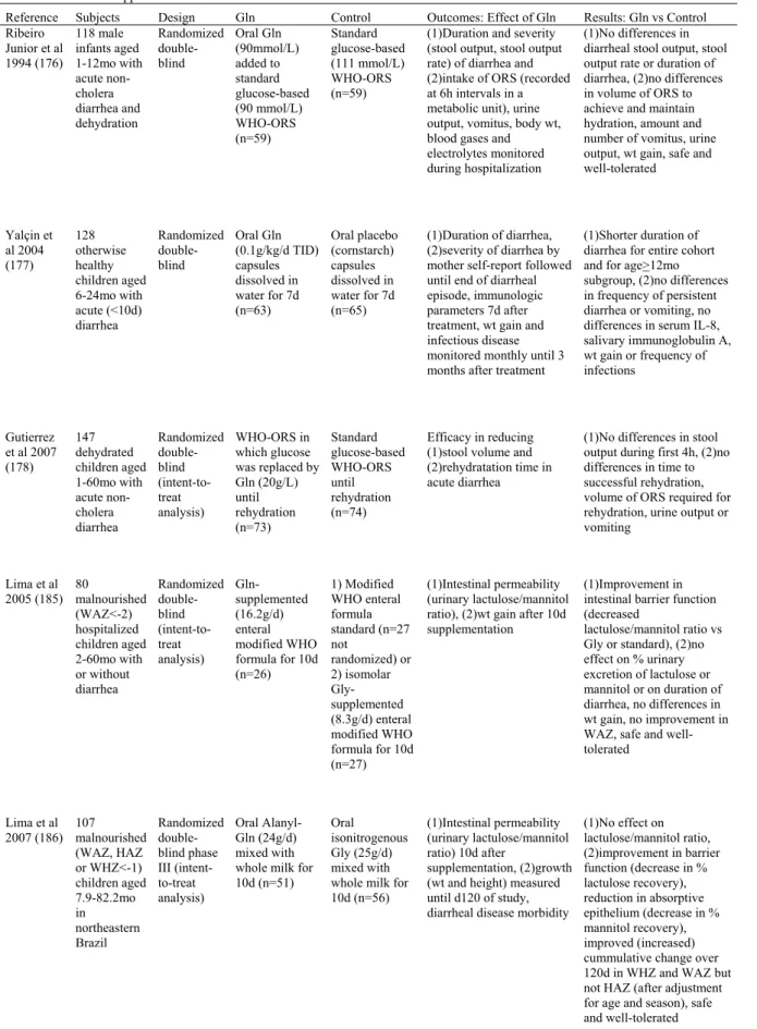
In premature human infants many prospective double blind randomized controlled trials have examined the effects of Gln supplementation. In this section we summarize the clinical trials (published in English) that tested the benefits of parenteral (Table 1) and enteral (Table 2) Gln on clinical outcomes in premature infants, in which a meta-analysis of 7 randomized controlled trials has recently been updated (114). We also describe studies that examined the effects of Gln on protein metabolism in this population (Table 1, Table 2).
Parenteral glutamine in premature neonates
Clinical outcomes
The first evidence to suggest that parenteral Gln appears safe and maybe considered a conditionally essential amino acid in premature infants was put forth more than a decade ago by Lacey et al (115) (Table 1). Although efficacy was not demonstrated in the entire cohort of very low birth weight (VLBW) infants (N=44), subgroup analysis in the infants with birth weight <800 g (n=24) showed that those supplemented with Gln (20% of amino acids) had fewer d on PN (13 versus 21 d, p<0.05), required less time to full feeds (8 versus 14 d, p<0.05) and needed less time on a ventilator (38 versus 47 d, p<0.05) compared to standard
isonitrogenous isocaloric PN. The positive results however, were based on subgroup analyses, follow-up of recruited infants was incomplete and intention-to-treat analysis was not performed. Thompson et al (116) further demonstrated that parenteral Gln may reduce the time to establish full feeds and appears to be well-tolerated and safe in a group of 35 ill ELBW neonates randomized to standard PN supplemented with Gln (16% of amino acids) or standard PN containing an isocaloric isonitrogenous amino acid solution. The primary outcome (median d to achieve full enteral nutrition (EN)) was significantly shorter in the Gln group (Gln: 13 d versus control: 21 d, p<0.05), whereas other clinical outcomes (growth, infection, number of episodes of sepsis, NEC or age at discharge) did not differ. Because the study did not achieve the calculated sample size of 120 infants, the risk of type II error cannot be ruled out for these outcomes. It is also not clear whether groups differed with respect to enteral feeding with either breast milk or preterm formula (that was started on d-3 of life) and if this was controlled for in the analysis. Although sample sizes were small, both trials were generally of good quality and provided evidence for improved feeding tolerance with parenteral Gln.
More recently Li et al (117) examined the effects of PN supplemented with alanyl-Gln dipeptide for more than 2 weeks in 53 premature infants of LBW. Gln-supplemented infants required fewer d on PN (24.8 versus 30.8d, p<0.05), had shorter hospital stays (32.1 versus 38.6d, p<0.05) and fewer episodes of hospital-acquired infections (0.96 versus 1.84 times, p<0.0001) compared to infants who received routine PN. They also regained birth weight sooner (8.1 versus 10.4 d, p<0.05), whereas there were no differences between groups for body weight or head circumference. The results should be interpreted with caution due to limitations in the methodology. It was not clear whether treatment allocation was randomized or whether care-givers or assessors were blinded to the intervention. In addition, follow-up of
recruited infants was incomplete. Although 68 infants were enrolled, 15 infants were excluded from the analysis because of insufficient time on PN (<2 weeks) and intention-to-treat analysis was not reported.
Pointdexter et al (118) performed the largest multi-centre trial to determine whether early PN supplemented with Gln reduces the risk of mortality or late onset sepsis in ELBW infants. Within 72 h after birth, 1433 infants were randomly assigned to receive either a standard IV amino acid solution (control) or an isonitrogenous amino acid solution with 20% Gln, whenever they received PN, up to 120 d of age, death or discharge from hospital. Safety was also assessed in a subset of 141 ELBW infants by measuring plasma concentrations of amino acids and ammonia after infants had received study PN (2.3+1.0 g/kg/d amino acids) for approximately 10 d (119). While parenteral Gln supplement increased plasma Gln concentrations with no apparent biochemical risk in ELBW infants, Gln did not reduce the incidence of death or late onset sepsis (Gln: 51% versus control: 48%; RR [95% CI]: 1.07 [0.97-1.17]). There were no differences between groups in the number of episodes of late onset sepsis, NEC, d on ventilator, length of hospital stay, d to first and full enteral feeds, feeding intolerance or growth. Moreover, infants who received Gln required more d of PN support. Although apparently safe in ELBW infants, the authors concluded that parenteral Gln supplementation does not reduce mortality or late onset sepsis and its routine use cannot be recommended. The lack of significant effect could be explained by a number of factors. Firstly, the primary outcome (death or late onset sepsis) could be influenced by other factors during the clinical course. In addition, as in previous studies (115-117) that used isonitrogenous controls (to ensure the specific effect of Gln), the overall amino acid intake may have been inadequate in the Gln group, as a consequence of the substitution of 20% of the standard amino acids with Gln. Specifically, in order to make the supplements
isonitrogenous, amino acids (including essential) were removed from the Gln-containing supplement, which could exacerbate specific amino acid deficiencies (especially if the PN period is prolonged). And although plasma amino acids were similar between groups, comparing plasma amino acid concentrations may not represent a valid marker for nutrient equivalence, since plasma amino acids may not reflect whole body amino acid concentrations or tissue concentrations. Furthermore, because infants in both groups did not receive the targeted amino acid intake of 3.0 g/kg/d until 10 d of age and most had also received small volumes of EN, the delivery of a sufficient dose of Gln may have been inconsistent. Moreover differences in enteral intake (formula or breast milk) may limit comparability of nutrient intakes between study groups.
Protein metabolism
2 small randomized controlled trials in LBW infants examined the effects of Gln-supplemented PN on whole body protein metabolism using primed continuous IV infusions of essential amino acid tracers (70, 120) (Table 1). Des Robert et al (120) studied 13 LBW neonates on postnatal d-3, while they received exclusive PN that was supplemented with Gln (0.5 g/kg/d) or an isonitrogenous Gln-free amino acid solution for 24 h. Compared to an isonitrogenous amino acid supplement, Gln decreased the rate of plasma Leu appearance, Leu release from protein breakdown (an index of whole body proteolysis; -16%, p<0.05) and rate of Leu oxidation (-35%, p<0.05). There was also however a decrease in non-oxidative Leu disposal (an index of whole body protein synthesis; -20%, p<0.05) and thus net Leu balance (protein balance) did not differ between groups. Plasma Gln concentrations were higher in Gln versus control, whereas plasma ammonia did not differ. Although parenteral Gln failed to enhance estimates of protein synthesis, Gln may preserve body protein as it suppressed Leu
oxidation and protein breakdown in LBW infants. In addition to the small sample size, the failure to enhance protein synthesis, may have also been due to insufficient amino acid availability since whole body protein kinetics were assessed on d-4 of life (when amino acid intake was 2 g/kg/d in both groups) before infants received an optimal amino acid intake of 3 g/kg/d (121).
Kalhan et al (70) examined the effect of 0.6g/kg/d Gln-supplemented PN for 3-5 d on whole body protein and Gln kinetics in a carefully selected population of 20 clinically stable LBW infants, between d-1 and -2 after birth. Compared to an isonitrogenous control, Gln-supplemented PN resulted in significantly lower rates of appearance of phenylalanine (Phe) and Leu nitrogen and a non-significant decrease in the rate of appearance of Leu carbon. Gln also suppressed the endogenous rate of Gln synthesis. There was no significant difference in urea turnover between the 2 groups. The results suggest that parenteral Gln supplementation at 0.6 g/kg/d decreases whole body protein breakdown and Gln de novo synthesis in clinically stable LBW infants and may be beneficial in selected populations of LBW infants. The carefully selected population of clinically stable infants limits the application of the results to other groups of premature neonates. Moreover, the use of a higher dose of Gln makes comparisons with other studies difficult.
Interestingly, the same group demonstrated that the suppression of proteolysis and protein oxidation in response to an acute increase in parenteral amino acids (without Gln) was not evident when the amino acid infusion was continued for a prolonged period in both acutely ill (71) and clinically stable LBW infants (72). The only exception was when amino acids were supplemented with Gln, whereby a prolonged infusion resulted in a sustained inhibition of whole body proteolysis and reduced Gln de novo synthesis. Taken together with previous
studies (70, 120), Gln supplementation may have a protein-sparing effect in preterm infants decreasing whole body protein breakdown and Gln de novo synthesis thereby “sparing” the increased amino acids for protein synthesis.
Enteral glutamine in premature neonates
Clinical outcomes
Neu et al (122) conducted a double-blind randomized trial to test whether enteral Gln supplementation for VLBW infants decreases morbidity (Table 2). 68 premature neonates were assigned to a Gln-supplemented premature formula or a non-supplemented standard premature formula between postnatal d-3 to d-30. The Gln supplemented group initially received a dose of 0.08 g/kg/d Gln which was increased to a maximum of 0.31 g/kg/d Gln by d-13. The Gln group had better tolerance to enteral feedings (fewer % of d with no oral intake in Gln: 8.8% versus controls: 23.8%, p<0.01). Episodes of hospital-acquired sepsis were 4/35 and 10/33 in Gln and control group, respectively. Moreover, when controlling for birth weight, the estimated odds of developing sepsis was 3.8 times higher for control versus Gln (95% CI: 1.01-14.18). Analysis of T cell subsets showed a blunting of the rise in HLA-DR+ and CD16/CD56 in the Gln group. There were however, no significant differences between groups for cases of NEC, growth or length of stay. Whereas the plasma concentrations of alanine (Ala), glycine (Gly), Ser, threonine (Thr), Phe and total non-essential amino acids were lower in the Gln-supplemented infants after 2 weeks supplementation, there were no differences between groups for plasma concentrations of Gln, Glu or ammonia (123). The authors speculated that the lower plasma amino acid concentrations in infants fed Gln were the result of enhanced uptake of these amino acids for gluconeogenesis and provide evidence
of reduced tissue catabolism. A secondary analysis of the initial trial also provided evidence for decreased hospital costs (124). While the control used is comparable to routine clinical practice, the study design cannot ensure the specific effect of Gln as the differences between feeding groups might result from higher intakes of nitrogen or energy with Gln supplementation. The authors however, chose not to use a third group with an isonitrogenous control due to recruitment constraints. Although the trial was small, these initial results provided evidence for better tolerance to enteral feedings and lower sepsis rates in VLBW infants receiving enteral Gln supplementation.
Barbosa et al (125) conducted a randomized controlled pilot study to evaluate the tolerance and clinical impact of enteral formula supplemented with 0.3 g/kg/d Gln for 5 days versus an equal dose of casein in 9 critically ill infants aged 1-24 months. Although Gln was well-tolerated, the study was underpowered to detect differences in septic complications (control: 3/4 versus Gln: 1/5, p=0.10), mortality (control: 2/4 versus Gln: 0/5, p=0.10) or other outcomes (ventilator use, length of stay in intensive care unit (ICU) or in hospital). It was also not reported in the inclusion criteria whether the study population of infants were preterm or term.
Vaughn et al (126) conducted a large multi-centre trial to test whether enteral Gln supplement decreases the incidence of hospital-acquired infection and other morbidities in 649 VLBW infants. Within the first 7 d of age, infants were randomly assigned to enteral Gln supplement (0.3 g/kg/d, 3% Gln in sterile water) or placebo (sterile water) given at the same time but separate from feedings for the first 28 d. There were no differences between groups for the primary outcome (nosocomial sepsis between 7 d and 36 weeks postmenstrual age; Gln: 30.9% versus control: 33.7%). However, gastrointestinal dysfunction (2.5 versus 7.5%,
p<0.01) and severe neurological sequelae among survivors (Grades 3 and 4 intraventricular hemorrhage and paraventricular leukomalacia; 10.4 versus 15.1%, p=0.08) were less frequent in Gln versus control, respectively. There were no differences in the occurrence of suspected sepsis, pneumonia, urinary tract infection, meningitis, NEC, retinopathy of prematurity, oxygen use at 36 weeks or mortality. Growth, age and weight at discharge were also similar. Whereas enteral Gln does not appear to decrease nosocomial sepsis in VLBW infants, the study may have been underpowered to detect a significant difference, as dropout rate was higher than anticipated (i.e. 105 infants exited the study before completion). Also the centre to centre variation in this and other multi-centre trials (118) may have blunted differences in outcomes (e.g. sepsis), since nutrition and infection control practices may differ among centres and may mask some of the differences that might be apparent in a single facility. While the study provides further evidence to suggest that enteral Gln improves feeding tolerance and may prevent central nervous system (CNS) morbidity these positive results are based on secondary endpoints and subgroup analyses. Furthermore, the Gln dose was based on birth weight and was not adjusted for interval changes in weight. Therefore the dose administered may have been inadequate due to rapid growth during the early neonatal period.
The apparent improved feeding tolerance in VLBW infants receiving enteral Gln in previous studies (122, 126) cannot be explained by enhanced mesenteric blood flow (127). It seems that in premature infants without acute illness and tolerating exclusive EN, mesenteric blood flow remains stable after 14 d of age and does not appear to be influenced by enteral Gln.
In contrast to previous reports on Gln-enriched EN in VLBW infants (122, 126), Van den Berg et al (128) found no improvement in feeding tolerance, as assessed by the median d to reach full enteral feeds (Gln:13 d versus control:13 d; hazard ratio [95%CI]: 1.19[0.79-1.79]).
In this randomized controlled trial, 102 VLBW infants received either enteral Gln supplementation or an isonitrogenous control (Ala) added to breast milk or preterm formula in increasing doses from d-3 to d-30 of life to a maximum dose of 0.3 g/kg/d Gln. There were also no differences between groups for other variables of feeding tolerance (age at which PN was discontinued, d of no enteral feeding), NEC or growth. However, the Gln-supplemented group had a lower incidence of >1 serious infections (sepsis, meningitis, pyelonephritis, pneumonia, arthritis) compared with the isonitrogenous control group (Gln: 50% versus control: 76%; OR[95%CI]: 0.32[0.14-0.74]. Other short-term outcomes (patent ductus arteriosus treated with indomethacin or surgical ligation, mechanical ventilation, supplemental oxygen, retinopathy, age at discharge from NICU, age at discharge from hospital or death) were not significantly different. Gln did not alter plasma concentrations of Gln, Glu or other amino acids (129). Although safe at the dose provided, Gln-enriched EN did not improve feeding tolerance or other short term outcomes in VLBW infants. However, because Gln reduced infectious morbidity, the use of Gln-enriched EN in VLBW infants deserves further consideration. Comparison however with other studies is made difficult because of the use of different feeding guidelines for the introduction or with-holding of enteral feeds. Whereas the choice of isonitrogenous control prevented the removal of amino acids from the Gln supplement in the present study, groups were made isonitrogenous by adding more amino acid (Ala) to the control group. The control group then received additional amino acid/nitrogen, which is not representative of daily practice. However given the limited sample size, a third comparison group (enteral formula routinely used in an NICU) was not feasible. The beneficial effect of enteral Gln on infection rate could not be explained by an increased number of bifidobacteria or lactobacilli in the intestinal microflora as demonstrated by a secondary analysis in a subset of 86 VLBW infants (130). Furthermore, Gln did not enhance the postnatal decrease in intestinal permeability, as assessed by the urinary lactulose/mannitol
ratio in a subset of 90 VLBW infants. Specifically, supplementation with Gln or isonitrogenous control equally decreased urinary concentrations of lactulose and increased urinary mannitol (131). More recently, follow-up of all surviving participants (n=77) revealed that Gln-enriched EN in VLBW infants may lower the incidence of atopic dermatitis (OR [95%CI]: 0.13 [0.02-0.97]) during the first year of life, but has no effect on the incidence of bronchial hyperactivity or infectious diseases (132). Although outcomes were assessed by validated questionnaires, parental report of physician diagnosis of disease could be subject to reporting/information bias. Furthermore, allergic and infectious diseases can manifest themselves at an older age than at 1 y, which may have been too early to investigate these outcomes. Finally, the same group studied neurodevelopmental outcome at 2 y corrected age in a subgroup of 72 VLBW infants and found no beneficial effect of Gln-enriched EN during the neonatal period (133).
The effect of Gln-supplementation on long-term outcome of VLBW infants has also been reported by Korkmaz et al (134) who studied the effect of 4-months enteral Gln supplementation on growth. From d-8 through d-120 of life, 69 VLBW infants were assigned to enteral Gln (0.3 g/kg/d) supplement or placebo (sterile water) according to the order of admission to the NICU. Whereas growth parameters did not differ during the first 2 months of life, by the end of the third and fourth month, infants treated with Gln showed higher weight, length, head circumference, mid-upper-arm circumference and mid-thigh circumference compared to controls. The authors concluded that long-term enteral Gln in VLBW infants may lead to improvements in growth in a time dependent manner without any signs of Gln toxicity. Although this was a prospective controlled study, a complete description of masking, blinding or randomization procedures was not provided. Also, because the placebo (sterile water) was neither isocaloric nor isonitogenous to the Gln treatment, the enhanced growth
could have resulted from the effect of increased amino acid/nitrogen, since early provision of parenteral amino acids (without Gln) has been shown to improve growth parameters in VLBW infants (135).
Protein metabolism
Darmaun et al (136) determined the effect of enteral Gln on Leu and Gln metabolism in a subset of 11 VLBW neonates from the larger trial (122) (Table 2). Enteral Gln supplementation provided at low doses (<0.2 g/kg/d) from d-3 to d-10 of life did not inhibit whole body protein breakdown in VLBW infants. Leu release from protein breakdown (an index of whole body protein breakdown) was slightly but not significantly lower in the Gln group versus controls. Plasma Gln concentration, Gln release from protein breakdown or Gln
de novo synthesis did not significantly differ between groups. However, there was a trend
toward lower rates of Gln de novo synthesis in infants receiving Gln supplement. The failure to detect a significant effect of Gln on its own metabolism or on whole body protein breakdown could be explained by the small number of patients. Alternatively, the dose of Gln used in the current study was lower than that (0.5 g/kg/d Gln) previously shown to inhibit proteolysis in LBW infants (120).
Parimi et al (137) examined the effect of enteral Gln on whole body Gln and nitrogen kinetics in healthy growing LBW infants during the fasting (3 h after the last meal) and fed state. This study was the only to have 3 groups, where Gln-supplemented group was compared with an isonitrogenous control and enteral formula routinely used in the NICU. Between 10-74 d of age, infants were randomly assigned to: formula supplemented with Gln (0.6 g/kg/d; n=9), isonitrogenous amounts of Gly (n=9) or unsupplemented formula (n=8) for 5 d. During
fasting, the rate of appearance of Phe, Leu carbon and Leu nitrogen (measures of proteolysis) were not significantly different between groups. Compared with controls, enteral Gln resulted in an increased rate of urea synthesis, no change in Gln rate of appearance or plasma Gln concentrations. Similar effects were observed with Gly supplement but the magnitude was less. The authors concluded that enteral Gln does not affect Gln rate of appearance or whole body protein turnover in a specific group of healthy growing LBW infants, thus suggesting that Gln is primarily metabolized in the gut (and liver) and is associated with an increased rate of urea synthesis. Alternatively, because in healthy preterm newborns there is already a high rate of Gln turnover (85% of which is contributed by de novo synthesis) (138), this specific population of neonates could have been less sensitive to enteral Gln. This is in contrast to premature neonates with acute illness, whereby catabolic stress may provoke a greater need for exogenous Gln. In summary, while parenteral Gln seems to have beneficial effects on protein metabolism in preterm infants, no such effects have yet been reported with enteral Gln in this population. Hence the route of administration may need to be considered when interpreting the effect of Gln on outcome in premature infants.
Discussion
Although methodologically sound randomized controlled trials consistently demonstrate safety in VLBW infants (70, 115, 116, 118, 120, 122, 126, 128, 137), parenteral or enteral Gln supplementation does not appear to affect mortality (116, 118, 122, 126, 128), NEC (116, 118, 122, 126, 128), length of stay (115, 116, 118, 122, 126, 128) or growth (116, 118, 122, 126, 128). Moreover, the results are conflicting for other short-term clinical outcomes such as feeding tolerance (115, 116, 118, 122, 126, 128), serious infections/sepsis (116, 118, 122, 126, 128), ventilator use (115, 118, 122, 128) and severe neurological sequelae (126). Few


![Figure 1. Estimation du métabolisme protéique par la perfusion de [1- 13 C]leucine.](https://thumb-eu.123doks.com/thumbv2/123doknet/7978906.267346/122.892.104.792.512.994/figure-estimation-métabolisme-protéique-perfusion-leucine.webp)