HAL Id: dumas-02421795
https://dumas.ccsd.cnrs.fr/dumas-02421795
Submitted on 20 Dec 2019HAL is a multi-disciplinary open access archive for the deposit and dissemination of sci-entific research documents, whether they are pub-lished or not. The documents may come from teaching and research institutions in France or abroad, or from public or private research centers.
L’archive ouverte pluridisciplinaire HAL, est destinée au dépôt et à la diffusion de documents scientifiques de niveau recherche, publiés ou non, émanant des établissements d’enseignement et de recherche français ou étrangers, des laboratoires publics ou privés.
Quantitative measurement of lung vasculature on CT in
pulmonary fibrosis: correlation with clinical parameters
and prognosis
Loïc Falque
To cite this version:
Loïc Falque. Quantitative measurement of lung vasculature on CT in pulmonary fibrosis: correlation with clinical parameters and prognosis. Human health and pathology. 2018. �dumas-02421795�
AVERTISSEMENT
Ce document est le fruit d'un long travail approuvé par le
jury de soutenance et mis à disposition de l'ensemble de la
communauté universitaire élargie.
Il n’a pas été réévalué depuis la date de soutenance.
Il est soumis à la propriété intellectuelle de l'auteur. Ceci
implique une obligation de citation et de référencement
lors de l’utilisation de ce document.
D’autre part, toute contrefaçon, plagiat, reproduction illicite
encourt une poursuite pénale.
Contact au SID de Grenoble :
bump-theses@univ-grenoble-alpes.fr
LIENS
LIENS
Code de la Propriété Intellectuelle. articles L 122. 4
Code de la Propriété Intellectuelle. articles L 335.2- L 335.10
http://www.cfcopies.com/juridique/droit-auteur
1
UNIVERSITÉ GRENOBLE ALPES
Année : 2018
UFR DE MÉDECINE DE GRENOBLE
Mesure quantitative de la vascularisation pulmonaire en tomodensitométrie
dans les fibroses pulmonaires: corrélation avec les paramètres cliniques et
fonctionnels et le pronostic des patients
THÈSE
PRÉSENTÉE POUR L’OBTENTION DU TITRE DE DOCTEUR EN MÉDECINE
DIPLÔME D’ÉTAT
Loïc FALQUE
THÈSE SOUTENUE PUBLIQUEMENT À LA FACULTÉ DE MÉDECINE DE GRENOBLE Le 30/10/2018
DEVANT LE JURY COMPOSÉ DE Président du jury : Pr Christophe PISON Membres : Pr Gilbert FERRETTI Pr Bruno DEGANO Dr Sébastien QUETANT
Dr Julien COHEN (directeur de thèse)
L’UFR de Médecine de Grenoble n’entend donner aucune approbation ni improbation aux opinions émises dans les thèses ; ces opinions sont considérées comme propres à leurs auteurs.
6
Remerciements
En ce jour symbolique, j’adresse mes remerciements sincères aux membres du jury et à toutes les personnes présentes aujourd’hui et au quotidien.
Au Docteur Julien Cohen mon directeur de thèse pour son aide précieuse et ses conseils avisés. Il a une part importante dans la réalisation de ce projet.
Au Docteur Sébastien Quétant et au Professeur Sam Bayat, qui m’ont apporté leurs
grandes connaissances en Pneumologie et en Physiologie et avec qui j’ai eu plaisir à travailler.
Au Professeur Pison, qui m’a fait l’honneur d’accepter d’être le Président de ce jury. Il m’a apporté et transmis un enseignement et une formation de grande qualité pendant ces 4 années d’internat. Je le remercie pour son côté humain et la confiance qu’il m’a accordée.
Au Professeur Gilbert Ferretti et au Professeur Bruno Degano qui m’ont fait l’honneur d’accepter de faire partie de ce jury.
A Hélène Pluchart et Sébastien Chanoine, docteurs en pharmacie, à Cevdet Ozcelik, Meriem Benmerad et Ludovic Broche, pour leur contribution efficace dans ce projet.
A Marie Peeters et Galina Penkov, attachés de recherche clinique du service d’explorations fonctionnelles et respiratoires.
Aux praticiens hospitaliers avec qui j’ai eu la chance de travailler et d’apprendre : l’équipe de Pneumologie et de Physiologie respiratoire, mes chefs d’infectiologie qui m’ont permis de prendre du temps pour avancer dans ce travail et l’équipe de réanimation médicale et de médecine interne du CHU.
7 A l’équipe de Pneumologie de Chambéry, pour son accueil et sa très bonne ambiance de travail.
A mes parents, sans qui tous ces efforts n’auraient pas été possibles. Ils ont été un soutien sans faille tout au long de ces années.
A ma sœur Elodie, toujours présente, et pour nos moments de complicité.
A ma grand-mère, à Pascale, à mes oncles qui m’ont toujours soutenu et souvent encouragé, à mes cousins et cousines.
A ma grand-mère et mes deux grands-pères disparus et pour lesquels aujourd’hui j’ai eu une immense pensée.
A mes amis, au premier rang desquels figure « la meute » : Jeremy sans lequel le duo J-man et Chany n’existerait pas, Kevin pour la seguridad et « tu joues jeudi ? » et Sébastien présent depuis maintenant plus de 25 ans à mes côtés. Merci pour cette amitié très précieuse. Merci aussi pour leur bonne humeur et leur soutien.
A Damien pour ses répliques cultes et qui a encore du ketchup sur le front, Jérémie (à ben i’m semble) pour ce voyage aux US qui restera dans les mémoires, Clément R et ses programmes qui marchent quand même un peu, Nicolas L qui ne sous-estime jamais la première soirée, Audrey et Caroline, déjà présentes sur ma photo de classe de maternelle et Thomas D pour ses « pays » et « clients ». Merci pour leur fidélité, leur présence et pour les excellents moments que nous partageons toujours.
Aux « carabins » devenus aujourd’hui des amis : Yann pour toutes ces années de coloc et d’amitié, Asticot pour toutes ces soirées et ces fous rires, Clément dit Messmer pour ces précieux conseils à la salle, Kevin même s’il est pour l’OM, Arnaud F même s’il préfère le GET27, Arnaud P le beau gosse de la team, Cédric, Mariette et Cloé pour les sessions à Tencin, Lisa, Noémie et Marion pour leur amitié depuis maintenant près de 10 ans, Aline toujours présente, des sous conf aux baptêmes, Renaud pour sa solidité, Marvin pour ces 6 mois à Chambéry, Thibault A pour cette année partagée entre Chambéry et la réa, Kash et Antho pour les soirées ligue des champions, Guigui pour les petits dej au relais H, Matthieu M pour ces 6 mois de folie à Chambéry, Natacha pour ces 6 mois de co-interne, Manon, Estelle, Justine, Gulay, Clémence, Charlotte, Quentin, Kursat… pour tous les moments passés ensemble (à la BU, à la fac, en soirées, en vacances à Hossegor…).
8 A mes amis et collègues de la « Pneumo Team », avec lesquels j’ai un réel plaisir à
travailler : Hubert pour tous nos semestres partagés, Victor pour avoir été mon premier contact avec la pneumologie grenobloise et sa présence en soirée jamais rassasiée, Julian (Tu vas ?) pour son humour, sa bonne humeur et ses conseils, Louis Marie avec qui j’ai
probablement passé mes meilleurs gardes, Thomas à qui j’attends de remettre une fessée au foot, Justin pour ces soccers, Geoffroy Merycopter pour les hélicos et les nombreuses imitations, Margaux, Elodie, Léonie, Micka, Héloïse, Salomé, Malik, Giulia, Wassila, Romain, Marie, Cécile, Pauline, Julie, Mathilde, Johanna, Anne Laure…
A mes co-internes actuels en infectiologie et à tous mes anciens co-internes, que j’ai eu la chance de rencontrer et de côtoyer au cours de ces belles années.
A Lélé, pour m’avoir supporté et motivé ces dernières semaines.
A mes coéquipiers du Foot : Simon, Thomas R, Loïc L, Guillaume C, Benjamin N, Augustin, Florian D, Ramzy, Guillaume D, Florent L, Dysmas, Florian T…
Au personnel médical, infirmiers, infirmières, aides-soignantes, kinés, secrétaires pour leur travail quotidien. A leur façon ils m’ont aussi accompagné dans ce parcours.
9
TABLE DES MATIERES
Résumé ……… 10
Abstract ………....11
Généralités ...
Fibrose pulmonaire ………12
Vascularisation pulmonaire ………..16
Quantitative measurement of lung vasculature on CT in pulmonary
fibrosis: correlation with clinical parameters and prognosis ………. 18
Introduction ……….19
Material and methods ……….21
Results ………..25
Discussion ………36
Conclusion ………...39
Conclusion générale ………40
Conclusion signée par le doyen ………..41
Bibliographie ………...42
10
Résumé
Objectifs : Étudier la relation entre la mesure scannographique du volume, du nombre et de la
surface des vaisseaux pulmonaires et les paramètres cliniques et le pronostic des fibroses pulmonaires.
Matériel et méthodes : Les paramètres vasculaires suivants ont été rétrospectivement extraits
des scanners thoraciques de 24 patients présentant un pattern de pneumopathie interstitielle commune (PIC) probable ou certaine: volume vasculaire pulmonaire total (VVP), nombre de vaisseaux (NoV), nombre de vaisseaux <5 mm² (NoV5). Nous avons étudié les corrélations de ces variables avec le déclin de la CVF, de la DLCO et du KCO entre le diagnostic et le suivi. De plus, nous avons évalué pour chaque patient les corrélations de ces paramètres vasculaires avec les données démographiques des patients, les comorbidités, les événements aigus, le test de marche de 6 minutes (TM6) et le recours à l’oxygénothérapie.
Résultats : 24 patients avec pattern de PIC ont été inclus. L'âge médian était de 76 ans (ratio
interquartile = 12) avec un ratio M/F de 3. Il existait des corrélations significatives entre le VVP (R = -0,51, p = 0,01), NoV (R = 0,5, p = 0,02), NoV 5 (R = 0,46, p = 0,03) et le déclin annuel de la DLCO et entre le VVP et le déclin du KCO (R = -0,56, p = 0,009). En outre, il existait une association significative entre la désaturation au TM6 et le VVP (R = -0,52, p = 0,03) et entre le VVP et le TM6 pour la distance parcourue en pourcentage de la prédite (R = -0,59, p = 0,009).
Conclusions : Les paramètres vasculaires pulmonaires du scanner sont significativement
corrélés à la diffusion des patients atteints de pathologies infiltratives diffuses avec pattern scannographique de PIC.
11
Abstract
Objectives : To investigate the relationship between CT measurement of pulmonary vessels
volume, number and area and clinical parameters and prognosis in pulmonary fibrosis.
Material and methods : The following CT vascular parameters were retrospectively
extracted from the unenhanced chest CTs of 24 consecutive patients presenting with a CT-pattern of probable or certain UIP: pulmonary vessel volume (PVV), number of vessels (NoV), number of vessels <5 mm². We studied the correlations of those quantitative CT variables with the following functional follow-up parameters: yearly decrease of raw and percentage of decline of FVC, DLCO and KCO. Additionally, we checked the correlations of those parameters with patients’ demographics, relevant medical history, clinical events, the 6 minute walk test and needing of oxygen therapy.
Results : 24 patients with UIP scan pattern were included. The median age was 76 years old (Interquartile ratio =12) with a M/F ratio of 3. There were significant correlations between PVV (R= -0.51, p= 0.01), NoV (R= 0.5, p= 0.02), NoV 5 (R= 0.46, p= 0.03) and the yearly decline of DLCO and between PVV and the decline of KCO (R= -0.56, p= 0.009). Furthermore, there was a significant association between PVV and 6 MWT desaturation (R= -0.52, p= 0.03) and between PVV and 6 MWT for the distance walked in percentage predict (R= -0.59, p= 0.009).
Conclusions : Quantitative CT lung vascular parameters are significantly correlated to DLCO
12
Généralités sur la fibrose pulmonaire
La fibrose pulmonaire idiopathique (FPI) est la plus fréquente des pneumopathies interstitielles diffuses (PID) chroniques de l’adulte. La prévalence rapportée est de 10 à 60 cas/ 100 000 habitants, avec une incidence en augmentation en Europe et aux Etats Unis (3 à 9 nouveaux cas par an pour 100 000 habitants) (1). Elle répond à un pattern scannographique de pneumopathie interstitielle commune (PIC) qui lorsqu’il est dit typique signe le diagnostic de fibrose pulmonaire (2), idiopathique ou non selon qu’une étiologie est retrouvée lors du bilan (connectivites, médicaments, expositions professionnelles ou domestiques). Le scanner thoracique est donc un élément clé du diagnostic dans cette pathologie puisqu’il permet de sursoir, dans ce cas, à une preuve histologique, difficile à obtenir et risquée, par biopsie chirurgicale le plus souvent (3). Cette pathologie, bien qu’accessible à certains traitements anti fibrosants sous certaines conditions, est chronique et évolutive, le plus souvent vers l’insuffisance respiratoire chronique terminale et le décès, lorsque le patient n’est pas éligible à une transplantation pulmonaire. Son pronostic est péjoratif avec un âge moyen des patients au diagnostic entre 60 et 70 ans et une médiane de survie estimée à 3,8 ans (4). Les principaux critères pronostics de cette maladie sont l’âge, le sexe masculin, un faible périmètre de marche ou une désaturation inférieure à 88% lors du test de marche de 6 minutes, une diminution de 10% de capacité vitale en 6 mois, une diminution de 15% de DLCO en 6 mois, des images en rayons de miel étendues sur le scanner ou encore une hypertension pulmonaire (5). En revanche l’histoire naturelle et notamment la vitesse d’évolution de cette maladie et du déclin de la fonction respiratoire sont imprévisibles et très variables selon les patients (2). Avec des patients relativement stables dans le temps, d’autres dont la maladie progresse lentement et certains avec une aggravation à court terme et un déclin de la fonction respiratoire rapide. Selman et al en 2007, rapportaient les importantes variations dans
13 l’évolutivité des fibroses pulmonaires idiopathiques et cela, indépendamment des caractéristiques des patients au diagnostic (6). Cette évolution dans le temps est également majorée en fonction des exacerbations respiratoires présentées par les patients, dont on ne sait pas prédire la survenue. Ceci est récapitulé dans la figure 1.
Figure 1 : Différentes vitesses d’évolution et de déclin respiratoire dans les
fibroses pulmonaires idiopathiques
14 La vitesse d’évolution des patients n’est pas prévisible au moment du diagnostic de FPI. Pouvoir anticiper la rapidité de progression de la maladie est un objectif important qui permettrait de rapprocher la surveillance des patients rapidement évolutifs, de mieux sélectionner ceux qui sont éligibles à un traitement anti fibrosant ou qui doivent être rapidement référés à un centre de transplantation. Enfin cela permettrait également d’améliorer notre connaissance de l’histoire naturelle de la maladie.
Le scanner thoracique est de plus en plus utilisé, non seulement comme élément diagnostic ou de classification des pathologies interstitielles mais également comme outil d’évaluation du pronostic de ces maladies : l’importance de l’étendue des images de fibrose, ou une proportion élevée de réticulations ou de rayon de miel sont des facteurs impactant la mortalité (7) (8). Récemment, Jacob et al. ont montré que l’évaluation visuelle des images de fibroses était variable selon l’opérateur (9) et pouvait être moins précise que l’évaluation des images scannographiques assistée par ordinateur à l’aide de logiciels type Caliper (10). Caliper est un outil logiciel novateur développé par le Laboratoire de ressources d'imagerie biomédicale de la Mayo Clinic Rochester. Il permet la segmentation des zones du poumon en petits volumes et attribue à chacun le pattern prédominant. Parmi les paramètres analysés par Caliper, le plus prédictif de la mortalité des patients atteints de FPI était le volume vasculaire pulmonaire (VVP) (11). Celui-ci semble être également bien corrélé au pronostic des fibroses inclassables (12) et des pneumopathies d’hypersensibilités (13). Cependant ce logiciel n’est pas aisément disponible en pratique quotidienne, et certains paramètres vasculaires tels que le nombre et la taille des vaisseaux n’ont pas été testés.
15
Figure 2
Mortalité élevée dans
les fibroses
pulmonaires
Paramètres dynamiques :
- Déclin CVF +++
- Autres : Déclin
DLCO, TM6 …
Comment anticiper
l’évolution de ces
paramètres dans le
temps ?
Vascularisation
pulmonaire ?
Flaherty et al. « Prognostic Implications of Physiologic and Radiographic Changes in Idiopathic Interstitial Pneumonia ». AJRCCM 168, no 5 (septembre 2003): 543-48.
16
Vascularisation pulmonaire et volume vasculaire pulmonaire
La vascularisation des poumons est composée de 2 systèmes que sont la circulation bronchique et la circulation pulmonaire. La circulation bronchique est à visée nutritive pour le poumon. Elle lui apporte l’oxygène nécessaire à son fonctionnement. Les artères bronchiques naissent de l’Aorte puis suivent le trajet des bronches jusqu’au bronches terminales où elles donnent naissance aux capillaires puis aux veines bronchiques qui feront le chemin inverse jusqu’à la veine cave supérieure.
La circulation pulmonaire est un système circulatoire fonctionnel. Son but est de permettre l’oxygénation de tout l’organisme. L’artère pulmonaire, riche en CO2, fait suite au ventricule droit, pénètre dans les poumons au niveau du hile puis se ramifie jusqu'aux capillaires pulmonaires qui vont être au contact direct avec les alvéoles (par contact externe) et se charge en oxygène. L'artère pulmonaire contient du sang non oxygéné, après les échanges au niveau des alvéoles, les veines pulmonaires ramènent du sang oxygéné jusqu'au cœur gauche.
Les pathologies interstitielles diffuses sont dorénavant de plus en plus évaluées par des techniques de tomodensitométrie informatisée. Celles-ci permettent, de différencier de manière automatisée, les différents patterns scannographiques que l’on rencontre dans les PID et de les quantifier de manière précise. Parmi ces patterns, on retrouve le volume vasculaire pulmonaire. Celui-ci correspond au volume des artères et veines pulmonaires excluant les vaisseaux du hile pulmonaire. Le score de volume vasculaire pulmonaire est subdivisé en fonction de la taille du vaisseau (< 5 mm2, > 5 mm2, < 10 mm2). Ceux-ci sont ensuite additionnés et exprimés en volume. Les mesures dérivées de ces analyses informatisées ont
17 été testées pour leur capacité à prédire la mortalité des patients atteints de fibrose pulmonaire. Etonnement, le paramètre le plus fortement associé à la mortalité des patients était le volume vasculaire pulmonaire, avec un hazard ratio à 1.52 (1.4-1.65, p < 0.0001). En comparaison avec cela, dans cette même étude de Jacob (11), le rayon de miel évalué par logiciel automatisé avait un hazard ratio à 1.11 (1.03-1.2, p < 0.006) et la capacité vitale forcée à 0.97 (0.96-0.98 p < 0.0001). Le mécanisme à l’origine de ce résultat n’est pas encore bien compris, plusieurs hypothèses sont avancées comme un excès de pression négatif intra vasculaire lors de l’inspiration du fait d’une rigidité pulmonaire accrue ou bien encore une redistribution de la vascularisation vers des zones indemnes de fibrose du fait d’une surpression des vaisseaux engrainés dans celle-ci. Une autre étude, de 2007, s’intéressait aux lésions vasculaires de patients avec une pathologie infiltrative fibrosante en émettant l’hypothèse que celles-ci pouvaient précéder les lésions de fibrose. Une augmentation de la paroi vasculaire des artères et des veines avec un rétrécissement liminal sévère étaient retrouvée dans les zones de poumons atteints de fibrose. Dans les zones indemnes de fibroses étaient retrouvés des atteintes veineuses avec des occlusions veinulaires sans modification importante au niveau artériel (14). La signification de ces lésions reste indéterminée. Néanmoins, ces différents résultats suggèrent un lien entre vasculopathie et pronostic des pathologies infiltratives fibrosantes qui pourrait être une piste d’étude intéressante pour prédire l’aggravation ou la stabilité de ces pathologies. D’autres études ont également montré un lien entre pronostic et atteinte vasculaire pulmonaire : Shin et al, en 2016, avait montré chez des patients atteints de FPI, une corrélation de la survie avec le diamètre de l’artère pulmonaire. En effet la survie des patients était supérieure lorsque le diamètre de l’artère pulmonaire était inférieur à 32.3 mm ou que le ratio artère pulmonaire/aorte était inférieur à 1 (15). Ces résultats rejoignent indirectement ceux rapportant un pronostic défavorable de la présence d’une hypertension pulmonaire chez des patients atteints de fibrose pulmonaire.
18
Quantitative measurement of lung vasculature on CT in
pulmonary fibrosis: correlation with clinical parameters and
prognosis
Keywords : Pulmonary fibrosis; Computed tomography, lung; Pulmonary circulation; Image
processing; Prognosis.
Abbreviations
6 MWT: 6-minute walk test
CI 95%: 95 percent confidence interval CPI: Composite physiologic index
DLCO: Diffusion capacity for carbon monoxide FEV1: Forced expiratory volume in one second FVC: Forced vital capacity
HRCT: High resolution computed tomography ILD: Interstitial lung disease
IPF: Idiopathic pulmonary fibrosis
KCO: coefficient of diffusion of carbon monoxide normalized to alveolar volume NoV: Number of vessels
NoV5: number of vessels with a cross-section < 5 mm2 PAH: Pulmonary arterial hypertension
PF: Pulmonary fibrosis
PFT: Pulmonary function tests PVV: Pulmonary vessel volume TLC: Total lung capacity
TLVV: Total lung vessel volume UIP: Usual interstitial pneumonia
19
Introduction
Idiopathic pulmonary fibrosis (IPF) is the most common chronic interstitial lung disease in
adults. Its reported prevalence is 10 to 60 cases per 100,000, with an increasing incidence in
Europe and the United States (3 to 9 new cases per year per 100,000) (1). Chest CT
assessment is of key importance in this disease since a certain UIP pattern enables to avoid
lung biopsy, to confirm diagnosis and orient treatment (3). Even though IPF now benefits
from several specific antifibrotic medications, the natural history of the disease is still
characterized by progression, most often towards terminal respiratory failure and death, when
the patient is not eligible for a lung transplantation. The prognosis of IPF remains pejorative
with a mean patient age at diagnosis between 60 and 70 years and a survival median of 3,8
years (4).
The main prognostic criteria of IPF are age, male sex, low walking distance or desaturation of
less than 88% in the 6-minute walk test (6 MWT), a 10% decrease in forced vital capacity in 6
months, 15% decrease in DLCO in 6 months, the extent of honeycombing on the CT scan and
pulmonary hypertension (5). Collard and al demonstrated that evolution at six and twelve
months of physiologic variables such as FVC, DLCO and KCO are the best predictors of
survival in this pathologic condition (16). Despite our knowledge in terms of prognostic
factors in this disease, IPF is known to be highly variable in term of clinical evolution
independently of baseline patient characteristics, with slow progression of the disease in some
patients, and rapid progression in others. Better predicting the disease evolution may be of
key clinical importance since it may enable physicians to select the patients which are eligible
for anti-fibrotic treatment or which must quickly be referred to a specialized center.
Chest CT scan is increasingly used not only as an ILD diagnostic and classification tool but
20
and honeycombing on HRCT have shown a significant relationship with mortality (6) (7).
Recent studies have found that the visual evaluation of fibrosis images on the CT scanner is
observer-dependent (8) and less accurate than automated quantitative CT measurements (9).
Among those quantitative CT parameters, pulmonary vessel volume (PVV) has been shown to
be strongly predictive of mortality (10). However, the software used in those studies is not
readily available for clinical use and several parameters remain untested, such as the number
and size of vessels. Moreover, the relationship between the pulmonary vascular volume and
mortality has been assessed, but the relationship between this parameter and the decline of
pulmonary function and other clinical parameters remains unknown.
In this study, we hypothesized that in UIP patients, quantitative CT parameters such as total
lung volume, the number of pulmonary vessels (NoV), the number of pulmonary vessels with
a cross-section < 5 mm2 (NoV5) are associated with decline in lung function, specifically
forced vital capacity (FVC), Diffusion capacity for carbon monoxide (DLCO) and DLCO
corrected for alveolar volume (KCO). Our second objective was to check whether these
vascular parameters were related to patient demographics, patients comorbidities (pulmonary
hypertension, emphysema, oxygen-requiring), the 6 minute walk test, prognostic scores used
in these pathologies, and the occurrence of an acute event during the follow-up.
21
Material and methods
Study population
42 patients with interstitial lung disease followed up at the Department of Pulmonary
Medicine of Grenoble University Hospital were initially screened for inclusion in this study.
Inclusion criteria were the availability of an unenhanced chest CT, a probable or certain UIP
pattern on CT and a minimum follow up of 6 months.
Study design
We conducted a monocentric, retrospective study. Lung CT images were analyzed to assess
pulmonary vascular morphological parameters. We collected PFT data from the patients’
electronic charts, at 3 time points (initial visit, one year and final follow-up which could vary
from one to eight years after diagnosis in our patients) and we correlated the decline of FVC,
DLCO and KCO, known as major prognostic markers in interstitial lung disease (17) (18) to
vascular data as primary outcome. Our secondary outcomes were to evaluate an association
between demographic, biometric and clinical data with vascular parameters and between the
Composite Physiologic Index (CPI), a prognostic marker in IFP (19) and vascular parameters.
CT imaging
All patients underwent a chest CT scan with the following protocol: 120kV, slice thickness 0,625 mm spaced by 0,625 mm intervals. There were slight changes in CT-dosimetry during the length of the study, due to technical evolutions of the CT machines.
22
Images were processed with a custom made software based on the python programming
language (Python Software Foundation; Python Language Reference, version 2.7).
Segmentation of the aerated lung volume from the CT images was performed using a region
growing algorithm which included all voxels spatially connected with a density value ranging
from -1023 to -224 HU. Using a morphological operation, the blood vessels and bronchial
structures were included into the image mask. This consisted in a spherical morphological
closing with a 5 mm diameter, followed by an erosion with a 2 mm diameter. Within the lung
mask the blood vessels were extracted with an active contour segmentation initialized by
manually selected seed points within the structure. The segmented vessel isosurfaces were
smoothed by a Laplacian filter to remove potential pixelation effects. Vessel number and
surface were measured in three separate axial cross sections, manually positioned within the
superior, midle and inferior lobes. Only vessel cross sections crossing the reference planes
with a circularity higher than 0.8 were selected for measurement, in order to avoid artefacts
due to angular sectioning. Cross-section was computed by fitting the vessel contour with an
23
Figure 3 : Segmented pulmonary vascular structures in 1 ILD patients
24 Biometric, lung function and clinical parameters
Age, sex, BMI, smoking status, performance at the 6 minute walking test, need of oxygen
therapy, acute events (hospitalization, respiratory exacerbation, death), auto immune context,
emphysema, pulmonary hypertension (evaluated approximately most often from transthoracic
echocardiography) and the final clinical diagnosis were retreived from the medical record.
Initial lung function parameters: FVC, DLCO, KCO, TLC and FEV1 within 3 months before
or after CT evaluation were collected from the medical record. These were also collected at
one year, and on the most recent available visit. The annual percent decline in these
parameters were calculated as: (final – initial)/final 100.
Statistical analysis
For the primary outcome, we analysed the correlation between metrics of CT vascular morphology (PVV, NoV and NoV5) and yearly declines of the PFT parameters (FVC, KCO, DLCO). For the secondary outcome, we assessed the association between the same CT values to the following parameters: the 6 minute walk distance, presence of pulmonary hypertension, smoking history, acute events, the composite physiologic index and the final clinical diagnosis retained. Since all variables had a normal distribution, continuous values were compared using Pearson’s product-moment correlations. Fisher’s exact test was used for categorical variables. Satistical analysis was performed using R (version 3.5.1, R Foundation for Statistical Computing, Vienna, Austria).
25
Results
Population characteristics
Of the 42 patients initially screened, 18 were excluded for the following reasons: Missing data
(n= 3, lack of functional reevaluation for no show at follow-up appointments), indeterminate
UIP pattern or alternate diagnosis on CT (n= 11),insufficient quality of the CT images (n= 3,
motion artifacts on inspiration or expiration judged non optimal) and refusal to consent (n= 1)
(Figure 4). The characteristics of our patients are reported in table 1. The median age of the
included patients was 76 [69,5 – 81,5] with a sex ratio of 3 (M/F). 16 patients out of 24
(67%) had an idiopathic pulmonary fibrosis as final diagnosis, among which 10/16 (62,5%)
were taking an anti-fibrotic treatment. 12/24 (50%) of patients were oxygen-dependent, 42%
had a pulmonary hypertension, 21% had emphysema and 25% had an identified auto-immune
26
Figure 4 : Flow Chart
Screened ILD patients (n=42)
- 11 indeterminate UIP or alternative diagnosis - 3 with lack of data
- 3 with insufficient quality of chest CT
- 1 withdrawal of consent
Patients with UIP pattern CT and available clinical and PFT
27
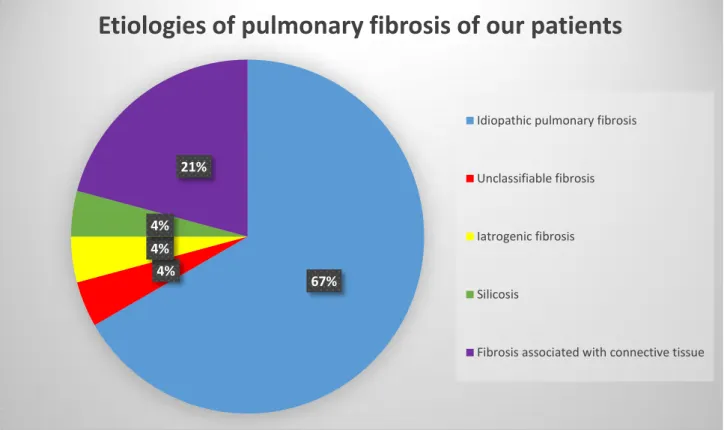
Figure 5 : Etiologies of our patients
67% 4%
4% 4%
21%
Etiologies of pulmonary fibrosis of our patients
Idiopathic pulmonary fibrosis
Unclassifiable fibrosis
Iatrogenic fibrosis
Silicosis
28
Table 1 : Population characteristics
Variables
N (%)
Median [Q1 – Q3]
Age 76 [69,5 – 81,5] Sexe (M/F) 18/6 (75/25) BMI (Kg.m2-) 23,4 [22 – 27,75] Smoking (MD=1) Never 10 (42) Sevre 15 (62) Actif 0 (0)Auto immune context 6 (25)
Emphysema 5 (21)
Pulmonary hypertension 10 (42)
Oxygen needed 12 (50)
Acute event Hospitalization 4 (16,7)
Exacerbation 3 (12,5)
Death 2 (8,3)
Anti-fibrotic treatment Pirfenidone 7 (29)
Nintedanib 3 (12,5)
Final diagnosis retained Idiopathic pulmonary fibrosis 16 (67)
ILD associated with connective tissue 5 (21)
Iatrogenic fibrosis 1 (4.2) Unclassifiable fibrosis 1 (4.2) Silicosis 1 (4.2)
29
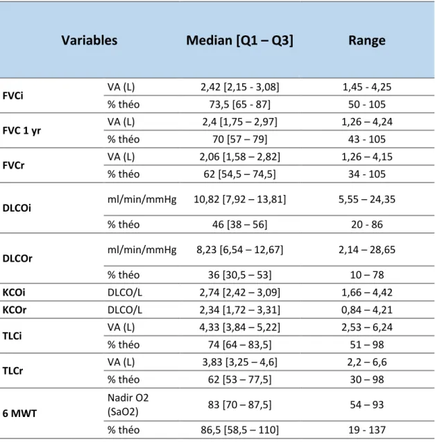
Table 2 : Functional characteristics in our patients:
Variables
Median [Q1 – Q3]
Range
FVCi VA (L) 2,42 [2,15 - 3,08] 1,45 - 4,25 % théo 73,5 [65 - 87] 50 - 105 FVC 1 yr VA (L) 2,4 [1,75 – 2,97] 1,26 – 4,24 % théo 70 [57 – 79] 43 - 105 FVCr VA (L) 2,06 [1,58 – 2,82] 1,26 – 4,15 % théo 62 [54,5 – 74,5] 34 - 105 DLCOi ml/min/mmHg 10,82 [7,92 – 13,81] 5,55 – 24,35 % théo 46 [38 – 56] 20 - 86 DLCOr ml/min/mmHg 8,23 [6,54 – 12,67] 2,14 – 28,65 % théo 36 [30,5 – 53] 10 – 78 KCOi DLCO/L 2,74 [2,42 – 3,09] 1,66 – 4,42 KCOr DLCO/L 2,34 [1,72 – 3,31] 0,84 – 4,21 TLCi VA (L) 4,33 [3,84 – 5,22] 2,53 – 6,24 % théo 74 [64 – 83,5] 51 – 98 TLCr VA (L) 3,83 [3,25 – 4,6] 2,2 – 6,6 % théo 62 [53 – 77,5] 30 – 98 6 MWT Nadir O2 (SaO2) 83 [70 – 87,5] 54 – 93 % théo 86,5 [58,5 – 110] 19 - 137
FVCi VA : absolute value of the initial forced vital capacity; FVCi %: forced vital capacity initial as a percentage predict; FVC 1 yr VA: absolute value of forced vital capacity at one year; FVC 1 yr %: forced vital capacity at one year in percentage predict; FVCr VA: absolute value of the reevaluated forced vital capacity; FVCr %: reevaluated forced vital capacity in percentage predict; TLCi VA: absolute value of initial total lung capacity; TLCi %: Initial total lung capacity in percentage predict; TLCr VA: Absolute value of reevaluated total lung capacity; TLCr %: Reevaluated total lung capacity in percentage predict; DLCOi: Initial DLCO in percentage predict; DLCOr: Reevaluated DLCO in percentage predict; KCOi: coefficient of carbon monoxide diffusion at the diagnosis; KCOr: coefficient of carbon monoxide diffusion at the reevaluation; 6 WMT nadir O2: Maximal oxygen desaturation during the 6-minute walk test; 6 WMT %theo: Distance walked to the 6-minute walk test as a percentage predict.
30
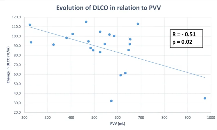
Correlation with lung function decline
There was a significant inverse correlation between the pulmonary vessel volume and yearly decline of DLCO (R= -0.51, 95%CI [-0.76; -0.11], p=0.01) (Figure 6) and KCO over time (R= -0.56 [CI 95% -0.8 - -0.16], p = 0.009) (Table 3). There was a significant positive correlation between NoV and NoV5 and the deterioration of DLCO: R= 0.5 (CI 95%: 0.09 – 0.76), p = 0.02 and R= 0.46 (CI 95%: 0.05 – 0.74), p = 0.03. There was also a significant inverse correlation between pulmonary vessel volume and the oxygen desaturation at the 6 minute walk test (R= -0.52 [CI 95% -0.79 - -0.07], p = 0.03) and with the distance of walking in percentage predict (R= -0.59 [CI 95% -0.83 - -0.18], p = 0.009) (Figure 7). Interestingly, at baseline the vascular parameters were not correlated to the diffusion capacity but to FVC (R= 0.46 [CI 95% 0.07 – 0.73], p= 0.02) with the number of vessels and the CPI (R= 0.51 [CI 95% 0.13 - 0.76], p= 0.01 with the PVV (Table 4). Other parameters tested (Forced vital capacity, presence of emphysema, smoking or auto immune context, needing of oxygen therapy, occurrence of an acute event during the follow up or the final clinical diagnosis retained) were not correlate to the vascular parameters (Table 5).
31
Figure 6 : Correlation between PVV and yearly decline of DLCO in
percentage of absolute value:
20,0 30,0 40,0 50,0 60,0 70,0 80,0 90,0 100,0 110,0 120,0 200 300 400 500 600 700 800 900 1000 C h an ge in D LC O ( % /y r) PVV (mL)
Evolution of DLCO in relation to PVV
R = - 0.51
p = 0.02
32
Table 3 : Correlation between initial vascular parameters and respiratory
functional yearly decline in percentage of absolute value :
Variables
PVV
NoV
NoV5
R [CI 95%] p-value R [CI 95%]
p-value R [CI 95%] p-value
FVC -0.33 [-0.66 – 0.1] 0.13 -0.04 [-0.46 – 0.38] 0.84 -0.02 [-0.44 – 0.4] 0.92
DLCO -0.51 [-0.77 - -0.11] 0.02 0.5 [0.09 – 0.76] 0.02 0.46 [0.05 – 0.74] 0.03
KCO -0.56 [-0.8 - -0.16] 0.009 0.05 [-0.4 – 0.47] 0.83 0.04 [-0.4 – 0.46] 0.87
PVV: Pulmonary vessel volume; NoV: Number of vessels; NoV5: Number of vessels with a cross section < 5 mm2; FVC: Forced vital capacity; DLCO: diffusion capacity of carbon monoxide; KCO: coefficient of diffusion capacity of carbon monoxide
33
Figure 7 : correlation between the distance walked at the 6 MWT in
percentage predict and vascular parameters
0 20 40 60 80 100 120 140 200 300 400 500 600 700 800 900 1000 6m WD (% Predicted ) PVV (mL) 6mWD
R = - 0.59
p = 0.009
34
Table 4 : Correlation between vascular parameters and baseline
characteristics
Variables
PVV
NoV
NoV5
R [CI 95%] p R [CI 95%] p R [CI 95%] p
FVC -0.22 [-0.58 – 0.2] 0.29 0.46 [0.07 – 0.73] 0.02 0.38 [-0.02 – 0.68] 0.06
DLCO -0.26 [-0.6 – 0.17] 0.24 0.4 [-0.1 – 0.7] 0.06 0.38 [-0.03 – 0.69] 0.07
KCO 0 [-0.42 – 0.42] 0.99 0.15 [-0.29 – 0.54] 0.5 0.14 [-0.3 – 0.53] 0.53
CPI 0.51 [0.13 – 0.76] 0.01 -0.11 [-0.49 –
0.31] 0.62 -0.1 [-0.49 – 0.31] 0.63
PVV: Pulmonary vessel volume; NoV: Number of vessels; NoV5: Number of vessels with a cross section < 5 mm2; FVC: Forced vital capacity; DLCO: diffusion capacity of carbon monoxide; KCO: coefficient of diffusion capacity of carbon monoxide
35
Table 5 : Correlation between clinical and paraclinical parameters and
vascular values :
Variables
PVV
NoV
NoV5
R [CI 95%] p R [CI 95%] P R [CI 95%] P
6 MWT Nadir O2 -0.52 [-0.79 - -0.07] 0.03 0.41 [-0.07 – 0.74] 0.09 0.45 [-0.02 – 0.76] 0.06 6 MWT distance % theo -0.59 [-0.83 - -0.18] 0.009 0.43 [-0.05 – 0.74] 0.08 0.44 [-0.03 – 0.75] 0.07 Pulmonary hypertension 0.74 0.57 0.03 Acute Events 0.67 0.57 0.66 Smoking 0.23 0.16 0.72 Clinical diagnosis 1 0.38 0.51 CPI r 0.46 [0 – 0.75] 0.05 -0.56 [-0.81 - -0.14] 0.01 -0.52 [-0.79 - -0.09] 0.02
PVV: Pulmonary vessel volume; NoV: Number of vessels; NoV5: Number of vessels with a cross section < 5 mm2; R: correlation coefficient; p: p value; CPI r: composite physiologic index after reevaluation; 6 MWT Nadir O2: maximal desaturation at the 6 minute walk test; 6 WMT distance % théo: Distance walked at the 6 MWT in percentage predict.
36 Discussion
This study was undertaken to explore the relation between CT vascular morphological parameters and clinical outcome as assessed by PFT decline, as well as by the occurrence of acute events, in ILD patients. Our main finding was that PVV and NoV/NoV5 were respectively negatively and positively correlated with the yearly decline of KCO and DLCO (p= 0.009 and p= 0.02; p= 0.02 and p= 0.003, respectively).
While the exact pathophysiology of those vascular changes in ILD patients is not known, it is known that in fibrotic ILDs, regions of significant fibrosis manifest with significantly reduced blood perfusion (20) with redistribution of the blood flow to the functional lung (21) (22). We speculate that this vascular redistribution may lead to enlargement of the large vessels and therefore a higher PVV, which may indirectly also correspond to infraclinical forms of PAH.
Our results are similar to those found in the current literature. Indeed, Jacob and al. also found in a series of 283 IPF patients a significant correlation between PVV and both pulmonary functional tests and mortality, and that semi-automatic parameters such as PVV were superior to those obtained by traditional visual CT scoring (10) (11). Our study was the first to investigate the number of vessels and small vessels in ILDs. Interestingly those parameters showed significant positive correlations with the decline of DLCO and the presence of PAH. While these parameters weren’t previously analyzed in ILDs, they have previously been analyzed in COPD patients in whom they showed significant positive relationship with PAH and lung functional parameters (23) (24). We hypothesize that the number of vessels may be indirectly linked with the vasoconstriction and/or destruction of small vessels in fibrotic areas.
37 This hypothesis might be supported by a study by Renzoni and al in 2003 which showed a significant decrease in small vessel density in the most extensively fibrotic regions (20).
There was also a significant correlation between PVV and the walking distance (p= 0.009) and extent of desaturation (p= 0.03) in the 6 MWT. The 6 MWT is known to be a reliable indicator of quality of life in ILD patients (26) (27) as well as a prognostic factor (28) (29). To our knowledge, no previous studies assessed the correlation between pulmonary vascularization and exercise capacity. Unlike the results of a study of Wemeau-Stervinou and al published in 2012 (25), which found a link between decreased capillary blood volume and exercise desaturation, there were no significant links in our study between small vessels measurement and 6MWT parameters. Furthermore, our results also showed significant correlations between PVV, NoV, NoV5 and CPI at the final evaluation, which stresses the fact that all those vascular parameters have the potential to reliably predict patient prognosis.
Our study had several limitations. First, since it is a retrospective monocentric study involving only a small number of patients, the statistical power of this study is limited. However, the fact that we found significant correlations may be indicative of strong underlying statistical. Moreover, some of the patients received antifibrotic treatment during follow-up which may have slightly biased the evaluation of PFT decline. However, up-to-date recommendations were followed for selecting patients for treatment. The decision of giving no treatment was made on the following reasons: alternate diagnosis than IPF, FVC ≤ 50% or DLCO ≤ 30% or clinical stability. Follow-up time also varied depending on patients’ survival, but this bias was corrected by using a yearly correction of the PFT decline. Finally, and similarly to all quantitative CT studies, variations in CT protocols such as X-ray tube current, intensity,
dose-38 reducing procedures or the choice of reconstruction algorithms may play an important role in the vascular parameters’ measurement. Such variations are unavoidable due to the timespan of this retrospective study, during which technical improvements were gradually achieved. It is also important to note that 4 different CT scanners were used. While this may be the cause of added variations, we believe it has the benefit to reproduce routine clinical conditions. According to our positive results, we believe that CT vascular parameters deserve further analysis. Especially, natural variations in healthy patients as well as CT parameters’ influence on their measurement may be especially interesting topics to investigate in the future.
39 Conclusions
In this study, we found that pulmonary vascular volume and the number of vessels estimated
by quantitative lung CT analysis is significantly correlated with the yearly decline in lung
function and may be a simple surrogate prognostic marker in ILD patients. These data merit
40
Conclusion générale
Les fibroses pulmonaires et notamment la fibrose pulmonaire idiopathique restent des maladies rares avec un pronostic péjoratif. Pouvoir anticiper le déclin de la fonction respiratoire dans ces maladies a une importance en termes de prise en charge et de suivi. Mais à l’heure actuelle l’évolution naturelle des patients avec une pathologie infiltrative fibrosante est propre à chacun et celle-ci est encore mal comprise, avec une vitesse d’aggravation variable selon les patients. De plus en plus d’études essaient de trouver des marqueurs prédictifs de l’évolution et du pronostic de ces patients sans résultats probants pour l’instant. Les nouvelles techniques d’imagerie assistées par ordinateur sont précises dans la quantification des lésions de fibrose et pourraient avoir un rôle dans la prédiction de l’évolution des patients. Ceci a déjà été évoqué, notamment par le biais de l’analyse de la vascularisation pulmonaire et du volume vasculaire pulmonaire, comme élément de mauvais pronostic. Notre étude renforce l’hypothèse d’un lien entre la vascularisation pulmonaire présente sur le scanner des patients avec une pathologie infiltrative fibrosante au moment du diagnostic et l’évolution de leur maladie. En effet, nous avons mis en évidence une corrélation inverse entre le volume vasculaire pulmonaire et le pronostic des patients qui pourrait être la conséquence d’une altération de la diffusion, en lien ou non avec une hypertension pulmonaire, connue comme un critère pronostic important. Il serait nécessaire de confirmer ce résultat par d’autres études plus robustes afin de confirmer la capacité de ces paramètres à prédire l’évolution de la fibrose et d’amélioration la compréhension des mécanismes physiopathologiques sous-jacents.
42
Références
1. Hutchinson J, Fogarty A, Hubbard R, McKeever T. Global incidence and mortality of idiopathic pulmonary fibrosis: a systematic review. Eur Respir J. sept
2015;46(3):795‑806.
2. Raghu G, Collard HR, Egan JJ, Martinez FJ, Behr J, Brown KK, et al. An Official ATS/ERS/JRS/ALAT Statement: Idiopathic Pulmonary Fibrosis: Evidence-based Guidelines for Diagnosis and Management. Am J Respir Crit Care Med. 15 mars 2011;183(6):788‑824.
3. Raghu G, Remy-Jardin M, Myers JL, Richeldi L, Ryerson CJ, Lederer DJ, et al.
Diagnosis of Idiopathic Pulmonary Fibrosis. An Official ATS/ERS/JRS/ALAT Clinical Practice Guideline. Am J Respir Crit Care Med. 1 sept 2018;198(5):e44‑68.
4. Raghu G, Chen S-Y, Yeh W-S, Maroni B, Li Q, Lee Y-C, et al. Idiopathic pulmonary fibrosis in US Medicare beneficiaries aged 65 years and older: incidence, prevalence, and survival, 2001-11. Lancet Respir Med. juill 2014;2(7):566‑72.
5. Barlo NP. Predicting prognosis in idiopathic pulmonary fibrosis. :11.
6. Selman M, Carrillo G, Estrada A, Mejia M, Becerril C, Cisneros J, et al. Accelerated variant of idiopathic pulmonary fibrosis: clinical behavior and gene expression pattern. PloS One. 30 mai 2007;2(5):e482.
7. Best AC, Meng J, Lynch AM, Bozic CM, Miller D, Grunwald GK, et al. Idiopathic pulmonary fibrosis: physiologic tests, quantitative CT indexes, and CT visual scores as predictors of mortality. Radiology. mars 2008;246(3):935‑40.
8. Lynch DA, Godwin JD, Safrin S, Starko KM, Hormel P, Brown KK, et al. High-Resolution Computed Tomography in Idiopathic Pulmonary Fibrosis: Diagnosis and Prognosis. Am J Respir Crit Care Med. 15 août 2005;172(4):488‑93.
9. Watadani T, Sakai F, Johkoh T, Noma S, Akira M, Fujimoto K, et al. Interobserver Variability in the CT Assessment of Honeycombing in the Lungs. Radiology. mars 2013;266(3):936‑44.
10. Jacob J, Bartholmai BJ, Rajagopalan S, Kokosi M, Nair A, Karwoski R, et al. Automated Quantitative Computed Tomography Versus Visual Computed Tomography Scoring in Idiopathic Pulmonary Fibrosis: Validation Against Pulmonary Function. J Thorac Imaging. sept 2016;31(5):304‑11.
11. Jacob J, Bartholmai BJ, Rajagopalan S, Kokosi M, Nair A, Karwoski R, et al. Mortality prediction in idiopathic pulmonary fibrosis: evaluation of computer-based CT analysis with conventional severity measures. Eur Respir J. janv 2017;49(1):1601011.
12. Jacob J, Bartholmai BJ, Rajagopalan S, Egashira R, Brun AL, Kokosi M, et al. Unclassifiable-interstitial lung disease: Outcome prediction using CT and functional indices. Respir Med. sept 2017;130:43‑51.
43 13. Jacob J, Bartholmai BJ, Egashira R, Brun AL, Rajagopalan S, Karwoski R, et al. Chronic
hypersensitivity pneumonitis: identification of key prognostic determinants using automated CT analysis. BMC Pulm Med [Internet]. déc 2017 [cité 2 oct 2018];17(1). Disponible sur: http://bmcpulmmed.biomedcentral.com/articles/10.1186/s12890-017-0418-2
14. Colombat M, Mal H, Groussard O, Capron F, Thabut G, Jebrak G, et al. Pulmonary vascular lesions in end-stage idiopathic pulmonary fibrosis: Histopathologic study on lung explant specimens and correlations with pulmonary hemodynamics. Hum Pathol. janv 2007;38(1):60‑5.
15. Shin S, King CS, Puri N, Shlobin OA, Brown AW, Ahmad S, et al. Pulmonary artery size as a predictor of outcomes in idiopathic pulmonary fibrosis. Eur Respir J. mai
2016;47(5):1445‑51.
16. Collard H. R, King T.E, Bartelson B.B, Vourlekis J.S, Schwarz M.I, Brown K.K, Changes in Clinical and Physiologic Variables Predict Survival in Idiopathic Pulmonary Fibrosis, American Journal of Respiratory and Critical Care Medicine. 168 (2003) 538–542. doi:10.1164/rccm.200211-1311OC.
17. du Bois RM, Weycker D, Albera C, Bradford WZ, Costabel U, Kartashov A, et al. Forced Vital Capacity in Patients with Idiopathic Pulmonary Fibrosis: Test Properties and
Minimal Clinically Important Difference. Am J Respir Crit Care Med. 15 déc 2011;184(12):1382‑9.
18. Flaherty KR, Mumford JA, Murray S, Kazerooni EA, Gross BH, Colby TV, et al. Prognostic Implications of Physiologic and Radiographic Changes in Idiopathic Interstitial Pneumonia. Am J Respir Crit Care Med. sept 2003;168(5):543‑8.
19. Wells A.U, Desai S.R, Rubens M.B, Goh N.S.L, Cramer D, Nicholson A.G, Colby T.V, du Bois R.M, Hansell D.M, Idiopathic Pulmonary Fibrosis: A Composite Physiologic Index Derived from Disease Extent Observed by Computed Tomography, American Journal of Respiratory and Critical Care Medicine. 167 (2003) 962–969.
doi:10.1164/rccm.2111053.
20. Renzoni EA, Walsh DA, Salmon M, Wells AU, Sestini P, Nicholson AG, et al. Interstitial vascularity in fibrosing alveolitis. Am J Respir Crit Care Med. 1 févr 2003;167(3):438‑43. 21. Ebina M, Shimizukawa M, Shibata N, Kimura Y, Suzuki T, Endo M, et al. Heterogeneous
increase in CD34-positive alveolar capillaries in idiopathic pulmonary fibrosis. Am J Respir Crit Care Med. 1 juin 2004;169(11):1203‑8.
22. Cosgrove GP, Brown KK, Schiemann WP, Serls AE, Parr JE, Geraci MW, et al. Pigment epithelium-derived factor in idiopathic pulmonary fibrosis: a role in aberrant
angiogenesis. Am J Respir Crit Care Med. 1 août 2004;170(3):242‑51.
23. Coste F, Dournes G, Dromer C, Blanchard E, Freund-Michel V, Girodet P.-O, Montaudon M, Baldacci F, Picard F, Marthan R, Berger P, Laurent F, CT evaluation of small
pulmonary vessels area in patients with COPD with severe pulmonary hypertension, Thorax. 71 (2016) 830–837. doi:10.1136/thoraxjnl-2015-207696.
44 24. Matsuoka S, Washko G.R, Dransfield M.T, Yamashiro T, San Jose Estepar R, Diaz A,
Silverman E.K, Patz S, Hatabu H, Quantitative CT Measurement of Cross-sectional Area of Small Pulmonary Vessel in COPD, Academic Radiology. 17 (2010) 93–99.
doi:10.1016/j.acra.2009.07.022.
25. Wémeau-Stervinou L, Perez T, Murphy C, Polge A-S, Wallaert B. Lung capillary blood volume and membrane diffusion in idiopathic interstitial pneumonia. Respir Med. avr 2012;106(4):564‑70.
26. Visca D, Mori L, Tsipouri V, Fleming S, Firouzi A, Bonini M, et al. Effect of ambulatory oxygen on quality of life for patients with fibrotic lung disease (AmbOx): a prospective, open-label, mixed-method, crossover randomised controlled trial. Lancet Respir Med. 28 août 2018;
27. Nishiyama O, Taniguchi H, Kondoh Y, Kimura T, Ogawa T, Watanabe F, et al. Health-related quality of life in patients with idiopathic pulmonary fibrosis. What is the main contributing factor? Respir Med. avr 2005;99(4):408‑14.
28. Flaherty KR, Andrei A-C, Murray S, Fraley C, Colby TV, Travis WD, et al. Idiopathic pulmonary fibrosis: prognostic value of changes in physiology and six-minute-walk test. Am J Respir Crit Care Med. 1 oct 2006;174(7):803‑9.
29. Lama VN, Flaherty KR, Toews GB, Colby TV, Travis WD, Long Q, et al. Prognostic value of desaturation during a 6-minute walk test in idiopathic interstitial pneumonia. Am J Respir Crit Care Med. 1 nov 2003;168(9):1084‑90.
30. Hallstrand TS, Boitano LJ, Johnson WC, Spada CA, Hayes JG, Raghu G. The timed walk test as a measure of severity and survival in idiopathic pulmonary fibrosis. Eur Respir J. janv 2005;25(1):96‑103.