The role of Tbf1 in telomere homeostasis in Saccharomyces cerevisiae
Par
Erin Bonnell
Département de Microbiologie et d’Infectiologie
Mémoire présentée à la Faculté de Médecine et des Sciences de la Santé
en vue de l’obtention du grade de Maître ès sciences (M. Sc.) en Microbiologie
Sherbrooke, Québec, Canada
Juin, 2017
Membres du jury d’évaluation
Pr. Raymund Wellinger, Département de Microbiologie et d’Infectiologie
Pr. Brendan Bell, Département de Microbiologie et d’Infectiologie
RÉSUMÉ
The role of Tbf1 in telomere homeostasis in Saccharomyces cerevisiae Par
Erin Bonnell
Département de Microbiologie et d’Infectiologie
Mémoire présenté à la Faculté de médecine et des sciences de la santé en vue de l’obtention du diplôme de maître ès sciences (M.Sc.) en microbiologie Faculté de Médecine et des Sciences de la
Santé, Université de Sherbrooke, Sherbrooke, Québec, Canada, J1H 5N4
En différenciant les extrémités chromosomiques des cassures d’ADN internes, les télomères empêchent l'activation de la signalisation d’un dommage à l'ADN et fournissent une protection contre des activités inappropriées qui sont associées à une réparation de l'ADN. Une telle réparation pourrait en fait créer une instabilité génomique. Chez Saccharomyces cerevisiae, un nombre de protéines sont impliquées dans la structure du télomère et / ou la fonction de la télomérase. On pense que la protection des télomères est gérée par les répétitions télomériques et les protéines associées, mais il y a de plus en plus d’indices que la région sous-télomérique joue également un rôle. Cette région contient des sites de liaison pour plusieures protéines, notamment pour Tbf1. TBF1 est un gène essentiel et la protéine est impliquée dans l'homéostasie des télomères et dans la réponse aux dommages de l’ADN. Toutefois, les mécanismes moléculaires restent à être précisés.
Mon projet de Maîtrise est basé sur l’observation que dans les cellules qui ont un allèle thermosensible (tbf1-ts), les télomères sont anormalement courts. Malheureusement, les 4 allèles mutants de tbf1 connus présentent tous des mutations ponctuelles multiples ce qui rend leur analyse difficile. Pour clarifier l'origine des variations phénotypiques de ces mutations, la mutagenèse dirigée a été utilisée pour créer des allèles tbf1 avec une seule mutation. Mes résultats montrent que deux mutations spécifiques, tbf1-82 et tbf1-453, causent des défauts de croissance cellulaires, ainsi qu'une sensibilité aux drogues qui endommageant l'ADN.
Une analyse détaillée de ces nouveaux allèles de tbf1 a montré que la protéine pourrait avoir un rôle direct dans le maintien de la stabilité des télomères. Par exemple, en absence de la télomérase qui est responsable du maintien des télomères, les cellules entrent en sénescence réplicative après environ 60 générations et arrêtent de se diviser. Par contre, une petite fraction de la population est capable de contourner cet arrêt de croissance car ces cellules maintiennent les télomères par un processus dépendant de la recombinaison homologue. L'introduction de mutations tbf1 dans des souches sans télomérase provoque une accélération d’entrée en sénescence ; donc Tbf1 est un régulateur précédemment inconnu de la sénescence. Divers tests génétiques avec des gènes de recombinaison homologue et des régulateurs de chromatine ont été effectués pour aider à caractériser TBF1 et ses interactions. La caractérisation de ces nouveaux allèles a permis de mieux comprendre les multiples rôles de Tbf1.
SUMMARY
By differentiating chromosomal ends from internal breaks, telomeres prevent DNA damage checkpoint activation and provide protection from inappropriate DNA repair activity that could create genomic instability. In Saccharomyces cerevisiae, a large number of genes have been identified that are implicated in telomerase and telomere structure and/or function. However, a comprehension of the mechanism of action of these genes and how they relate to other genes is lacking. The function of end protection is based on the telomeric repeats and associated proteins, but evidence is accumulating that the subtelomeric region also plays a role. This region contains binding sites for various proteins, notably Tbf1.
TBF1 is an essential gene and the protein has been implicated in telomere homeostasis,
chromatin remodelling, and the DNA damage response. My master’s project is based on the observation that cells harbouring a thermosensitive (tbf1-ts) allele have abnormally short telomeres. However, all four known mutant tbf1 alleles have multiple point mutations, which renders their analyses difficult. In order to be able to more precisely determine the origin of the phenotypic variations, we used site-directed mutagenesis to create single point mutation tbf1 alleles. These experiments yielded two particular mutations, tbf1-82 and tbf1-453, which were found to have growth defects at various temperatures as well as increased sensitivity to DNA damaging drugs.
Although the alleles had only minor telomere length phenotypes, it was discovered that Tbf1 could have a direct role in telomere stability in special situations. For example, in the absence of telomerase, which normally maintains telomeres, cells enter replicative senescence after about 60 population doublings and stop dividing. A small subset of the cellular population is able to evade this growth arrest by maintaining telomeres via a recombination-dependent process. An introduction of the tbf1-82 or tbf1-453 mutation into strains that also lacked telomerase caused a dramatic advance in time of onset of senescence. Thus this work uncovered that Tbf1 is a previously unknown regulator of senescence. Various genetic assays with homologous recombination genes and chromatin regulators were performed to help further characterize TBF1 and its interactors. Characterization of these novel tbf1 alleles has given new insights into the multiple roles of Tbf1.
LIST OF TABLES AND FIGURES
Tables
Table 1: Strains and plasmids that are referred to throughout this work. Table 2: Primers used in this study.
Table 3: Average bulk telomere lengths at beginning and end of ELY065 senescence assay. Table 4: Average bulk telomere lengths at beginning and end of ELY067 senescence assay. Table 5: Average bulk telomere lengths at beginning and end of tlc1∆ tbf1-82 rpd3∆ senescence assay.
Table 6: Average bulk telomere lengths at beginning and end of tlc1∆ tbf1-453 rpd3∆senescence assay.
Figures
Figure 1: Organization of telomeric sequences in S. cerevisiae and H. sapiens. Figure 2: Nucleoprotein schematic of the telosome in S. cerevisiae.
Figure 3: Telomeres differ by their subtelomeric element composition.
Figure 4: Semi-conservative DNA replication generates telomeric sequence losses. Figure 5: Ribonucleoprotein complex telomerase in S. cerevisiae.
Figure 6: pRS304-TBF1-NatMX plasmid.
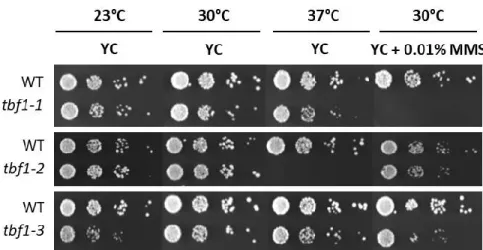
Figure 7: Sample spot test comparing growth in different conditions.
Figure 8: Schematic of XhoI restriction sites and terminal restriction fragments (TRF). Figure 9: Example of Southern blot to measure telomere length.
Figure 10: Example graph of liquid senescence assay
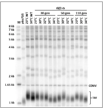
Figure 11: Southern blot of DNA derived from cells with the tbf1-ts allele.
Figure 12: Schematic representation of the Tbf1 protein and amino acid substitutions of tbf1 alleles.
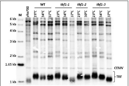
Figure 13: Serial dilution growth tests of cells with the tbf1-1, tbf1-2, or tbf1-3 alleles. Figure 14: Southern blot of telomere lengths of tbf1-1, 2, and 3 alleles.
Figure 15: Effect of TBF1 mutations on senescence timing in telomerase-negative cells. Figure 16: Spot tests of cells with the tbf1-82 mutation vs WT cells.
Figure 17: Southern blots of DNA derived from cells with the tbf1-82 allele. Figure 18: Spot growth tests of tbf1-299 cells vs WT cells.
Figure 19: Southern blot of DNA derived from cells with the tbf1-299 allele. Figure 20: Spot growth tests of cells with the tbf1-398 allele vs WT cells.
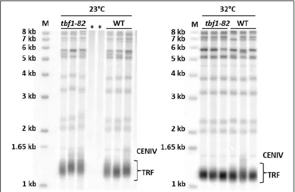
Figure 21: Southern blot of DNA derived from cells with the tbf1-398 allele. Figure 22: Spot growth tests of cells with the tbf1-453 allele vs WT cells. Figure 23: Southern blot of tbf1-453 at various temperatures.
Figure 24: Southern blot of tbf1-453 at 23°C. Figure 25: Spot tests of tbf1-480 vs WT. Figure 26: Southern blot of tbf1-480.
Figure 27: Spot tests of two alleles derived from tbf1-ts.
Figure 28: Microdissection plates derived from spore tetrads of tbf1-451::NatMX/TBF1 diploids. Figure 29: Spot growth tests of BY4705 background cells with the tbf1-453 mutation.
Figure 30: Western blot of Rad53 phosphorylation in cells with tbf1-82 and tbf1-453 mutations. Figure 31: Effect of the tbf1-82 mutation on senescence rate in telomerase-negative cells. Figure 32: Southern blot of DNA extracted from tbf1-82 tlc1∆ senescence assay.
Figure 33: ssDNA in-gel assays of tbf1 mutants in comparison to wildtype.
Figure 34: Spot test analysis of haploids from tbf1-82/TBF1 rad52∆/RAD52 tlc1∆TLC1 diploid. Figure 35: Spot test analysis of haploids from tbf1-453/TBF1 rad52∆/RAD52 tlc1∆/TLC1 diploid. Figure 36: Negative interaction between tbf1-82 and sgs1∆.
Figure 37: Effect of SGS1 deletion on tbf1-82 senescence rate in telomerase-negative cells. Figure 38: Liquid growth assay of ELY065 telomerase-positive cells.
Figure 39: Southern blot of DNA extracted from tbf1-82/TBF1 sgs1∆/SGS1 tlc1∆/TLC1 senescence assay.
Figure 40: Genetic interactions of tbf1-453 with sgs1∆.
Figure 41: Effect of SGS1 deletion on senescence rate of tbf1-453 in telomerase-negative cells. Figure 42: Southern blot of DNA extracted from tbf1-453/TBF1 sgs1∆/SGS1 tlc1∆/TLC1 senescence assay.
Figure 43: Genetic interactions of tbf1-453 with mre11∆.
Figure 44: Southern blot of clones from ELY078 tbf1-453/TBF1 mre11∆/MRE11. Figure 45: Interactions between tbf1-82 and rpd3∆.
Figure 46: Effect of RPD3 deletion on senescence rate of tbf1-82 in telomerase-negative cells. Figure 47: Liquid growth assay of telomerase-positive cells.
Figure 48: Southern blot of DNA extracted from tbf1-82 rpd3∆ tlc1∆ senescence assay. Figure 49: Interactions of tbf1-453 with rpd3∆.
Figure 50: Effect of RPD3 deletion on senescence rate of tbf1-453 in telomerase-negative cells. Figure 51: Liquid growth assay of telomerase-positive cells.
Figure 53: Functional interaction of tbf1-82 and htz1∆ alleles.
Figure 54: Southern blot of telomere length of tbf1-82 and htz1∆ mutants compared to WT. Figure 55: Functional interactions of tbf1-453 and htz1∆ alleles.
Figure 56: Southern blot of telomere length of tbf1-453 and htz1∆ mutants compared to WT. Figure 57: Proposed model of role of Tbf1 in the subtelomeric region.
TABLE OF CONTENTS
INTRODUCTION
Telomeres ... 1 Sequence and structure of telomeres... 1-2 Functions of telomeres – end protection ... 3-4 Subtelomeres ... 5-6 Telomeres and subtelomeric chromatin ... 6-7 Telomere position effect ... 7 Functions of telomeres – end replication ... 7-9 Telomerase ... 9-11 Replicative senescence & survivors ... 11-13 DNA damage response & telomeres ... 13 Tbf1 ... 14-17 Hypothesis & objectives ... 17-19 MATERIALS AND METHODS
Yeast strains and plasmids ... 20-22 Construction of TBF1 alleles ... 22-24
E. coli transformation ... 25
Yeast transformation ... 25 Genomic DNA extraction and quantification ... 25-26 Sporulation and microdissection ... 27 PCR-mediated gene deletion ... 27 Serial dilution growth tests on solid plates ... 27-28 Southern blot & telomere length analysis ... 28-30 In-gel hybridizations ... 30-31 Western blots ... 31-32 Senescence curves ... 32-33 Primers used for the studies ... 33-35 CHAPTER I – Creation and characterization of TBF1 mutants
Preamble ... 36 Results ... 37-61
CHAPTER 2 – Functional interactions of TBF1 with homologous recombination genes
Preamble ... 62-63 Results ... 63-79 CHAPTER 3 – Genetic interactions of TBF1 with chromatin maintenance genes
Preamble ... 80 Results ... 80-97 DISCUSSION & CONCLUSIONS
Discussion & conclusions ... 98-109 Proposed model ... 110 REFERENCES
LIST OF SYMBOLS, ABBREVIATIONS, AND ACRONYMS
∆: delta, identifies a deletion °C: degrees Celsius
μg: microgram μl: microliter μM: micromolar
3’: 3’ extremity of a nucleotide, free OH group on the 3’ carbon of ribose ring
5’: 5’ extremity of a nucleotide, free OH group or phosphate ester on 5’ carbon of ribose ring 5-FOA: 5-Fluoroorotic acid
A: adenosine b: base(s) bp: base pair(s)
BIR: Break-induced replication C: cytosine
ChIP: Chromatin Immunoprecipitation
ChIP-seq: Chromatin Immunoprecipitation-sequencing Ci: Curie
Cm: centimeter
cpm: counts per minute C-terminal: carboxy-terminal dCTP: deoxycytidine triphosphate dGTP: deoxyguanosine triphosphate DNA: Deoxyribonucleic acid
DSB: Double-strand break dsDNA: double-stranded DNA dNTP: deoxynucleoside triphosphate DTT: Dithiothreitol
ECL: Electrochemiluminescence or electro generated chemiluminescence EDTA: Ethylenediaminetetraacetic acid
EST: Ever Shorter Telomeres G: guanosine
h: hour
HO: Homothallic switching endonuclease KAc: Potassium Acetate
kb: kilobase kDa: kiloDalton
LB: Luria Broth medium LiCl: lithium chloride M: molar
mg: milligram min: minute
mini-prep: mini-preparation of plasmid DNA ml: milliliter
mm: millimeter mmole: millimole
MMS: Methyl methanesulfonate m/v: mass per volume
NaCl: sodium chloride NaOAc: sodium acetate NEB: New England Biolabs ng: nanograms
NHEJ: Non-Homologous End-Joining nm: nanometer
nM: nanomolar nt: nucleotide
N-terminal: amino terminal ORF: Open Reading Frame PBS: phosphate-buffered saline
PBS-T: phosphate-buffered saline with 0.1% Tween-20 PCR: Polymerase Chain Reaction
Pfu: High-fidelity DNA polymerase from Pyrococcus furiosus pH: measure of acidity or basicity of a solution
Phleo: Phleomycin RNase: Ribonuclease SDS: sodium dodecyl sulfate
SDS-PAGE: SDS-Polyacrylamide Gel Electrophoresis snoRNA: small nucleolar RNA
SSC: Saline sodium citrate ssDNA: single-stranded DNA
STARs: SubTelomeric Anti-silencing Regions T: Thymidine
TAE: 40 mM Tris, 20 mM glacial acetic acid, 1 mM EDTA pH 8.0 Taq: DNA polymerase from Thermophilus aquaticus
TBE: 40 mM Tris, 20 mM borate, 1 mM EDTA pH 8.0 TCA: Trichloroacetic acid
TPE: Telomere Position Effect TRF: Terminal Restriction Fragment ts: temperature sensitive
v/v: volume per volume V: Volt
YC: Yeast Complete synthetic media
ACKNOWLEDGEMENTS
I would foremost like to thank my research director Pr. Raymund Wellinger for having welcomed me into his laboratory, first as an intern and then as a masters student, as well as for always having an open door for conversations scientific or not. I would also like to thank all the members of the Wellinger lab, past and present, for all their scientific and moral support, as well as being a great group of people to work with. I would particularly like to thank Isabelle Dionne for much help and advice getting my project started, and Nancy Laterreur for being an invaluable source of guidance and troubleshooting. A big thanks goes out to my family and friends for their enthusiastic encouragement and many hours on the phone entertaining me during my long commute. I would especially like to thank Dave, who provided an immense amount of encouragement, support, and who was endlessly patient and understanding. I thank Dr. Maria Pia Longhese for the strains provided. Finally, I would like to thank Pr. Guylain Boissonneault and Pr. Brendan Bell for agreeing to be part of my jury.
INTRODUCTION
Telomeres
Eukaryotic genomes are highly complex and divided into linear chromosomes. Therefore unlike for most prokaryotic genomes that are circular, a unique problem arises; the necessity of protecting the integrity of the chromosome ends. This is accomplished by telomeres, aptly named by Hermann Muller in the late 1930’s as it originates from the ancient Greek “telos” (end) and “meros” (part) (Muller, 1938). The earliest efforts to describe chromosomes were based on characteristics visible with the technology of the time. Muller discovered that a highly stable specialized structure existed at the ends of chromosomes through his work in Drosophila, studying X-ray-induced breakage and rejoining of chromosomes (Muller, 1938). Fortuitously, at around the same time, Barbara McClintock came to a similar conclusion regarding the high stability of native chromosome ends while using the breakage-fusion-bridge cycle to study newly formed ends of chromosomes in maize (McClintock, 1939; McClintock, 1941). Little did they know at the time, but they were on to something big. Today, telomeres are recognized as essential for eukaryotic genomic stability, as accomplished through their specialized nucleoprotein structure and their characteristics are associated with human disease and aging.
Sequence and structure of telomeres
In eukaryotes, telomeric DNA is generally composed of G-rich double-stranded repetitive sequences and a 3’ G-rich single-stranded overhang (Wellinger and Zakian, 2012). Protein complexes bind to the DNA to help protect the structural integrity of telomeres, creating a unique nucleoprotein complex called the telosome (Zakian, 1995). The telosome provides a capping effect that is essential for not only telomere homeostasis, but for stability of the entire chromosome. Despite some minor sequence variations between species, this arrangement is highly conserved.
The DNA of human telomeres consists of 5-15 kilobases (kb) of double-stranded TTAGGG telomeric repeats abbreviated T2AG3 (de Lange et al., 1990) and a 50-300 nucleotide (nt) 3’ single-stranded overhang (McElligott and Wellinger, 1997). This sequence is conserved in mammals, although the length of the repeats may vary significantly between species. The double-stranded
repeats are protected by the shelterin complex, which consists of six protein subunits. Of these, TRF1 and TRF2 directly bind the double-stranded repeats and POT1 binds the single-stranded overhang. The complex is interconnected and stabilized by three additional proteins, TIN2, TPP1, and RAP1 (De Lange, 2005). Shelterin is a dynamic complex with DNA modeling capabilities that acts in conjunction with other associated DNA repair factors to facilitate DNA end protection and replication (De Lange, 2005; Palm and de Lange, 2008). Fascinatingly, the general telomeric arrangement of a repetitive DNA structure with binding and accessory proteins is conserved even down to single-celled eukaryotic species, such as budding yeast Saccharomyces cerevisiae.
Like most eukaryotic organisms, the telomeres of S. cerevisiae are heterogeneous in length. They consist of 300 ± 75 bp of degenerate repeat sequences abbreviated (TG)1-6TG2-3, often simplified as TG1-3 (Shampay et al., 1984; Wellinger and Zakian, 2012). The G-rich single strand overhang consists of 12-15 nucleotides for most of the cell cycle (Larrivée et al., 2004). However, during a short period in late S/G2 phase of the cell cycle G tails are longer, with a size of ≥ 30-100 nt (Wellinger et al., 1993). It is important to note that the budding yeast telomeric sequence differs from the mammalian one described above, although they serve the same purpose. Like mammalian telomeres, the unique DNA structures at yeast chromosome ends are bound by a variety of proteins that facilitate the two main functions of telomeres – end protection and end replication. A schematic comparing the general organization of telomeric repeats is seen in figure 1.
Figure 1: Organization of telomeric sequences in S. cerevisiae and H. sapiens.
Telomeric structure is highly conserved and is characterized by a variable length of TG-rich repeats and a G-rich single-stranded 3’ overhang.
Functions of telomeres – end protection
The nucleoprotein structure of a telomere provides a capping function that permits telomeres to safeguard the integrity of chromosome by preventing the end from being recognized by DNA repair machinery as a double-strand break. This, in turn, prevents DNA damage checkpoint activation and provides protection from inappropriate DNA repair activity such as non-homologous end joining (NHEJ) or non-homologous recombination (HR). Failing to prevent recognition by DNA repair mechanisms could lead to end-to-end fusions, recombination, or resection events that could create genomic instability (Sandell and Zakian, 1993; Zakian, 1995). Undoubtedly, the function of end protection is critical not only for protecting telomeres but also sustaining the integrity of the entire genome.
In the budding yeast Saccharomyces cerevisiae, several proteins bind to the unique telomeric DNA sequences in order to maintain a capping function. Rap1 is responsible for binding the telomeric TG1-3 repeats, which contain high-affinity Rap1 binding sites approximately every 20 bp (Conrad et al., 1990; Gilson et al., 1993). Interestingly, like many proteins involved in telomere homeostasis, Rap1 performs double duties in the cell. It was discovered first as a major transcription factor in yeast with the ability to repress or activate gene expression (Shore and Naysmith, 1987). The functional domains of Rap1 have been extensively studied, and it has been shown that Rap1 binds DNA via an essential double Myb domain (Graham et al., 1999). In addition, the C-terminus is critical for activities related to telomere maintenance, as the Sir3 and Sir4 silencing proteins and the length regulators Rif1 and Rif2 bind this region (Hardy et al., 1992a, 1992b; Buck and Shore, 1995; Wotton and Shore, 1997). Rap1 in association with the Rif proteins acts as a type of counting mechanism of end repeats that helps to negatively regulate telomerase (Marcand et al., 1997). Furthermore, the association of Rap1 with Rif2, and to a lesser extent, Rif1, is important for preventing telomere fusions and limiting Exo1-mediated end resection (Marcand
et al., 2008; Vodenicharov et al., 2010). Another factor important for telomere capping is the
heterodimer ring called yKu and comprised of the yKu70 and yKu80 proteins (Boulton and Jackson, 1996; Gravel et al., 1998). The yKu complex associates with double-stranded DNA ends such as those found at a double-strand break or the double-strand – single-strand junction of telomeres, however it does not show any sequence specificity (Gravel et al., 1998; Downs and Jackson, 2004). At telomeres, yKu is implicated in inhibition of 5’-end resection, telomere position
effect (TPE), and positioning of telomeres within the nucleus (Boulton and Jackson, 1998; Gravel
et al., 1998; Laroche et al., 1998; Polotnianka et al., 1998). Recently, it has also been
demonstrated that the Yku complex is able to bind telomeric repeats at sites distal from the chromosome ends, possibly due to breaks caused by difficulties experienced by replication forks, although this remains to be fully elucidated (Larcher et al., 2016). YKu also has a non-capping role in telomere homeostasis through its interaction with a stem loop of the RNA scaffold of telomerase, promoting its nuclear localization (Gallardo et al., 2008).
In addition to the double-stranded repeats being bound by protective protein complexes, the single-stranded repeats require protection as well. This is accomplished largely due to Cdc13 and its interacting proteins. Cdc13 plays an essential role in telomere protection via specific binding to the single-strand 3’ overhang (Lin and Zakian, 1996). Loss of CDC13 leads to extensive resection and cell cycle arrest (Garvik et al., 1995). In addition, it has a role in regulating access to the 3’ overhang, making it important for coordination of end-replication events including recruitment of telomerase (Lin and Zakian, 1996). Cdc13 forms the CST complex through interactions with two other essential proteins, Stn1 and Ten1, which may also have roles independent of Cdc13 (Grandin et al., 1997, 2000, 2001; Petreaca et al., 2006). Figure 2 provides a simplified graphic representation of the telomeric proteins as they are thought to occur in S.
cerevisiae.
Figure 2: Nucleoprotein schematic of the telosome in S. cerevisiae.
The double-stranded repeats are bound by Rap1, which negatively regulates telomerase through association with Rif1 and Rif2. Rap1 also associates with the heterochromatin-forming Sir2/3/4 proteins. The CST complex is formed by ssDNA-binding protein Cdc13 associating with Stn1 and Ten1 and protects the 3’ single-stranded DNA overhang from degradation. Adapted from Wellinger and Zakian, 2012 and Larcher et al., 2016.
Subtelomeres
The function of end protection is generally thought to be managed by the telomeric repeats and associated proteins, however, evidence is accumulating that the subtelomeric region plays a role as well, perhaps as a regulatory region or as a backup mechanism when a critically short telomere occurs. In S. cerevisiae, this subtelomeric area is composed of two classes of subtelomeric elements, X and Y’. The X element is heterogeneous in size and sequence, with a core X region consisting of approximately 500 bp. One core X-element is present at all telomeres and it is associated with a variable number of particular repeats, causing X elements to range between ~ 0.5-4 kb (Chan and Tye, 1983b; Walmsley et al., 1984; Louis et al., 1994). Y’ elements occur in up to four tandem copies and are found at about half of all telomeres (Chan and Tye, 1983a, 1983b).Y’ elements are highly conserved in size, and are referred to as Y’ short and Y’ long. Y’ short is 5.2 kb and Y’ long is 6.7 kb, only differing from each other due to small insertions and deletions (Chan and Tye, 1983a, 1983b; Louis and Haber, 1992). When subtelomeric regions are comprised of both elements, X is centromere proximal and Y’ is located next to the terminal telomeric repeats. In addition, short tracts of telomeric DNA repeats can be found at junctions of X and Y’ elements, and between Y’ repeats (Walmsley et al., 1984). Interestingly, potential replication origins and binding sites for several transcription factors have been found in both types of elements (Louis et al., 1994; Mak et al., 2009). This supports the idea that subtelomeric structures have the capacity to play a significant role in telomere maintenance as a regulatory region. As well, it must be noted that the variability seen in sequence and proteins binding the subtelomeric regions of different telomeres leads to a certain inherent heterogeneity in telomeric behavior.
Figure 3: Telomeres differ by their subtelomeric element composition.
All telomeres contain a core X sequence (solid blue) and most contain a variable repeat region indicated in striped blue, creating an X element of variable size and sequence. In addition to the X
element, approximately 50% of telomeres contain one to four Y’ elements (green) that are highly conserved in size and sequence. Tracts of TG1-3 repeats are indicated by black arrows and are found at the ends of telomeres as well as in between X and Y’ elements and Y’-Y’ repeats.
Telomeric and subtelomeric chromatin
End protection is also derived from the arrangement of telomeric chromatin. Telomeres have a non-nucleosomal heterochromatic structure due to protection from a variety of protein complexes that cover the terminal repeats, as previously discussed. In contrast, subtelomeric DNA has been found to be largely arranged in nucleosomes (Wright et al., 1992). However, this chromatin organization differs between subtelomeric elements. The cores of X elements are histone-poor and lack a defined nucleosome structure, with adjacent nucleosomes subject to histone modifications characteristic of silenced regions (Zhu and Gustafsson, 2009). Conversely, Y’ elements are composed of arrays of nucleosomes and are transcriptionally active, as well as having a characteristic that limits the spread of silent chromatin (Zhu and Gustafsson, 2009). These differences between the two elements are due to a variety of chromatin-modulating factors. For example, telomeric Rap1 and the yKu heterodimer have been shown to bind to telomeric DNA and participate in recruitment of the Sir protein complex, propagating a heterochromatic conformation towards the subtelomeres through interactions between the Sir proteins and histone tails (Piña et al., 2003). In short, the histone deacetylase (HDAC) Sir2 deacetylates H4K16, facilitating binding of Sir3 and Sir4 to hypoacetylated histone tails (Hecht et
al., 1995; Strahl-Bolsinger et al., 1997). Repetition of this process facilitates spreading of
deacetylated chromatin and occupancy by Sir proteins. This heterochromatic environment is echoed in the X elements, which show enrichment for Sir2 and Sir3 and lack of H4K16 acetylation (Zhu and Gustafsson, 2009; Takahashi et al., 2011). In contrast, Y’ elements are more similar to euchromatic regions, characterized by a lack of silencing Sir proteins, significant H4K16 acetylation, and transcriptional activity (Zhu and Gustafsson, 2009; Takahashi et al., 2011). Interestingly, some subtelomeric nucleosomes contain the histone H2A variant H2A.Z, encoded by
HTZ1. H2A.Z has a demonstrated involvement in biological processes such as efficient
transcriptional activation of gene promoters and formation of heterochromatin-euchromatin boundaries, where it can have an anti-silencing function and is able to counteract Sir-dependent silencing (Meneghini et al., 2003; Guillemette et al., 2005). In addition to the presence of H2A.Z, Sir spreading is counteracted by histone acetyltransferase (HAT) Sas2, which antagonizes Sir2
deacetylation by acetylating H4K16 (Kimura et al., 2002). As well, it has been shown that the histone deacetylase Rpd3 can counteract the propagation of Sir2-dependent silent chromatin (Zhou et al., 2009). At first glance, this may seem counterintuitive, given that it is responsible for histone deacetylation of lysine residues within the N-terminus of core histone proteins present in subtelomeric regions and throughout the genome (Rundlett et al., 1996; Kadosh and Struhl, 1998; Vogelauer et al., 2000). Indeed, deletion of RPD3 causes an increase in global acetylation levels (Vogelauer et al., 2000). However, it has been proposed that Rpd3-mediated histone deacetylation removes the substrate required by Sir2 to further propagate Sir spreading (Ehrentraut et al., 2010). It is evident that given the complexities of chromatin regulation, the mechanisms behind many of its intricacies and its role in telomere homeostasis still remain to be fully uncovered.
Telomere Position Effect
It can be inferred from their complex chromatin environment that telomeres may have some control over gene expression. This has indeed been shown to be the case. A telomere position effect (TPE), initially described by Gottschling and Zakian, is proposed to be caused by the chromatin structure found on telomeric DNA and which could prevent the transcription of genes located near telomeres (Gottschling et al., 1990). However, it has been found that this effect varies significantly from telomere to telomere, and silencing is discontinuous throughout the X and Y’ subtelomeric elements (Fourel et al., 1999; Pryde and Louis, 1999). This is bolstered by the fact that genome-wide analyses have shown a discontinuous binding pattern for Sir3 and Sir4 at native chromosome ends (Radman-Livaja et al., 2011). It is a distinct possibility that differences between telomeres in regulation of TPE could be a mechanism to regulate different groups of genes found in these regions in response to varying biological or environmental conditions.
Functions of telomeres – end replication
In addition to the role of end protection, telomeres have a primordial role in the cell – namely that of mediating DNA end replication. The linearly organized genome of eukaryotic organisms leads to an inherent difficulty in replication. Due to the facts that conventional DNA polymerases cannot replicate linear DNA completely to the ends (Watson, 1972; Olovnikov, 1973),
and chromosomes end with 3’ extensions (Lingner et al., 1995) chromosome ends require a specialized replication machinery. Replication fork passage at the end of the chromosome generates two different types of DNA ends – those replicated by the leading strand and those replicated by the lagging strand. The lagging strand is synthesized discontinuously using short RNA primers and degradation of the last terminal primer generates the requisite 3’ single-stranded overhang (Soudet et al., 2014). Leading strand synthesis occurs continuously in the 5’ to 3’ direction using the C-rich parental strand as a template (Olovnikov, 1973). Despite the fact that leading strand synthesis can proceed to the template end, a problem arises due to the fact that the template strand is shorter than the template for lagging strand synthesis. The newly synthesized leading strand end therefore will be shortened and theoretically be blunt-end. In order to regenerate the 3’ overhang, this blunt end has been shown to be a replication intermediate and in fact must undergo an exonucleolytic resection of about 30-40 nt before being filled in to leave the required 3’ overhang (Soudet et al., 2014). As a consequence, the leading strand will undergo a slight loss of terminal sequences due to shorter template length. This process is represented in figure 4 below.
Figure 4: Semi-conservative DNA replication generates telomeric sequence losses.
Parental red strands indicate 3’ ends and the blue strands indicate 5’ ends. Daughter strands are seen in pink and green The RNA primer is indicated by a thicker black arrow. The vertical dashed lines indicate the length daughter strands need to achieve to avoid sequence losses. The 3’ end needs to meet the red line and the 5’ end needs to meet the gray line. Adapted from (Wellinger, 2014).
Hayflick and Moorhead first demonstrated that even in culture, human cells do not possess unlimited replicative potential, but are limited to a finite number of cell divisions, spawning the term “Hayflick limit” (Hayflick and Moorhead, 1961). It took many years for it to become apparent that the molecular basis for this phenomenon may be the progressive losses of telomeric repeats found at the ends of chromosomes (Lundblad and Szostak, 1989). The elucidation of the end-replication problem paved the way for the need to find an activity that could add telomeric DNA to the G-rich strand in the absence of a template. The solution was found in a specialized reverse transcriptase, telomerase. In seminal work by Greider and Blackburn using a ciliate as the model organism (Tetrahymena thermophila), it was demonstrated that this progressive loss of telomeric repeats could be counteracted by telomerase (Greider and Blackburn, 1985). Indeed, the unique discoveries of the protection of chromosome ends by telomeres and the replication of telomeres by telomerase were significant enough to merit the award of the 2009 Nobel Prize in Physiology or Medicine to E. Blackburn, C. Greider, and J. Szostak (https://www.nobelprize.org/nobel_prizes/medicine/laureates/2009/press.html). Although telomerase was first discovered and characterized in the ciliate T. thermophila, it was later found to be conserved from humans to yeast (Morin, 1989; Cohn and Blackburn, 1995). It followed that the problem of progressive telomere shortening could be solved by the fact that the generation of 3’ single-stranded overhangs allows the binding of telomerase (Greider and Blackburn, 1987). After the 3’ strand is elongated by telomerase, the complementary C strand is filled in by conventional DNA polymerase, thus providing a mechanism for complete replication of the ends of linear chromosomes. Telomeric overhangs are important for cellular proliferation as they provide a substrate for telomerase-mediated elongation, therefore preventing loss of telomeric sequences that would eventually lead to the process of replicative senescence.
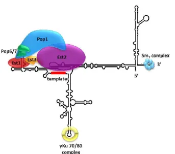
Telomerase
Telomerase is a ribonucleoprotein reverse transcriptase, requiring an RNA template, catalytic protein subunit, and accessory proteins (figure 5). In telomerase of budding yeast, the flexible RNA scaffold, TLC1, is a large 1158 nt non-coding RNA which has a region that acts as an intrinsic template for the de novo synthesis of telomeric repeats being added to the G-rich 3’ overhang (Blackburn et al., 1989; Singer and Gottschling, 1994). Telomerase also contains a trio of Est (Ever Shorter Telomere) proteins. The Est2 catalytic subunit binds to a central domain of TLC1,
comprised of a template region, a pseudoknot, and a template limiting stem (Singer and Gottschling, 1994; Seto et al., 2003; Chappell and Lundblad, 2004). Est1 and Est3 are accessory telomerase subunits required for telomerase function in vivo (Lendvay et al., 1996). Est1 binds to a highly conserved stem-bulge region of TLC1 (Seto et al., 2002), and the interaction of Est3 is thought to be mediated by other protein factors (Tucey and Lundblad, 2014). Recently, the essential proteins Pop1, Pop6, and Pop7 were identified as indispensable for in vivo telomerase activity through their role in stabilization of Est1 and Est2 (Lemieux et al., 2016). In addition, bound to TLC1 is the SM7 protein complex which aids in stability (Seto et al., 1999), as well as the yKu70/80 heterodimer which is involved in the nuclear import and retention of telomerase (Seto
et al., 1999; Stellwagen et al., 2003; Gallardo et al., 2008). In summary, telomerase plays an
important role in genomic stability at each replication round by preventing loss of the terminal chromosome repeats which are required to facilitate the protective capping function of telomeres and avoid loss of genetic information.
Figure 5: Ribonucleoprotein complex telomerase in S. cerevisiae. See text for description of different subunits. Adapted from Lemieux et al., 2016.
Telomerase activity varies by developmental stage and cell type, with high levels of activity seen in mammalian germ and stem cells, allowing for significant proliferative capacity (Bodnar et al., 1998). However, unlike germ and stem cells, telomerase is not active in most human somatic cells. This lack of telomerase activity leads to a process called replicative senescence, which will be discussed in more detail further on. It functions as a way to limit the
accumulation of harmful genetic instabilities, thus acting as a barrier to carcinogenesis. This barrier can be overcome in some cases, with cancer cells upregulating telomerase as a way to enable replicative immortality, making the study of telomeres and telomerase critical in understanding the molecular mechanisms enabling cancer progression (Shay et al., 1991; Kim et
al., 1992; Shay and Wright, 2011). Much of the work on telomeres and telomerase has been
accomplished in model organisms such as S. cerevisiae, which is particularly attractive for studying mechanisms of telomere maintenance due to the fact that it constitutively expresses telomerase. In yeast, telomere length acts as a telomerase regulatory mechanism. Not all telomeres are elongated at every cell cycle and it has been shown that short telomeres undergo preferential targeting and elongation by telomerase (Teixeira et al., 2004). It is clear that telomere length homeostasis involves a complex and dynamic interplay of regulatory mechanisms involving many different factors. Indeed, genome-wide screens have identified a large number of genes other than telomerase components that affect telomere length, but the mechanisms by which they do this often still remain to be fully elucidated (Askree et al., 2004; Gatbonton et al., 2006).
Replicative senescence & survivors
Telomerase-null mutants can be created in budding yeast by elimination of the non-coding RNA scaffold TLC1, or by deletion of genes ennon-coding the catalytic subunit Est2 and other regulatory proteins Est1 and Est3, leading to what has been termed the Est (ever shorter telomere) phenotype. These mutants suffer from progressive telomere shortening, and enter a permanent cell cycle checkpoint arrest at around 60-80 generations, leading to a loss of viability (Lundblad and Szostak, 1989; Singer and Gottschling, 1994; Lendvay et al., 1996). Intriguingly, it has been found that in telomerase-negative cultures, some cells stop growing even when the expected average telomere length is wildtype (Lundblad and Blackburn, 1993; Abdallah et al., 2009; Khadaroo et al., 2009). This indicates that the gradual attrition of bulk telomere length is not necessarily the primary determinant for arrested growth. Indeed, previous results show that the presence of a single critically short telomere is sufficient to initiate checkpoint activation and accelerate senescence (Abdallah et al., 2009; Khadaroo et al., 2009; Xu et al., 2015). It is likely that a critically short telomere arises due to the inherent difficulties experienced during telomeric DNA replication and cannot be elongated in the absence of telomerase, thus triggering a growth arrest (Hackett et al., 2001; Hackett and Greider, 2003).
After telomerase-negative cell cultures enter senescence, not all cells lose viability at the same time and a small subset of the cells eventually regains proliferative capability and the ability to maintain their telomeres through telomerase-independent mechanisms involving homologous recombination. This capacity to avoid replicative senescence earned these cells the name “survivors” (Lundblad and Blackburn, 1993; Teng and Zakian, 1999). Homologous recombination is critical for survivor formation, given that there are no survivors in telomerase-negative cells in which RAD52 is also deleted (Lundblad and Szostak, 1989). These double mutants display a strong synthetic growth defect as soon as telomerase is removed, even before displaying the progressive telomere shortening associated with entering senescence (Kass-Eisler and Greider, 2000). In addition, a homologous recombination-based DNA synthesis pathway, termed break-induced replication (BIR), is also needed for formation of survivors. BIR repairs one-sided DSBs as may occur at collapsed replication forks or by an erosion of an uncapped telomere. This mechanism requires the DNA polymerase subunit POL32, and upon deletion of this gene, survivor formation is abolished (Lydeard et al., 2007).
There are two subtypes of survivors, type I and type II, differing in the composition of their terminal DNA arrangements (Lundblad and Blackburn, 1993; Teng and Zakian, 1999). Type I survivors are characterized by amplification of subtelomeric tandem Y’ element repeats and short tracts of double-stranded telomeric DNA with a normal single-strand 3’ overhang, as well as extrachromosomal circular Y’ elements (Lundblad and Blackburn, 1993; Larrivée and Wellinger, 2006). In addition to RAD52 and POL32, type I survivor formation requires RAD51, RAD54, RAD55, and RAD57. In the absence of these genes, only type II survivors can form (Le et al., 1999; Chen et
al., 2001). Type II survivors differ from type I in that they have only slight amplification of Y’
elements, but extensive and heterogeneous expansions of telomeric TG1-3 repeats, ranging from very short to over 12 kb in size (Teng and Zakian, 1999; Teng et al., 2000). Type II survivor initiation most likely involves rolling circle replication, as the telomeres in these cells experience progressive shrinking and then random and dramatic lengthening events. In these cells, extrachromosomal telomeric DNA circles do also occur (Teng et al., 2000; Larrivée and Wellinger, 2006). Type II survivor formation is dependent on the presence of RAD59, the MRX complex (MRE11, RAD50 & XRS2), TEL1, and SGS1. As above, in the absence of these genes, only type I survivors appear (Le et al., 1999; Teng et al., 2000; Chen et al., 2001; Huang et al., 2001; Johnson
et al., 2001). It must be noted that although type I survivors initially occur more frequently, they
experience very slow growth with intermittent intervals of growth arrests, and often eventually convert to type II survivors, which have a faster growth rate and outcompete type I in liquid culture if both are present (Lundblad and Blackburn, 1993; Teng and Zakian, 1999; Teng et al., 2000).
Surprisingly, there has been relatively little investigation into the chromatin environment of telomeres in senescence and survivor formation. However, emerging evidence indicates that chromatin components active in the subtelomeric and telomeric regions such as histone acetyltransferases and deacetylases are important for regulating senescence (Kozak et al., 2009). Telomeric transcription, which would require chromatin reorganization to allow the binding of transcription factors, has been shown to influence senescence as well (Maicher et al., 2012; Pfeiffer and Lingner, 2012).
DNA damage response & telomeres
The relationship between DNA damage response, checkpoint activation, repair mechanisms and telomere homeostasis is complex and still far from being fully understood. The DDR pathway involves the rapid response of sensors, mediators, and effectors at the sites of DNA damage. Triggers include DSBs, ssDNA, replication fork stalling, or other constraining structures (Teixeira, 2013). This takes on a special significance at telomeres, as telomeric DNA is difficult to replicate due to its GC-rich nature and is prone to replication fork slowing and/or stalling (Ivessa et
al., 2002). Around the same time that it was discovered that one of the main telomere functions is
to inhibit inappropriate checkpoint activation, it was also observed that DNA damage checkpoint proteins are involved in telomere homeostasis (Sandell and Zakian, 1993; Greenwell et al., 1995). Over the years, the list of proteins involved in both DDR and telomere maintenance has grown significantly and the relationship between the two is not always clear and at times can even appear contradictory. For example, the MRX complex and Tel1 associate with both DSBs for repair and during replication to short telomeres in order to initiate resection events and also telomere elongation by telomerase (Ritchie and Petes, 2000; Tsukamoto et al., 2001). However, unlike at DSBs, at telomeres this ssDNA generation does not activate Mec1, thus avoiding triggering a DDR phosphorylation cascade that would initiate DNA repair mechanisms (Mcgee et al., 2010). Other
significant proteins involved in both processes include Yku70/80, Sae2, Sgs1, Dna2, and Exo1 (Wellinger et al., 1996; Gravel et al., 1998; Larrivée et al., 2004; Bonetti et al., 2009). As mentioned previously, further complicating this picture is the fact that many of these DDR proteins are also key players in telomere maintenance in telomerase-negative cells.
Tbf1
In terms of telomere biology and homeostasis, much attention is paid to telomeric end-binding factors and their accessory proteins, and less consideration has been given to factors influencing telomere homeostasis via subtelomeric regions. For example, one of the proteins that has been implicated in both the DNA damage response and telomere maintenance is Tbf1 (Berthiau et al., 2006; Arneric and Lingner, 2007; Ribaud et al., 2011; Bonetti et al., 2013; Di Domenico et al., 2013). Tbf1 binds to subtelomeric elements and specifically to TTAGGG repeats. It was first discovered as Tbfα (Telomere-binding factor) through DNase I footprinting in 1991 (Liu and Tye, 1991). A series of experiments indicated that it was able to bind T2AG3 repeats, the same sequence as mammalian telomeres, found in yeast at the junction between the subtelomeric region and the telomeric repeats (Liu and Tye, 1991; Brigati et al., 1993). It was proposed that due to the conservation of T2AG3 repeats at the telomere or subtelomeric regions of many organisms, this sequence must be bound by a highly conserved protein important for genomic stability (Brigati et al., 1993). Indeed, TBF1, a 562 amino acid DNA binding protein in yeast, was found to be an essential gene (Brigati et al., 1993). Interestingly, around the same time, in vitro studies confirmed the existence of mammalian TTAGGG repeat binding factors that we now know as the human telomere binding proteins TRF1 and TRF2, yielding a first glimpse into potential binding similarities of factors that bind to T2AG3 repeats (Zhong et al., 1992; Brigati et al., 1993). These similarities were further elucidated with the discovery of a Myb-related telomeric DNA binding motif conserved across a variety of species from yeast to plants to humans, named the telobox (Bilaud et al., 1996, 1997; Vassetzky et al., 1999). The telobox is considered an atypical Myb-binding domain as Myb domains typically consist of three slightly variable tandem repeats (R1, R2 and R3), as seen in the prototypical member of the Myb family, human c-Myb (Ogata et al., 1994; Ko et al., 2008). Tbf1 contains a single Myb repeat analogous to the R3 repeat, but does not bind typical Myb DNA binding sites, instead having a variable consensus sequence with an essential core TAGGG motif, similar to other telomere-binding proteins such as TRF1 and TRF2 in mammals
and Taz1 in Schizosaccharomyces pombe (Bilaud et al., 1996; Vassetzky et al., 1999; Koering et al., 2000). The Tbf1 Myb-like binding domain is an approximately 60 amino acid helix-turn-helix sequence located in the C-terminus of the protein from residues 404-464 although the exact beginning and end positions are slightly variable depending on the sequence alignment database used (http://www.yeastgenome.org/locus/S000006049/protein). Although Tbf1 demonstrates high affinity for T2AG3-like repeats, it shows no binding ability to the degenerate TG1-3 telomeric repeats bound by Rap1 in S. cerevisiae (Bilaud et al., 1996; Koering et al., 2000).
As previously mentioned, S. cerevisiae subtelomeric regions are composed of the obligatory X elements and a variable number of Y’ elements (Louis and Haber, 1992; Louis et al., 1994). In both elements, the distal ends contain Tbf1 binding sites and Tbf1 has been shown to bind in vitro and in vivo, suggesting that it plays a role at chromosome ends due to its subtelomeric binding affinity (Brigati et al., 1993; Bilaud et al., 1996; Fourel et al., 1999; Koering et
al., 2000). It was shown that Tbf1 binding is able to counteract silencing events such as the
telomere position effect (TPE). Furthermore, it was found that Tbf1, along with the transcription factor Reb1, is a key component in subtelomeric anti-silencing regions (STARs). It was proposed that these regions facilitate chromatin boundary formation in order to limit silent chromatin propagation and possibly to protect the telomere from subtelomeric influence or vice versa (Fourel et al., 1999, 2001; Koering et al., 2000). As well, using artificial telomere constructs with and without Tbf1 binding sites, it has been demonstrated that due to its subtelomeric location, Tbf1 is involved in partially redundant mechanisms of discriminating short from wild-type telomeres (Arneric and Lingner, 2007). Another type of artificial telomere construct that has been used to study Tbf1 is humanized yeast telomeres. The telomerase RNA gene TLC1 was altered in its templating region so that telomere elongation by telomerase produces the human T2AG3 sequence in place of yeast TG1-3 repeats (Henning et al., 1999). Due to Tbf1 and Cdc13 binding, these humanized telomeres are short but remain stably maintained, indicating that Tbf1 has the capacity to participate in telomeric capping under certain circumstances (Alexander and Zakian, 2003; Bah et al., 2011). Using a similar artificial construct with inducible insertion of T2AG3 repeat arrays, Tbf1 binding was shown to regulate telomere elongation in a length-dependent manner and again act as a de facto capping mechanism in the absence of Rap1 (Ribaud et al., 2011).
Immunofluorescence studies showed that Tbf1 not only binds repeats at the subtelomeric regions but also at internal chromosomal sites, indicating that it most likely has a role outside of telomere maintenance (Koering et al., 2000). Indeed, the essential function of Tbf1 was proposed to be due to a non-telomeric role which was suggested to be related to regulation of transcription, much like the dual roles of the canonical telomere binding protein Rap1 which also is a transcription factor (Bilaud et al., 1996). Changes in gene expression result from both transcription factor binding to regulatory sequences and modifications to nucleosome organization in regulatory regions (Preti et al., 2010). The possibility that Tbf1 was involved in transcriptional processes was later confirmed by analyses of in vivo transcription, chromatin immunoprecipitation (ChIP), and nucleosome occupancy analyses that were performed to identify and delineate transcriptional control elements located upstream of snoRNA genes (Preti et al., 2010). Remarkably, Tbf1 was found to bind at on the promoters of approximately 80% of all snoRNA genes (Preti et al., 2010). Most likely its function there is to be a transcription activator, involved in the fine tuning and full expression of snoRNA levels (Preti et al., 2010). However, in addition to its role at snoRNA promoters, Tbf1 was also found to bind upstream of over 200 protein-coding genes, including its own promoter (Lavoie et al., 2010; Preti et al., 2010). However, its roles at snoRNA promoters versus the promoters of protein-coding genes seem to be slightly different. At the promoters of protein-coding genes, Tbf1 binding, together with the two Tbf1-interacting proteins, Vid22 and Env11, was found to foster the formation of a 5’ nucleosome-free region (Preti et al., 2010). However, the same work showed at snoRNA targets it did not appear to facilitate nucleosome exclusion or associate with these two proteins. Finally, in concordance with earlier studies promoting a subtelomeric regulatory role for Tbf1, ChIP-seq analysis displayed single or clustered Tbf1 association peaks at 22 out of 32 telomeric sites on 15 out of 16 chromosomes (Preti et al., 2010).
Interestingly, recent work by the Longhese lab about the involvement of Tbf1 in DNA damage repair further pointed towards a role in modulating chromatin organization as part of the DNA damage response (Bonetti et al., 2013). As TBF1 cannot be deleted, mutant tbf1 alleles were created by low-fidelity PCR mutagenesis coupled with screening of clones for decreased viability either at high temperatures or in response to DNA damaging agents such as the DNA break-inducing drug phleomycin. This led to the selection of three mutant alleles containing 2-4 point mutations, tbf1-1, tbf1-2, and tbf1-3 (Bonetti et al., 2013). These alleles were shown to be
sensitive to both phleomycin and MMS, a drug that leads to replication fork stalling through alkylation of DNA. Genetic assays revealed that combining tbf1-1 and tbf1-3 with various homologous recombination mutants such as rad52∆ (roles in multiple HR pathways), mre11∆ (initiation of HR end-processing), sgs1∆ and mms4∆ (processing HR intermediates) led to synthetically sick double-mutants with significant growth defects and increased sensitivity to DNA-damaging agents (Bonetti et al., 2013).This indicated that Tbf1 is involved in supporting cell viability when homologous recombination mechanisms are impaired. They further demonstrated that Tbf1 was recruited near HO endonuclease-induced double-strand breaks and mutants were defective in end processing of these breaks (Bonetti et al., 2013). Given that previously, Tbf1 had been shown to participate in nucleosome exclusion at promoter regions (Preti et al., 2010), it was postulated that it might have a role in promoting chromatin remodeling at DSBs as well. Indeed, Tbf1 was found to functionally interact with chromatin regulation proteins, with mutants showing a synthetic negative interaction with increased sensitivity to phleomycin when combined with an
esa1 mutant allele, the catalytic subunit of the NuA4 acetyltransferase complex involved in DSB
repair (Bird et al., 2002; Bonetti et al., 2013). In contrast, when the histone deacetylase RPD3, which counteracts Esa1 acetylation, was deleted in tbf1-1 cells, a marked decreased sensitivity to phleomycin was documented (Bonetti et al., 2013). This study therefore highlighted Tbf1 as having a previously unrecognized role in the response to DNA damage, most likely due to a direct role in chromatin organization at a double-strand break.
Hypothesis and objectives
Previous work clearly indicates that Tbf1 exercises multiple functions in the cell. It is implicated in processes such as transcription, the DNA damage response and DNA repair pathways, and finally, telomere homeostasis. Despite this, surprisingly little is known about how it is involved in these processes and what it interacts with to fulfill these varied roles. Many of the experiments involving the role of Tbf1 at telomeres were carried out using artificial telomeric constructs from which functions at native telomeres were inferred. Hence there is a lack of information characterizing the role of Tbf1 in telomere maintenance at native telomeres, rendering it an interesting candidate to study in this context. I hypothesize that Tbf1 exerts effects on telomere regulation due to its ability to bind to subtelomeric elements, where it interacts with known regulatory mechanisms. The primary objective of my master’s project is to better
understand the role that Tbf1 plays in telomere maintenance due to its subtelomeric location. This was accomplished through a variety of genetic analyses.
Given that TBF1 is an essential gene, the typical preliminary genetic analyses used in S.
cerevisiae are more complicated to achieve, as it cannot be deleted to examine potential
phenotypes. A gene that is essential for cell viability often is involved in a function in one or more highly conserved processes, making it a particularly interesting avenue of research (Giaever et al., 2002). This essentiality in turn renders a gene quite difficult to study, given the problems inherent in examining the function of essential genes. What amino acid residues or domains are critical for function? What modifications effect phenotypic changes, and why? When looking for phenotypic effects of an essential gene, one of the main approaches is to use hypomorphic alleles. This type of allele reduces how well the gene is expressed. In fact, there are collections of hypomorphic essential gene mutants available that now cover almost every essential gene (Schuldiner et al., 2005; Ben-Aroya et al., 2008; Ungar et al., 2009). These alleles were generated via a variety of methods. For example, one group constructed titratable alleles by placing the gene under an inducible promoter, so that gene expression is regulated by the addition of a substance such as an antibiotic or carbon source (Mnaimneh et al., 2004). This has the benefit of being able to control gene expression at will. A drawback of this method is that the gene is not under the control of its native promoter, and overall regulation and changes in expression are hard to link to a specific phenotype.
Another way to obtain hypomorphic alleles is to generate them by random mutagenesis. This allows screening of large numbers of mutagenized genes based on certain criteria such as temperature or drug sensitivity (Ben-Aroya et al., 2008). Often, these alleles contain multiple randomly-generated point mutations that can be identified through genomic sequencing. Again, the phenotype of these alleles can be complicated to interpret, given that the contribution of each mutation to the phenotype is not known. However, such alleles can be a good starting point for phenotypic analysis. Once the individual point mutations are known via sequencing, mutants can be constructed that contain only one point mutation, which will allow the analyses of specific mutations and how they induce phenotypic variation. In an ideal scenario, this leads to a single mutation causing an identifiable phenotypic change.
In order to examine the function of Tbf1, I initially obtained and analyzed conditional mutants that were obtained from other research labs. These included a hypomorphic thermosensitive allele, tbf1-ts, as well as a trio of alleles with multiple mutations generated through random mutagenesis: tbf1-1, tbf1-2, and tbf1-3 (Ben-Aroya et al., 2008; Bonetti et al., 2013). Once certain phenotypic variations caused by these alleles were identified, I then generated novel alleles with single point mutations. These alleles were combined with known mutants of telomere maintenance in order to look for genetic interactions between Tbf1 and other genes. Through these genetic analyses, specific individual residues were linked to phenotypic effects and a previously unknown role for Tbf1 in telomere homeostasis was uncovered.
MATERIALS AND METHODS
Yeast strains and plasmids
There are several commonly used S. cerevisiae laboratory strains that have slight differences in their genetic backgrounds. S288c (Mortimer and Johnston, 1986) was the original strain sequenced by Sanger sequencing. Several strains have been derived from the S288c background, including the haploid strains BY4741, BY4742 and diploid strain BY4743 found in the systematic deletion project that created the commercially available YKO collection of non-essential gene deletions (Brachmann et al., 1998) and other BY background strains such as BY4705. W303 is another commonly used laboratory strain, originally created by Rodney Rothstein (Rothstein, 1983). The W303 genome is approximately 85% derived from S288c, part of the other regions are similar to other laboratory strains (Ralser et al., 2012). About 800 coding regions differ between W303 and S288c, but often by very few residues. The original W303 strain carries a rad5-535 point mutation (Fan et al., 1996) that has been corrected in the W303 derivative W3749 (Lisby et al., 2004).
Table 1: Strains and plasmids that are referred to throughout this work. All strains without a reference listed were constructed for this study.
Name Genotype Reference
tbf1-ts
MATa ura3Δ0 leu2Δ0 his3Δ1 lys2Δ0 (or
LYS2) met15Δ0 (or MET15) can1Δ::LEU2-MFA1pr::His3 tbf1-ts::URA3
Ben-Aroya et al., 2008.
W303
MATa ade2-1 can1-100 his3-11,15 leu2-3,112 trp1-1
ura3-1 rad5-535 Bonetti et al., 2013.
tbf1-1
MATa ade2-1 can1-100 his3-11,15 leu2-3,112 trp1-1
ura3-1 rad5-535 tbfura3-1-ura3-1::LEU2 Bonetti et al., 2013.
tbf1-2
MATa ade2-1 can1-100 his3-11,15 leu2-3,112 trp1-1
ura3-1 rad5-535 tbfura3-1-2::LEU2 Bonetti et al., 2013.
tbf1-3
MATa ade2-1 can1-100 his3-11,15 leu2-3,112 trp1-1
ura3-1 rad5-535 tbfura3-1-3::LEU2 Bonetti et al., 2013.
ELBY63 W303 tbf1-1::LEU2/TBF1 tlc1∆::KanMX4/TLC1 +pAZ1 ELBY54 W303 tbf1-2::LEU2/TBF1 tlc1∆::KanMX4/TLC1 +pAZ1 ELBY76 W303 tbf1-3::LEU2/TBF1 tlc1∆::KanMX4/TLC1 +pAZ1
W3749
MatA/α can1-100/can1-100 ura3-1/ura3-1
his3-11,15/his3-11,15 leu2-3,112/leu2-3,112 trp1-1/trp1-1
bar1Δ::LEU2/bar1Δ::LEU2 Lisby et al., 2004.
BY4705
ade2::hisG/ade2::hisG his3∆200/his3∆200 leu2∆0/leu2∆0 lys2∆0/lys2∆0 met15∆0/met15∆0 trp∆63/ trp∆63 ura3∆0/ura3∆0 Brachmann et al. 1998. ELY030 W3749 tbf1-299::NatMX/TBF1 ELY045 W3749 tbf1-82::NatMX/TBF1 ELY046 W3749 tbf1-453::NatMX/TBF1 ELY047 W3749 tbf1-398::NatMX/TBF1 ELY048 W3749 tbf1-480::NatMX/TBF1
ELY050 W3749 tbf1-453::NatMX/TBF1 tlc1Δ::KanMX4/TLC1 ELY052 W3749 tbf1-82::NatMX/TBF1 rpd3Δ::TRP1/RPD3 ELY054 W3749 tbf1-82::NatMX/TBF1 tlc1Δ::KanMX4/TLC1
ELY058
W3749 tbf1-82::NatMX/TBF1 rad52::TRP1 /RAD52
tlc1Δ::KanMX4 /TLC1+pAZ1.R52-URA3 ELY059 W3749 tbf1-453::NatMX/TBF1 rad52∆::TRP1/RAD52 tlc1Δ::KanMX4 /TLC1+pAZ1.R52-URA3 ELY065 W3749 tbf1-82::NatMX/TBF1 sgs1∆::TRP1/SGS1 tlc1Δ::KanMX4/TLC1 ELY067 W3749 tbf1-453::NatMX/TBF1 sgs1∆::TRP1/SGS1 tlc1Δ::KanMX4/TLC1 ELY069 W3749 tbf1-82::NatMX/TBF1 tlc1Δ::KanMX4/TLC1 rpd3Δ::TRP1/RPD3 ELY070 W3749 tbf1-453::NatMX/TBF1 tlc1Δ::KanMX4/TLC1 rpd3Δ::TRP1/RPD3
ELY076 BY4705 tbf1-453::NatMX/TBF1
ELY078 BY4605 tbf1-453::NatMX/TBF1 mre11∆/MRE11 ELY083 W3749 tbf1-82::NatMX/TBF1 htz1Δ::URA3/HTZ1 ELY084 W3749 tbf1-453::NatMX/TBF1 htz1Δ::URA3/HTZ1 ELY085 W3749 tbf1-305::NatMX/TBF1 ELY090 W3749 tbf1-447::NatMX/TBF1 ELY093 W3749 tbf1-451::NatMX/TBF1 ELY095 W3749 tbf1-451::NatMX/TBF1 EPY072 W3749 htz1Δ::URA3 E.Pasquier, Wellinger lab
mre11∆ Mat A mre11∆::KanMX his3∆1 leu2∆0 met15∆0 ura3∆0
YKO library, open Biosystems
JNY61
MATa ade2-1 can1-100 his3-11,15 leu2-3,112 trp1-1 ura3-1 tlcura3-1∆::KanMX4 + pAZura3-1
J.F. Noel, Wellinger lab
1 rad52∆::TRP1 tlc1∆::KanMX4 + pAZ1 lab
Plasmids used in this study:
pCT300
∼300 bp C1-3A repeats EcoRI fragment of pYLPV in
the BamHI/SalI site of the cloning vector pVZ1 Bourns et al., 1998
pAZ1 URA3 – CEN – TLC1 Beeler et al., 1994
pAZ1.R52
pVL191-Sal1 fragment (3.3kb) containing RAD52 cloned
into pAZ1 at XhoI site Wellinger lab
pAG25 pAgTEF1-natMX-tAgTEF1
Goldstein and McCusker, 1999
pRS304
TRP1 - f1 ori (NaeI) - T7 promoter - lacZ'/MCS - T3
promoter - pMB1 ori - bla
Brachmann et al., 1998
pRS304-TBF1::NatMX
TRP1 - f1 ori (NaeI) - T7 promoter - lacZ'/MCS - T3
promoter - pMB1 ori – bla – TBF1 ::NatMX
Construction of TBF1 alleles
Three fragment Gibson assembly was used to clone upstream sequences of TBF1, a nourseothricin (ClonNAT) antibiotic resistance cassette (NatMX), and sequences downstream of
TBF1 into the pRS304 plasmid containing the TRP1 auxotrophic marker (Brachmann et al., 1998).
Fragment 1 consisted of the sequence of TBF1 -165 bp to TBF1 + 93 bp PCR amplified from genomic DNA derived from strain W3749 (see Table 1). Fragment 2 was the NatMX cassette amplified from plasmid pAG25 (Goldstein and McCusker, 1999). Fragment 3 was a 128 bp sequence downstream of TBF1 also amplified from genomic DNA of W3749. PCR conditions for fragment amplification are described below. Upstream sequence in fragment 1 and downstream sequence in fragment 2 were included to provide homology for genomic integration. The pRS304 vector plasmid was linearized with BamHI and NotI restriction enzymes. The linearized vector and PCR fragments were purified on 0.6% agarose gel prior to the Gibson assembly reaction, which was performed according to the manufacturer’s instructions
(https://www.neb.com/applications/cloning-and-synthetic-biology/dna-assembly-and-cloning/gibson-assembly). The pRS304-TBF1::NatMX plasmid was cloned in DH5α Escherichia coli cells (see transformation protocol below). Clones were grown in liquid Luria broth (LB) + ampicillin overnight and the plasmid was extracted using EZ-10 spin column plasmid DNA mini-preps kit (Biobasic). The plasmid was sequenced to ensure correct assembly and absence of unwanted