Université de Montréal
AXL receptor tyrosine kinase in breast cancer: Defining
novel substrates and pathways involved in cell motility and
invasion
Par Afnan Abu-Thuraia
Programmes de Biologie Moléculaire
Faculté de Médecine
Thèse présentée
en vue de l’obtention du grade de de doctorat en Biologie Moléculaire
August 2018
Résumé
Le cancer du sein est le cancer le plus fréquemment diagnostiqué et le plus mortelle chez la femme, où sa progression vers le stade métastatique constitue une menace pour la vie des patientes. La présence de métastases représente le défi clinique central de l'oncologie des tumeurs solides, de sorte que les mécanismes et les voies sous-jacents au processus métastatique doivent être mieux définis. L'expression aberrante du récepteur tyrosine kinase (RTK) AXL a été liée cliniquement à la formation de métastases et à l'acquisition d'une résistance aux médicaments contre le cancer. AXL est un membre de la sous-famille des récepteurs tyrosine kinase TAM et intervient dans plusieurs processus biologiques tels que l'atténuation de la réponse immunitaire, l'élimination des cellules apoptotiques et la promotion de la survie cellulaire. L'expression d'AXL dans les tumeurs primaires humaines corrèle avec la faible survie des patients. Malgré sa régulation positive préférentielle dans les lignées cellulaires triple négatives / basales B, des études ont montré que l’expression d’AXL est indépendante du sous-type de la tumeur mammaire des patients. AXL peut être activé par son ligand GAS6 ou par d'autres RTK. Lors de son activation, AXL induit une signalisation en aval entraînant l'activation d'intermédiaires de signalisation canoniques, notamment MAPK, AKT et PI 3-kinases. Cependant, les voies de signalisation spécifiques engagées par AXL pour conférer un tel pouvoir pro-invasion ne sont pas connues. Ainsi, le but de cette thèse est d'identifier des substrats spécifiques d’AXL et des voies en aval qui jouent un rôle important dans le maintien d'un état « EMT » et d'un renforcement du phénotype mésenchymal dans les cellules cancéreuses.
À la recherche de régulateurs en amont du complexe ELMO/DOCK1 impliqués dans l’activation de RAC, nous présentons au chapitre 2 les protéines d’échafaudage ELMO en tant que substrats directs et partenaires de liaison d’AXL. Grâce à des approches de protéomique et de mutagenèse, nous révélons que la kinase AXL phosphoryle ELMO1/2 sur un résidu tyrosine carboxy-terminal conservé. Dans les cellules cancéreuses du sein, l'activation d'AXL dépendante de GAS6 a conduit à la phosphorylation endogène d'ELMO2 sur Tyr-713, menant ainsi à la formation du complexe AXL/ELMO. En outre, l'activation de RAC induite par GAS6 dans les cellules cancéreuses du sein dépendait de l'expression d'ELMO2. Semblable au blocage
d’AXL, l'inhibition d’ELMO2 ou l'inhibition pharmacologique de DOCK1 supprime l'invasion des cellules du cancer du sein, qui, selon nous, dépendait de l'état de phosphorylation d'ELMO. Notre travail au chapitre 2 définit un nouveau mécanisme par lequel AXL favorise la prolifération et l'invasion cellulaire et identifie l'inhibition de la voie ELMO/DOCK comme une cible thérapeutique potentielle pour arrêter les métastases induites par AXL.
Bien qu'il soit encore difficile de savoir comment les signaux d’AXL induisent son phénotype pro-invasif, notre travail au chapitre 3 vise à identifier des substrats et des voies de signalisation spécifiques qui sont significativement modulés lors de l'activation d'AXL. Pour y remédier, nous avons défini le phosphoprotéome de la régulation d’AXL dans des cellules cancéreuses du sein triple-négatives en utilisant une approche quantitative. Nous révélons qu’AXL module de manière robuste, parmi de nombreux processus et voies biologiques importants, la phosphorylation d'un réseau de protéines d'adhésion focale (FA) aboutissant à un désassemblage plus rapide des FA. De manière intéressante, nous avons trouvé que la modulation de la voie FA était unique à AXL par rapport à d'autres RTK tels que l'EGFR. En particulier, nous avons trouvé qu’AXL phosphoryle la protéine NEDD9, modulant la formation du complexe NEDD9/CRKII/DOCK3, qui orchestre la phosphorylation de la pseudo-kinase PEAK1 médiée par AXL. Nos données révèlent un mécanisme distinct par lequel les complexes PEAK1 avec la kinase CSK médient la phosphorylation de PXN et le renouvellement des FA induit par AXL. En utilisant l'injection orthotopique de cellules cancéreuses du sein dans le tissu adipeux mammaire des souris et dans la veine de la queue, nous révélons que l'inactivation de PEAK1 par CRISPR diminue la croissance tumorale et les métastases in vivo. De plus, notre travail au chapitre 3 révèle une contribution unique et inattendue de la signalisation d’AXL à la dynamique des FA, révélant un mécanisme longtemps recherché sous-tendant l'activité invasive d'AXL. Cette compréhension approfondie des réseaux de signalisation régulés par AXL identifie PEAK1 comme une nouvelle cible thérapeutique dans les tumeurs AXL positives.
En conclusion, cette thèse a identifié, pour la première fois, le phosphoprotéome d’AXL et des voies de signalisation spécifique à AXL, pouvant justifier le rôle du récepteur en tant que promoteur de métastases et de résistance aux médicaments. Notre travail révèle de nouvelles cibles thérapeutiques qui pourraient avoir un grand potentiel si elles sont utilisées en thérapie
combinatoire avec l’inhibition d’AXL pour prévenir la formation de métastases des tumeurs AXL positives.
Mots clés: Métastases, Voies de signalisation récepteur de tyrosine tinase, Protéomique,
Abstract
Breast cancer is the most frequently diagnosed cancer in women where its progression to the metastatic stage poses a threat to the life of patients. The metastatic disease represents the central clinical challenge of solid tumor oncology such that mechanisms and pathways underlying the metastatic process must be better defined. The aberrant expression of the receptor tyrosine kinase (RTK) AXL has been linked clinically to metastasis and acquisition of drug resistance. AXL is a member of the TAM subfamily and functions in several biological processes such as dampening the immune response, clearing apoptotic cells and promoting cell survival. Despite its preferential upregulation in triple negative/basal B cell lines, studies have shown AXL expression in the clinic to be subtype independent. AXL can be activated by its ligand GAS6 or by a crosstalk with other RTKs. Upon its activation, AXL induces downstream signaling resulting in the activation of canonical signaling intermediates including MAPKs, AKT and PI 3-kinases. However, the specific signaling pathways engaged by AXL to confer such enhanced pro-invasion power are not known and the goal of this thesis is to identify AXL-specific substrates and downstream pathways that are behind AXL’s significant role in maintaining an EMT state and reinforced mesenchymal phenotype in cancer cells.
In search of upstream regulators of ELMO/DOCK1 complex involved in RAC activation, we reported ELMO scaffolds as direct substrates and binding partners of AXL. Through proteomics and mutagenesis approaches, we revealed phosphorylation of ELMO1/2 by AXL kinase on a conserved carboxyl-terminal tyrosine residue. In breast cancer cells, GAS6-dependent activation of AXL led to endogenous ELMO2 phosphorylation on Tyr-713 and AXL/ELMO complex formation. In addition, GAS6-induced RAC activation in breast cancer cells was dependent on ELMO2 expression and phosphorylation. Our work in chapter 2 defines a new mechanism by which AXL promotes cell proliferation and invasion and identifies inhibition of ELMO/DOCK pathway as a potential therapeutic target to stop AXL-induced metastases.
While it still remains elusive how AXL signals to induce its pro-invasive phenotype, our work strove to identify specific substrates and signaling pathways that are significantly modulated upon AXL activation using a quantitative phosphoproteomics approach. By
generating GAS6-induced AXL phosphoproteome, we found that AXL robustly modulates, among many different significant biological processes and pathways, the phosphorylation of a network of focal adhesion (FA) proteins culminating in faster FA disassembly. Interestingly, we found AXL modulation of FA pathway to be unique to AXL in comparison with other RTKs such as EGFR. NEDD9 FA protein was identified to be a direct substrate of AXL, where its phosphorylation modulates its complex formation with CRKII/DOCK3, and this subsequently orchestrates the AXL-mediated phosphorylation of the pseudo-kinase PEAK1. Our data revealed a distinct mechanism by which PEAK1 complexes with CSK kinase, mediating PXN phosphorylation and AXL-induced FA turnover. Using in vivo assays such as tail-vein metastasis assay and tumor growth assay, we revealed that gene inactivation of PEAK1 by CRISPR CAS9 decreased tumor growth and metastasis. Furthermore, our work in chapter 3 uncovers an unexpected and unique robust contribution of AXL signaling to FA dynamics revealing a long sought-after mechanism underlying AXL pro-invasive activity. This in-depth understanding of AXL regulated signaling networks identifies PEAK1 as a new therapeutic target in AXL positive tumors.
In conclusion, this thesis identified, for the first time, AXL phosphoproteome and AXL specific downstream signaling pathways that may justify AXL’s role as a promoter of metastasis and drug resistance. Our work reveals novel therapeutic drug targets that may hold a great potential if used in combinational therapeutics with AXL inhibition to prevent metastasis of AXL positive tumors.
Keywords: Metastasis, RTK signaling, Proteomics, Focal adhesion, Breast Cancer, Cell
Preface
This thesis is written in a manuscript format by articles and is divided into three chapters followed by a discussion. It contains one published article and two articles prepared for submission.
CHAPTER 1: INTRODUCTION
Section 1.1, 1.3, and 1.4 contain a literature review on breast cancer and metastasis, dynamics of actin cytoskeleton, and cancer cell migration and invasion, respectively. Section 1.5 contains our research hypothesis and objectives. Section 1.2 contains a manuscript of a review to be submitted for publication:
Abu-Thuraia A*, Goyette MA*, Delliaux C and Côté JF (2018) Dissecting AXL’s role in
cancer progression. *Equal contribution
CHAPTER 2: AXL phosphorylates ELMO scaffold proteins to promote RAC activation and cell invasion.
This chapter contains a published article:
Abu-Thuraia A, Gauthier R, Chidiac R, Fukui Y, Screaton RA, Gratton JP and Côté JF (2015).
Axl phosphorylates Elmo scaffold proteins to promote Rac activation and cell invasion. MCB 35, 76-87.
CHAPTER 3: AXL confers cell migration and invasion by hijacking a PEAK1-regulated focal adhesion protein network.
This chapter contains a submitted manuscript to Nature Communication and is in revision:
Abu-Thuraia A, Goyette MA, Delliaux C, Boulais J, Chidiac R, Bagci H, Davidson D, Veillette
Table of Content
Résumé ... 1 Abstract ... 4 Preface... 6 Table of Content ... 7 List of tables ... 10 List of figures ... 11 List of abbreviations ... 13 Acknowledgments... 19 CHAPTER 1 ... 21 INTRODUCTION ... 211.1 The Central Dogma of Cancer ... 22
1.1.1 Metastasis ... 24
1.1.2 Breast Cancer and its molecular subtypes ... 27
1.1.3 Receptor Tyrosine Kinases: key players in cancer biology ... 29
1.2 Dissecting AXL’s role in cancer progression ... 33
Preface... 34
Introduction ... 35
Regulation of TAM Expression ... 35
Mechanisms of activation ... 38 Ligand-dependent ... 38 Ligand-independent ... 39 Regulation of activation ... 41 Signaling in cancer ... 42 Survival ... 42 Migration... 43
TAMs in human cancer ... 47
Expression of TAM receptor ... 47
Tumor growth and survival ... 47
Metastatic progression of solid cancer ... 48
EMT and metastasis ... 48
AXL in multiple steps of the metastatic cascade ... 48
Dormancy ... 49
Tumor microenvironment ... 49
Angiogenesis ... 49
Antitumor immunity ... 50
Mediators of resistance ... 53
Chemotherapy and antimitotic drugs ... 53
Targeted therapy ... 53
Immunotherapy and radiation ... 55
Future perspectives ... 58
1.3 Dynamics of the actin cytoskeleton ... 59
1.3.1 Regulation of actin cytoskeleton dynamics ... 60
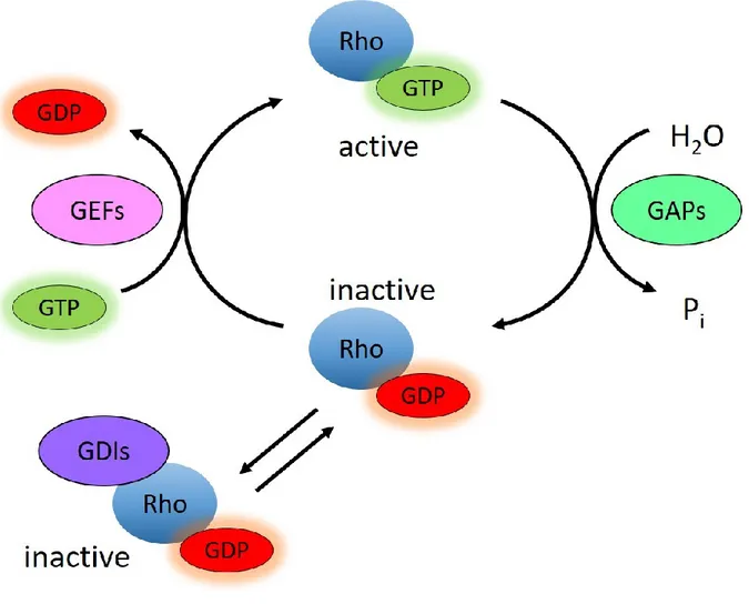
1.3.1.1 RHO Family of GTPases ... 63
1.3.1.1.1 Regulation of RHO GTPases ... 66
1.3.1.1.2 DOCK family of atypical GEFs ... 69
1.3.1.1.2.1 Binding Partners... 71
1.3.1.1.2.2 Regulation of DOCK/ELMO complex ... 73
1.3.1.1.2.3 Role of DOCK/ELMO complex in cancer ... 74
1.3.2 Conclusion ... 76
1.4 Cancer Cell Migration and Invasion ... 77
1.4.1 Cell Polarization and Protrusions... 80
1.4.2 Focal Adhesions ... 82
1.4.2.1 Regulation ... 85
1.4.3 Conclusion ... 87
1.5 Research hypothesis and objectives ... 88
Objective 1 (CHAPTER 2): ... 89
Objective 2 (CHAPTER 3): ... 89
CHAPTER 2 ... 90 AXL phosphorylates ELMO scaffold proteins to promote RAC activation and cell invasion 90
Contributions... 91
AXL phosphorylates ELMO scaffold proteins to promote RAC activation and cell invasion ... 92 Abstract ... 93 Introduction ... 94 Results ... 96 Discussion ... 110 Methods... 114 Acknowledgments... 119 Supplementary Information ... 120 CHAPTER 3 ... 136
AXL confers cell migration and invasion by hijacking a PEAK1-regulated focal adhesion network ... 136 Contributions... 137 Abstract ... 140 Introduction ... 141 Results ... 143 Discussion ... 168 Methods... 170 Acknowledgments... 181 Data Availability ... 181 Supplementary Information ... 182 DISCUSSION ... 197 References ... i
List of tables
Table 1. I Biological roles of GTPases in the cell ... 65
Table 1. II Post-translational modifications of GTPases ... 68
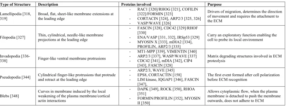
Table 1. III Various types of protrusions formed by the cell upon cell movement... 81
Table 1. IV Major players involved in the regulation of Focal Adhesion dynamics ... 86
Table 2. SI: GST-kinase library (List of human protein kinases in the GST-kinase expression library used in Figure 2.1 to screen.)... 128
Table 2. SII: List of kinases capable of phosphorylating Elmo1 identified in the screen for kinases. ... 134
Table 2. SIII: Primers used for different procedures ... 135
Table 3. SI: List of antibodies ... 193
Table 3. SII: List of siRNA ... 194
Table 3. SIII: List of plasmids ... 195
List of figures
Figure 1.1.1 The invasion-metastasis cascade ... 26
Figure 1.1.2 Molecular subtype of breast cancer ... 28
Figure 1.1.3 Receptor tyrosine kinases ... 31
Figure 1.2.1 Schematic representation of TAM family of receptors and their ligands .... 37
Figure 1.2.2 Mechanisms of AXL activation ... 40
Figure 1.2.3 AXL signaling cascades ... 46
Figure 1.2.4 Role of AXL in metastasis and tumor microenvironment ... 52
Figure 1.2.5 Mediators of Resistance ... 57
Figure 1.3.1 GDP-GTP cycle of RHO GTPases ... 62
Figure 1.3.2 Phylogenetic tree of the RHO GTPase family... 64
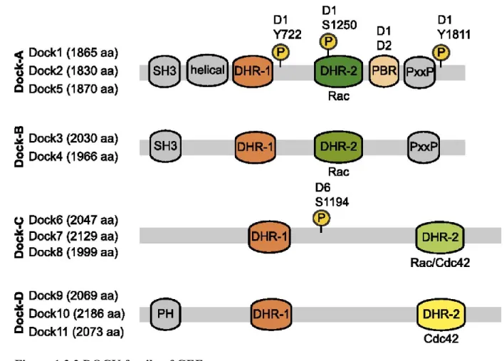
Figure 1.3.3 DOCK family of GEFs ... 70
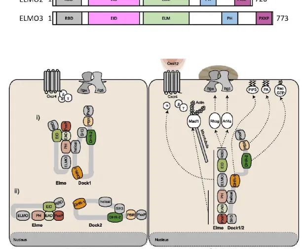
Figure 1.3.4 ELMO family of proteins and their regulation of DOCK protein localization ... 72
Figure 1.4.1 Cell movement mechanism ... 79
Figure 1.4.3 Focal Adhesion Structure ... 83
Figure 1.4.4 Focal adhesions turnover in a directed cell migration ... 84
Figure 2.1 The TAM receptors phosphorylate Elmo proteins ... 98
Figure 2.2 Elmo1 is phosphorylated on Tyrosine 720 and 724 by TAM receptors ... 100
Figure 2.3 Elmo Modulation by Axl is dependent on Axl’s catalytic activity ... 103
Figure 2.4 Rac activation in Hs578T cells is Axl- and Elmo2-dependent ... 106
Figure 2.5 Cell invasion and proliferation of MDA-MB-231 cells is Elmo2 and Axl dependent ... 108
Figure 2. S1 Elmo phosphorylation by the TAM receptors ... 121
Figure 2. S2 Specificity of pY713 Elmo2 antibody... 123
Figure 2. S3 Tyrosine 773, 821 and 860 in Axl C-terminal are not required for Elmo2 binding and phosphorylation ... 124
Figure 2. S4 Expression Profile of the TAM receptors and Elmo proteins in MDA-MB-231 and Hs578T cells ... 125
Figure 2. S6 Inhibition of activation or knockdown of Axl, Elmo2, Dock1, and Dock5 alter Vimentin Expression ... 127 Figure 3.1 Phosphoproteomic analyses of the receptor tyrosine kinase AXL in TNBC model. ... 146 Figure 3.2 High-resolution overview of AXL signaling revealed from a phosphoproteomic screen in TNBC cells ... 149 Figure 3.3 AXL localizes at FA sites and modulates their turnover. ... 151 Figure 3.4 AXL phosphorylates NEDD9 and regulates its localization at FA ... 154 Figure 3.5 AXL interacts with PEAK1 and modulates its phosphorylation and localization in the cell ... 157 Figure 3.6 CRKII direct binding to PEAK1 is necessary for AXL-mediated PEAK1 phosphorylation and for CRKII localization at FA. ... 159 Figure 3.7 PEAK1, in complex with CSK, regulates FA turnover by PXN phosphorylation downstream of AXL. ... 162 Figure 3.8 AXL/PEAK1 complex regulate recruitment of FA disassembly complex... 165 Figure 3.9 PEAK1 expression regulates tumor growth and metastasis of TNBC in vivo.
... 166
Figure 3.10 Schematic model of AXL and EGFR signaling in a cancer cell, where EGFR modulates adheren junctions and AXL modulates the FA turnover. ... 167 Figure 3. S1 Overview of the GAS6 phosphoproteome profiling and validations. ... 183 Figure 3. S2 Dot plot representation of significantly enriched KEGG pathways of GAS6 regulated phosphoproteins at the three different time points of stimulation ... 184 Figure 3. S3 AXL modulates FA turnover in Hs578T cells ... 185 Figure 3. S4 AXL modulates NEDD9 phosphorylation levels in vitro and its complex formation ... 187 Figure 3. S5 PEAK1 interacts with CRKII and mediates PXN phosphorylation. ... 189 Figure 3. S6 AXL/PEAK1 modulate βPIX/GIT1/PAK complex. ... 191 Figure 3. S7 PEAK1 is required for wound healing, proliferation and tumorsphere formation in vitro... 192
List of abbreviations
ABP: Actin Binding ProteinsADAM10: ADAM Metallopeptidase Domain 10 ADAM17: ADAM Metallopeptidase Domain 17 ADP: Adenosine Diphosphate
AKT: AKT Serine/Threonine Kinase ALK: Anaplastic Lymphoma Kinase, RTK ARMs: Armadillo Repeats
ATP: Adenosine Triphosphate
AXL: AXL Receptor Tyrosine Kinase
BAD: BCL-2 Associated Agonist of Cell Death BAX: BCL-XL Associated Protein
BCAR1: Breast Cancer Antiestrogen Resistance 1 BCL-2: B-cell CLL/Lymphoma-2
BCL-XL: BCL-2 Like Protein
B-CLL: B-cell Chronic Lymphocytic Leukemia βPIX: β-PAK Interacting Exchange Factor
C1-TEN: C1-Domina containing Phosphatase and Tensin Homolog CAF: Cancer-Associated Fibroblasts
CBLB: CBL proto-oncogene B
CRKII: CRK proto-oncogene, Adaptor protein CSK: C-terminal SRC Kinase
CXCR4: C-X-C Motif Chemokine Receptor 4 DH: Dbl Homology
DHR: Dbl Homology Region DOCK: Dedicator of cytokinesis DTC: Disseminated Tumor Cells ECM: Extracellular Matrix
EGFR: Epidermal Growth Factor Receptor EID: ELMO Inhibitory Domain
ELMO: Engulfment and cell motility
EMT: Epithelial-to-Mesenchymal Transition EPHA2: Ephrin Receptor 2
ER: Estrogen Receptor
ERK: Extracellular Signal-Regulated Kinase FA: Focal Adhesions
FAK: Focal Adhesion Kinase FRA-1: FOS Related Antigen-1 GAP: GTPase Activating Protein GAS6: Growth Arrest Specific 6 GDI: GDP Dissociation Inhibitor GDP: Guanosine Diphosphate
GEF: Guanine nucleotide Exchange Factor
GIT1: G-protein coupled receptor kinase Interacting ArfGAP1 GnRH: Gonadotropin-Releasing Hormone
GRB2: Growth Factor Receptor Bound Protein 2 GTP: Guanosine Triphosphate
HER2: Human Epidermal Growth Factor Receptor 2 HR: Homologous Recombination
KID: Kinase Insert Domain KO: Knockout
LPA: Lipoprotein A
MCL1: Myeloid Cell Leukemia 1 MEK: MAPK/ERK kinase
MERTK: MER Proto-oncogene, Receptor Tyrosine Kinase MET: Mesenchymal-to-Epithelial Transition
MET: MET proto-oncogene
MHC1: Major Histocompatibility Complex 1 MMP: Matrix Metallopeptidase
MYPT1: Myosin phosphatase, Target Subunit 1 NCK: NCK adaptor protein
NEDD9: Neural precursor cell Expressed, Developmentally Downregulated 9 NF-KB: Nuclear Factor Kabba B subunit
NK: Natural Killer Cells
OPCML: Opioid Binding Protein/Cell Adhesion Molecule-Like PAK: P21 Activated Kinase
PDGFR: Platelet Derived Growth Factor Receptor PD-L1: Programmed Death-Ligand 1
PI3K: Phosphatidylinositol-4,5-Bisphosphate 3-Kinase PKA: Protein Kinase A
PKC: Protein Kinase C PLC-γ: Phospholipase C-γ PR: Progesterone Receptor
PRAG1: PEAK1 Related-Kinase Activating Pseudokinase 1 PRR: Proline-Rich Region
PS: Phosphatidyl Serine
PTK2: Protein Tyrosine Kinase 2
PTPRG: Protein Tyrosine Phosphatase, Receptor Type G PUMA: P53 Up-regulated Modulator of Apoptosis PXN: Paxillin
PyMT: Polyoma Middle T oncoprotein RAS: RAS proto-oncogene GTPase RBD: Ras binding domain
RTK: Receptor Tyrosine Kinase
SDF-1α: Stromal cell-Derived Factor 1α SH3: SRC Homology Domain 3
SH2: SRC Homology Domain 2
SRC: SRC proto-oncogene, nonreceptor tyrosine kinase STAT: Signal Transducer and Activator of Transcription TAM: Tyro3 AXL MER
TGF-β: Transforming Growth Factor β THBS2: Thrombospondin 2
TKD: Tyrosine Kinase Domain TKI: Tyrosine Kinase Inhibitor TLR: Toll-Like Receptor TMA: Tissue Microarray
TNBC: Triple Negative Breast Cancer TYRO3: TYRO3 Protein Tyrosine Kinase
VEGFR-2: Vascular Endothelial Growth Factor Receptor - 2 VSMC: Vascular Smooth Muscle Cells
WASP: Wiskott-Aldrich Syndrome Protein
WAVE2: Wiskott-Aldrich Syndrome Protein family member 2 ZEB1/2: Zinc Finger E-box Binding Homeobox 1/2
Acknowledgments
First and foremost, I would like to thank GOD for giving me the will and power to believe in myself and pursue my goals in attaining a doctorate degree. I could never have done this without the faith I have in you.
I take immense pleasure to express my sincere gratitude to my guiding supervisor and mentor Dr. Jean-Francois Cote for his continuous guidance and mentorship, for believing in who I can become, and for his creative suggestions and motivation throughout my doctoral research. I thank him for developing my scientific curiosity and critical thinking. I submit my humble thanks to him for bringing my dreams into reality.
I offer my gratitude to the members of my thesis committee, Dr. David Hipfner, Dr. Peter Siegel and Dr. Philippe Roux, for their support and encouragement, as well as their continuous interest and follow up in my doctoral work.
Thanks to our funding agencies, CIHR and Quebec Breast Cancer Foundation, who this work would not have been possible without. I also want to thank the funding who provided my doctoral scholarships: Internal IRCM award offered by Emmanuel Triassi and FRQS doctoral award. In addition, thanks to Universite de Montreal and IRCM for the poster and oral talk awards I received throughout my doctoral years during conferences.
I would like to extend my gratitude to Universite de Montreal and the department of Biologie Moleculaire for supporting me in attaining my doctoral degree. I am also thankful to the IRCM community for making these past 6 years memorable, Virginie Leduc for her help and cooperation to reach my academic expectations and IRCM platform facilities, specifically Dr. Dominic Filion in Microscopy platform and Dr. Denis Faubert in Proteomics department. Huge thanks go to Dr. Jonathan Boulais, our computational analyst, who helped in analyzing my mass spectrometry data and bringing it into a visual model. Without them, our work wouldn’t have advanced technically.
Thanks to our collaborators: Dr. Jean-Philippe Gratton, Dr. Rob Screaton, Dr. Roger Daly, Dr. Andre Veillette, Dr. Yoshinori Fukui and Dr. Anne-Claude Gingras for their time and
resources. Special thanks go to Rosemarie Gauthier, Dr.Halil Bagci, Dr. Rony Chidiac and Dr. Carine Delliaux for their contributions in our work.
I sincerely acknowledge my previous and current colleagues that shared with me, the ups and downs, throughout my doctoral years. Thanks to Marie-Anne for the good teamwork on collaborations we have made as well as the insightful discussions on our protein AXL. Thanks to Viviane for always making me critical of my data and for being interested to give me some scientific input. Thanks to Noumeira for being a friend and for sharing a cup of coffee or a walk with me. Thanks to our previous and current research assistants, Ariane Pelletier, and Marie-Pier Thibault, for their continuous support, availability, and interest.
I cherish friendship and will take this opportunity to thank my friends and specifically Kathy Malas who has been an encouragement every time and who put confidence in me and helped me reach here. Thank you for your continuous support, whole-hearted solidarity and for bearing with me throughout my tough days.
Thank you to my amazing husband Mohamed, who has supported me and pushed me throughout these years to be my best. I could not have done this without his support, love, and encouragement.
Last but not least, my greatest gratitude and for whom I owe so much, is my beloved parents, Nabil Abu-Thuraia and Shamieh Al-Khatib. I can not find the words to express the wisdom, love, and support I have had from them throughout my bachelor, master, and Ph.D. I am in debt for your unconditional love, encouragement, and endurance. You have always been there with me and my family and supported us throughout those years. Without them, I would have never achieved what I achieved today. My heartful gratitude also goes to my beautiful siblings who always have been there through the thick and thin. Thank you for continuous support and encouragement!
There are so many others whom I may have not mentioned, and I sincerely thank all of them for their support.
CHAPTER 1
INTRODUCTION
Cancer is a neoplastic disease that is very complex. It consists of an uncontrolled growing tumor that represents a complex tissue where multiple cell types are involved participating in heterotypic interactions with one another. Cancer occurs when a normal cell acquires a neoplastic state due to oncogenic and tumor suppressor mutations that will enable it to become tumorigenic and malignant. To understand the complexity of this disease, hallmarks of cancer have been proposed and comprise of eight biological capabilities that are acquired during the progress of the disease. These hallmarks include sustaining proliferative signaling, evading growth suppressors, resisting cell death, enabling replicative immortality, reprogramming of energy metabolism, evading immune destruction, inducing angiogenesis, activating invasion and metastasis and cellular dormancy [1-3]. These biological capabilities allow the cancer cell to survive, proliferate and disseminate via distinct mechanisms in diverse tumor types at various times during the process of tumorigenesis. The acquiring of these hallmarks is made possible by the enabling characteristics of the neoplasia. One of which is genome instability and mutability in the cancer cells, which generates random mutations and genetic alterations including chromosomal rearrangements, that will allow tumor progression [2, 3]. These will in turn foster and orchestrate the hallmark capabilities. Another enabling feature or characteristic of the neoplasia involves the inflammatory state of the premalignant neoplasia that is driven by the innate immune cells to promote inflammation that will support multiple hallmark capabilities.
Apart from these biological capabilities acquired by the cancer cells, normal cells in the vicinity of the cancer cells are also used by the cancer cells to contribute to tumorigenesis and acquire their hallmarks. These cells are known to create what is known as “tumor-associated stroma” or “tumor microenvironment” [2, 3]. This tumor microenvironment consists of many different cell types such as cancer-associated fibroblasts, cancer stem cells, endothelial cells, pericytes, and immune inflammatory cells. The interactions and associations between these diverse cell types in a tumor environment are orchestrated and maintained by heterotypic signaling interactions that are of a great importance for tumor progression. Interestingly, these heterotypic intracellular signaling are not static and change along the way the tumor progresses due to the reciprocal interactions between the cancer cells and the stromal cells, where stromal cells enhance the neoplastic phenotypes of the cancer cells and the cancer cells evolve to
reprogram the stromal cells to support the neoplasm growth and ultimately its dissemination into adjacent tissues.
1.1.1 Metastasis
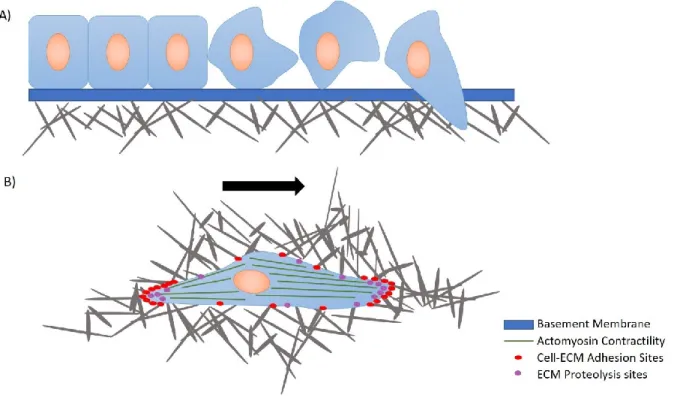
Metastasis is the final stage of multistep tumor progression. It is divided into two steps: the step where the tumor disseminates from its primary tissue and colonize into a distant tissue and the step where the disseminated tumor has to successfully colonize and form metastases. The process of dissemination starts when the epithelial cancer cell becomes invasive and goes through a process of alterations in shape and its attachment with other cells and the extracellular matrix (ECM). One of the best-characterized features of this process is the change in expression of an adhesion molecule named E-Cadherin, which is responsible for the formation of adherens junctions in cell-cell contact. Reduction of its expression is known to potentiate the process of invasion and metastasis [4, 5]. In contrast, another adhesion molecule named N-Cadherin, known to be expressed in migrating neurons and mesenchymal cells is upregulated in invasive tumor cells.
The invasion metastasis cascade is a multistep process [6, 7] (Figure 1.1.1). In specific, epithelial cells in primary tumors invade locally the surrounding tissue by degrading the ECM and the stromal cell layer. They then intravasate into the lumina of the blood vessels and survive in the circulation to adhere to the vessel at a distant organ site to extravasate into the parenchyma of the distant tissue. Once extravasated, the tumor cells must survive the foreign environment in order to colonize and form micrometastases. To further grow and form macrometastases, the tumor needs to go through a step named metastatic colonization, where the tumor cells reinitiate their proliferative programs to grow and proliferate. These steps of the invasion metastasis cascade are influenced by intrinsic molecular pathways orchestrated in the carcinoma cells, as well as extrinsic signaling cascades from non-neoplastic stromal cells [7].
Furthermore, epithelial cells co-opt a cell biological process named Epithelial-Mesenchymal Transition (EMT) that facilitates its invasion process throughout the metastatic cascade. This process involves the dissolution of cell-cell contact, cell-ECM attachment and loss of cell polarity [8]. Several transcription factors such as SNAIL, SLUG, TWIST, and ZEB1/2 have been characterized and identified as the major players behind the EMT process
[9]. Among the biological traits evoked by these transcription factors, are the downregulation of E-Cadherin and upregulation of N-Cadherin expression, expression of matrix metalloproteases, increased invasiveness and resistance to apoptosis.
Another factor that contributes to the invasion metastasis cascade is heterotypic interactions of cancer cells with the stromal cells, mentioned above [10]. The reciprocal interactions between cancer and the stromal cells that defined the multistep progression of the tumor at the primary site can be reinitiated in the distant tissue to promote the colonization of the disseminated cancer cell at the new organ sites. In comparison to this logic, others have stated that microenvironments of certain tissues named “metastatic niches”, can support the growth of the seeded cancer cells at distant sites, independent of the cancer cell-induced stroma [11, 12]. Furthermore, for the cells to grow and colonize at distant sites, cancer cells might revert from the invasive phenotype and go through the reversible process of EMT named “Mesenchymal-to-Epithelial Transition” (MET) or they can colonize and grow independently of MET due to acquired genetic alterations that confer all the necessary traits for metastatic seeding. Mesenchymal cells are known to cycle slowly or are dormant and reverting back to MET from EMT has been proven necessary for the mesenchymal dormant cell to become reactivated and proliferate rapidly at the distant site [13]. In MET-independent metastasis, genetic alterations or epigenetic reprogramming can induce a state in which EMT-fixed cells have a high invasive and proliferative potential which will lead them to become more aggressive and more likely to seed at a different organ. Altogether, both mechanisms of metastasis can then subsequently result in colonization and the formation of macrometastases.
Figure 1.1.1 The invasion-metastasis cascade
Adapted from Laura Gómez-Cuadrado et al. in Dis. Model Mech. Review 2017 [14]
Metastasis is a multistep process. Initially, tumor cells migrate into adjacent tissues, referred to as local invasion. This involves the breakdown of the basement membrane and invasion into the surrounding ECM. In fact, invasive tumor cells recruit macrophages (Mɸ) and cancer-associated fibroblasts (CAFs), which are part of the tumor microenvironment, to produce promigratory and pro-invasive factors and cytokines that will facilitate their invasion and metastasis. Intravasation then allows cells to enter the circulation. In blood vessels, circulating tumor cells exist as single cells or clusters, coated with platelets. They need to survive shear stress and evade clearance by the immune system to successfully reach distant organs. Tumor cells then attach to endothelial cells, which facilitates their extravasation. After settling in the metastatic target organ, tumor cells must survive in this foreign environment and establish micrometastases. These disseminated tumor cells (DTCs) can remain dormant for many years before proliferating into large macrometastases in a process termed colonization. The primary site also regulates the development of metastasis via secretion of factors (such as cytokines and exosomes) that can prime a pre-metastatic niche and support survival of DTCs.
1.1.2 Breast Cancer and its molecular subtypes
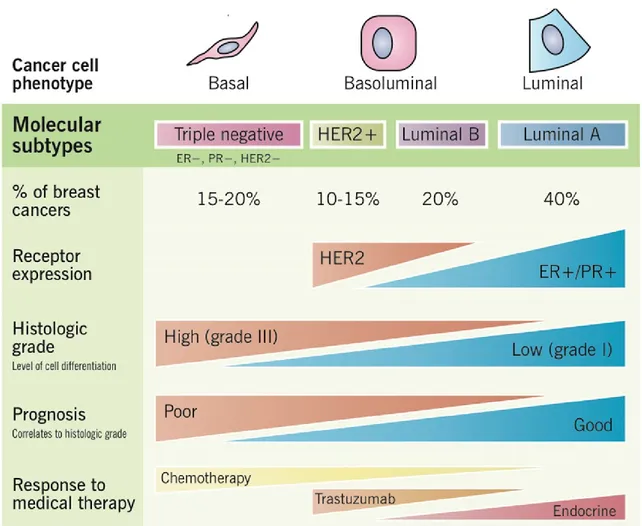
Breast cancer is the most frequently prevalent cancer in woman and is the most leading cause of cancer death in most developed countries [15]. It is now recognized that breast cancer is not a single disease and comprises of many biological entities. Studies have shown that breast cancer with different biological and histopathological features exhibit different behaviors and thus respond differently to treatments. Hence breast cancer is grouped into different subtypes to provide a more efficient therapeutic strategy. The classification of breast cancer into various subtypes is determined by the molecular and genetic features of the tumor cells (Figure 1.1.2). It is classified into Luminal A and Luminal B, which are hormone receptor-positive tumors (Estrogen Receptor+ or Progesterone Receptor+), HER2+ and Basal-like or Triple negative (TNBC; ER-, PR-, HER2-). Several groups have put some effort into identifying further varying gene signatures that define each of these molecular subtypes [16]. With the development of tissue microarray technology (TMA), these gene signatures of the different molecular subtypes have been validated at the translational level, confirming the biological heterogeneity of breast cancer. Interestingly, Luminal A and B cancers have the highest prevalence and the best prognosis. Among breast cancers, HER2+ and TNBC subtypes have a poor prognosis due to their tendency for metastasis. In HER2+ cancer, HER2 receptor which is an EGFR receptor tyrosine kinase family member is amplified or overexpressed to transmit signals that will mediate tumor growth, invasion, and metastasis. However, TNBC subtype of breast cancer represents 10-20% of breast cancer diagnosis and has the worst prognosis in the clinic. In comparison to other breast cancer subtypes, TNBC breast cancer lacks any targeted therapy treatment. The only effective therapy for TNBC breast cancer in the clinic is currently chemotherapy and unfortunately, 50% of the patients develop resistance and relapse to develop metastasis. Hence, it still remains elusive and is of a great significance to investigate in defining the molecular mechanisms that are behind TNBC aggressiveness and what novel key players could be used as future drug targets for therapy.
Figure 1.1.2 Molecular subtype of breast cancer
1.1.3 Receptor Tyrosine Kinases: key players in cancer biology
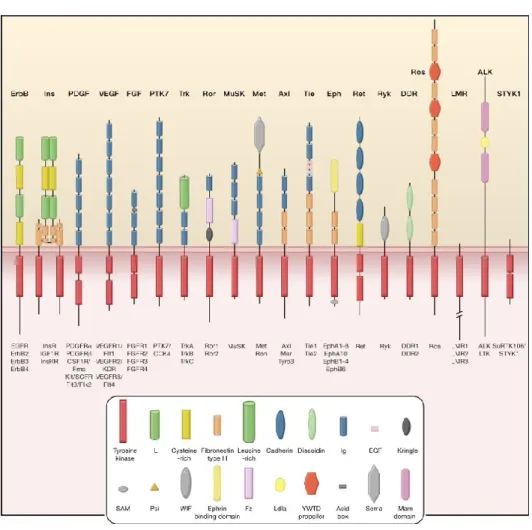
Receptor protein tyrosine kinases (RTKs) are a subclass of tyrosine kinases that are cell surface receptors and are known to be key regulators of different cellular processes such as cell migration, cell cycle, cell survival, metabolism, and differentiation [17]. Overexpression of many RTKs such as epidermal growth factor receptors (EGFRs), vascular endothelial growth factor receptors (VEGFRs) and platelet-derived growth factor receptors (PDGFRs) have been found in many types of cancer, such as breast cancer, and are correlated with cancer aggressiveness and decreased overall and disease-free survival [18]. They are known to respond to environmental cues to initiate the appropriate signaling pathways in tumor cells. In fact, RTKs may transduce their downstream signal via another class of protein tyrosine kinases named non-RTKs, which are predominantly cytoplasmic and contain a kinase domain with a catalytic activity regulated by phosphorylation upon external cues. Moreover, RTKs regulate many signaling pathways that may play a pivotal role in regulating cancer stemness, angiogenesis, and metastasis.
RTKs consist of 58 members that are subdivided into 20 subfamilies (Figure 1.1.3A) [19]. They all possess an extracellular domain, containing different components, that bind an activating ligand, a single transmembrane helix and an intracellular domain that contains the tyrosine kinase domain (TKD). In some cases, TKD is interrupted by a sequence known as a kinase insert domain (KID) that can regulate the function of TKD. Generally, all RTKs are activated upon growth factor binding that induces receptor dimerization. It is important to note, however, that some RTKs form oligomers in the absence of a ligand. Whether the “inactive” RTK is in a monomer or an oligomer form, the binding of a bivalent ligand is still required to induce a structural change in the “inactive” state which will stimulate the tyrosine kinase activity and become “active” to subsequently serve as a site of assembly for intracellular proteins to induce downstream cell signaling. Extensive structural studies have shown a range of mechanisms for ligand-induced dimerization of the extracellular regions of RTKs. This dimerization could be ligand-mediated, receptor-mediated or a combination of ligand- and receptor-mediated [19]. In all cases, the dimerization of the extracellular domain leads to the activation of the intracellularly TKD. All TKDs contain an N-lobe, a C-lobe and an activation loop. The N-lobe contains a glycine-rich loop which is followed by a lysine that is important for
ATP binding. The C-lobe however, contains a conserved aspartic acid that is important for the catalytic activity of the TKD. These residues are required for ATP binding, metal ion (Mg+) binding, and phosphoryl group transfer. Some kinases lack at least one of the motifs required for catalysis and have been termed pseudokinases [20]. They are seen as signal transducers by bringing together components of signaling complexes. However, it still remains elusive whether pseudokinases are true pseudokinases and lack any catalytic activity since some have reported some pseudokinases to have catalytic activity under certain conditions [20]. Hence, pseudokinases should be studied individually by probing for their activity with direct methods and having their structures determined.
Furthermore, before activation of an RTK, each TKD is cis-autoinhibited by intramolecular interactions unique for each receptor, which is released by ligand binding and dimerization of the extracellular regions of RTKs. Three different forms of cis-autoinhibition exist that are induced by various intramolecular interactions (Figure 1.1.3B) [19]. TKD autoinhibition is mediated by the activation loop occluding the ATP and substrate binding sites. This form of autoinhibition could be stabilized by the juxtamembrane regions of the kinase that can make extensive contacts with the TKD domain to stabilize the activation loop in an inactive conformation. In addition, the C-terminal tail can also play a role in cis-autoinhibition of TKD by blocking the substrate binding pocket. Releasing all these forms of autoinhibition by ligand binding and dimerization of the extracellular regions of RTKs can lead an active form of a TKD that can bind ATP and a substrate of interest to induce downstream signaling.
Under normal physiological conditions, RTKs function is tightly balanced. When they acquire transforming abilities through different mechanisms such as gain-of-function mutations, genomic amplifications, chromosomal rearrangements, or autocrine activation by an upregulation of their ligands, this will lead to the dysregulated function of the RTKs and their constitutive activation [21]. This can ultimately trigger oncogenic properties and RTK-induced oncogenesis. Due to the pivotal roles, they play in tumorigenesis and metastasis in breast cancer, RTKs are used as drug targets for therapy. However, resistance to anti-RTK therapy has been prevalent due to the acquired mechanisms they uptake to remain constitutively active.
A)
B)
Adapted from Mark A. Lemmon and Joseph Schlessinger in Cell Reviews (2010) [19]
A) Human receptor tyrosine kinases (RTKs) contain 20 subfamilies, shown here schematically with the family members listed
beneath each receptor. Structural domains in the extracellular regions, identified by structure determination or sequence analysis, are marked according to the key. The intracellular domains are shown as red rectangles. B) Activation loop inhibition: In the activation loop interacts directly with the active site of the kinase and blocks access to protein substrates or to both ATP and protein substrates. Phosphorylation of key tyrosines (“Y”) disrupts these autoinhibitory interactions and allows the kinase to “relax” to the active state. Juxtamembrane inhibition: the juxtamembrane region (red) interacts with elements within the active site of the kinase (including the αC helix and the activation loop) to stabilize an inactive conformation. Phosphorylation of key tyrosines in the juxtamembrane region destabilizes these autoinhibitory interactions and allows the TKD to assume an active conformation. C-terminal tail inhibition: C-terminal tail (red) interacts with the active site of the TKD to stabilize an inactive conformation.
Preface
A review to be submitted once CHAPTER 3 of my thesis is in the revisions process of a publication.
Dissecting AXL’s role in cancer progression
Afnan Abu-Thuraia1,2*, Marie-Anne Goyette1,2*, Carine Delliaux1, Jean-Francois Côté1,2,3,4# 1 Montreal Clinical Research Institute (IRCM), Montréal, QC, H2W 1R7, Canada.
2 Molecular Biology Programs, Université de Montréal, Montréal, QC, H3T 1J4, Canada. 3 Department of Anatomy and Cell Biology, McGill University, Montréal, QC, H3A 0C7,
Canada.
4 Department of Biochemistry and Molecular Medicine, Université de Montréal, Montréal,
QC, H3C 3J7, Canada.
*First co-authors, contributed equally to the work #Corresponding author:
Jean-François Côté IRCM
110 avenue des Pins Ouest Montréal (QC) Canada H2W 1R7
Email: [email protected] Phone: (514) 987-5647
Introduction
The TAM receptor tyrosine kinases (RTKs) comprised of TYRO3, MER and AXL are one of the latest family of RTKs to evolve and be identified since they are not known as strong oncogenic drivers [22, 23]. They have been largely identified in neoplastic cell lines, yet they are not frequently mutated in cancer. TAMs are known for their involvement in inflammation, autoimmunity and recently in cancer progression. They are ectopically expressed and highly expressed in many cancer types. TAMs are defined by their similar overall domain structure and a unique KWIAIES, a conserved amino acid sequence found in their catalytic kinase domain [24] (Figure 1.2.1). Their extracellular domain consists of two tandem immunoglobulin-like domains (Ig1 and Ig2) followed by two tandem Fibronectin type 3 (FN-III)-like domains. Their ligands for their activation are Growth Arrest Specific factor 6 (GAS6) and Protein S, which require Vitamin-K dependent γ-Carboxylation to have the ability to activate the TAMs [25]. Their structure consists of an N-terminal γ-carboxyl glutamic acid (GLA) domain, 4 tandem Epidermal Growth Factor (EGF)-like repeats, and a C-terminal Sex Hormone-Binding Globulin-like region consisting of two tandem Laminin G-like (LG) repeats. They bind the receptor with their carboxy-terminal domain and are known to bind the lipid moiety Phosphatidylserine (PS) with their amino terminus. PS is abundant in the body but can only activate TAMs when externalized on apoptotic cell membranes, aggregating platelets, exosomes, and invading virus envelopes [26-28]. While GAS6 and Protein S share common domains and require γ-carboxylation to activate TAM receptors, they have different affinities to different TAM receptors. GAS6 binds all TAM receptors, with the highest affinity for AXL, whereas Protein S only binds MER and TYRO3 [25]. PS increases GAS6 and Protein S activation of MER and to a lesser degree TYRO3. However, it still remains unknown and requires further experimentation how the PS/Gla binding translates into the LG binding to the receptor to induce receptor activation. Other than GAS6 and Protein S, novel TAM ligands have been identified recently that are tissue and receptor-specific [29-31].
Regulation of TAM Expression
TAMs overexpression has been observed in many cancer types promoting the survival, chemo-resistance, motility, and invasion of the tumor cells. Their expression has been shown to
correlate with poor prognosis in various tumor types. For example, MER kinase is aberrantly expressed in B-cell acute lymphoblastic leukemia (B-ALL) and T-ALL, whereas AXL is less observed in ALL, but highly observed in acute myeloid leukemia (AML) and chronic lymphoid leukemia (CLL) and chronic myeloid leukemia (CML) [24]. In addition, TYRO3 is also observed to be expressed in some leukemia samples. Hence, there seems to be a preferential for TAM receptor expression in different types of cancers. However, it still remains understudied how TAM expression is induced in cancer cells. Some evidence suggests that the expression of the different TAM receptors is regulated differently. For instance, dexamethasone increases MER expression and eliminates AXL expression in bone-marrow derived macrophages, yet TLR agonists increase AXL expression without altering MER expression [32]. In addition, previous studies have shown MER kinase transcription is upregulated in macrophages upon Liver X receptor (LXR) activation by its ligand, where ligand-bound LXR binds to the promoter of MER to enhance transcription [33]. Epstein-Barr virus (EBV) lytic transcription factors have been also demonstrated to substantially increase MER expression as well [34]. In terms of AXL expression, hypoxic conditions in the tumor increase HIF1α expression levels, which directly binds to the promoter of AXL to induce its expression [35]. TYRO3 also contains similar HIF1α responsive elements in its promoter, suggesting its expression upregulation under hypoxic conditions. Moreover, epigenetic regulation of expression has been observed in relapsed AML patients where hypomethylation of AXL promoter was shown to correlate with its high expression [36]. In addition, MZF1 transcription factor has been shown to bind AXL promoter to result in a dose-dependent increase in AXL mRNA levels [37]. TAM expression can also be regulated post-transcriptionally by miRNAs, where for instance, miRNA 355 known to negatively regulate MER kinase, is suppressed in breast cancer [38-42].
Although TAMs are overexpressed in multiple tumor types, genetic mutations or amplification of the genes encoding TAM RTKs are relatively rare. In some instances, point mutations and translocations creating fusion genes have been reported [24], yet the functional importance of these mutations remains elusive.
Mechanisms of activation
Ligand-dependent
AXL activation mechanisms are unique and can be achieved in many ways (Figure
1.2.2). To become active, AXL binds the C-terminal of its ligand GAS6, an interaction that is
of a strong affinity and independent of the presence of GAS6 γ-carboxylated N-terminal Gla domain [43]. However, this ligand interaction does not translate into AXL autophosphorylation and activation since Vitamin K-dependent γ-carboxylation of GAS6 Gla domains is necessary for GAS6-induced AXL autophosphorylation and activation [44, 45]. In fact, there is some evidence showing that warfarin treatment, which depletes Vitamin K levels, decreases the γ-carboxylation process of GAS6 and inhibits AXL autophosphorylation [46]. Upon GAS6 binding to AXL, GAS6 induces AXL homodimerization in a 2:2 stoichiometry where one GAS6 binds one AXL monomer, and this leads to its homodimerization, autophosphorylation, and activation to induce downstream signaling [43]. GAS6 binding and induced homodimerization of AXL is a unique activation mechanism to AXL that is different from MER and TYRO3 where GAS6 binds in a 1:1 stoichiometry manner. To become activated, AXL is auto-phosphorylated on Y702/Y703 in mammals, which then leads to the autophosphorylation of three other tyrosine residues in its intracellular domain (Y779, Y821, Y866) that act as scaffold hubs for proteins such as p85 (the regulatory subunit of PI3K), Grb2, and phospholipase C-γ (PLC-γ) to induce downstream signaling [47].
The γ-carboxylated Gla domain of GAS6 is known to bind, in a Ca2+-dependent manner,
the lipid moiety phosphatidylserine (PS), which is externalized on membranes of apoptotic cells, exosomes, and enveloped viruses [48]. It is believed that PS exposed membranes facilitate the dimerization or oligomerization of MER and TYRO3 to induce signaling [24]. However, there seems to be a controversy for the role of PS in AXL activation. Some have shown that GAS6-induced activation of AXL requires the PS binding to GAS6 to have complete activation of AXL [32, 49]. They suggest that AXL is assumed to be constitutively in complex with GAS6 due to their high-affinity binding and act as a hybrid receptor to detect PS and subsequently become activated [32, 50]. Others, however, have shown that GAS6-induced activation of AXL does not require PS binding to GAS6 and was not further enhanced by the presence of PS [43]. A
more recent study has demonstrated a model that provides a quantitative and mechanistic understanding of efficient GAS6 activation of AXL [51]. They demonstrated AXL activation to be observed only when localized concentrations of GAS6 is high. This localized ligand concentration drives a diffusional influx of AXL from regions of low to high GAS6 concentrations which will allow GAS6 binding and result in receptor aggregation and dimerization to induce efficient AXL signaling. Ultimately, this localized signal of receptor function can be a marker for PS exposure in cell clearance.
Furthermore, AXL activation can also be achieved by forming heterodimers with other TAM members (MER or TYRO3) to induce signaling [52, 53]. This heterodimerization could be mediated by GAS6 binding, where AXL can bind GAS6 and activate MER or TYRO3 kinases, which have less affinity in GAS6 binding.
Ligand-independent
AXL activation could also be atypical and mediated by a ligand-independent approach. One way this can occur is by homophilic dimerization and autophosphorylation during pathophysiological conditions due to excess receptor expression or oxidative stress [54-56]. Another way GAS6-independent AXL activation can occur is by heterodimerizing or clustering with other RTKs, such as EGFR, MET, PDGFR and VEGFR-2, to become auto-phosphorylated and activated to induce downstream signaling [57-61]. In the squamous cell cancer of head and neck, AXL heterodimerizes with EGFR to confer resistance to PI3K inhibition. A similar mechanism was seen also in EGFR-mutant lung cancer, whereupon EGFR tyrosine kinase inhibition, AXL dimerizes with MET receptor to bypass EGFR inhibition and confer resistance.
Furthermore, these molecular mechanisms of AXL activation shed light into the diverse approaches AXL can take to become activated and function to induce downstream signaling. It also complexifies the mechanisms of its regulation during physiological and pathophysiological conditions.
Figure 1.2.2 Mechanisms of AXL activation
AXL activation can take place in various manners. Ligand-mediated activation of AXL depends on AXL binding to GAS6 ligand to lead to its homodimerization and autophosphorylation (A). In addition, phosphatidylserine (PS) exposed on a membrane (ex. apoptotic cells) can induce GAS6 binding and lead to more efficient and localized AXL activation (B). In some cases, AXL can also heterodimerize with other TAM receptors in order to activate signaling (C). For cases when the ligand is absent, AXL can be activated in a ligand-independent manner in several ways. When AXL is expressed at high levels due to a disease condition or is induced by a specific stimulus, its high expression can lead to its dimerization and autophosphorylation (A). In many cancers, AXL has been shown to be transphosphorylated by many RTKs such as EGFR, HER2, MET, VEGFR-2 and PDGFR to diversify signaling. In ovarian cancer, AXL has been shown to converge with β3 signaling to induce tumor cell adhesion.
Regulation of activation
AXL activation and function can regulate cytoskeletal functions, intracellular signaling and gene expression [24]. Its aberrant expression or activity can dysregulate physiological functions and lead to a disease. Thus, the regulation of its activity is a crucial process to keep AXL function appropriate. Upon its activation, AXL has been shown to be negatively regulated in several ways. The proteolytic shedding of its extracellular domain mediated by the metalloproteinases ADAM10 and ADAM17 has been shown to play a role in decreasing its activity at the membrane [62]. In fact, MEK inhibitor resistance was associated with a decreased circulating level of AXL ectodomain in melanoma patients, which represents reduced RTK shedding as a mechanism in which cancer cells bypass signaling to attain high AXL activity and drug resistance. This suggests that soluble AXL ectodomain could be used as a diagnostic biomarker for clinical use.
In addition to AXL shedding, AXL can be negatively regulated by its binding partner C1 domain-containing phosphatase and tensin homolog (C1-TEN). C1-TEN can negatively regulate AKT activation downstream of AXL activation to inhibit survival and motility [63, 64]. Moreover, AXL, MER, and TYRO3 are targets of E3 ubiquitin ligase CBLB [46]. A study has emphasized on the role of TAM RTK ubiquitination by CBLB ligase in natural killer (NK) cells. Genetic deletion of cblb in NK cells prevented TAM RTK ubiquitination and degradation and hence inhibited NK cell activity in rejecting tumor and metastatic growth and this was reversed by TAM RTK inhibition [46]. Recently, another study has identified a novel mechanism in negatively regulating AXL oncogenic signaling in ovarian cancer by the GPI-anchored tumor suppressor OPCML [65]. Once activated by GAS6, AXL directly interacts with OPCML and subsequently gets accumulated in cholesterol-rich lipid domains where OPCML resides. This brings AXL in proximity with a lipid residing phosphatase named PTPRG, where AXL gets dephosphorylated by this phosphatase and can not transactivate other RTKs such as MET and EGFR to induce ERK signaling to induce EMT and cell invasion.
It still remains elusive how the cell can shuttle and traffic AXL from the cell membrane to regulate its activity. In addition, it will be interesting to explore if AXL once shuttled, gets
degraded in the endosomes via lysosomal degradation or continue to signal intracellularly in the endosome vesicles until vesicles merge back with the cell membrane.
Signaling in cancer
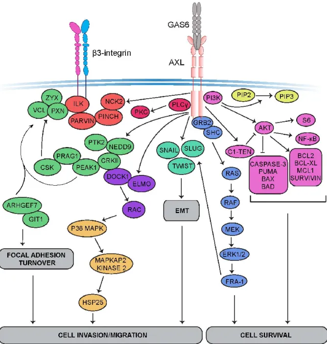
Several studies in many cancers have shown different roles of AXL signaling in cancer cells that positively correlate with chemo-resistance, targeted-therapy resistance, metastasis, and poor patient outcome. AXL signaling in cancer is deregulated and is known to induce downstream oncogenic signaling in the cell that leads to the promotion of survival and motility of the tumor cells (Figure 1.2.3).
Survival
AXL was first shown to play a role in survival in leukemia cells in which GAS6 stimulation prevented apoptosis upon growth factor deprivation [66, 67]. Treatment of B-CLL cells with AXL tyrosine kinase inhibitor led to apoptosis induction [68]. Another study demonstrated that inhibition of AXL expression by shRNA in leukemia cells decreased the cells ability to form colonies [69]. As with other RTKs, AXL promotes survival and proliferation by harnessing the RAS/ERK cascade and PI3K/AKT kinase pathway [70, 71]. AXL inhibition in cancer cells abrogates the activation of these pathways upon ligand stimulation [66, 72-75]. In fact, AXL induces a survival signal by upregulating the expression of anti-apoptotic proteins such as BCL-2, BCL-XL, MCL1 and SURVIVIN, and inhibiting the proapoptotic protein Caspase-3, BAD, BAX, and PUMA [68, 76-79]. For instance, AKT signaling downstream of AXL led to the increase of BCL-2 levels and inactivated the pro-apoptotic BAD proteins [78]. In addition, STAT proteins are also involved in the transcription of survival genes. AXL activation induces tyrosine phosphorylation of STAT3 to regulate its transcriptional activity [23, 38, 80]. Other pathways are also stimulated including those that involve NF-kB and p38 signaling that leads to the promotion of survival and regulation of survival and pro-migratory gene expression [23, 81, 82]. Notably, NF-kB activity in schwannoma cells has been shown to be a mediator of GAS6/AXL induced overexpression of pro-survival genes to enhance survival of merlin-deficient tumors [82]. Altogether, these pathways induced downstream of
AXL activation in many cancer types play a role in promoting cancer cell survival in response to apoptotic stimuli.
Migration
Apart from survival signaling, AXL has been shown to play a significant role in the metastasis of many cancer types. Whether it's in patient samples or cell lines, AXL expression correlates with invasion and metastasis [24]. Initially, AXL was first demonstrated to play a role in cell migration of GnRH neurons to the hypothalamus [83, 84]. Another study in AXL-expressing glioblastoma cells portrayed the role of AXL in cell migration, where transfection of the dominant negative form of AXL lacking the kinase domain resulted in reduced motility and filopodia formation and loss of cell-cell contact [85]. In fact, AXL activity in glioblastoma cells was shown to play a role in tumor growth and invasion. Studies have further shown a role for AXL in a facilitating a process named epithelial-to-mesenchymal transition (EMT) that is required in metastasis. The first in vivo evidence that links AXL to metastasis was shown in breast cancer where dissemination of highly metastatic breast cancer cells and EMT-driven motility was AXL-dependent [86, 87]. Similarly, AXL inhibition in ovarian cancer prevented dissemination and establishment of metastatic lesions [88]. EMT gene expression inducers such as TWIST, SNAIL, and SLUG are induced upon AXL activation or overexpression. These transcription factors, TWIST, and SNAIL, in return, can induce the expression of AXL as a positive feedback loop system to reinforce EMT [72, 89-91]. In fact, a positive correlation between AXL expression and mesenchymal phenotype was present in human cancer cell lines, particularly in breast cancer and non-small cell lung cancer [92]. A study has defined the mesenchymal subtype tumor cells of ovarian cancer to have an enrichment of AXL expression that positively correlated with EMT and poor patient outcome [93]. In this mesenchymal subtype of ovarian tumor cells, AXL co-clusters with and activates EGFR, HER2 and MET to induce protracted ERK signaling to promote motility and invasion by inducing the expression of the ERK pathway effector FRA-1, which in return can induce the gene expression of the EMT inducer SLUG [94]. Overall, AXL expression and signaling in many cancer types is necessary, not only to sustain an EMT state and allow cancer cells to disseminate and invade to form metastatic lesions but also to acquire resistance to targeted drug therapies.
The mechanisms in which AXL sustains an EMT state remain to be elusive. Initially, AXL signaling through PI3K/RAC pathway was shown to cause actin rearrangement and an increase in motility [83, 84]. This was recently explained in part in breast cancer cells where AXL mediated phosphorylation of ELMO proteins bound to DOCK1, promoted RAC-mediated cytoskeleton changes and induced breast cancer cell migration [95]. In addition, AXL was reported to regulate cell adhesion by modulating the signaling complex ILK/PINCH/PARVIN found at adhesion sites via its interaction with NCK2 protein [96]. It has also been shown to localize to active myosin filaments and phosphorylate tropomyosin at sites critical for adhesion [97]. In fact, a recent study has demonstrated how AXL signaling in ovarian cancer can sustain an EMT state by converging with β3 integrin signaling pathway to promote adhesion to the extracellular matrix and induce invasion [98]. These different mechanisms may suggest AXL as a regulator of cell adhesion during cell invasion process. In support of this notion, AXL invasive activity in tumor cells was further demonstrated to be mediated through the activation of matrix metalloproteases (MMPs) which are required to break down the extracellular matrix during cell adhesion process. Indeed, AXL activation enhances the expression of MMP9, known to promote tissue remodeling and invasion, and this was shown to be reversed upon AXL inhibition [99].
These attempts in defining mechanisms behind AXL’s significant role in maintaining an EMT state and reinforced mesenchymal phenotype do not justify the role it plays in the invasiveness of the tumor to become invasive and metastasize. More recently, a novel approach was taken in defining signaling pathways and mechanisms that are specifically modulated by AXL activation. The first phosphoproteome of AXL was characterized in triple negative breast cancer cells where AXL is highly expressed. Interestingly, this study emphasized AXL’s role in robustly regulating actin cytoskeleton rearrangements and in specific focal adhesion dynamics. Upon AXL activation or inhibition, focal adhesion disassembly rate was increased or decreased, respectively. The process of disassembly was found to be modulated by AXL-mediated recruitment of the disassembly complex GIT/βPIX/PAK to focal adhesions. Furthermore, the scaffold focal adhesion protein NEDD9 was identified as a novel direct substrate of AXL upon its activation by GAS6. AXL-mediated phosphorylation of NEDD9 induces its complex recruitment with CRKII/DOCK3 to induce RAC activation and modulate its role at focal
adhesions. This complex recruitment orchestrates AXL-mediated phosphorylation of the focal adhesion pseudokinase protein named PEAK1, via binding to the middle SH3 domain of CRKII. In this study, PEAK1, known to lack a kinase activity, was shown to recruit CSK kinase to modulate PAXILLIN phosphorylation levels at focal adhesion sites and regulate AXL-induced focal adhesion turnover. Notably, CRISPR CAS9 deletion of PEAK1 in triple negative breast cancer cells decreased tumor growth and metastasis in vivo. This study emphasizes AXL’s role in exploiting NEDD9/CRKII/PEAK1/CSK module to regulate focal adhesion turnover in breast cancer cells to promote and sustain their mesenchymal phenotype and progress to metastasis. In addition to focal adhesion dynamics, other pathways and processes were revealed to be modulated by AXL activation such as RNA transport, vesicle trafficking, and phagocytosis. It remains elusive if these pathways modulated by AXL contribute to AXL-induced survival and migration in invasive cancer cells or affect a different biological process. Moreover, this is the first time that AXL effector pathways are defined in a quantitative manner that may explain and justify its invasive role in cancer cells. The global view of AXL signaling will aid in defining the mechanisms AXL acquires to promote metastasis and attain drug resistance. This will ultimately bring forward novel potential therapeutic targets that may hold promise in the clinic to be used in drug combination therapy.
TAMs in human cancer
Expression of TAM receptor
TAM receptors and their ligands are expressed in a variety of human cancers and their expression correlates with a decrease in survival in solid malignancies and blood cancer. An exhaustive table of their expression in cancers and their significance in prognostic, function, metastasis and chemoresistance roles was reviewed in [24]. However, GAS6 contribution during cancer progression in human cancer still remains to be unclear. In non-small cell lung cancer, its expression was linked to increased metastasis and AXL decoy receptors that trap GAS6 were shown to reduce metastasis in vivo [100, 101]. On the other hand, other studies have shown GAS6 expression to correlate with a positive outcome in breast cancer and we have recently shown that GAS6 was not required, in contrast to AXL, for the metastatic progression of HER2+ breast cancer in vivo [58, 102].
Tumor growth and survival
TAMs are defined as proto-oncogenes because their overexpression leads to signals that can contribute to cancer by various mechanisms like resistance to apoptosis and proliferation. Indeed, it was shown that tumor cells educate infiltrating leukocytes to secrete GAS6 to promote tumor cells proliferation via activation of the TAMs [103]. In addition, AXL and MER have been shown to be important for tumor growth of various cancers [73, 81, 104, 105]. However, AXL contribution to tumor growth seems to be context dependent. For instance, our group and others have observed that genetic ablation of AXL has no or minor effect on tumor growth and that its role is mostly on the metastatic progression [58, 86].
AXL as a prognostic marker
The question remaining unanswered in the field is whether the TAMs can be used as prognostic markers. Some studies strongly suggest that AXL can be used as a biomarker for the survival of different cancer types and a recent meta-analysis support these observations [100, 106, 107]. Nonetheless, a further large-scale analysis will be required to validate the prognostic value of AXL and its oncogenic status.