Université de Montréal
Study of nuclear receptor dynamics by BRET
David Cotnoir‐White
Biologie moléculaire, Faculté de médecine
Thèse présentée à la Faculté de médecine en vue de
l’obtention du grade de doctorat en biologie moléculaire
Avril 2014
© David Cotnoir‐White, 2014
Résumé
Les récepteurs nucléaires (RN) sont des facteurs de transcription ligand dépendants qui contrôlent une grande variété de processus biologiques de la physiologie humaine, ce qui a fait d'eux des cibles pharmacologiques privilégiées pour de nombreuses maladies. L'un de ces récepteurs, le récepteur de l’œstrogène alpha (ERα), peut activer la prolifération cellulaire dans certaines sections de l'épithélium mammaire tandis qu’un autre, le récepteur de l'acide rétinoïque alpha (RARα), peut provoquer un arrêt de la croissance et la différenciation cellulaire. La signalisation de ces deux récepteurs peut être altérée dans le cancer du sein, contribuant à la tumorigénèse mammaire. L’activité d’ERα peut être bloquée par les anti‐oestrogènes (AE) pour inhiber la prolifération des cellules tumorales mammaires. Par contre, l’activation des voies de RARα avec des rétinoïdes dans un contexte clinique a rencontré peu de succès. Ceci pourrait résulter du manque de spécificité des ligands testés pour RARα et/ou de leur activité seulement dans certains sous‐types de tumeurs mammaires.Puisque les récepteurs nucléaires forment des homo‐ et hétéro‐dimères, nous avons cherché à développer de nouveaux essais pharmacologiques pour étudier l'activité de complexes dimériques spécifiques, leur dynamique d’association et la structure quaternaire des récepteurs des œstrogènes. Nous décrivons ici une nouvelle technique FRET, surnommée BRET avec renforcement de fluorescence par transferts combinés (BRETFect), qui permet de détecter la formation de complexes de récepteurs nucléaires ternaires. Le BRETFect peut suivre l'activation des hétérodimères ERα‐ERβ et met en évidence un mécanisme allostérique d'activation que chaque récepteur exerce sur son partenaire de dimérisation. L'utilisation de BRETFect en combinaison avec le PCA nous a
permis d'observer la formation de multimères d’ERα fonctionnels dans des cellules vivantes pour la première fois. La formation de multimères est favorisée par les AE induisant la dégradation du récepteur des oestrogènes, ce qui pourrait contribuer à leurs propriétés spécifiques.
Ces essais de BRET apportent une nette amélioration par rapport aux tests de vecteurs rapporteur luciférase classique, en fournissant des informations spécifiques aux récepteurs en temps réel sans aucune interférence par d'autres processus tels que la transcription et de la traduction. L'utilisation de ces tests nous a permis de caractériser les propriétés de modulation de l’activité des récepteurs nucléaires d’une nouvelle classe de molécules hybrides qui peuvent à la fois lier ER ou RAR et inhiber les HDACs, conduisant au développement de nouvelles molécules prometteuses bifonctionnelles telles que la molécule hybride RAR‐agoniste/HDACi TTNN‐HA.
Mots clés : Récepteur nucléaire, pharmacologie, FRET, BRET, estrogène, acide rétinoïque.
Abstract
Nuclear receptors (NRs) are ligand‐dependent transcription factors that control a wide variety of biological processes in human physiology, which has made them preferred pharmacological targets for many diseases. One such receptor, the estrogen receptor alpha (ERα), can activate cell proliferation in some sections of the mammary epithelium while another, the retinoic acid receptor alpha (RARα), can cause growth arrest and cellular differentiation. Signalling by these receptors can be altered in breast cancer, contributing to tumorigenesis. ERα can be blocked by antiestrogens (AEs) in the clinical setting to inhibit tumor cell proliferation. However, attempts to activate the RARα pathway with retinoids have not proven beneficial in clinical trials. This may result from the lack of specificity of the tested ligands for RARa and/or from their activity only in a subset of breast tumors.
Since nuclear receptors form homo‐ and heterodimers, we sought to develop novel pharmacological assays to study the activity of specific receptor‐dimer complexes, their dynamics and quaternary structure. We report here a new FRET technique dubbed BRET with fluorescence enhanced by combined transfers (BRETFect) that can detect the formation of ternary nuclear receptor complexes. BRETFect can monitor the activation of ERα‐ERβ heterodimers and highlights an allosteric mechanism of activation that each receptor exerts on its dimer partner. Use of BRETFect in combination with PCA‐BRET has allowed us to observe the formation of functional ERα multimers in live cells for the first time. The formation of multimers is favored by AEs which induce receptor degradation, and may underlie their specific properties.
These assays are a net improvement over the classical luciferase‐reporter experiment as they deliver real‐time receptor specific information with no interference by other process such as transcription and translation. Using these BRET assays we developed a new class of NR hybrid ligands that can modulate ER or RAR activity and inhibit HDACs. This has allowed for the development of promising new bifunctional molecules such as the RAR‐ agonist/HDACi hybrid molecule TTNN‐HA. In conclusion, the work presented here brings new insight in NR dynamics and quaternary structure and offers novel tools to study their mechanism of action or design new modulators of NR activity such as hybrid AE‐HDACis and Retinoid‐HDACis.
Key words: Nuclear receptors, pharmacology, FRET, BRET, estrogen, retinoic acid.
Table of Content
Résumé ... iii Abstract ... v Table of Content ... vii List of Tables ... x List of Figures ... xi List of Abbreviation ... xiv Acknowledgements ... xvii Chapter 1: Introduction ...1 Receptors ...2 Membrane receptors ...2 Intracellular receptors ...5 Nuclear receptor signaling ...6 Nuclear receptor domain organization ... 7 Nuclear receptor 3D structure ... 13 Nuclear receptor interactome ... 15 Genomic action of nuclear receptors ... 22 Class I Nuclear Receptors ... 23 Class II Nuclear Receptors ... 24 Chromatin action ... 26
Nuclear receptors and cancer ... 29 Breast cancer and the estrogen receptor‐alpha ... 31 Antiestrogens ... 31 Aromatase inhibitors ... 34 Histone deacetylases inhibitors ... 35 Experimental approaches to nuclear receptor study ... 36 FRET ... 37 Objectives ... 40 Chapter 2: Monitoring ternary complex formation in live cells by BRETFect ... 42 Chapter 3: Ligand‐specific modulation of estrogen receptor conformation and multimerization in live cells ... 72 Chapter 4: Investigation of SHR multimerization capacity and heteromultimer properties. ... 111 Chapter 5: Antiestrogen and histone deacetylase inhibitor molecular hybrids. ... 124 Chapter 6: A Hybrid Retinoic Acid Receptor Agonist / Histone Deacetylase Inhibitor as an Antiproliferative Agent in Breast Cancer ... 149 Chapter 7: Discussion ... 180 On the quaternary structure of the estrogen receptors α and β ... 180 Estrogen receptor dimerization and localization ... 180 PCA‐BRET for heterodimer studies ... 187 Nuclear receptor higher order complexes ... 188
Potential roles for multimerization in nuclear receptor action and biology ... 188 Hybrid HDACi‐NR ligands ... 194 AE‐HDACi molecules ... 194 Ret‐HDACi molecules ... 198 References ... 203
List of Tables
CHAPTER 1 TABLE 1. Sampling of various nuclear receptor expression and action in breast cancer. 31 CHAPTER 2 TABLE 1. Excitation and emission wavelengths of ID, IA and TA used in this study.
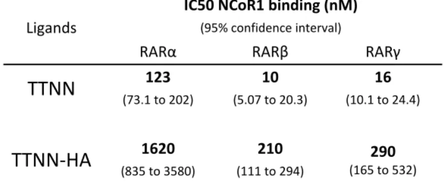
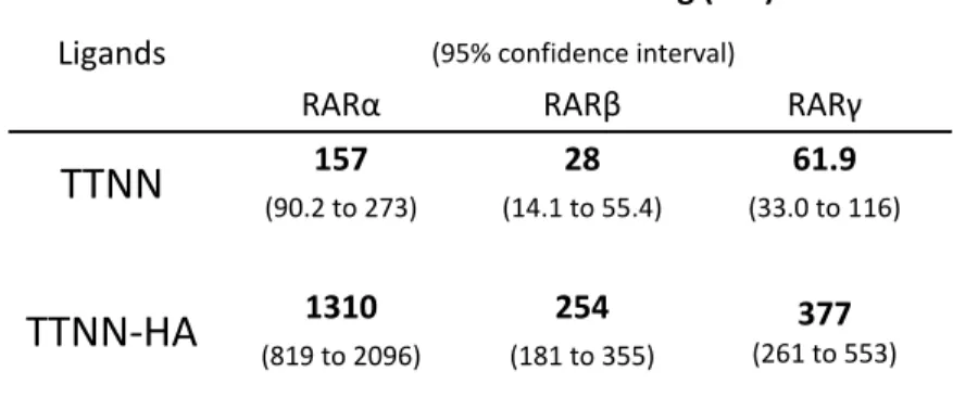
70 TABLE 2.Allosteric effects of heterodimeric partners for recruitment of CoApep 71 TABLE 3. Allosteric effects of heterodimeric partners for recruitment of NCoA1 72 CHAPTER 5 TABLE 1. IC50s of hybrids in luciferase assays 137 TABLE 2. In vitro HDACi activity of hybrids using purified HDAC3 and HDAC6. 139 TABLE 3. Antiproliferative activity of hybrids in MCF7 cells. 140 CHAPTER 6 TABLE 1. Efficiency of CoRNBOX motif release from RARs by TTNN and TTNN‐HA 174 TABLE 2. Efficiency of NCoA1 domain recruitment to RARs by TTNN and TTNN‐HA 175 TABLE 3. In vitro inhibition of HDAC3 and 6 by TTNN‐HA 176 TABLE 4. Growth IC50 of TTNN, SAHA and TTNN‐HA 177
SUPPLEMENTARY TABLE 1. Result of NCI 60 screen 180
List of Figures CHAPTER 1 Figure 1. Nuclear receptor structure. ...8 Figure 2. ERα’s LBD with coactivator binding groove and lining helices identified. ... 14 Figure 3. Example of nuclear receptor activation on the chromatin. ... 18 Figure 4. The estrogen receptor acting at an enhancer and looping chromatin to promoter. ... 27 Figure 5. Basic principle of the Förster Resonance Energy Transfer (FRET). ... 38 CHAPTER 2 Figure 1. BRETFect monitors ternary complex formation in live cells. ... 57 Figure 2. Comparison of net trimer signals in the BRETFect and SRET setups. ... 58 Figure 3. Activation of ERα‐ERβ heterodimers by selective ligands. ... 59 Supplementary Figure 1. Absorption and emission spectra of different donors and acceptors used in BRETFect and SRET. ... 60 Supplementary Figure 2. Use of coelenterazine H instead of coelenterazine 400 greatly increases signal output. ... 61 Supplementary Figure 3. FRET measurement of ERα and ERβ recruitment of CoApep‐ Venus. ... 62 Supplementary Figure 4. Specific interactors are required to achieve BRETFect ... 63 Supplementary Figure 5. ERα dimer interaction with CoApep‐Venus is concentration‐ dependent and saturable. ... 64 Supplementary Figure 6. BRETFect assays detect ERα‐ERβ heterodimer activation. .... 65 Supplementary Figure 7. Comparison of CoApep recruitment by ERα‐ERα or ERβ‐ERβ homodimers in the presence of E2 and PPT or DPN, respectively. ... 66 Supplementary Figure 8. Comparison of CoApep or SRC1‐RID recruitment by ERα‐ERβ heterodimers in the presence of E2, PPT or DPN. ... 67 Supplementary Figure 9. BRETFect assays are excellent assays for high‐throughput screening of heterodimer activity. ... 68
CHAPTER 3 Figure 1. Different tools can monitor the conformation of ERα dimers under different treatment. ... 98 Figure 2. SERMs and SERDs prevent recruitment of a coactivator peptide to ERα in the presence of E2 but allow for the binding of additional ERα partners. ... 99 Figure 3. SERDs form different higher order ERα oligomers than pure SERMs and E2. 100 Figure 4. SERD induced oligomers are biochemically different from E2 induced counterparts. ... 101 Figure 5. Novel BRETFest experiments reveal the formation of heterotetramer composed of three ERα subunits and a coactivator peptide. ... 102 Supplementary Figure 1. The G400V mutant (HE0) fails to dimerize in absence of ligand. ... 103 Supplementary Figure 2. Treatment with geldanamycin does not prevent dimerization or recruitment of cofactors to ERα. ... 104 Supplementary Figure 3. Dimerization mutant ERα(L507R) does not properly recruit coactivator motifs. ... 105 Supplementary Figure 4. Receptor affinity for the LXXLL coactivator motif reflects transactivation potential. ... 106 Supplementary Figure 5. AF1 function is not disrupted by SERDs but SUMoylation and ubiquitination is increased. ... 107 Supplementary Figure 6. High oligomerization is a correlates to SERD activity of AEs. 108 Supplementary Figure 7. Increased oligomerization potential of molecules correlate with their ability to induce sumoylation and ubiquitination of the receptor. ... 109 Supplementary Figure 8. ERα oligomerization state reflects proper activity of the receptor and SERD‐susceptibility. ... 110 CHAPTER 4 Figure 1. Heterodimers are predicted to form between NR3C nuclear receptors and NR3A and NR3Bs. ... 117 Figure 2. Androgen Receptor PCA‐BRET shows the formation of receptor tetramers. . 118
Figure 3. Androgen and Glucocorticoid Receptor heterodimers show restrictive coactivator affinitie and heteromultimerization. ... 120 Figure 4. Promiscuity of interactions between SHR. ... 122 CHAPTER 5 Figure 1. Selected ER ligands ... 127 Figure 2. Selected HDACi’s ... 128 Figure 3. Pharmacophores of full antiestrogen ICI‐164384 and SAHA ... 131 Figure 4. Antiestrogenic activity of hybrids in a BRET assay for agonist‐induced coactivator recruitment. ... 136 Figure 5. Comparison of antiproliferative effects of SAHA, Faslodex and RMS‐575 on MCF‐7 and MDA‐MB‐231 breast cancer cells. ... 140 Figure 6. MDA‐MB‐231 cells treated with RMS‐575 ... 141 Figure 7. Docking overlay of RMS‐162 (22, yellow), RMS‐234 (24, violet) and RMS‐70 (21, orange) with ICI‐164,384 (green) ... 142 CHAPTER 6 Figure 1. Structure of hybrid molecule TTNN‐HA and parental molecule TTNN. ... 168 Figure 2. BRET assay reveal TTNN‐HA’s activation RARγ. ... 169 Figure 3. TTNN‐HA regulates retinoid and HDACi sensitive genes. ... 170 Figure 4. TTNN‐HA has HDACi activity similar to SAHA. ... 171 Figure 5. TTNN‐HA inhibits the growth of breast cancer cells but spares normal cells. 172 Supplementary Figure 1. TTNN‐HA selectively activates RARs. ... 177 Supplementary Figure 2. RAR antagonist BMS493 restores some growth to cells under TTNN‐HA treatment. ... 178 CHAPTER 7 Figure 1. Possible multimerization interface and complexes of ERα. ... 191 Figure 2. ER multimers looping chromatin and stabilizing a coactivator. ... 193 Figure 3. RMS‐70 modulates cell cycle similarly to SAHA. 197
List of Abbreviation
AF‐1/2 Activation function ½ AI Aromatase Inhibitor ATP Adenine triphosphate BRET Bioluminescence Resonance Energy Transfer CoA Coactivator CoR Corepressor CRABP Cellular retinoic acid binding protein DNA Deoxyribonucleic acid E1 Estrone E2 Estradiol E3 Estriol EGF Epidermal growth factor EGFR Epidermal growth factor receptor ERα Estrogen receptor alpha ERβ Estrogen receptor beta ERE Estrogen Response Element eRNA enhancer RNA ESC Embryonic Stem Cells FACS Fluorescence activated cell sorting FRET Förster Resonance Energy Transfer GR Glucocorticoïd receptor HAT Histone acetyl transferaseHDAC Histone deacetylase HER2 Human EGF receptor 2 HMEC Human mammary epithelial cell HMT Histone methyl transferase HRE Hormone Response Element IPA Ingenuity Pathway Analysis KDM Lysine specific demethylase LXR liver X receptor MAPK Mitogen activated protein kinase mRNA messenger ribonucleic acid NCOA Nuclear coactivator NCOR Nuclear corepressor PCA Protein complementation assay PPAR Peroxisome proliferator‐activated receptor PR Progesterone receptor PSC Pluripotent Stem Cells PXR pregnane X receptor RA Retinoic acid RAR Retinoic acid receptor RARE Retinoic acid response element ROR RA receptor‐related orphan receptor RTK Receptor Tyrosine Kinase RXR Retinoid X receptor SAHA Suberoylanilide hydroxamic acid
SAR Structure Activity Relationship SERD Selective Estrogen Receptor Down‐regulator SERM Selective Estrogen Receptor Modulator SHR Steroid Hormone Receptor SMRT Silencing mediator of RAR and TR SRC Steroid coreceptor TF Transcription Factor TR Thyroid hormone receptor TSA Trichostatin A VDR Vitamin D receptor
Acknowledgements
When I was fired from my Master’s I was told I “didn’t have it”. That was hard to accept. I couldn’t imagine who would take me on for PhD at that time. I don’t remember how I came across Dr. Mader’s lab page while browsing around for a future but there was something about nuclear receptors mechanism that seemed irresistible (it still is!). I was very lucky that Sylvie called me in for an interview and then doubly so to be offered a spot in her lab. The work reported here is not what I started out doing here; nothing on GPR30 or artificial transcription factor projects… Sylvie always stayed positive even when these projects crashed and/or burned and it was good to have that attitude guiding me. It’s important for a student to feel his supervisor thinks “he’s got it” and I never doubted that with Sylvie. I am also grateful to have been giving some liberties with my projects to just “try some things out” which led to a good deal of the stuff that is reported here. We had a great lab going during my stay. I’ll fondly remember the times spent with Alice, David, Khalid, Marieke, Martine, Maxime, Mohammed and Wayne talking science and kidding around as the minutes of an incubation crawl away.
Special thanks go out to Mohammed for all his work in developing our lab’s BRET assays with me.
My father (Dr. White) warned me when I got to university “Don’t do a PhD!” It was great advice and I relay it to every young student that comes through our lab. Yet, even though I disobeyed him, he remained supportive and shared quite a bit of insight on how to navigate grad school bureaucracies and other PhD candidate traps. Thank you dad.
My mom deserves much praise too. What I do isn’t obvious or easy to understand (That became very clear when reading reviewer #2’s criticism of Chapter 2…) Still, my mom made great efforts to understand my work (and life as a grad) and I am proud to say she gets estrogen receptor signalling and what is needed to study it. Merci maman.
Marieke and Wayne, I couldn’t have asked for better drinking buddies or companions in canoe adventures/culinary endeavours/absurdist comedy appreciation/Brazilian bare‐ knuckle death‐matches. I hope there is more to come. Finally, I would like to thank who wanted me to deposit this thesis more than me. Eve, you have been very patient, I wish I could tell you it is going to pay off now but this is a PhD; there is no pay out. Thank you for the daily support these last 6 years, I hope you have another 60 in you. I love you and I am grateful for everything you do for me, great (Olivia) and small (head scratch – more please). Olivia I swear by the Flying Spaghetti Monster if you don’t go back to bed this instan… Olivia you are the darling of my heart I know you will grow up to do great things but listen to your dad: DON’T DO A PhD!
CHAPTER 1: INTRODUCTION
Receptors
Health and disease of the human body are regulated by an intricate balance of signalling within and between the tissues. The overwhelming majority of signalling is regulated by families of receptors that respond in most cases to the presence or absence of their ligand [1‐3], a molecular trigger whose binding to the receptor causes a shift in their structural conformations that allows for the receptors activity to be modulated. Receptors can be divided into two categories [4‐6] dependent on their localization, i.e. as membrane receptors and intracellular receptors. Membrane receptors can be further split into three classes: G protein coupled receptor [7], receptor tyrosine kinase [8] and Cys‐loop (ionotropic) receptor [9].
Membrane receptors
Cys‐loop receptors, also known as ligand‐gated ion channels or ionotropic receptors, are a class of receptors mostly associated with the nervous system. They allow the exchange of ions such as Na+, Ca2+, K+ and Cl‐ between extracellular and intracellular space when bound by neurotransmitters to generate action potentials which will be further propagated by voltage‐gated ion channels or modulate cell biology [10]. Ligand binding will induce a shift in conformation, which will allow the free flow of the ions specific to the receptor along their gradient [11]. Ionotropic receptors are the target of most anaesthetics and certain psychotropic drugs [12‐14].
Receptors tyrosine kinase (RTKs) are membrane receptors characterized by an extracellular ligand‐binding/chaperone domain, a single transmembrane domain and an intracellular kinase domain [15‐18]. Notable examples of the RTKs are the vascular endothelial growth factor receptors (VEGFR1, 2 and 3) [19], ephrin receptors and the erbb family of receptors – erbb1 or epidermal growth factor receptor (EGFR), erbb2 or heregulin receptor 2 (HER2), erbb3 and 4 [20]. The ligands to these receptors are usually small peptides circulating in the extracellular space that cause receptor dimerization when binding to their target [21‐23]. The dimerization of the RTKs brings their intracellular domain into close proximity and leads to reciprocal phosphorylation on tyrosine residues of receptor monomers, which become activated [24]. The apo‐ligand‐ binding domain of the RTKs is inhibitory to that process [25] as extracellular domain deletion or specific point mutation of the receptor are often constitutively active, suggesting this domain acts as a chaperone to the receptors [26, 27]. After activation and autophosphorylation the modified tyrosine residues act as binding sites for adapter proteins which contain the src‐homology 2 (SH2) domain [28]. In turn the adapter proteins, which often have their own tyrosine kinase domain, can recruit and activate effector proteins to engage changes in plethora of cellular activities such as cell morphology, cell cycle control, metabolism etc… [29]. Several RTKs are disregulated in cancer. For instance, HER2 is amplified in 25‐30% of breast cancers and EGFR in many cancers such as breast and colorectal carcinomas [30‐32]. Targeting RTKs for drug development has led to novel cancer treatments such as the HER2 inhibitor herceptin (Trastuzumab) and various VEGFR inhibitors to treat renal cell carcinomas and various metastatic diseases [33]. The efficiency of these treatments is often limited by the inbuilt
redundancy of the signalling network they are part of and the often ubiquitous expression of some of the target receptors, as with the EGFR [34].
G protein‐coupled receptors (GPCRs), also known as seven transmembrane (7TM) domains receptor, are a family of approximately 800 genes [7] characterized by their signalling through the heterotrimeric G‐proteins to engage rapid signalling cascades of events through calcium release, cyclic‐AMP production [35], kinase activation[36], etc…[37]. GPCRs are classically activated by binding of extracellular ligands [38, 39] which can range greatly in size and nature from a small sugar molecule for the taste receptors TAS1R2/3 [40] to multi‐kilodalton proteins such as Wnt1 for the Frizzled receptor, which feeds into many cell proliferation and differentiation pathways [41]. While GPCRs have been shown to be active as monomers in vitro, in vivo dimeric signalling is a major component of signalling regulation and specificity. As previously mentioned, taste receptor TAS1R3 will be activated in the presence of sucrose when dimerized with TAS1R2 [42], while dimerization with TAS1R1 will cause activation in the presence of L‐ glutamate [40]. Adding to the layer of signalling regulation, different ligands binding to the same GPCR may stabilize different conformations of the receptor, which may in turn activate a sub‐selection of signalling pathways [43]. This emerging area of research is referred to as ligand‐biased signalling, which we will cover later on. It is acknowledged that over 50% of today’s approved pharmaceuticals target GPCRs yet their targeting in cancers has only recently started [44‐46]. One recent breakthrough has been the selective targeting and inhibition of the entothelin receptor A (ETaR), which has protumoral activity in gastroenteric tract cells [47]. Conversely, the activation of
receptors with pro‐apoptotic signalling such as 2AR could also provide novel treatment options for certain malignancies [48]. Intracellular receptors Intracellular receptors are ligand‐dependent transcription factors made up of the nuclear receptor super‐family and its lone cousin the aryl‐hydrocarbon receptor (AHR). The AHR is a basic helix‐loop‐helix transcription factor that is activated by planar aromatic hydrocarbons [49] such as natural plant flavonoids and phenols [50]. Upon binding to these ligands, the AHR will activate the transcription of specific target genes by recruiting cofactors to the promoter and enhancer regions of these genes [51, 52] (in a mechanism that highly resembles that of type 1 nuclear receptor which we will cover later) such as the P450 enzymes which metabolize xenobiotics [53]. The AHR also regulates a plethora of other pathways in the absence of xenobiotics such as cell growth, proliferation, differentiation and death [54‐56]. Interestingly the AHR has recently been identified as a key player in hematopoietic stem cell expansion, making its targeting an active area of research to develop novel ways to perform stem cell transplants [55].
Nuclear receptors, which this thesis focuses on, form a family of 48 genes in humans encoding for over two hundred transcription factors when accounting for different isoforms [57, 58]. While some of these are orphan‐receptors, meaning that no ligand have been assigned to them to date, they are overwhelmingly considered
chaperone [60] or co‐repressor proteins [61]. Once bound by their ligands, they shed these proteins. Nuclear receptors homo‐ or heterodimerize with partner receptors and bind key regulatory sites in the genome to modulate transcriptional activity [62]. Nuclear receptors influence many (if not all) signalling pathways such as placental development[63], metabolism [64] and circadian rhythms [65, 66]. Most receptors with known ligands already have FDA‐approved targeted therapies [67] or molecules in clinical trials [68], yet as we will see the scope of the targeting strategies that have been used to date are limited as they focus on individual receptors while those receptors actually act in concert with other NR partners in dimeric complexes.
Nuclear receptor signaling
Nuclear receptor superfamilyNuclear receptors are a hallmark of metazoans and are not present in lower eukaryotes [69, 70]. While the human genome harbors 48 nuclear receptor genes, other species will have evolved fewer, such as Drosophila melanogaster with only 18 genes [71], or many more like Caenorhabditis elegans that hosts an impressive 283 nuclear receptor‐ encoding genes [72]. It is theorized that nuclear receptors first evolved as “energy sensors” that could sense the availability of nutrients by binding free fatty acid derivatives in multi‐cell organisms to modulate their growth [69, 73]. Through duplication of the genes and mutations the family acquired the ability to sense different molecules, either exogenous or endogenous, to integrate physiological and environmental cues in multicellular organisms.
The first nuclear receptor was identified by Elwood Jensen in the 1950s when he showed using a radiolabeled ligand that only estrogen‐responsive tissues could bind and concentrate estrogen in cells [74]. Before this breakthrough, it was believed that steroid hormones acted as nutrients that delivered energy to specific tissues where they could be metabolized [75]. The discovery of a protein that bound hormones and translated this signal into physiological changes in target tissues eventually led to the identification of other hormone receptors such as androgen receptor (AR) [76, 77] and the progesterone receptor (PGR) [78, 79]. Pierre Chambon’s and Ron Evan’s group were the first to clone and sequence NR ‐ ERα [80] and GR [81] respectively and subsequently all NRs were characterized before genome sequecing by homology cloning.
Nuclear receptor domain organization
Nuclear receptors are defined by a modular structure composed of a ligand‐ independent activation function (AF‐1) [82], a zinc finger type DNA‐binding domain (DBD) [83], and overlapping ligand‐binding domain (LBD) and a ligand‐dependent activation (AF‐2) (Fig. 1A) [84, 85]. Notable exceptions to this rule are DAX1 and SHP, which constitute the NR0 sub‐family of receptors; these receptors do not have AF1 domains or DBDs [86, 87].
0.1
Figure 1. Nuclear receptor structure.
(A) Schematic view of the modular organization nuclear receptors with AF‐1 (yellow), DBD (red), hinge (green) and LBD/AF‐2 (blue) domains. (B) ERα DBD and LBD crystal structures. The DBD (red) is drawn bound to an ERE half‐site with the P‐box in yellow and D‐box in orange (adapted from structure 1HCQ). The E2‐ bound LBD (blue) is drawn with helix 12 in yellow and the helix 11 dimerizing interface in orange (adapted from structure 1ERE). DNA binding domainThe DNA‐binding domain is the most conserved domain within the nuclear receptor super‐family. All nuclear receptor DBDs possess two treble‐clef (or C4‐type) zinc finger motifs that allow for direct DNA interactions [88]. While both zinc fingers make stabilizing contacts, the specificity of DNA binding is determined by the amino acids of the P‐box at position 2‐6 in the first zinc finger (Fig. 1B). These amino acids make contact with 4 adjacent base pairs that form the core of the nuclear receptor response motif. The
response element is complemented by two flanking base pairs – one on each side – that make less crucial contacts with the DBD [89]. The P‐box sequence diverges between nuclear receptors and thus contributes for diversification of possible genomic sites of action for the receptors [90‐92]. The DNA binding domain also contains a dimerization interface called the D‐box (Fig. 1B) which, with other interfaces in the LBD, allows nuclear receptor to dimerize – a behavior typical of nuclear receptors [93]. Thus a typical nuclear receptor response element (RE) is composed of two motifs bound by one nuclear receptor each [94‐97]. The organization of the receptor dimers determines the REs that can be bound, with variable spacing and orientation of the motifs [98‐101] The C‐terminal part of the DBD is linked to a hinge region. While this domain was long thought to merely serve a string to tie the DBD to the LBD it has recently been shown that it is a critical hub that relays signal from LBD to DBD and can integrate signals coming from the two dimeric receptors to allow intra‐dimeric allosteric control [102‐104]. Ligand binding domain and AF2 transcriptional activation function
The ligand‐binding domain, which follows the hinge region, is structurally conserved within the NR super‐family. It is formed of a specific 12 alpha helix bundle structure also called alpha‐helical sandwich, which contains a hydrophobic ligand‐binding pocket [84]. Much of the surface‐exposed amino acids are conserved between nuclear receptors and play roles in receptor dimerization and cofactor recruitment [93, 105, 106] (Fig. 1B). The amino acids of the LBD core, whose side chains and backbones are buried
inside the LBD structure, form the lining of the ligand binding pocket [107, 108]. These amino acids define the shape, size and polarity of the ligand binding pocket, resulting in ligand binding specificity [109]. The binding typically occurs via hydrophobic water exclusion, aromatic stacking, and van der Waals forces [110]. Endogenous ligand binding to the receptor is usually selective ; for example endogenous estrogens come in three varieties: estrone (E1), estradiol (E2) and estriol (E3) that are all high affinity agonists of ER and ER Miller . Moreover the ERα ligand‐binding pocket can also bind 27‐hydroxycholesterol in the low micromolar range [111, 112]. 27‐ hydroxycholesterol (27HC) induces a different conformation of the LBD that is less conducive to activation, so that in absence of estrogens 27HC will cause partial activation of estrogenic signalling while in the presence of estrogens high levels of 27HC will inhibit full activation of the receptor [113]. 27HC is thus considered to be a partial agonist or an endogenous selective estrogen receptor modulator (SERM) [114, 115]. Other receptors such as the xenobiotic sensing receptors CAR and PXR [116, 117], on the other hand appear to bind with more promiscuity to allow the detection of a greater variety of molecules.
Ligand binding induces conformational changes in the LBD. These changes modulate the affinity of the receptor for partner proteins, which allows the receptor to switch from inactive to active state [118]. For instance, while in the inactive state, the steroid receptors interact with the chaperone protein Hsp90, which stabilizes the receptors and limits their DNA binding and cofactor recruitment [119‐122]. This interaction is required because of the inherent instability of the LBD in the apo‐state. When the receptor binds a ligand the conformational changes induced stabilize the LBD, cause the release of Hsp90 and expose a surface that can interact with cofactors.
The LBD overalps with the C‐terminal AF2 transcriptional activation region, which mediates cofactor recruitment [123, 124]. The core of the AF‐2 domain is contained in the terminal helix (H12) of the LBD, whcih acts as a lid to the ligand‐binding pocket and stabilizes the ligand [125‐127]. Upon ligand‐binding, helix H12 folds onto the to create with residues from helices 3‐5 a coactivator binding groove in which a coactivator motif (often corresponding to the consensus LXXLL) can bind to form a receptor‐coactivator complex [128‐130]. A ligand‐bound nuclear receptor homo or heterodimer contains two binding grooves, and may thus accommodate two motifs per receptor dimer. In addition, most coactivators contain more than one coactivator‐motif increasing the avidity of receptor‐coactivator interaction [131]. NR antagonists disrupt AF‐2 folding and prevent cofactor interactions [132, 133] [134‐138].
While AF‐2 domains have a conserved helical ternary structure they have some level of sequential diversity. This primary structure diversity allows different receptors to recruit different cofactors and to achieve different intramolecular conformations [139, 140]. The two main classes of coactivator motifs are the leucine and phenylalanine repeats – defined by the LXXLL and FXXLF sequences respectively. Receptors may bind both of them, like in the case of the glucocorticoid receptor (GR), or only one of them, like ER to the LXXLL motif and the androgen receptor (AR) to the FXXLF motif [140]. Moreover, AF‐2, along with H3 and H11, determines the capacity of the receptor to recruit, in the absence of ligand, corepressors through the aliphatic repeat corepressor nuclear receptor box motif (CoRNRBox) defined as (I/L)XX(V/I)I [139]. The AF‐2 domain can also mediate intramolecular interactions between the C‐terminal LBD and the N‐ terminal AF‐1 domain as is observed for ERα [141] and AR [142]. The ERα and β do not bind corepressors in the absence of ligand and rely on a variety of mechanisms such as
nuclear exclusion, chaperone protein binding and AF‐1 to AF‐2 looping [143] to exclude binding of the coactivator to the AF‐2 function and help keep the receptor in an inactive state.
AF1 transcriptional activation function
The AF‐1 domain is a loosely unstructured domain at the N‐terminus of the receptor [144] which is missing or incomplete in some receptors such as CAR [145] but can also comprise most of the receptor itself as in the case of Nur77 [146]. The diversity of sequence and structure imparts a high variety of function to the AF‐1 region. When isolated from the rest of the receptor, it functions as a ligand‐independent activation‐ function. Within the receptor, it may interact with cofactors in the absence of agonist and even in the presence of antagonist [144, 147]. Some receptors, such as Nur77, are highly dependent on AF‐1 activity as they do not mediate cofactor recruitment through the AF‐ 2 domain and may require a dimeric NR partner (such as RXR) to mediate AF‐2 contact with the coactivator [146, 148]. As described above the AF‐1 domain can bind the AF‐2 domain and often does so through coactivator like motifs encoded in its sequence – this conformation is observed in the receptors apo‐state[143]. When the receptor is activated, the AF1 domain is released and can interact with cofactors that contain a conserved glutamine rich region [149]. This alternate method of cofactor recruitment allows NRs to interact with a wide range protein. For example, while the AR does not strongly bind the LXXLL motifs of SRC‐1 through its AF‐2domain it can stably interact with SRC‐1’s glutamine rich region and recruit SRC‐1 to the chromatin, yet AR can also recruit cofactors such as RNF14 solely through the AF2‐domain binding a FXXLF motif of the cofactor [150].
Nuclear receptor 3D structure
Because of their modular nature and their domains’ small size many crystallographic and solution structures have been acquired for nuclear receptors in complex with DNA RE, cofactors, ligands and/or dimerization partners. Recently full length receptor dimers bound to DNA have also been solved for RXR heterodimers [151‐ 153].
The DNA binding domain structure of GR was first solved in 1991 and confirmed its helical zinc‐finger motifs. Further studies that co‐crystallized NR DBD with their response‐element highlighted the contacts made between the NR P‐box and DNA and how those defined the selectivity of receptors for their respective elements [154, 155].
The ligand binding domains of RXR, RAR and TR were also crystalized soon after and revealed the LBD’s helical bundle structure [156‐158]. The addition of coactivator peptide to ligand bound receptor generate crystal structure that showed how the AF‐2 domain folds down between helices 3 and 12 and is held in place in a charge clamp (amino acids E380, D538, D545, and R363 in of ERα) (Fig. 3). In this conformation helices 3, 4, and 12 create a coactivator binding groove where the LXXLL or FXXLF peptide lays down [159, 160]. In the case of the estrogen receptors, helix twelve also makes important contacts with asp351 that stabilizes it in its activated conformation [161]. Corepressor peptide were also crystalized with the RARa that define the structural basis for corepressor recruitment in the case of apo‐RAR receptor but not for apo‐RXR or apo‐ER complexes
0.2Figure 2. ERα’s LBD with coactivator binding groove and lining helices identified.
Electrostatic surface of ERα’s LBD with helical structure overlaid. The negative (red) and positive (blue) charges of the charge clamp are apparent around helices 4 and 12 (negative) and 3 (positive).
The full structure of AF‐1 and much of the C‐terminal of the LBD – which can stretch more than 50 amino acids past helix 12 – have not been solved and it is believed that they are naturally disordered in the absence of larger macro‐complexes [166, 167]. The main function of these domains is to act as a scaffold for cofactors to bind the nuclear receptor and recruti them to nuclear receptor binding sites to modulate transcription [168, 169]. Importantly, they contain sequences that are subject to post‐translational
CoA
groove
Helix
3
Helix
4
Helix 12
modifications such as acetylation, phosphorylation etc. that can alter receptor activity or turnover [170]. Nuclear receptor interactome Nuclear receptors coordinate the assembly of large transcription complexes at their sites of action, directly or indirectly recruiting a plethora of different cofactors and mediator proteins to transduce regulation of target genes [171‐173], which can in some instances be located many kilobases away from the response‐element [174]. The three most characterized interactions are the NR‐NR dimerization, the AF‐1 and AF‐2 cofactor recruitment. NR dimerization The dimerization of nuclear receptors happens in part through contacts of the helices 11 of both receptors in their LBDs and the DBD D‐box. This H11 to H11 contact is kept together by a leucine zipper‐like structure between the two receptors [106, 175]. Mutations in the leucine residues in H11 with large polar amino acids have been shown to destabilize or prevent dimerization [141], correlating with loss of transcriptional activity in dimerization dead mutants. This is of course due in part to lack of DNA binding of most monomeric receptors [176] but the contribution of regrouping two AF‐2 and AF‐ 1 domains in a single complex also increases the affinity of the cofactors for the nuclear receptor complex [177].
Dimerization of the receptors is also completed via contacts between the two DBDs and these interactions are stabilized by the binding of the DBDs to their response element [176]. The response elements are organized in two half‐sites, each one with a sequence recognized by one of the dimerization partner, and a nucleotide spacer that separates the two half‐sites. The spacer length is specific to the receptor pairing, as RXR can bind to RE with any spacer length between 1 and 5 nucleotides but RAR‐RXR heterodimers prefer 5 nucleotide spacers while RXR‐VDR pairs prefer 3 nucleotides [89, 98, 100, 178].
Moreover, the orientation of the half‐sites to one another is crucial for dimer recognition [179]. Half‐sites can have translational symmetry and be called direct repeats; these REs have both half‐sites on the same DNA strand separated by 1 to 5 nucleotides. Half‐sites can also be positioned with reflectional symmetry; if the sites are 5’‐3’ to 3’‐5’ the RE is considered an inverted repeat, if the orientation is 3’‐5’ to 5’‐3’ the RE is an everted repeat. The required orientation of the RE half‐sites is dictated by the identity of the NR dimer targeting the site [180].
Through literature mining, Amoutzias et al. [181] catalogued 88 certifiable NR dimers – meaning directly observed in two separate publications – and another 101 probable dimers that were characterized in a single publication between NRs of various phylogenetic commonalities.
The association of two receptors in vivo will however be limited by the co‐ expression of these receptors, as in most tissues only about half of all receptors are expressed. Moreover, the activity of the receptor dimer complex may be conditional on the presence of the ligand to one or both of the receptors [182].
Furthermore, some reports indicate that NRs may attain higher order complex through tetramerization, or dimer of dimers. While the tetrameric conformation has only been observed in vitro and for homotetramers of RXR [183] and TR [184], one could speculate that in vivo, in the presence of different NRs, heterotetramers could form to add another layer of possible NR‐NR interactions beyond dimers. Crystal structures have been generated of the RXR LBD tetramers in a repressed state showing the AF‐2 domains of a dimer being exchanged with AF‐2 domains of a second dimer binding and with the C terminal part of the four H11 helices coming together with stabilizing hydrophobic and aromatic interactions [185]. Cofactor recruitment NR receptors primarily – but not exclusively – interact with cofactors via their N‐ terminal AF‐1 and C‐terminal AF‐2 domains. In the absence of agonists, or in some cases of activating post‐translational modifications, the receptor cannot interact with co‐ activators and for a large subset of NRs, corepressors (NCoR and SMRT) are recruited to enforce a repressive activity on the target genes [61, 139, 165] (Fig 2). Alternatively, some corepressors such as LCoR [186] and RIP140 [187] contain LXXLL motifs and can bind to activated receptors – in this cases ligand activation of a receptor would lead to repression of transcription. Corepressors serve as platforms for the assembly of histone‐modifying enzymes that chemically modify histone tails to make the chromatin environment less favorable to transcription [188]. Examples of these enzymes are the histone deacetylases (HDACs), the histone methyltransferases (HMTs) and the histone demethylases.
HDACs remove acetyl groups from lysine residue side‐chains in the N‐terminal tail of core histone proteins: H2A, H2B, H3 and H4 (one report has identified a site of acetylation on histone H1) [189]. The removal of acetylation marks from lysine side chains restores their positive charge and allows stronger interactions between histones and DNA which in turn contracts chromatin and prohibits the binding of the transcriptional machinery and gene expression [190]. HDACs bind the corepressors’ repression domains (RDs) and also require the activity of the corepressors’ deacetylases activating domain (DAD) to properly function [191]. 0.3
Figure 3. Example of nuclear receptor activation on the chromatin.
A retinoic acid receptor(RAR)/retinoic acid X receptor(RXR) heterodimer recruits the corepressor SMRT (NCoR2) which in turns recruits HDAC complex to remove acetyl groups from histone tails. Binding of all‐trans retinoic acid (ATRA), the RAR ligand, causes release of SMRT and recruitment of the coactivator SRC‐1 (NCoA1), acetylation of histone tails and assembly of the p300/PCAF complex. A transcription start site (TSS) is then revealed to allow for transcription.HMTs play a more nuanced role in genomic regulation. While histone deacetylation is considered a purely repressive mark – note that this only refers to cis‐ repression; the mark can repress the chromatin at the site of a repressor complex binding site and lead to trans‐activation of a distal promoter [192] [193] – histone‐tail methylation can be an activating or repressing mark [194]. Methylation takes place on lysine or arginine side‐chains and modulates not so much the histone‐DNA contacts but instead leaves marks that can be recognized by other transcription factors. Methylation of histone‐tail residues further differentiates itself from acetylation as it can occur several times on the same residues; up to three methylation marks can be left on a lysine residues while only one acetylation mark can be fitted on. The level of methylation of the residues will affect what proteins are recruited to the chromatin [195, 196]. Generally, the methylation of H3K4 and H3K36 are associated with activation while H3K9 and H3K27 methylation is associated with repression. While histone methylation was once thought to be permanent because of their long half‐lives, several proteins with in vivo histone demethylase activity or lysine‐specific demethylase (KDM) have been identified [197, 198]. As with HMT, histone demethylases can act as coactivators or corepressors depending on which methylation mark they recognize and remove from histone‐tail residues [199]. The JARID family of histone demethylase specifically removes H3K4 methylation marks to silence active promoters, and as such they are considered corepressors [200, 201]. Removal of H3K36 leads to promoter inactivation yet H3K36 methylation is also a multidisciplinary mark recognized by other transcription and replication factors outside of the promoter area and can have
effects on splicing and elongation of transcripts as well as replication of origin firing [202‐ 205]. Thus, disruption of H3K36‐trimethyl marks outside of promoters may not directly lead to repression of expression.
Together the work of HDACs, repressive HMTs and KDMs recruited by corepressor complex will reshape the chromatin to an inactive state to prevent transcription. To maintain the active repression exerted by the nuclear receptor complexes, other transcription factors like heterochromatin protein 1 (HP1) will recognize the closed chromatin conformation, bind it and stabilize a repressed state that will be maintained even in the absence of NR action [205, 206].
To reverse the repression imposed on the chromatin, nuclear receptors must recruit coactivators to remove repressive marks and add activating ones. For a subset of receptors that do not interact with corepressors in their inactive states, such as the estrogen‐receptor, a basal interaction can be observed with coactivator in the absence of ligand because the cofactor binding groove is not obstructed by a corepressor and AF‐ 1 domain is available [207, 208]. As such, overexpression of coactivators, such as SRC‐1, 2 and 3, has been shown to activate the transcription of ER target genes in the absence of estrogen [209, 210]. Alternatively, coactivators can be recruited to ligandless NRs following post‐translational modification of the receptor; many kinase cascades can target the receptor leading to phosphorylation of residues in the LBD, which alters the conformation of the receptor to increase its affinity for coactivators [211]. Examples for ERs?
One major mechanistic difference between nuclear receptor corepressors and coactivators is that corepressors do not possess intrinsic enzymatic activity and rely on the recruitment of HDACs, whereas coactivators interacting with NRs can possess histone acetyltransferase (HAT) activity [212, 213], while serving as scaffolds for the recruitment of other cofactors with enzymatic activity such as HMTs [214] (Fig. 2). The action of cofactivators with HAT activity, such as SRCs, is rapid and causes acetylation on numerous histone tail residues across all core histones. This weakens association of DNA with histones and increases the repositioning or shuffling of the histone along the DNA, exposing TF binding sites that were hidden before [215, 216]. Moreover, acetylated histone can be specifically recognized by tandem bromodomains present in a variety of chromatin remodelling proteins, propagating the activation signal along the chromatin [217, 218]. Thus, recruitment of the p160/p300 compelx by the estrogen receptor to upstream promoter regions in an estrogen target gene will cause acetylation of neighboring histones on many residues, including the histone 4 tail residues K5, K6, K12 and K16 [219] which will be recognized by the general coactivator CBP bromodomain. The binding of CBP will propagate the acetylation marks to the promoter of the target gene, making it mnore accessible to transcription factors and the transcription machinery.
HATs are divided into two classes. Class A represents the nuclear HATs with genomic action and class B the cytoplasmic, novel histone‐modifying HATs [220, 221] [222]. While some HATs share properties of class A and B, generally nuclear receptors
recruit class A HATs. HATs can be further subdivided into different phylogenetic families: the Gcn5‐related N‐acetyltransferases (GNATs) family [223], the MOZ‐Ybf2‐Sas2‐Tip6 (MYST) family [224] and the “orphan‐class” family which groups together such proteins as p300 and the SRCs that show intrinsic HAT activity but do not possess the consensus domain found in both GNAT and MYST HATs [225]. Early activation events by NRs usually relies on the SRCs and other AF1‐ and AF2‐ interacting domain cofactors of the orphan‐class family [223]. The interaction between the NR and the SRCs is transient but allows for the recruitment of other HATs to the chromatin such as p300‐CBP associated factor (PCAF) of the GNAT family [226, 227]. Since not all HATs share the same acetylation specificity, the regulation of HAT composition of transactivation complexes allows for another level of transcriptional control. Genomic action of nuclear receptors Nuclear receptors are known to activate G‐proteins and kinase cascades in rapid, non‐genomic effects but the best characterized function they hold is to regulate genomic pathways [228]. Nuclear receptors act as powerful switches to determine cell‐fate, proliferation, energy consumption etc. The mechanisms used to regulate the gene expression required to oversee these functions separate nuclear receptors into two classes.
Class I Nuclear Receptors
Class I nuclear receptors are best exemplified by the NR3 subfamily that encompasses the steroid hormone receptors (SHRs) of which ER and AR are members [5]. Class I receptors are thought to be kept inactive in the absence of ligands as monomers in association with the chaperone protein HSP90, which usually plays a role in protein folding by facilitating the condensation of the hydrophobic core of nascent proteins in the cytoplasm’s hydrophilic environment [229]. Since unliganded SHRs have exposed hydrophobic surfaces (ligand‐binding pocket and cotactivator binding groove), they require stabilization to prevent misfolding and degradation. Thus, inhibition of HSP90 will lead to inhibition of SHR signalling for most receptors [230] [231]. Furthermore, HSP90 is often overexpressed in cancers either to generally handle the increased protein synthesis or to allow the proper function and expression of several oncogenic kinases such as AKT and the EGF‐family of RTKs [232‐234]. HSP90 inhibitors have thus been developed to treat certain forms of cancers and may in some cases result in SHR inhibition [235‐238]. Once released from chaperones, the receptors are free to dimerize with other activated receptors. While homodimerization is accepted as the norm for steroid receptors [239], heterodimer formation has also been reported [240, 241]. While in theory both receptors should have bound their ligands, reports have been published of heterodimers between two receptors following treatments with a selective ligand to one of the partners [242]. BE MORE SPECIFIC
After ligand binding and release from chaperone proteins the class I receptors are shuttled to the nucleus by the nuclear import machinery [243‐245]. This is achieved by exposing nuclear localisation signals that were concealed by the chaperone protein binding and it ensures that once active the nuclear receptors remain nuclear [246, 247]. This can be easily visualized with fluorescence tagged receptors that display a diffuse distribution in absence of treatment and concentrate in the nucleus in the presence of their ligands. Interestingly, while the estrogen receptors have initially been reported to follow this mechanism like other SHR, in breast cancer cells they are mostly nuclear in the absence of ligand, but can in certain situations be observed in the cytoplasm [248‐250].
When receptors are localized to the nucleus they are able to bind to their response elements (RE) or interact with other transcription factors already localized to the chromatin. In the case of direct DNA binding, the REs recruit receptor dimers via interactions with each units of the dimer. The receptor can also be tethered to the chromatin through other TFs bound to their own response elements [251, 252]. Liganded receptors can recruit cofactors to modulate the transcription of the targeted genes. It is still unclear whether receptors can recruit coactivators in the absence of DNA binding. Class II Nuclear Receptors
Nuclear receptors of the Class II variety are exemplified by the RXR dimer complexes. While class I receptors mostly homodimerize after ligand activation, class II receptors generally heterodimerize – often with RXR [253, 254] – and are bound to their
RE before ligand binding [255]. Following synthesis, the receptors are shuttled directly to the nucleus [256]. In the nucleus, they will dimerize with the available partners and bind to REs with motif sequences and spatial configurations compatible with the receptor heterodimer. All this can be achieved in the absence of ligand, although some reports show that ligand presence can alter the dimerization partner preferences of RXR (Giner et al, Manuscript in preparation)[257]. In their inactive states many class II complexes will recruit corepressors such as NCoR1 and SMRT [258, 259] through binding of the corepressor CoRNRbox motif to the NR AF‐2 domain [260]. This will allow the formation of a repressor complex on the chromatin and propagate an active inhibition of transcription on the target gene. This negative regulation is dependent on many factors such as the availability of the corepressor, the expression of competing receptors and the absence/presence of their ligands in the cells.
The identity of the receptors interacting with RXR will influence the permissiveness of the receptor heterodimer complex: in a permissive complex, binding of a ligand to either of the two partners activates the complex and exchanges the corepressor for a coactivator, while in a non‐permissive complex only one receptor – and its ligand – is responsible determining the activity of the receptor [164, 261‐263]. Note that the permissiveness of a complex is also determined by cellular context as the TR‐RXR dimer in thyroid cells is non‐permissive; only thyroid hormone binding to TR will activate the complex, RXR is a “silent partner” and binding of its ligand 9cis‐retinoic acid will not influence the complex activity [262, 264]. Yet in pituitary cells TR‐RXR is a permissive complex and the RXR ligand
9 cis‐retinoic acid will activate transcription. This variability may be due in part by the balance of corepressor and coactivator expression in different cell types. [262] Once class II receptor dimers are activated, they recruit cofactors and modulate transcription in the same fashion as class I receptors would [265]. Other smaller groups of receptors can be classified as class III, characterized by class II‐like receptors that only homodimerize and bind direct repeats, and class IV receptors which act as monomers [5].
Chromatin action
Nuclear receptors have been observed to bind all over the genome but their activity is mostly concentrated at the promoter and enhancers of target genes [261]. Of these two regions, enhancers are often considered the most crucial site of action [174, 266]. Enhancers are chromatin regions located at variable distances from the TSS that act as distal landing platforms for transcription factors. Enhancers are characterized by specific chromatin modifications such as H3K4mono‐methyl [267].
Nuclear receptors are in charge of activating large genomic programs that define cell fate, identity and function. Receptors such as the RARs are important triggers for embryonic stem cells (ESCs) differentiation into pluripotent stem cells (PSCs) [268, 269] and can further direct the neuronal differentiation of PSCs [270]. Yet, in those cases the
same RAR will be modulating a different set of target genes due to the different set of available enhancers it will be targeted to [271]. Enhancers themselves are rendered accessible by pioneering factors, transcription factors that bind to intergenic regions and lay down specific histone modification such as histone 3 lysine 4 monomethylation [272]. The sum of these modified regions forms a super‐enhancer which determines cellular identity [273‐275].
0.4
Figure 4. The estrogen receptor acting at an enhancer and looping chromatin
to promoter.
The estrogen receptor (ER) bound to its response element (ERE) recruits coactivators (CoA) and mediator complex that links the receptors to the transcription initation complex and RNA polymerase II (RNA pol II) at the transcription start site (TSS).
The enhancer’s mechanism of action is still not completely understood but its actions have been studied extensively in the case of nuclear receptor control. Upon
protein‐protein interaction with the transcriptional start site (TSS) of the gene the enhancer regulates [276, 277] (Fig. 2). This allows for the existence of cell‐specific enhancers for genes expressed in different cell types. Note that one sub‐region of the enhancer can also be looped to another part of the enhancer and not necessarily to the TSS directly, as shown in a previous study from our laboratory on the GREB1 gene , which contains multiple high affinity estrogen response elements in its upstream regulatory sequences [174]. How looping is coordinated and how contacts between two distal sites are initially made is not completely worked out but the presence of active transcription complexes on the enhancer is emerging as an important step for enhancer activity. Indeed, recent studies using genomic run‐on sequencing (GRO‐seq) and chromatin immunoprecipitation (ChIP) have shown how RNA polymerase II is recruited to and actively transcribes the enhancer sequence bidirectionally to produce enhancer RNA (eRNA). eRNA levels are correlated with mRNA levels of the enhancer’s target genes and eRNA knockdown by siRNA correlated with decreased mRNA levels of the target gene [278‐281]. The mechanism behind this is not yet understood but the eRNA may act as a scaffold for the assembly of TFs and may bridge the TF binding sites in the enhancer and promoter to loop the chromatin [282].
Gene ontology analysis for nuclear receptor target genes often indicates that large cellular programs such as proliferation [283], metabolism [284] or apoptosis [285] are regulated. It is thus hardly surprising that dysregulation of NRs can bring about catastrophic consequences such as cancer, aberrant development or a plethora of other pathologies [286‐288].