DOI: 10.5897/AJMR2013.6404
ISSN 1996-0808 ©2014 Academic Journals http://www.academicjournals.org/AJMR
African Journal of Microbiology Research
Full Length Research Paper
Characterization of Lactobacillus strains isolated from
Algerian children faeces for their probiotic properties
Fathia Bahri
1*, Annick Lejeune
2, Robin Dubois-Dauphin
2, Thami Elmejdoub
2,
Abderrahmane Boulahrouf
3and Philippe Thonart
21
Département de Biologie Cellulaire et Moléculaire, Faculté des Sciences de la Nature et de la Vie. Université de Jijel. BP 98 Jijel. 1800, Algeria.
2
Unité de Bio-Industries, CWBI, Gembloux Agro. Bio-Tech, Université de Liège, Passage des Déportés, 2. 5030 Gembloux, Belgium.
3
Laboratoire de Génie Microbiologique et Applications, Département de Microbiologie, Faculté des Sciences de la Nature et de la Vie, Université Constantine 1. Algeria.
Accepted 27 December, 2013
Lactic acid bacteria termed probiotics have preventive as well as curative effects on several types of diarrhoea of different aetiologies. The main objective of this study was to screen lactobacilli strains having probiotic traits, isolated from Algerian healthy children faeces on the purpose of using them further in children diarrheal illnesses. One hundred and twenty (120) lactic acid bacteria isolates were selected from faecal samples of healthy Algerian children aged between five and ten years. Gram positive rods and catalase negative bacteria (52 isolates) were screened, in vitro, for their probiotic potential properties including ability to survive in simulated gastro-intestinal conditions, adherence to Caco-2 cells and their antimicrobial activity. The results show that only five strains resisted in simulated gastric juice at pH 1.5 and pepsin. Four of them were resistant to simulated intestinal conditions at pH 8 and pancreatin and have a good adherence. In the end, three of them were retained as they display interesting probiotic profiles characterized by a strong antimicrobial effect against some intestinal pathogenic bacteria. They were identified by 16S rDNA sequencing as Lactobacillus plantarum F12, Lactobacillus brevis G6 and Lactobacillus paracasei B13.
Key words: Probiotics, Lactobacillus, selection criteria, human origin.
INTRODUCTION
The digestive tract of the newborn infant is almost sterile at the time of birth.However, it is rapidly colonized by diverse groups of microorganisms. First, the infant digestive tract colonization by vaginal mother lactobacilli flora is realized (Song et al., 1999). Then, this flora is rapidly replaced by a new flora composed of lactobacilli and other microorganisms originating from food and environment (Tannock et al., 1990). Among intestinal
biota, lactobacilli and bifidobacteria are the important organisms recognized for their health and nutritional benefits as probiotics (Rosenfeldt et al., 2002; Kesarcodi-Watson et al., 2008). Probiotics are defined as “live microorganisms that when ingested in adequate amounts, confer beneficial effects to the host by improving its intestinal microbial balance (Schrezenmeir and Vrese, 2001). Furthermore, for being selected, the *Corresponding author. E-mail: fa.bahri@gmail.com. Tel: 00213 777 16 37, 00213 32 44 80 04. Fax 00213 37 22 80 35.
probiotic strains must overcomesome barriers and perform some functions including, for example, human origin for Human use, resistance to gastrointestinal conditions such as gastric acidity and bile toxicity, the ability of adherence to the host’s epithelial cells, antimi-crobial activity against pathogens and the possession of a health-promoting effects on the host (Dunne et al., 2001; FAO/WHO, 2001). Probiotic strains should also have good technological properties (Parvez et al., 2006) and be safe without virulence factors, harmful activities and transmissible antibiotic resistances (Salyers et al., 2004). A number of clinical studies have demonstrated the efficacy of lactic acid bacteria (LAB) probiotics, particularly Lactobacillus strains, in the treatment and/or prevention of diarrhoea (Fuller, 1991; Rosenfeldt et al., 2002; Servin, 2004; Nomoto, 2005; Guo et al., 2010). In Algeria, as in some developing countries, despite declining cases, infectious diarrhoeas remain a major cause for infant mortality. This is because of a poor hygiene, a bad nutrition and sometimes a lack access to health structures (UNICEF/WHO, 2009).
According to the joint report of FAO/WHO (2001), probiotics can be an important means to reduce diarrhoeas in developing countries. Though the efficacy of probiotics needs to be demonstrated and controlled by clinical trials, their primary preselecting is based on in
vitro tests (Dunne et al., 2001; FAO/WHO, 2002). This
work aims to collect faeces from Algerian children cohabiting diverse rural areas in Jijel city (Eastern Algeria). The lactic acid bacteria were isolated from these samples, and screened for their probiotic traits, in order to use them further in child nutrition, to prevent diarrhoeas. The characterization of the screened bacteria was based on the study of several criteria, that is, the tolerance to simulated human gastric and intestinal juice, the adherence to Caco-2 cells and antimicrobial activity against some intestinal pathogens. The choice of different remote rural areas of Jijel city to collect faeces samples is not trivial; the objective was to target a diversity of strains based on the variability of natural food bowls.
MATERIALS AND METHODS
Isolation and identification of lactic acid bacteria
In this study, the samples were recovered twice from healthy children faeces (five to ten years old). One gram of each faecal sample was mixed with nine ml of 0.9% of sterile peptone water, and decimal dilutions were prepared. Then, 0.1 ml of appropriate dilutions was spread on Man Rogosa Sharp (MRS) agar. After 48 to 72 h of plate incubation, the obtained bacterial colonies were purified several times on MRS agar. Preliminary identification of the isolated bacteria was carried out using microscopic observation, Gram staining, catalase reaction and API 50 CHL test strip (Biomerieux, France).
Survival ability under gastric conditions
The ability of bacteria to survive under gastric conditions was
carried out according to (Huang and Adams, 2004) with slight modification. The simulated gastric juice was prepared (3 g/L of pepsin (EC 3.4.23.1, 7190 Merck) in 0.5% (w/v) NaCl), adjusted to pH1.5. Overnight cultures were prepared in MRS medium and 1 ml of each preculture strain was added to 9 ml of a simulated gastric juice and incubated at 37°C. One millilitre of culture was sampled at 0 and 2 h of incubation. The viable bacteria were determined by plating 100 µl of appropriate dilutions on MRS agar. The experiment was replicated three times.
Survival ability under intestinal conditions
The capacity of bacteria for surviving under intestinal conditions was carried out by using, simulated small intestinal juices (1 g/L pancreatin (EC 232-468-9, P7545 Sigma-Aldrich) in 0.5% NaCl), with and without 0.3% bile salts (Ox-gall, Sigma), and adjusted to pH 8. The total viable bacteria were determined, at 0 and 4h of incubation, as mentioned above (Huang and Adams, 2004).
In vitro adherence assay to Caco-2 cells
The ability of the strains to adhere to human cells was investigated in the Caco-2 intestinal cells following the procedure described by Sugimura et al. (2011) modified by Ren et al. (2012). Monolayers Caco-2 cells were grown in Dulbecco’s Modified Eagle’s Medium DMEM (Lonza BE12- 604F) supplemented with 10% heat-inactivated foetal bovine serum (Gibco 10270-106), penicillin (100
U ml-1; Lonza) and streptomycin (100 mg ml-1; Lonza), in 24-well
(Cellstar 662- 160) tissue culture plates, at a concentration of 1.0 x
105 cells ml-1. The medium of Cells were replaced by fresh
unsupplemented DMEM for 1h prior to the adhesion assay and then rinsed three times with DMEM. The cells were incubated with
approximately 109 CFU ml-1of LAB isolates. After 2 h of incubation
at 37°C under 5% CO2 atmosphere, the monolayers were washed
three times with sterile phosphate buffer saline (pH 7.4). The adherent bacteria were detached by repeated pipetting. The counting of adherent LAB cells was done by performing a series of dilutions and plating on MRS agar. The assay was repeated three times for every strain; counts were performed in duplicates. The percentage of adhesion (CFU/100 cells) was estimated using the formula (the adhered LAB cells/the Caco-2 cells x 100%). On the other hand, the adherent bacteria to Caco-2 cells were fixed with methanol for 30 min, Gram stained (0.5% crystal violet) and observed by microscopic examination under oil immersion (x 100 magnification).
Antimicrobial activity
The capacity of the strains for inhibiting a representative group of intestinal pathogens was determined using the well diffusion method described by Hechard et al. (1990).The tested pathogens were Escherichia coli O111, Escherichia coli O55, Salmonella typhimurium, Listeria monocytogenes, Bacillus cereus and Escherichia coli ATCC 29522. Twenty millilitres of MRS agar at 45°C were vigorously mixed with 200 µl of an overnight culture of the indicator strain and poured into Petri dishes. Supernatants were prepared by centrifugation of the culture at 8000 x g for 10 min, filter sterilized through a 0.22 µm pore membrane (Sartorius,
Germany), added to a sterile solution of catalase (1000 U ml-1,
Sigma) to eliminate the possible inhibition effect of hydrogen
peroxide and neutralized to pH 6.5 with NaOH mol-1. Wells were
made and 50 µl of overnight cultures and cell freesupernatants were placed in each well. Experiments were performed in three independent experiments, and each assay was performed in triplicate.
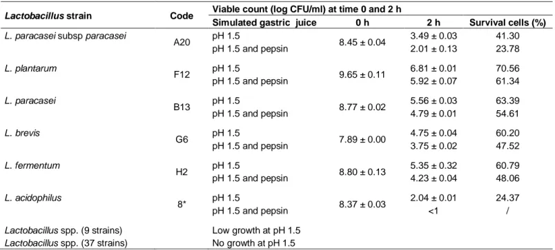
Table 1. Survival ability of lactic acid bacteriaisolates in simulated gastric conditions.
Lactobacillus strain Code Viable count (log CFU/ml) at time 0 and 2 h
Simulated gastric juice 0 h 2 h Survival cells (%)
L. paracasei subsp paracasei
A20 pH 1.5 8.45 ± 0.04 3.49 ± 0.03 41.30 pH 1.5 and pepsin 2.01 ± 0.13 23.78 L. plantarum F12 pH 1.5 9.65 ± 0.11 6.81 ± 0.01 70.56 pH 1.5 and pepsin 5.92 ± 0.07 61.34 L. paracasei B13 pH 1.5 8.77 ± 0.02 5.56 ± 0.03 63.39 pH 1.5 and pepsin 4.79 ± 0.01 54.61 L. brevis G6 pH 1.5 7.89 ± 0.00 4.75 ± 0.04 60.20 pH 1.5 and pepsin 3.75 ± 0.02 47.52 L. fermentum H2 pH 1.5 8.80 ± 0.13 5.35 ± 0.32 60.79 pH 1.5 and pepsin 4.23 ± 0.04 48.06 L. acidophilus 8* pH 1.5 8.37 ± 0.03 2.04 ± 0.01 24.37 pH 1.5 and pepsin <1 /
Lactobacillus spp. (9 strains) Low growth at pH 1.5
Lactobacillus spp. (37 strains) No growth at pH 1.5
Log CFU ml-1 values express the mean ± standard deviation; each data point is the average of repeated measurements from 03 independently replicated experiments, n = 3. P <0.05.
Identification of the potent probiotic strains by 16S rDNA analysis
The strains showing the best probiotic profiles were identified by the 16S rDNA analysis as described by Diop et al., (2008). Genomic
DNA from MRS broth cultures were extracted using the Wizard®
genomic DNA purification kit (Promega, Madison, USA), and used as a template for the amplification of 16S rRNA genes by the polymerase chain reaction (PCR). The primer pair 16SPO and 16SP6 was used. PCR products were resolved by electrophoresis in 1% (w/v) agarose gels and visualized by ethidium bromide staining and purified with Microcon YM-100 kit (Bedford, MA, USA). The BigDye Terminator sequence was performed using the Vector NTI (Version 8) software package (BD Biosciences, San Jose,
USA). Sequences were BLAST in GenBank database
(www.ncbi.nlm.nih.gov) for species assignment.
Statistical analysis
Thedatawerecalculatedwithmeanvalues,andstandarddeviations
(mean ± SD) were determined from triplicate trials. Statistical significance of the results was evaluated by ANOVA (analysis of variance). Statistical significance was attributed to (P<0.05).
RESULTS
Isolation and identification of lactic acid bacteria strains
One hundred and twenty (120) of LAB were selected and purified from seven samples collected twice from children faeces. Fifty two strains of them were retained for further work. They were rods, Gram positive, catalase negative and the fermentation profile obtained by the API50 CHLsystem, showed that they belonged to
the Lactobacillus genus (data no shown). They were closely related to L. fermentum (10 strains), L. casei (eight strains), L.gasseri (eight strains), L. salivarius (five strains), L. plantarum (five strains), L. vaginalis (four strains) L.paracasei subspparacasei (four strains), L. brevis (two strains), L. reuteri (two strains), L. acidophilus (2 strains) and L. ruminis (two strains).
Survival ability under gastric conditions
In this study, the effect of pH 1.5 was investigated with and without pepsin (0.3%) to test the ability of LAB to survive in simulated conditions of gastric juice. The results showed that pH 1.5 without pepsin, was critical forthe majority of isolates after 2 h of incubation. In fact, thirty seven isolates among the fifty two sited above, weretotally inhibited and nine manifested a low rate of growth. However, only six isolates were characterized by a good viability in low pH conditions (pH 1.5). The tolerance to simulated gastric conditions test (pepsin, pH 1.5) showed that five isolates of them supported such conditions with different degrees of viability. The highest survival percentage was observed with L. plantarum F12 (61.34%), followed byL. paracasei B13 (54.61%), L. fermentum H2 (48.06%), L. brevis G6 (47.52%) and L. paracasei subsp paracasei A20 (23.78%) (Table 1). The addition of pepsin to acid conditions causes a significant loss in isolates viability compared to the low acidity alone.
Survival ability under intestinal conditions
The most resistant LAB to simulated gastric conditions (F12, B13, H2, A20 and G6), obtained in the previous test, were assayed for their ability to survive in simulated intestinal conditions at pH 8, in the presence of pancreatin, with and without 0.3% of bile. The obtained results showed a decrease in growth of all tested LAB, depending on the strain, in comparison with their culture in the same conditions but without bile (Table 2). Four LAB tested had a
Table 2. Survival ability of lactic acid bacteria isolates in simulated intestinal conditions.
Strain Code
Viable count (log CFU ml-1)
Absence of bile Presence of 0.3% of bile
0 h 4 h 0 h 4 h
L. paracasei subsp. paracasei A20 8.35±0.02 7.43±0.10 8.35±0.03 2.90±0.12
L. plantarum F12 9.60±0.06 9.19±0.00 9.60±0.03 9.56±0.06
L. paracasei B13 8.83±0.04 9.65±0.32 8.83±0.12 7.01±0.05
L. brevis G6 7.91±0.09 7.97±0.08 7.91±0.10 6.89±0.00
L. fermentum H2 8.82±0.01 6.54±0.02 8.82±0.07 <1
Log CFU ml-1 values express the mean ± standard deviation; each data point is the average of repeated measurements from 03 independently replicated experiments, “n = 3. P < 0.05.
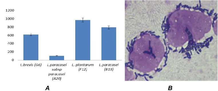
A B
Figure 1. (A) Adhesion of Lactobacillus strains to Caco-2 cells after 2 h of incubation at 37°C in a 5% CO2 atmosphere. (B) Adhesion
image of L.plantarum (F12) to Caco-2 cells observed by microscope (x 100) after staining cells with crystal violet (0.5%). Values (cells/100 Caco-2 cells) express the mean ± standard deviation; each data point is the average of repeated measurements from 3 independently replicated experiments, “n = 3. P< 0.05.
good tolerance to simulated intestinal juice after 4 h of exposure. L. plantarum F12 was the most resistant strain, followed by L. paracasei B13, L. brevis G6 and L. paracasei subsp paracasei A20. The strain L. fermentum H2 was completely inhibited.
In vitro adherence assay to Caco-2 cells
L. plantarum F12 L. paracasei B13, L. brevis G6 and L. paracasei subsp paracasei A20 were tested for their adherence to Caco-2 cells as they displayed high tolerance rates tosimulated gastro-intestinal juice. The results showed a difference in the abilities of this LAB to adhere to Caco-2 cells. However, 03 of them exhibited a good adherence. These were: L. paracasei B13, L. brevis G6, and particularly, F12 L. plantarum strain which presented the highest adherenceto Caco-2 cells (Figure 1).
Antimicrobial activity
The strains L. paracasei (B13), L. brevis (G6) and L. plantarum
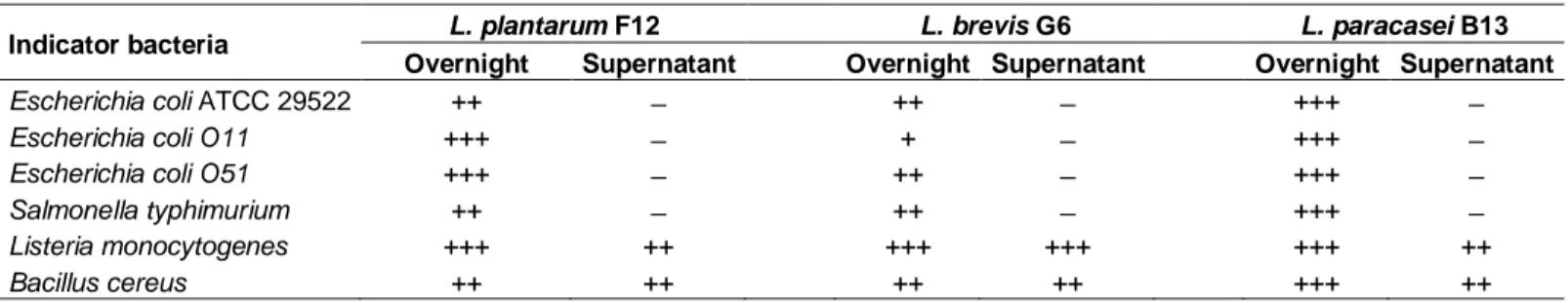
(F12) which exhibited a good adherence to Coco-2 cells were tested for their antimicrobial activity.In this study, the overnight cultures of the three tested strains demonstrated a strong antagonist activity against pathogens but their neutralized supernatants at pH 6.5 and treated with catalase (NSC) were unable to inhibit any pathogenic Gram-negative microbe. Moreover, the NSC of three LAB isolates had a good antimicrobial activity against Gram-positive bacteria (Table 3).
Identification of the potential probiotic strains by 16S rDNA sequencing
Based on the results of our study, three strains were retained as they displayed a high potential probiotic profile. Their identification by API CH 50 system was confirmed by 16S rDNA sequencing. BLAST analysis of their corresponding 16S rDNA sequences, in GenBank database, showed that they were closely related to L. plantarum (F12), L. brevis (G6) and L.paracasei (B13) (Table 4).
Table 3. Antimicrobial activity of lactic acid bacteriaisolates against bacterial pathogens.
Indicator bacteria L. plantarum F12 L. brevis G6 L. paracasei B13 Overnight Supernatant Overnight Supernatant Overnight Supernatant
Escherichia coli ATCC 29522 ++ ̶ ++ ̶ +++ ̶
Escherichia coli O11 +++ ̶ + ̶ +++ ̶
Escherichia coli O51 +++ ̶ ++ ̶ +++ ̶
Salmonella typhimurium ++ ̶ ++ ̶ +++ ̶
Listeria monocytogenes +++ ++ +++ +++ +++ ++
Bacillus cereus ++ ++ ++ ++ +++ ++
All indicators bacteria were tested for inhibition of growth microbes’ growth inhibition . Tested overnight cultures and supernatants were treated with catalase and neutralized to pH 6.5. Pathogen growth inhibition was determined by measuring inhibition zones . No inhibition ( ̶ ) ; diameter between 0 and 3mm (weak, +); diameter between 3 and 6 mm (good,++) and diameter larger than 6 mm (strong,+++). Each data point is the a verage of repeated measurements from 3 independently replicated experiments, n = 3. P< 0.05.
Table 4. Comparison between API and 16S rDNA identification of the potential probiotic strains.
Isolate API CHL identification identification Percent of
Identification by 16S rDNA
Identification number access Sequence Percent of match
CWBI\B-1515 (B13) Lactobacillus paracasei 99.3 Lactobacillus paracasei JQ436724 98% L.paracasei DQ462440* CWBI/B-1542 (G6) Lactobacillus brevis 91.3 Lactobacillus brevis JQ436725 98% L.brevis APO12167* CWBI/B-1543 (F12) Lactobacillus plantarum 97.2 Lactobacillus plantarum JQ436723 95% L.plantarumKF171888*
* Strains from GeneBank used for comparison with our strains.
DISCUSSION
The initial screening of strains using in vitro methods remains a useful preliminary step in the detection of probiotic candidates, despite the difficulties encountered to characterize reliable probiotic strains in this way (Campana et al., 2012). In this work, fifty two isolates were selected among one hundred twenty LAB, screened from faecal samples of healthy children aged between five and ten years. The selected bacteria were rods, Gram positive, catalase negative and non-motile. Their identification according to API test revealed its appur-tenance to Lactobacillus genus. They were tested, then, for their probiotic properties. In fact, human origin, resistance to simulated GI conditions, adherence to Caco-2 cells and antimicrobial activity against pathogens are the in vitro, tests suggested for preselction of the pro-biotic candidates in human use (FAO/WHO, 2002).
During fasting, the stomach pH can drop as low as 1.5 (Draser et al., 1969) and can dramatically affect bacterial growth. In the present study, among fifty two strains only six showed interesting viability at pH1.5 after 2 h of exposure. Previous reports indicated that pH (1.5-2) is the lethal acidity value for LAB isolated from human GI tract (Lankaputhra and Shah, 1995; Khalil et al., 2007; Xiaodong et al., 2009; Kirtzalidou et al., 2011). In the stomach, in addition to low pH, the probiotics are, also, subjected to the stress of proteolytic enzyme, pepsin
(Holzapfel et al., 1998). In the current study, the most resistant LAB (6 isolates) to pH 1.5 were tested for their tolerance to simulated gastric juice. At pH 1.5 with 0.3% pepsin, only five isolates were able to survive. The high survival rates to these conditions observed with the strains: L. plantarum F12, L. paracasei B13, L. brevis G6,
L. fermentum H2 and L. paracasei subsp paracasei A20.
This makes them, may be, able to transit the stomach and to reach the intestinal level.
One of the roles of bile acids is the inhibition of the proliferation of bacteria in the upper part of the digestive system. The mean intestinal bile concentration is believed to be 0.3% w/v, in addition, the transit time is suggested to be 4 h (Parasad et al., 1998). In this study, the most resistant strains (F12, B13, H2, A20 and G6) to simulated gastric juice were tested for their ability to survive in simulated intestinal juice. Four of the tested LAB had a good tolerance to simulated intestinal juice after 4 h of exposure and their growth decreased, in comparison with their culture in the same conditions but without bile. These findings were conforming to those of Haller et al. (2001), Vamanu and Vamanu (2010). The most resistant of them was the strain L. plantarum F12 followed by L. paracasei B13, L. brevis G6 and L.
paracasei subsp paracasei A20. The strain L. fermentum
H2 was completely inhibited.
The ability of probiotic bacteria to adhere to the intes-tinalepitheliumisprerequisiteforprobioticmicroorganisms
to be effective (Collado et al., 2005; Xiaodong et al., 2009). It was reported that adhered probiotic cells interact with the immunomodulatory cells of mucosal immune system, such as leucocytes, in order to stimulate their phagocytic activity against pathogens (Azacarate-Peril et al., 2009). In our study, four strains were tested for their adherence to Caco-2 cells as they showed high tolerance to simulated gastro-intestinal conditions. There was a significant difference (P<0.05) in their ability to adhere to Caco-2 cells, however, three of them exhibited a good adherence. They were L. paracasei B13, L. brevis G6 and particularly, the strain L. plantarum F12 which presented the highest adherence to Caco-2 cells, compared to that of L. rhamnosus GG reported by other studies (Thornton, 1996; Ren et al., 2012). The lowest adherence was observed with the strain L. paracasei subsp paracasei A20. Previous studies concluded that some Lactobacillus strains of human origin have good adherence to Caco-2 cells (Dunne et al., 2001; Campana et al., 2012). In our study, the high adherence of the strains B13, G6, and F12to Caco-2 cells suggests their ability to colonize the human intestinal epithelium tissue and to be in competition with pathogens. They were tested for their antagonistic effect against a represent-tative group of intestinal pathogens bacteria.
Antibacterial activity is vital for the successful colonization of lactobacilli in the intestinal mucosa as they provide a barrier effect and defence against pathogens (Vaughan et al., 1999). According to several studies, the antimicrobial activity of LAB may be due to a number of factors such as decreased pH levels, competition for substrates, production of H2O2 and bactericidal or
bacteriostatic substances, including bacteriocins (Servin, 2004). In our study, the overnight culture of the strains L.
plantarum F12, L. brevis G6, L. paracasei B13
demon-strated a strong antagonist activity against pathogens. Moreover, their supernatants neutralized andtreated with catalase were unable to inhibit any pathogenic Gram-negativetested. This suggests that antagonist activity of our LAB against Gram-negative rods is due to the com-bined action of low pH, organic acids and H2O2 or to one
of them.
Moreover, we found that the NSC of the three tested strains has a good antimicrobial activity on Gram-positive bacteria, Listeria monocytogenes and Bacillus cereus. This is probably due to substances supposed bacteriocins. Our results go in accordance with the data reported by Dortu and Thonart (2009), where the authors reported that no bacteriocin produced by lactic acid bacteria with activity against Gram-negative bacteria has been described.
Owing to the wall structure of Gram negative bacteria, the bacteriocins can’t cross the outer membrane to reach the inner membrane, the site of their activity. Similar results were obtained with the strains belonging to L.
paracasei, L. plantarum and L. brevis (Vaughan et al.,
1999; Tham et al., 2011; Rishi et al., 2011; Ren et al.,
2012; Russo et al., 2012).
The identification of probiotic strains by 16S rDNA sequencing was recommended by FAO/WHO (2002). In our study, the identification by API system, of the three potential screened probiotic strains, was confirmed by16S rDNA sequences analysis using BLAST analysis. They were L. brevis (G6) L. paracasei (B13) and L.
plantarum (F12).These strains were registered in the
GenBank database under access numbers respectively JQ436725, JQ436724 and JQ436723.
Conclusion
The faeces of healthy children living in Algerian rural areas seem to be a good source for isolating potential probiotic strains. In this study, we have selected three strains of Lactobacillus with high potential probiotic; L.
plantarum, L.brevis and L. paracasei. All the isolate
strains belong to the dominant lactobacilli of intestinal populations of healthy people and are usually recognized for their probiotics traits. Furthermore, their strong antimicrobial activity against the tested intestinal pathogens suggests their use in the prevention of bacterial childhood diarrhoea. However, further investigations, in vitro, and, in vivo, are needed to study their potential health benefits and their technological properties.
ACKNOWLEDGEMENTS
This research was realized at the University of Jijel/Algeria and at the University of Liege/Gembloux Agro Bio-tech/ Centre Wallon de Biologie Industrielle (CWBI)/ Belgium. The author wishes to acknowledge Professor Philippe Thonart and his team, Professor Willems Luc and Idoui Tayeb for their technical assistance.
REFERENCES
Azacarate-Peril MA, Tallon R, Klaenhammer TR (2009).Temporal gene expression and probiotic attributes of Lactobacillus acidophilus during growth in milk. J. Dairy. Sci. 92:870-886.
Campana R, Federici S, Ciandrini E, Baffone W (2012). Antagonistic Activity of Lactobacillus acidophilus ATCC 4356 on the Growth and Adhesion/Invasion Characteristics of Human Campylobacter jejuni. Curr. Microbiol. 64:371-378.
Collado MC, Gueimonde M, Hernandez M, Sanz Y, Salminen S (2005). Adhesion of selected Bifidobacterium strains to human intestinal mucus and its role in enteropathogen exclusion. J. Food Prot. 68:2672-2678.
Diop MB, Dubois-Dauphin R, Dortu C, Destain J, Tine E, Thonart P (2008). In vitro detection and characterization of bacteriocin-like inhibitory activity of lactic acid bacteria (LAB) isolated from Senegalese local food products. Afr. J. Microbiol. Res. 2:206-216. Dortu C, Thonart P (2009). Les bactériocines des bactéries lactiques:
caractéristiques et intérêts pour la bioconservation des produits alimentaires. Biotechnol. Agron. Soc. Environ. 3(1):143-154. Draser BS, Shiner M, McLeod GM (1969). Studies on the intestinal
achlorhydric persons. Gastroenterology 56:71-79.
Dunne C, O’Mahony L, Murphy L, Thornton G, Morrissey D, O’Halloran S, Feeney M, Flynn S, Fitzgerald, Daly C, Kiely B, O’Sullivan GC, Shanahan F, Collins JK (2001). In vitro selection criteria for probiotic bacteria of human origin: correlation with in vivo findings. Am. J. Clin. Nutr. 73(suppl):386-392.
FAO/WHO (2001). Health and Nutritional Properties of Probiotics in Food including Powder Milk with Live Lactic Acid Bacteria.Food and Agriculture Organization of the United Nations and World Health Organization.Working Group Report. Cordoba, Argentina.
FAO/WHO (2002). Guidelines for the evaluation of probiotics in food.Food and Agriculture Organization of the United Nations and World Health Organization. Working Group Report. London, Ontario, Canada.
Fuller R (1991). Probiotics in human medicine. Gut 32:439-442. Guo XH, Kim JM, Nam HM, Park SY, Kim JM (2010). Screeninglactic
acid bacteria from swine origins for multistrain probiotics based on in
vitro functional properties. Anaerobe 16:321-326.
Haller D, Colbus H, Ganzle MG, Scherenbacher P, Bode C, Hammes WP (2001). Metabolic and functional properties of lactic acid bacteria in the gastrointestinal ecosystem: A comparative in vitrostudy between bacteria of intestinal and fermented food origin. Syst. Appl. Microbiol. 24:218-226.
Hechard Y, Dherbomez M, Cenatiempo Y, Lettllier F (1990). Antagonism of lactic acid bacteria from goats’ milk against pathogenic strains assessed by the ‘sandwich method’. Lett. Appl. Microbiol. 11:185-188.
Holzapfel WH, Haberer P, Snel J, Schillinger U, Huisin’t Veld JHJ (1998). Overview of gut flora and probiotics. Int. J. Food Microbiol. 41:85-101.
Huang Y, Adams MC (2004). In vitro assessment of the upper gastrointestinal tolerance of potential probiotic dairy propionibacteria. Int. J. Food Microbiol. 91:253-260.
Kesarcodi-Watson A, Kaspar H, Lategan MJ, Gibson L (2008). Probiotics in aquaculture: the need, principles and mechanisms of action and screening processes. Aquaculture 274:1-14.
Khalil R, Mahrous H, El-Halafawy K, Kamaly K, Frank J, El Soda M (2007). Evaluation of probiotic potential of lactic acid bacteria isolated from faeces of breast-fed infants in Egypt. Afr. J. Biotechnol. 6(7):939-949
Kirtzalidou E, Pramateftaki P, Kotsou M, kyriacou A (2011).Screening for lactobacilli with probiotic properties in the infant gut microbiota. Anaerobe 17(6):440- 443.
Lankaputhra EV, Shah NP (1995). Survival of Lactobacillus acidophilus and Bifidobacterium spp. in the presence of acid and bile salts. Cult. Dairy Prod. J. 30:2-7.
NomotoK(2005). Review prevention of infections by probiotics. J. Biosci. Bioeng. 100:583-59
Parasad J, Gill H, Smart J, Gopal PK (1998). Selection and characterization of Lactobacillus and Bifidobacterium strains for use as probiotics. Int. Dairy. J. 8:993-1002.
ParvezS, MalikKA, Ah Kang S, Kim HY (2006). Probiotics and their fermented food products are beneficial for health. J. Appl. Microbiol. 100:1364-5072.
Ren D, Li C, Qin Y, Yin R, Li X, Tian M, Du S, Guo H, Liu C, Zhu N, Sun D, Li Y, Jin N (2012). Inhibition of Staphylococcus aureus adherence to Caco-2 cells by lactobacilli and cell surface properties that influence attachment. Anaerobe 18:508-515.
Rishi P, Preet S, Kaur P (2011). Effect of L. plantarum cell-free extract and co-trimoxazole against Salmonella Typhimurium: a possible adjunct therapy. Ann. Clin. Microbiol. Antimicrob. 10:9. Available from http://www.ann-clinmicrob.com/content/10/1/9.
Rosenfeldt V, Michaelsen KF, Jakobsen M, Larsen CN, Moller PL, Pedersen P, Tvede M, Weyrehter H, Valerius NH, Paerregaard A (2002). Effect of probiotic Lactobacillus strains in young children hospitalized with acute diarrhea. Pediatr. Infect. Dis. J. 21:411-416. Russo P, Fernández de Palencia P, Romano A, Fernández M, Lucas P,
Spano G. Lucas P, Spano G, Lopez P (2012). Biogenic amine production by the wine Lactobacillus brevis IOEB 9809 in systems that partially mimic the gastrointestinal tract stress.BMC.Microbiol. 12:247. [Available from http://www.biomedcentral.com/1471-2180/12/247].
SalyersAA, Gupta A, Wang Y (2004). Human intestinal bacteria as reservoirs for antibiotics genes. Trends Microbiol. 12:412-416 Schrezenmeir andVrese M (2001). Probiotics, prebiotics, and
synbiotics-approaching a definition.Am. J.Clin.Nutr. 73(suppl):361-4. Servin AL (2004).Antagonistic activity of Lactobacilli and Bifidobacteria
against microbial pathogens. FEMS. Microbiol. Rev. 28:405-440. Song YL, Kato N, MatsumiyaY, Liu CX, Kato H, Watanabe K (1999).
Identification of anhydrogen peroxide productionby fecal and vaginal lactobacilli isolate from Japanese women and newborninfants. J. Clin. Microbiol. 37:3062-3064.
Sugimura Y, Hagi T, Hoshino T (2011). Correlation between in vitro mucus adhesion and the in vivo colonization ability of lactic acid bacteria: screening of new candidate carp probiotics. Biosci. Biotechnol. Biochem. 75(3):511-515.
Tannock GW, Fuller R, Smith SL, Hall MA (1990). Plasmid profiling of members of the familyEnterobacteriaceae, lactobacilli, and bifidobacteriato study the transmission of bacteriafrom mother to infant. J. Clin. Microbiol. 28:1225-1228.
Tham CSC, Peh KK, Bhat R, Liong MT (2011). Probiotic properties of bifidobacteria and lactobacilli isolated from local dairy products. Ann. Microbiol. 62(3):1079-1087.
Thornton GM (1996). Probiotic bacteria selection of Lactobacillus and
Bifidobacterium strains from the healthy human gastrointestinal tract;
characterization of a novel Lactobacillus-derived antibacterial protein. PhD. Thesis. National University of Ireland, Cork, Ireland.
Vamanu E, Vamanu A (2010). Viability of the Lactobacillus rhamnosus IL1 strain in simulated gastrointestinal conditions. Int. J. Pharmacol. 6:732-737.
United Nations Children’s Fund/World Health Organisation (UNICEF/WHO) 2009. Diarrhoea: why children are still dying and what can be done. Lancet. 14:1-2
Vaughan EE, Mollet B,Devos WM (1999). Functionality of probiotics and intestinal lactobacilli: light in the intestinal tract tunnel. Cur. Opin. Biotechnol. 10:505-510.
Xiaodong P, Fenqin C,Tianxing W, Honggang T, Zhanyu (2009). The acid, bile tolerance and antimicrobial property of Lactobacillus