Perception of vertebrate volatiles
in the tropical bont tick,
Amblyomma variegatum
Fabricius
CHO
ffiyr^jpar
Pascal Steullet
V
e
Thèse présentée à la Faculté des Sciences de l'Université de Neuchâtel pour obtenir le grade de docteur es sciences
Université de Neuchâtel
Faculté des Sciences
Perception of vertebrate volatiles
in the tropical bont tick,
Amblyomma variegatum
Fabricius
par
Pascal Steullet
Thèse présentée à la Faculté des Sciences de l'Université de Neuchâtel
pour obtenir le grade de docteur es sciences
Perception of vertebrate volatiles in the
.. .tropical, ..bant... txc^....Amblyomma-. .varxegatum...
•Fabricius
de M>nsieur...p.as.cal. .Steullet.
UNIVERSITE DE NEUCHATEL
FACULTÉ DES SCIENCES
La Faculté des sciences de l'Université de Neuchâtel sur le rapport des membres du jury,
MM. P. Guerin, P.A. Diehl, E. Stadler (Wädenswil) e t R.A. Steinbrecht
..^^^^!^^-!!(^^^^!!^,....seeWiesen^
autorise l'impression de la présente thèse.
Neuchâtel, le 2?._.noye^re__ 1993_
1
...La tique solitaire, concentrée et cachée dans son arbre, aveugle sourde et muette, tout occupée pendant des années, à flairer sur des lieues à la ronde le sang des
animaux qui passent et qu'elle n'atteindra jamais par ses propres moyens Mais la tique, butée, bornée et répugnante, reste embusquée, et vit, et attend. Attend jusqu'à ce qu'un hasard extrêmement improbable lui amène le sang juste sous son
arbre, sous la forme d'un animal. Et c'est alors seulement qu'elle sort de sa réserve, se laisse tomber, se cramponne, mord et s'enfonce dans cette chair inconnue...
...Jl (Jean-Baptiste Grenouille) se mit à s'introduire nuitamment dans les étables, pour y envelopper pendant quelques heures des vaches, des chèvres ou des cochons avec des linges enduits de graisse, ou pour les emmailloter dans des bandages huileux. Ou bien il se glissait furtivement dans un enclos à brebis pour y tondre clandestinement un agneau, dont ensuite il lavait à l'esprit-de-vin la laine odorante....Les résultats ne furent d'abord guère satisfaisants. Car, à la différence d'objets dociles comme un bouton de porte ou une pierre, les animaux se montraient récalcitrants au prélèvement de leur odeur....
extrait de "Le Parfum" de Patrick Siiskind
(traduit de l'allemand par Bernard Lortholary) édition Fayard (Le livre de poche)
CONTENTS
I. INTRODUCTION 4 1.1. Systematic position of ticks and Amblyomma variegatum 4
1.2. Biology of Amblyomma variegatum 4
1.3. Host-seeking in Amblyomma variegatum 6
1.4. Olfactory sense organs in ticks and Amblyomma variegatum 8
1.5. Ultrastructure of olfactory sensilla in Amblyomma variegatum 10
1.5.1. Wall-pore single-walled A sensilla 10 1.5.2. Wall-pore single-walled B sensilla 11 1.5.3. Wall-pore double-walled A sensilla 11 1.5.4. Wall-pore double-walled B sensilla 11 1.5.5. Wall-pore double-walled C sensilla 11
1.6. Specificity of tick olfactory receptors 11
1.7. Outline of the present study 12
II. MATERIALS AND METHODS 13
2.1. Animals- 13
2.2. Electrophysiology 13
2.3. Stimulation 14
2.4. Gas chromatography-coupled electrophysiology 14
2.5. Gas chromatography-coupled mass spectrometry 17
2.6. Behavioural bioassays 17 2.6.1. Wind tunnel 17 2.6.2. Activation bioassay 17 2.6.3. Locomotion compensator (Kramer's sphere) 19
CONTENTS
3
RESULTS 21
3.1. Perception of breath components by the tropical bont tick,
Amblyomma variegatum Fabricius (Ixodidae). I CO^receptors.
J. Comp. Physiol. A (1992) 170:665-676 22 3.2. Perception of breath components by the tropical bont tick,
Amblyomma variegatum Fabricius (Ixodidae). Il Sulfide receptors.
J. Comp. Physiol. A (1992) 170.-677-685 34 3.3. Identification of vertebrate volatiles stimulating olfactory receptors
on tarsus of the tick Amblyomma variegatum Fabricius (Ixodidae). I Receptors within the Halter's organ capsule.
J.Comp. Physiol. A (in press) 43 3.4. Identification of vertebrate volatiles stimulating olfactory receptors
on tarsus of the tick Amblyomma variegatum Fabricius (Ixodidae). II Receptors outside the Halter's organ capsule.
J.Comp. Physiol. A (in press) 55
IV. GENERAL DISCUSSION 64
4.1. Location and number of tick olfactory sense organs 64 4.2. The capsule of Halter's organ, a complex olfactory sense organ 65
4.3. Specificity and sensitivity of the olfactory receptors 66 4.4. Correlation between receptor specificity and sensillum ultrastructure 69
4.5. Conservatism in the evolution of the tick olfactory system 71 4.6. Perception of vertebrate volatiles and host-finding 73
V. SUMMARY-RESUME 82
VI. REFERENCES 88
VIL REMERCIEMENTS 104
I. INTRODUCTION
Whenever the word tick is pronounced, a host of fears, legends and questions about these blood-sucking animals invariably arise. " A dangerous
insect that falls from trees onto humans and mammals to feed on blood " would
likely be the erroneous definition of a tick given by many people. The usual ignorance about how ticks recognize and find a host contributes to the legends on these small ectoparasites. The present work on the chemical ecology of ticks, more precisely on host-odour detection in the tropical bont tick
Amblyomma variegatum, consequently needs a short introduction on tick
systematic, the biology of A variegatum, and different aspects of olfaction and the host-finding strategy in this tick species.
1.1. Systematic position of ticks and Amblyomma variegatum
As members of the Order Acari ticks (900 sp.) are distinguishable from mites by stigmata located behind the fourth leg pair, but also have a highly expandible fused body (the idiosoma), and possess mouth parts adapted to haematophagy (the gnathosoma) equipped with 2 sharp chelicerae, 1 hypostome covered with recurved teeth, and 2 palps. These blood-sucking arthropods comprise 3 families: Nuttalliellidae, Argasidae (soft ticks), and Ixodidae (hard ticks) to which the genus Amblyomma belongs (Rg. 1.1.). The Ixodidae, subdivided into 5 subfamilies (Rg. 1.1.), are characterized by a sclerotized plate, which in the female and immature stages is limited to the anterior dorsal region, while in males it covers the entire back thus restricting male body extension during a blood meal. The subfamily Amblyomminae includes the genus Amblyomma (102 sp) which are considered primitive ticks because of their large size and their three-host life-cycle pattern (Sonenshine 1991).
1.2. Biology of Amblyomma variegatum
A. variegatum is a large tick widely distributed in most tropical and
subtropical regions of continental Africa, Madagascar, the South-West Arabic Peninsula, and the Cape Verde Islands (Hoogstraal 1956; Morel 1969). This species, also called the tropical bont tick, has been noted in the Caribbean Islands since the end of last century (Morel 1969) and threatens to colonize Central and South America. A. variegatum is a typical three-host tick with each stage (larva, nymph, adult) parasitizing mammals for a blood-meal of variable duration (larvae 5-8 days; nymphs 6-13 days; females 14-22 days) (Morel
INTRODUCTION
5
1969). A blood meal is indeed a requirement for successful moulting of immatures, spermiogenesis and ovogenesis in adults, and for oviposition in females.
Phylum: Arthropods Class: Chelicerata Sub-class: Arachnida Order: Acari
Sub-order: Metastigmata = ticks
Family: Ixodidae (hard ticks), NuttaEliellidae, Argasidae (soft ticks) Amblyomminae Haemaphysalinae Hyatomminae
Aponomma
Amblyomma Haemaphysalis Hyabmma
Rhiptcephalinae Ixodinae Dermacentor Cosmiomma Nosomma Rhipicephalus Anomabhytnalaya Rhipicentor Boophilus Margaropus Ixodes
Fig. 1.1. Systematic position of ticks (ca. 900 sp) and the genus Amblyomma (102 sp).
Immature stages of the tropical bont tick parasitize a wide range of reptiles, birds, and mammals (Matthysse and Colbo 1987), but Barré (1989) noticed a preference for ungulates. Adults for their part feed principally on large ruminants (Morel 1969; Matthysse and Colbo 1987; Barré 1989). Although large wild mammals such as buffaloes and antelopes are thought to be the natural hosts, domestic animals (cattle, sheep, goats) are also strongly infested in many areas (Hoogstraal 1956; Matthysse and Colbo 1987). Aeschlimann (1967) suggested that the distribution of this tick species has been expanded by the development of cattle ranching. A varìegatum, a vector of various pathogens such as Cowdria ruminantium (heart-water), Coxiella burnetii (Q-fever) and Dermatophilus congolensis (dermatosis), also causes anaemia and skin damage (Aeschlimann 1976, Barré 1989). It has thus become a serious life-stock pest responsible for much animal weakness and illness.
1.3. Host-seeking In Amblyomma variegatum
Parasitizing vertebrates is a very tough task for all tick life-stages, since the potential host is much bigger and more mobile. Thus, specific physiological capabilities and adequate host-seeking strategies are the keys to successful completion of the life-cycle. Tremendous resistance to desiccation and starvation allow ticks to survive for months during non-parasitic phases. While some tick species live within nests or burrows and thus maintain a close relation with their host, others, such as A variegatum, quest along pathways or areas frequented by their hosts. Consequently, the latter need well-adapted host-seeking strategies and efficient sense organs to optimize chances of meeting a host. Waladde and Rice (1982) reviewed the different senses involved during the successive behavioural steps of host-seeking, from tick arousal to its attachment on a host. In this context, olfaction is considered a major sense. Transported by wind and air turbulence, odour molecules can be detected over great distances. Odour quality as well as concentration constitute potential sources of information, i.e. estimation of the distance to a host, host recognition, host location.
Immature A. variegatum quest on vegetation and grasp any mammal which passes nearby (Barré 1989). Mechanical disturbance causes significant activation of immature stages (Barré 1989). By contrast, adult A. variegatum, which are hunter ticks (Morel 1969; Barré 1989), rest in the litter zone until a host in the close vicinity activates them (Rg. 1.2.). Excited ticks then start to walk about on the ground in order to locate the vertebrate, orient to it and grasp it, and then find a suitable feeding-site (Rg. 1.2.). Activation of resting adults is mainly mediated by host odour such as CO2, while vibrations, temperature, infrared radiation, and visual cues do not play a primordial role during the arousal steps of host-finding (Stämpfii 1987; Steullet unpublished; Poffet unpublished; Kaltenrieder et al. 1989; Yunker and Norval 1991). On the other hand, odours along with vertebrate-emitted infrared radiation (Poffet, unpublished), and visual cues (Kaltenrieder et al. 1989; Kaltenrieder 1990) intervene in host location. Males are first to infest mammals at the end of the dry season (Morel 1969), and females appear a few days later on the infested hosts (Hoogstraal 1956; Morel 1969; see Rg. 1.2.). Others have observed that attached males (the primary colonizers) of different species of Amblyomma attract conspecific females and other males (the secondary colonizers) to the same feeding-site (Gladney et al. 1974; Rechav et al. 1977; Norval and Rechav 1979). Schoeni et al. (1984) and Apps et al. (1988) identified the components of
INTRODUCTION 7
A
uninfested hostHost location /
AV
/
Contact with the host
PIONEERCOLONIZERS ïï Arousal "resting V male AAP Attachment Sexual maturation
Emission of pheromone and feeding
Feeding-site selection
B
infested host AAPHost location onj
SECONDARY COLONIZERS
Contact with the host Attraction to mature male
resting
female Attachment
and feeding
AAP: aggregation-attachment pheromone
the aggregation-attachment pheromone released by fed male A variegatum and A. hebraeum. Diehl et al. (1991) found that the amount of pheromone emitted by a male of both species is considerable. Thus, a male A. variegatum can secrete in one hour more than 1 ug of 2-nitrophenol, one of the major component of the aggregation-attachment pheromone. The combination of host odours and the aggregation-attachment pheromone leads other members of at least some species of Amblyomma to a suitable host on which blood-feeding and mating can be accomplished (Rg. 1.2.). Nevertheless, for pioneer
Amblyomma males, host odours are the only olfactory cues available during
host-seeking. Studies on the sensitivity and the specificity of olfactory sense organs in A. variegatum should thus provide information on host-odour cues employed by this parasite.
1.4. Olfactory sense organs in ticks and Amblyomma variegatum
With the exception of special olfactory organs such as the sensilla placodea in honeybees (Boeckh et al. 1965), olfactory sense organs in arthropods are normally innervated setae with pore-walls. The latter favour diffusion of molecules from the air to the dendritic membrane of the olfactory receptor cell(s) within the sensillum via the receptor lymph. Sensillum ultrastructure and morphology reviewed by Altner and Prillinger (1980), and Zacharuk (1985), and olfactory transduction processes such as odour molecule diffusion, odour molecule-receptor affinity, enzymatic cascade, electrical events, adaptation and specificity of olfactory receptors have been most extensively investigated in insects (reviews by Kaissling 1986; Kaissling et al. 1987; de Kramer and Hemberger 1987; Vogt et al. 1987a and b; Lerner et al. 1990; Stengl et al. 1992). However, only some works on the ultrastructure of olfactory sensilla and only a few studies on receptor specificity have been published in ticks.
Odour-stimulated ticks often raise the first pair of legs in the air as insects do with their antennae. This behaviour soon suggested to acarologists that the first leg pair bears crucial sense organs. Haller (1881) first described a capsule equipped with numerous setae which he suspected to be a hearing organ on the tarsus of the first leg pair. However, further behavioural experiments in which different parts of tick appendages were amputated or masked revealed that the tarsus of the first leg pair was in fact the main site of odour perception (Hindle and Merriman 1912, Lees 1948). The morphology and ultrastructure of tarsal sense organs, and principally the Haller's organ in ticks have been intensively studied (i.e. Schulze 1941; Leonovitch 1979, 1980;
INTRODUCTION 9
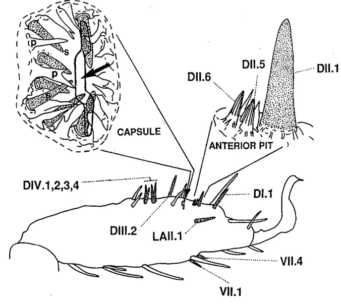
Fig. 1.3. Right tarsus of the first leg pair of an adult A. uariegatum and location of the wall-pore sensilla with their respective names (according to the nomenclature proposed by Hess and Vlimant, 1982). A brief description of the morphological properties of each olfactory sensillum is given in the text. Â detailed diagram of the anterior pit (lateral view) and the capsule (dorsal view) of Haller's organ is also provided. The arrow indicates the slit-like opening of the capsule. The seven wall-pore single walled sensilla (s) and cuticular pleomorphs (p) enclosed in the capsule are represented under the capsule opening.
Roshdy and Axtell 1972; Roshdy et a!. 1972; Foeiix and Axtell 1972; Waiadde 1976,1977). Haller's organ which contains almost half of all olfactory receptors of the tarsus (Hess and Vlimant 1986) has two dense groups of olfactory sensilla, the anterior pit and the capsule (Fig. 1.3.). Hess and Vlimant (1982, 1983, and 1986) described 5 different types of olfactory sensilla on the tarsus of
the idiosoma, Vlimant, personal communication). Three types of olfactory sensilla (wall-pore single-walled A and B; wall-pore double-walled C) are restricted to Haller's organ, whereas two other types (wall-pore double-walled A and B) are located on other parts of the tarsus. Adults and nymphs have 19 olfactory sensilla sharing a total of 68-94 suspected olfactory receptors per tarsus, while larvae possess only 14 olfactory sensilla with 57-77 olfactory receptors (Hess and Vlimant 1986). Furthermore, the number and location of tarsal sensilla is highly conserved between Ixodidae. However, primitive ticks possess slightly more olfactory sensilla, i.e. 20 in Ixodes ricinus, 19 in A.
variegatum, and 17 in Dermacentor marginatus compared to 14 in Boophilus microplus (Hess and Vlimant 1986). Nevertheless, the total number of olfactory
receptors is remarkably similar for the same life-stages of different tick species. Reduction of sensillum number results from the fusion of sensilla (Hess and Vlimant 1982). Figure 1.3. shows the position of each olfactory sensillum on the tarsus of an adult A. variegatum and a brief description of these sensilla is provided in the following section (for further details, see Hess and Vlimant
1982, and 1986).
1.5. Ultrastructure of olfactory sensilla in Amblyomma variegatum 1.5.1. Wall-pore single-walled A sensilla (L. N. A: 2V
These sensilla basiconica (50-60 u,m long) have relatively thick walls (0.6-1 u,m thick) with a large number of plugged pores (0.1 urn diameter). One of the two present, the Dl. 1 sensillum, is located on the dorsal side distalIy from the anterior pit of Haller's organ (Fig. 1.3.) and includes 5 neurones with branched dendrites. The other, the Dl 1.1 sensillum, belongs to the anterior pit (Fig. 1.3.) and is innervated by 3 sets of neurones (2x5 + 1 x 4) with branched dendrites. The latter is thought to result from the fusion of 3 separate sensilla. Indeed, each set of neurones possesses a thecogen cell and a trichogen cell, while the tormogen cells form a common outer receptor lymph cavity (Hess and Vlimant 1982). Interestingly, this fusion is not complete in the related species A
americanum where two wall-pore single-walled A sensilla are found in the
anterior pit, one with two sets of neurones and the other with only one set of neurones (Foelix and Axtell 1971).
INTRODUCTION
11
1.5.2. Wall-pore single-walled B sensilia (L: 4/ N. A: 7)Wall-pore single-walled B sensilia, occasionally bifid, are deeply enclosed in a cavity, the capsule of Haller's organ (Rg. 1.2). Projecting from the floor or from the proximal or lateral cuticular walls of the capsule towards the slit opening, these sensilia have thin walls (0.08-0.14 jam tick) with wide plugged pores (0.1-0.16 urn) and are innervated by 3 to 5 neurones with dendrites showing various degrees of branching.
1.5.3. Wall-pore double-walled A sensilia (L: 2/ N. A: 4)
Called DIV, 1, 2, 3, and 4 respectively, these sensilia trichodea (35-40 \i m long) are located very close together on the dorsal side of the tarsus, praxi mally from the capsule (Fig. 1.3.). They are innervated by 1 or 2 neurones with unbranched dendrites and have grooved walls. Radial canals lead from the central cavity of the sensillum to narrow pores, situated in the longitudinal grooves.
1.5.4. Wall-pore double-walled B sensilia (L. N. A: A)
These sensilia chaetica resemble the wall-pore double-walled A type but have two neurones forming a tubular body (mechanosensory units) and 4 to 7 neurones with unbranched dendrites. One of these sensilia (Dlll.2) is located proximally from the capsule, another (LAI 1.1) is inserted on the lateral side, and the other two (Vl 1.1 and 4) are found on the ventral side of the tarsus (Fig. 1.3.).
1.5.5. Wall-pore double-walled C sensilia (L. N. A: 21
Both wall-pore double-walled C sensilia basiconica are confined to the anterior pit (Fig. 1.3.) and have walls with longitudinal but interrupted grooves. Three neurones with unbranched dendrites innervate these sensilia.
1.6. Specificity of tick olfactory receptors
Despite several studies, our current knowledge regarding specificity of tick olfactory receptors is rather limited and fragmented. Only a few receptors have been properly characterized. Receptors sensitive to 2,6-dichlorophenol, a tick pheromone component, have been identified in wall-pore single-walled A sensilia of A americanum (Haggart and Davis 1981), and A variegatum (Waladde 1982; Schoeni 1987), and in the wall-pore single-walled C sensillum of I. ricinus (Thonney 1987). Wall-pore single-walled A sensilia of A variegatum also contain receptors which respond to 2-nitrophenol (Schoeni 1987) or to short-chain fatty acids (Hess and Vlimant 1980). Other receptors confined to the anterior pit are excited by NH3 in Rhipicephalus sanguineus (Haggart and
Davis 1980), by breath and cattle wash in B. microplus (Waladde and Rice 1982). Furthermore, methylsalicylate, an aggregation-attachment pheromone component in A variegatum (Schoeni et al. 1984), stimulates a receptor in the capsule of Haller's organ of A variegatum (Hess and Vlimant 1986), but also in
I. ricinus (Guerin, unpublished) and B. microplus (de Bruyne, unpublished).
Other capsule receptors were found to respond to breath and CO2 in B,
microplus (Waladde and Rice 1982), while Sinitsina (1974) obtained responses
to mouse odour, pentanoic acid, breath, but not to CO2 in capsular receptors of
Hyalomma asiaticum. Holsher et al. (1980) also reported presence of
CO2-excited receptor(s) with a tungsten electrode inserted proximalIy in the capsule of three tick species. Finally Haggart and Davis (1980) found a receptor sensitive to high concentrations of NH3 in a wall-pore double-walled A sensillum of R. sanguineus. The latter is the only report of a response to an olfactory stimulant by receptors in wall-pore double-walled sensilla of ticks.
1.7. Outline of the present study
Although breath, mouse odour, and the pelage wash of cattle can stimulate receptors of some tick species, CO2, NH3, and short-chain fatty acids are currently the only vertebrate-associated volatiles known to stimulate tick olfactory receptors. Considering the total number of olfactory receptors present on the tarsus (68-94 in A. variegatum), it would be very surprising if no other host-odour compounds could be detected. Although Haller's organ is generally considered as the main site of host-odour detection, the exact location and number of host-odour receptors are not known. The present work aims to systematically investigate the specificity of olfactory receptors of A. variegatum, an ideal tick model for such a study. The morphology and ultrastructure of olfactory receptors in this tick species are well known (Hess and Vlimant 1982,
1983, and 1986), the size of A variegatum (up to 1 cm) renders electrophysiological preparation relatively easy, and olfaction plays a primary role in host-finding for this hunter tick. This study should permit identification of different classes of vertebrate-associated volatiles perceived by A. variegatum and allow us to understand how finely tuned the olfactory system of A.
variegatum is to the odour bouquet of various vertebrates. Finally, the
odour stimulants identified will be considered as behavioural cues in host-finding.
METHODS 13
II. METHODS
In this chapter, only the main principles of the Methods used are described. Further details are provided in the corresponding publications presented in chapter 3.
2.1. Animals
Male of A. variegatura were principally used because of their pioneer role in parasiting hosts. Rearing conditions are described in chapter 3.1..
2.2. Electrophysiology (chapters 3.1., 3.2., 3.3., 3.4.)
Since Monta and Yamashita's pioneering work on Bombyx (1961), classic electrophysiology methods have been used in arthropods to pick up such electrical events as receptor potentials and action potentials generated at the level of olfactory receptors for the study of their specificity and sensitivity. In the present work, electrical activity of tick olfactory receptors has mostly been captured by contacting the cut tip or the pored wall of a sensillum with a glass electrode. A tungsten electrode inserted into the receptor lymph at the base of a sensillum was also employed for specific purposes in this study (see chapter 3.1.). Glass electrodes show several advantages over tungsten electrodes. A recording glass electrode permits recording of shifts in potential and action potentials associated with receptor cells, whereas a tungsten electrode will cut off lower frequencies (slow potential shifts). Furthermore, receptor cells can be more easily destroyed by tungsten than by glass electrodes
Typically, glass electrodes are glass capillaries filled with an electrolyte solution which is electrically connected via Ag/AgCI wires to the amplification circuit. Small voltage changes between the reference glass electrode (ground) inserted in the tick haemolymph and the recording electrode in contact with the
sensillum were thus amplified (10 to 100Ox) via a 101 2 Ohm high input
impedance preamplifier and an AC/DC amplifier. Signals were then displayed on an oscilloscope, printed on paper, stored on video tapes, or fed to a computer for further analysis (Fig. 2.1.). In many cases, analysis of spike shapes and amplitudes (extracellular recordings of action potentials generated by each receptor cell in response to stimulation with volatiles) permits characterization of the sensitivity and specificity of each responding receptor cell of a sensillum. Indeed, spike shapes and amplitudes generated by the separate receptor cells in the same sensillum often differ. Specific passive properties (impedance and capacitance) of the electrical circuitry within the
sensillum (through receptor cells and accessory cells) may indeed contribute to the shape and amplitude of the extracellularly recorded spikes (de Kramer
1985; Rumbo 1989).
2.3. Stimulation (chapters 3.1., 3.2., 3.3., 3.4.)
Air from a cartridge containing volatiles was briefly swept via electrically controlled valves into a charcoal-filtered air stream leading to the tick preparation. Shifts in potential and spike frequency changes of the olfactory receptors resulting from stimulation were analysed. The specificity of the receptors was tested with a series of well-known vertebrate-associated volatiles representing different- chemical classes (0(¾, CH4, NH3, acetone,
4-heptanone, y-butyrolactone, y-valerolactone, pentanol, 1-octen-3-olf
4-methylphenol, hexanal, propanoic acid, butanoic acid, 2-methylpropanoic acid, 3-methylbutanoic acid, pentanoic acid, heptanoic acid, L-lactic acid, ), with some tick pheromone components (nonanoic acid, 2,6-dichlorophenol, 2-nitrophenol, methylsalicylate), some other volatiles (3-pentanone, 6-caprolactone, 1-octene, octylamine, hexyl acetate) and with vertebrate odours (human breath, human axillary secretions, bovine breath, bovine skin wash, odours collected around steers and rabbits). Vertebrate-associated volatiles were collected on a charcoal trap (for breath), on the porous polymer adsorbent Porapak Q (for breath and odours of steers and rabbits, see Rg. 2.2.), or in a cold trap (for headspace over cotton pads used to wash bovine skin, see Fig. 2.3.) and extracted with solvent for use as host-odour stimuli. Breath as well as cotton pads soaked with human axillary secretions were also introduced into the stimulus cartridge and used directly as stimuli.
2.4. Gas chromatography-coupled electrophysiology (chapters 3.2., 3.3., 3.4.)
Whenever a host-odour extract stimulated receptor(s), the next step consisted of identifying the stimulant(s) among the numerous constituents of the extract. These volatiles were separated by gas chromatography on a high-resolution capillary column coupled to an electrophysiological preparation of a tick olfactory sensillum, thus permitting observation of the effect of each component of the extract on the activity of the receptors (Wadhams 1982).
METHODS
15
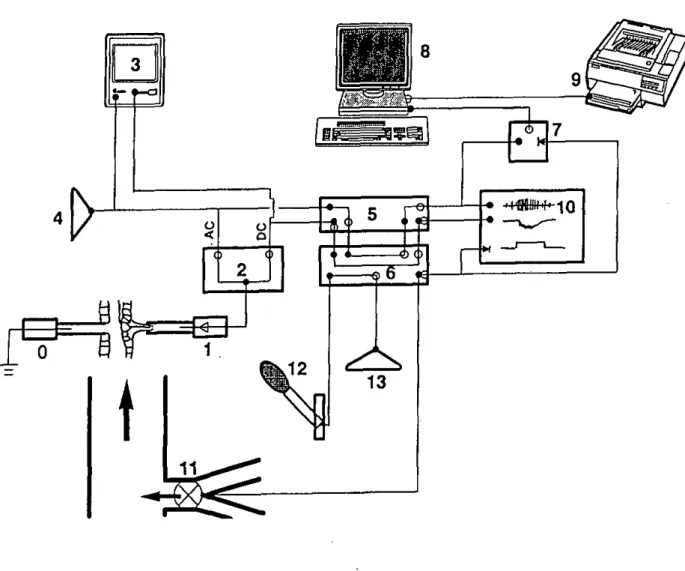
Fig. 2.1. Diagram of the electrophysiology set-up used to study olfactory receptors of A.
variegatum. (0): reference glass electrode impaled in the coxa of one of the first legs. (1):
Recording glass electrode in contact with the cut tip of the sensillum. The electrode is electrically connected via an Ag/AgCl wire to a high input impedance preamplifier (symbolized by a triangle). (2): Universal AC/DC amplifier with separate outputs for the AC and DC parts of the recorded signal. (3): Oscilloscope to visualize the AC and DC signals. (4): Loudspeaker to render spikes audible in the AC signal. (5): PCM-I Digital-VCR instrumentation recorder adaptator for analogue/digital or digital/analogue conversion of the AC and DC signals. (6): Video tape recorder used to store digitized signals and to play them back for further analysis via digital/analogue conversion in (5). (7): "Go-Box" which contains the circuitry necessary to provide the trigger to start digitization on the computer of the analogue AC signal played back from the video tape recorder via (5). (8): IBM-compatible computer equipped with an analogue/digital board to digitize the analogue AC signal and with software (SAPID tools) to visualize and analyse the digitized signal, (9): Laser printer to print signals and analyses. (10): Recorder to print signal. (11): Stimulus delivery controlled by solenoid valves. The stimulation period is indicated by a pulse signal which can be stored on an audio channel of (6) and used subsequently as a trigger signal, i.e. in (7) or in (10). (12): Microphone used to add comments stored on the second audio channel of (6). (13): Loudspeaker to listen to comments during play-back. Bold circles: inputs to each instrument; open circles: outputs of each instrument; bold triangles: trigger inputs.
1 odour collection 2 extraction 3 extract concentration
CH2Cl2
air from the room wHh cattle
V
N2h
v
concentrated extract extractFig. 2.2. Odour collection using adsorbent material (charcoal or Porapak Q). This method was used to collect volatiles in the air of rooms with either cattle or rabbits.
1 odour collection
N2
70
'
C
J
cotton pad rubbed on bovine skin
L
flowmeter -7O0C 2 solvent extraction CH2Ci2V cold trapW
acetone + dry-ice \ extract extractV
liquid (organic phase) -1O0CO
1S
N2 N y ^ (water phasePy/ TJphase separation extract concentration
Fig. 2.3. Odour collection using a cold trap. This method was used to collect volatiles from cotton pads rubbed on bovine skin (skin wash).
METHODS
17
2.5. Gas chromatography-coupled mass spectrometry (chapters 3.3., 3.4.) Chemical analysis of the active component(s) of an extract, located by gas chromatography-coupled electrophysiology, was then tackled with gas chromatography-coupled mass spectrometry. After separation of the extract components by gas chromatography, the mass spectrum of the active component(s) (located on the chromatogram with reference to its Kovat's
index1) was then analysed. Identification of the unknown was based on the
match of its mass spectrum with that of a known product stored in a computer-based library. The mass spectrum and the calculated Kovat's index of the unknown were then compared with those of the library-proposed synthetic analogue, injected under the same conditions. Finally, identification was confirmed by testing the electrophysiological activity of the synthetic substance on the relevant receptor.
2.6. Behavioural bioassays (chapters 3.1,3.2., 3.3., and discussion)
Behavioural bioassays were designed to investigate some identified olfactory stimulants, namely breath stimulants (CO2, and H2S), as behavioural cues for adult A variegatum.
2.6.1. Wind tunnel (chapter 3.1.)
Resting adult A variegatum were placed in a wind tunnel and then stimulated with CO2 as described in chapter 3.1.. Activation as well as positive anemotaxis were observed and quantified as described in chapter 3.1..
2.6.2. Activation bioassav (chapter 3.2.1
Resting adult A. variegatum were enclosed in glass flasks continuously swept with charcoal-filtered and humidified air. Every 24 hours, ticks are confronted with short stimulations of two olfactory stimulants identified by electrophysiology, i.e. CO2, H2S, and mixture of both. The tick's degree of
'The Kovat's index indicates the retention characteristic of a product on a column with respect to n-alkanes. It is expressed as following;
/ = 100[ n{(log V* - log V0V(IOg Ve+11 - log Ve)! + c]
where Vx is the retention time of the unknown compound, Ve is the retention time of the alkane elu ting before the unknown, V0+11 is the retention time of the alkane eluting after the unknown, c is the carbon number of the alkane eluting before the unknown, n refers to the difference in the number of carbon atoms for the two n-alkanes used as reference.
Fig. 2.4. Diagram of the gas chromatography-coupled electrophysiology set-up (GC-EL). (0): Reference glass electrode impaled in the coxa of one of the first legs. (1): Recording glass electrode in contact with the cut tip of the sensillum. The electrode is electrically connected via an Ag/AgCl wire to a high input impedance preamplifier (symbolized by a triangle). (2): Universal AC/DC amplifier with separate outputs for the AC and DC components of the recorded signal. (3): Oscilloscope to visualize AC and DC signals. (4): Window discriminator apparatus and frequency/voltage converter to sort spikes from background noise of the AC signal and to transform the frequency of the spikes into a DC voltage. (5): Loudspeaker to render spikes audible in the AC signal. (G): Chart recorder which printed the DC signal, the frequency to voltage converted signal, and the response of the chemical detector ( 17) on separate channels. (7): PCM-1 Digital-VCR instrumentation recorder adaptator for analogue/digital or digital/analogue conversion of the AC signal. (8): Video tape recorder used to store the AC signal and to play it back for further analysis via digital/analogue conversion in (7). (9): "Go-Box" which contains the circuitry necessary to provide the trigger to start digitization on the computer of the analogue AC signal played back from the video tape recorder. (10): IBM-compatible computer equipped with an analogue/digital board to digitize analogue AC signal and software (SAPID tools) to visualize and analyse the digitized signal. (11): Laser printer to print signals and analyses. (12): Microphone used to add comments stored on an audio channel of (8). (13): Loudspeaker to listen to comments during play-back. (14): Pulse signal stored on the second audio channel of (8) which is used to mark stimulation period and to subsequently trigger via (9) the digitization of the analogue AC signal in the computer. (15): High-resolution gas capillary column enclosed in the oven of a gas Chromatograph. (16): Splitter delivering the column effluent {thin arrow) to the chemical detector (17) and the electrophysiological preparation via the heated transfer line (18) of the Chromatograph. (19): Water-jacketed glass tube to maintain the humidified main air-stream (large arrow) flowing over the preparation at a constant temperature. Bold circles: inputs to each instrument; open circles: outputs of each instrument; bold triangles: trigger inputs.
METHODS 19
arousal is evaluated immediately after each stimulation as described in chapter 3.2..
2.6.3. Locomotion compensator (Kramer's sphere! (chapter 4.6.1
Preliminary experiments on the triggering and maintenance of orientation in excited male A. variegatura by CO2, H2S, and mixture of both were undertaken. Only the most relevant results of these behavioural bioassays are given in chapter 4.6. and briefly discussed. As it is not described elsewhere, details of the method and procedure employed for these preliminary experiments are provided here.
A breath-excited tick was placed on the top of a sphere (50 cm diameter) which rotated so that the animal was always kept at the sphere north pole. A light beam perpendicularly illuminated the sphere pole (field diameter: 3 cm). A small retroflective foil (n0 7610, 3M, Switzerland), glued to the dorsal side of the tick, reflected light back to a sensor which continuously evaluated the deviation of the reflective foil from the sphere pole. According to these deviation evaluations, signals were sent to two servomotors placed orthogonally on the equator of the sphere to recenter the reflective foil and hence the tick (Kramer 1976, Rickli et al. 1992). Displacement of the sphere was registered by two incremental pulse generators with a resolution of 0.1 mm. The X and Y coordinates of the tick's position were sampled at 0.1 s intervals and fed into a computer for track analysis. The sphere was enclosed in a styropore chamber to isolate the experimental area from the open laboratory. An upright aluminium cylinder (12 cm diameter, 11 cm high) with an upwind and a downwind aperture surrounded the north pole of the sphere to render the environment near the tick uniform. Experiments were accomplished during the last 6 hours of light of the daily cycle (LD 12:12) with unfed 6-7 month old males. Breath-excited individuals were placed on the sphere and allowed to walk for 2 min. Animals which regularly stopped were discarded. Walking responses were then recorded for 90 consecutive s divided into 30 s prestimulation control followed by 30 s of stimulation and a further 30 s poststimulation. The tick behaviour was also recorded on video for further analysis with a CCD camera focused on the tick.
A humidified charcoal-filtered air stream was directed at the north pole of the sphere in a 3.6 cm diameter water-jacketed stainless steel tube to provide a constant air flow of 0.1 m/s at 25 ± 1°C and 70 ± 5% RH. Stimuli carried by solenoid-activated air flow(s) were added to the main air stream at 14 cm from the outlet of the tube situated at 3 cm from where the tick walked. H2S
stimulation was provided by air (120 ml/min) which passed through a 50-ml gas-wash flask with 1 ml of an aqueous solution of Na2S (1-100 mg/ml). CO2 stimulation was made with various additions of a flow from a 5% C02/95% N2 gas cylinder which passed through a 50-ml gas-wash flask with 1 ml distilled water added to provide concentration of 0.15-0.6% COg at the north pole of the sphere. CO2/H2S stimulation was made with synthetic 5% CC>2/95% N2 from a gas cylinder mixed with H2S vapours from the flask described above. Polluted air was evacuated via a funnel placed 20 cm downwind from where the tick walked and cleaned over charcoal.
RESULTS
21
III. RESULTS
3.1. Perception of breath components by the tropical bont tick,
Amblyomma variegatum Fabricius (Ixodidae). I C02-receptors. J.
Comp. Physiol. A (1992) 170:665-676
3.2. Perception of breath components by the tropical bont tick,
Amblyomma variegatum Fabricius (Ixodidae). Il Sulfide receptors. J.
Comp. Physiol. A (1992) 170:677-685
3.3. Identification of vertebrate volatiles stimulating olfactory receptors on tarsus of the tick Amblyomma variegatum Fabricius (Ixodidae). I Receptors within the Haller's organ capsule. J.Comp. Physiol. A (in press)
3.4. Identification of vertebrate volatiles stimulating olfactory receptors on tarsus of the tick Amblyomma variegatum Fabricius (Ixodidae). Il Receptors outside the Haller's organ capsule. J.Comp. Physiol. A (in press)
J Comp Physiol A (1992) 170:665-676
Journal of
Comparative SET Physiology A SSSt
© Springet-Verlag 1992
Perception of breath components by the tropical bont tick.
Amblyomma variegatum Fabricius (Ixodidae)
L C02-excited and C02-inhibited receptors Pascal Steullet and Patrick M. Guerin
Institute of Zoology, University of Neuchâtel, Chantemerle 22, CH-2007 Neuchâtel, Switzerland Accepted March 23, 1992
Summary. Wall-pore olfactory sensilla located in the
capsule of Haller's organ on the tarsus of Amblyomma
variegatum ticks bear cells responding to vertebrate
breath: one of these sensilla contains a C02-excited re-ceptor and a second sensillum has a CO2- inhibited recep-tor. Each of these antagonistic C02-receptors) which display typical phasic-tonic responses, monitors a dif-ferent CO concentration range. The C02-inhibited re-ceptor is very sensitive to small concentration changes between 0 and ca. 0.2¾, but variations of 0.01% around ambient (ca. 0.04%) induce the strongest frequency mod-ulation of this receptor. An increase of just 0.001-0.002% (10-20 ppm) above a zero C02-level already inhibits this receptor. By contrast, the C02-excited receptor is not so sensitive to small CO2 shifts around ambient, but best monitors changes in CO2 concentrations above 0.1%. This receptor is characterized by a steep dose-response curve and a fast inactivation even at high CO2 -concentrations (>2%). In a wind-tunnel, Amblyomma
variegatum is activated from the resting state and
at-tracted by CO2 concentrations of 0.04 to ca. 1%, which corresponds to the sensitivity range of its C02-receptors. The task of perceiving the whole concentration range to which this tick is attracted would thus appear to be divided between two receptors, one sensitive to small changes around ambient and the other sensitive to the higher concentrations normally encountered when ap-proaching a vertebrate host.
Key words: Tick - C02-excited receptor- C02-inhibited receptor - Haller's organ - Host finding
Introduction
The CO2 contained in vertebrate breath is an activating stimulus or attractant for most blood-sucking arth-ropods (e.g. mosquitoes: Gillies and Wilkes 1968; tsetse
Correspondence to: P. SlcuHel
flies: Turner 1971; Stomoxys calcitrarti.• Warnes and Finlayson 1985; Simulidae: Fallis and Raybould 1975; Tabanidae: French and Kline 1989; Reduvidae: Bernard 1974; Siphonaptera: Osbrink and Rust 1985). Some C02-sensitive receptors have been described on palps of mosquitoes (Kellogg 1970) and on antennae of tsetse flies (Bogner 1989). Ticks also respond strongly to breath and CO2, and Garcia (1962) has shown that CO2 attracts many different tick species. Since that first report several authors have devised CO2 baited traps for field sampling (e.g. Garcia 1965; Wilson et al. 1972; Gray 1985; Gu-glielmone et al. 1985; Norval et al. 1987, 1988), or have studied the effects of CO2 on tick behaviour in the lab-oratory (Nevill 1964; Sauer et al. 1974).
In spite of the above, our knowledge of breath and CO2 perception in ticks is fragmentary. Breath-stimulated ticks lift their first pair of legs in the air to sample the surroundings as insects do with their anten-nae. From these observations, and various behavioural experiments where different parts of tick appendages were amputated or masked (Hindley and Merriman 1912; Lees 1948), we know of the primordial role of the tarsus of leg pair 1 for host odour perception. A large number of ultrastructural studies have described dif-ferent kinds of olfactory sensilla located on the tarsus of the first leg pair (reviews: Waladde and Rice 1982; Hess and Vlimant 1986), and the Haller's organ situated on the dorsal side of the tarsus (Fig. IA and B) bears a significant proportion of all tick olfactory sensilla. Thus, among the 19 tarsal olfactory sensilla of Amblyomma
variegatum, 3 belong to the anterior pit of Haller's organ
and 7 to the capsule of Haller's organ. Nevertheless, few investigations on physiological and functional charac-teristics of the tarsal olfactory sensilla have been under-taken in relation to host odour perception. Sinitsina (1974) using electrophysiological methods found olfac-tory cells responding to breath, mice odours, and n-vale-ric acid in the capsule of the Haller's organ in Hyalomma
asiaticum, but he failed to account for a C02-receptor. On the other hand, Waladde and Rice (1982) mentioned the presence in Boophilus microplus of cells responding to
RESULTS-CHAPTER 3.1. 23
666 P. Steullet and P.M. Guerin: Perceplion of breath components by Ambtyomma I
breath and cow wash in the anterior pit of Haller's organ, and changes in the activity of cells from the capsule when stimulated with either breath or CO2. But these studies were mainly qualitative and apparently involved few recordings. The small number of physiological studies on olfactory sensilla of ticks may be ascribed to their limited accessibility, especially for those located inside the cap-sule, as well as the added complication of the high num-ber of cells in many of these sensilla. Several questions remain unresolved. Do any of these olfactory cells re-spond to breath? Where are they located? What are the stimuli contained in breath which induce the response? Based on complete ultrastructural studies on all tarsal sensilla of Amblyomma variegatum (Hess and Vlimant 1982, 1983, 1986), we have systematically searched for tarsal olfactory sensilla responsive to breath and its com-ponents.
Materials and methods
Tick rearing
Experiments were mainly undertaken with unfed Amblyomma
va-riegatum males but unfed females were also used for some
record-ings. Originating from West Africa (Adiopodoumé, Ivory Coast), ticks were reared at the Agricultural Research Centre of Ciba-Geigy Ltd. (St-Aubin, Switzerland). AU stages (immatures and adults) were fed on Simmcntal calves at 22 to 24 0C. Ticks were kept under
constant darkness at 28 °C/8O-909É RH except during moulting when conditions were 29 0C and 90¾ RH. Finally, adults were
maintained in this laboratory in an environmental cabinet under the following conditions: 10 h of darkness at 18 0C/ 95« RH and 10
h of light at 25 °C/85% RH separated by 2 h "dusk" and "dawn" transition periods.
Light and scanning electron microscopy
Scanning electron microscope examination was made on ticks which were killed and fixed in 80% ethanol for several days, cleaned with ether/chloroform in a soxhlet extractor for 12 h, dehydrated in acetone, and critical point dried in CO2 with a Balzers CPD
device. The mounted specimens were gold sputtered in a Balzers sputtering apparatus, and then observed in a Philips 500 PSEM. Light microscopy examination was made on sections of cut tarsi which were fixed in 2% glutaraldehyde (Sabatini et al. 1963), post-fixed in 2% OsO4 (Palade I952), dehydrated in acetone, and
em-bedded in SPURR. Emem-bedded tarsi were cut, at the level of the capsule of the Haller's organ, in either transversal or sagittal sec-tions of 0.5 u.m and observed under a light microscope (Vanox-S, Olympus, Japan) after toluidine blue staining.
Tick preparation
The tick was immobilized on a perpex holder on double-sided sticky tape. Pedal nerves were destroyed by pinching coxa of the forelegs with fine forceps ; this prevented muscle activity during electrophysi-ological recordings. To make proper recordings from the 7 olfactory sensilla located in the capsule of Haller's organ (an olfactory pit some 80 urn deep and 60 urn wide), dissection was needed to improve their accessibility as the opening of this capsule is just a narrow slit of ca. 5 urn wide and ca. 50 urn long across the tarsus (Fig. IB). The cuticular roof was removed (Fig. IC) with a piece of razor blade in a holder (John Weiss & Son LTD., England) mount-ed on a Leitz micromanipulator under an Olympus SZH stereomi-croscope at a magnification 192 x (working distance: 48.5 mm).
Electrophysiology
In order to improve contact, the tips of sensilla not located in the capsule were cut with the flame-pulled tip of a glass rod (1.5 mm dia.) oscillating in the ultrasound frequency range (ca. 120 kHz) as induced by a piezoelectric transducer disk (n0 4322 020 177721,
Philips, The Netherlands) (Gödde 1989). The recording glass elec-trode filled with 0.2 M KCl was brought into contact with the cut tip of the sensillum with a Leitz micromanipulator, and the refer-ence glass electrode, filled with 0.2 M NaCl, was inserted in the coxa of one of the anterior legs. Electrical activity of capsular sensilla was also recorded with glass electrodes gently introduced into the dis-sected capsule until cell activity was captured. These recording electrodes also contained 1% polyvinylpyrrolidone K90 (Fluka, Switzerland), in order to prevent electrolyte flowing from the tip. Indeed, tips sometimes broke when they touched cuticular plec-morphs located between sensilla (Fig. IC). Nevertheless, it was still possible to record properly with broken tips of up to ca. 5 um. With some experience it was possible to recognize patterns typical of different sensilla according to 1) electrode position, orientation and depth inside the capsule, as the relative position of each sensillum in the cavity was indeed very consistent between individuals, 2) typical spontaneous activity of its cells, 3) spike shapes, and 4) behaviour of these cells to various stimuli. Tungsten electrodes were also used in some cases when the preparation was exposed lo a dry air stream.
Recorded signals were fed via a IO12 Cl input impedance
pream-plifier into a universal AC/DC ampream-plifier (UN-03, Syntech, The Netherlands) and registered on video tapes via a PCM-I Digital VCR-instrumentation recorder adaptator (Medical System Corp. Grecnvale, USA) onto a video cassette recorder (Grundig VS540 Monolith, Germany) (Gödde I985). Records were visualized either by playing them back onto a paper recorder (Graphtec WR7600, Japan) used in memory mode, or by using the plot or the view option of the spike analysis programme SAPID (Smith et al. 1990). For the latter, the recordings were fed into a 386 IBM compatible computer (Mandax) via the DAS 16 analogue/digital plug-in board (MctraBytc Corporation, USA) at a digitizing rate of 10 kHz. Discrimination for (he activated cells according to their amplitudes, shapes, and spike frequencies was made by eye. This simple method was found to be the most appropriate one for the multicellular responses evoked by breath in these sensilla. SAPID was quite inadequate to properly analyse these multicellular responses be-cause of the large number of overlapping spikes and, moreover, because of the change in amplitude and/or shape of some spikes. The length of spike trains employed for determining activity will be indicated for each case in Results.
Nevertheless, for experiments with long CO1 stimulation, the
clear nature of the response of CO1-CXCiICd or COz-inhibited
recep-tors in their respective sensilla allowed us to sort spikes of CO1
-receptors with a window discriminator (model 121, W-P Instru-ments Inc., USA), whose frequency was converted into a DC vol-tage by a frequency-volvol-tage converter (time constant: I s) in the UN-03 amplifier. Visualisation of the window discriminator upper and lower levels on the oscilloscope (Tektronix 5112, USA) allowed us to sort spikes properly for unambiguous discrimination for those of a C02-receptor from others in a record. The firing rate of other
cells was rather low, thus inducing few or no overlapping spikes. Nevertheless, some rare spikes, not typical of C02-receptors, were occasionally counted. But the error was estimated at less than 556.
Stimulation
Tarsal sensilla of ticks frequently contain receptors for different modalities, i.e. apart from chcmorcccptors they may also support thermoreceptors and hygroreceptors other than those reported by Hess and Loftus (1984). In order to discriminate for responses induced primarily by the chemicals being tested here, it was neces-sary to maintain the sensillum. as far as was practically possible, in
Hg. I. A Tarsus of the foreleg of a Iemale Anihlyoiiinia varivqatitm. The white arrow shows Nailer's organ on the dorsal side with olfacior) sensilla and the opening of the capsule; scale bar: 200 pm. B Mailer's organ with the group of anterior pit sensilla (/) and opening of the capsule [J). scale: 30 um. arrow proximal end of the iarSUS. C" Capsule of I lallei s organ w uh culiculai roof removed revealing two wafl-pore capsular sensilla white arrows) andcuticu-lar pleomorphe <*). The 3 dashed white arrow s indicate approximate electrode positions and angles of approach towards sensilla with
breath-sensitive cells : a lor one inhibited by C O2. h l'or one excited
by C O2. f for one insensitive to C O2 (described as a Il ,S-sensilive cell m Steatiti and Guelfa 1992): scale bar: 20 urn. Nodi arrow proximal end of the tarsus. D Longitudinal section through the capsule showing a sensillum (hlaik arrow ) ; the base of a second sensillum is also visible (while arrow i ; *: culiculai pleomorph. scale bar: M) um. A1 B, and C are scanning electron micrographs.
RESULTS - CHAPTER 3.1. 25
668 P. Steullet and P.M. Guerin: Perception of breath components by Amblyomma I a controlled air stream. For that purpose, air scrubbed in charcoal
and silicagel, and humidified to 80% RH, at 22 ± I *C in a water bath, was continuously blown at 10 ml/s through a glass tube onto the tarsus. The outlet of the tube (3 mm i.d.) was 5 mm from the tarsus, providing an air speed at the level of the preparation of about 1.5 m/s. The tip of a 5-mI polypropylene syringe containing the odour (breath, CO2, or other volatiles) was introduced through
a septum-covered hole in the tube, 3 cm or 25 cm from its outlet, depending of the experiment (see Results). A charcoal-filtered air pulse, delivered by a solenoid valve, was administered via a stopper at the back of the syringe, so that 2 ml of the syringe content was injected in 1 s into the glass tube. To prevent changes in air flow during stimulation, a charcoal-filtered air flow of 2 ml/s was de-livered via another solenoid valve through a blank syringe into the glass tube, and at the same distance from the preparation, during stimulus off. Stimulations followed at 3 min intervals.
CO2. A range of concentrations of CO2 were produced by mixing
the manometer-controlled outflows from 100% CO2 or 5% CO2/
95% O1 gas cylinders in fixed proportions to pure N2. A 5-ml
syringe with a rubber stopper in place of the plunger was filled with precise concentrations of CO2, as confirmed with an IR-gas
analyser (Binosl, Leybold-Heraeus, FRG). The lip of the syringe was then introduced into the glass stimulus-delivery-tube and 2 ml of its content flushed over the preparation as described above. As the flow rate of the main humidified air flow was 10 ml/s, CO2
concentration of the stimulus pulse was diluted 6 times in its passage to the preparation to provide a range of concentrations from ca. 0.04% to 5% CO2. In longer experiments with continuous or pulsed
CO2 stimulation, a mixture of 5% C02/95% O2 from a gas cylinder
was injected directly into the glass stimulus-delivery-tube. Various concentrations of CO2 were obtained by regulation of a
voltage-pressure converter which controlled the flow rate of the C 02/ 02
mixture into either the continuous humidified air stream of ca. 0.04% CO2 or into a dry synthetic air stream of 20% O2/80% N2
which was free OfCO2. In order to prevent changes in air speed at
the level of the tarsus, two solenoid valves operated alternatively, permitting delivery of either the C 02/ 02 mixture or an equivalent
charcoal-filtered air stream into the continuous air flow.
Breath. Human breath was blown into the barrel of a 5-ml syringe
used as stimulus cartridge and delivered to the preparation as de-scribed above. The CO1 concentration of breath was likewise
mea-sured with the IR-CO2 analyser. Taking into account a dilution
factor of 6 in the delivery tube, the estimated concentration at the level of the tarsus was therefore ca. 0.6%.
Other volatiles tested. The following volatiles were also tested:
methane, ammonia, acetone, 3-pentanone, 4-heptanone, g-buty-rolactone, g-valeg-buty-rolactone, g-capg-buty-rolactone, hexanal, pentanol, 1-oc-ten-3-ol, 1-octene, propionic acid, n-butyric acid, ìso-butyric acid, n-valcric acid, iso-valcric acid, heptanoic acid, L-lacttc acid (all vertebrate-associated volatiles), nonanoic acid, 2-nitrophenoI, 2,6-dichlorophenol, methylsalicylatc (tick phcromonc components), dichloromethane and distilled H2O (solvent blanks). The purity of
these products, except for the first two gases, was >99% as in-dicated by GC. Ten ul of a 10"3 or 10"2 M stimulus solution in
dichloromethane (Merck analytical grade) or in distilled H2O was
deposited on a piece of filter paper and enclosed in a stoppered 5-ml syringe (stimulus cartridge) after evaporation of the organic solvent. Three min later, 2 ml of the syringe content was evacuated into the delivery lube as described above. Methane was taken from the mains. A stock solution of 35% NH4OH diluted 10 or 100 limes was
used as ammonia stimulus.
Spatial field of perception of the capsule of Holler's organ
Stimulation of the C02-excited receptor found in one of the
cap-sular sensilla was made from different points in space around the
tarsus to determine whether preferential directions exist in the capsule's field of perception. For this purpose, female ticks, which are slightly bigger than males, were mounted on a pointed perpex holder allowing stimulation from almost any direction. A tungsten electrode, chosen to economise space in the capsular slit, was gently introduced through the slit-like opening of an undissected capsule until good contact was made with the sensillum bearing the CO2
-excited receptor. The recording electrode did not occupy more than 10% of the slit opening, and thus had a minimal effect on the way air could enter the capsule. The reference tungsten electrode was fixed in the coxa of one of the anterior legs. CO2 stimulation was
administered in I s into the delivery stream (40 cm/s) blowing from the directions indicated in Fig. 7 onto the tarsus, which was placed at 15 mm from the orifice of the stimulus-delivery-tube.
Wind-tunnel experiments
Behavioural experiments were made in a wind-tunnel (111 cm long, 45 cm wide, and 28 cm high) at 22 ± 2 0C and ca. 45% RH. Room
air was blown down the tunnel by a fan through a charcoal filter (Therma, Switzerland) and a glass-fibre net (1.4 mm mesh). Five resting males or females, with legs folded under the body, were introduced 80 cm downwind from the stimulus source. Ticks were discarded unless they remained immobile during the 2 min prior to stimulation. The stimulus source was a humidified synthetic air flow bearing various CO2 concentrations which was introduced via a
nozzle centrally at floor level in the upwind part of the tunnel. This stimulus was immediately carried by the wind flowing down the tunnel at 20 cm/s to the resting ticks. Laminarity of lhe flow, wind speed, and plume characteristics at the floor were defined with cigarette smoke. The stimulus was diluted 25 to 30 times as mea-sured by CO2 indicator tubes ±5-10% error (Dräger, Germany),
to give a mean concentration from ambient control of ca. 0.04% to 0.35% CO2 over the resting ticks at the highest level tested. The
mean CO1 concentration within 8 cm of the source ranged from
ambient control to 1.1 % at the highest level tested. Nevertheless, it is important to mention that the plume, as indicated by cigarette smoke, had a disrupted structure with some twofold differences in concentration around the mean concentration. Ticks were observed during 5 min of stimulation. Individuals initiating locomotion as well as those walking upwind to within 8 cm of the source were counted. Experiments were replicated 20 times for each CO2
con-centration and each sex.
Results
Breath only elicited responses in sensory cells of 3 of the wall-pore-single-walled sensilla according to Altner's et al. classification (1977) in the capsule of Haller's organ. Two of the 7 capsular sensilla as well as some pleo-morphs are shown in Fig. IC, along with the approxi-mate orientation of the recording electrodes used to cap-ture electrical activity of cells which were responsive to breath. The activity pattern of cells recorded from each of these 3 electrode orientations was distinctive and con-sistent between ticks, and between the left and the right tarsus of the same individual in terms of the number of cells, the spike amplitudes and the spike shapes. Other precise orientations of electrode insertion permitted cap-ture of other patterns of olfactory cell activity which were altered by other stimuli such as methylsalicylate (Hess and Vlimant 1986) or by vertebrate body odours (Steul-let, unpublished). Indeed, the different orientations of the electrodes to where cell activities were recorded corre-sponded to the sensilla locations within the capsule as
P. Steullel and P.M. Guerin: Perception of breath components by Amblyomma I 669
observed by microscopy. The pattern of cell activity was the same whether the tip of the capillary remained un-broken (tip diameter < I ^m) or broke on contact (diam-eter up to 5 urn). This result indicated that simple contact of the electrolyte with the wall of the sensi Hum sufficed for a good recording. The walls of these sensilla are indeed very thin (0.08-0.14 urn) with large and numerous pores (0.1-0.16 urn dia.) (Hess and VIimant 1982). Re-cordings generally displayed activity of 3 to 5 cells, an observation which correlated well with ultrastnictural studies showing that capsular sensilla in A. variegation contain 3 to 5 sensory cells (ibid.). This and the fact that the sensilla were never seen to touch one another in sections under high magnification lead to suggest that the spikes observed in a given recording were picked up from a single sensillum. The fact that electrophysiological ac-tivity of cells responding to breath was captured with the electrode inserted in 3 different orientations proximally in the capsule suggests that 3 different sensilla were im-plicated. Each type displayed a characteristic multicellular response. CO2 excited a cell in one of these sensilla (Fig. 2A), and inhibited a cell in another sensillum (Fig. 6), whereas cells of the third type of breath sensillum did not respond to CO2. In the latter, one cell is described as being a sulfide-sensitive cell (Steullet and Guerin 1992). Despite the difficulty associated with working blindly within the capsule, many reproducible recordings were obtained by
judicious placement of the electrode. This permitted re-cordings from 65 breath sensilla bearing the C02-excited cell, 17 breath sensilla with the C02-inhibited cell, and 37 breath sensilla with no C02-receptor, in different ticks.
C02-excited receptor
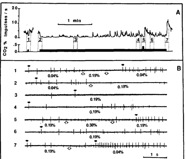
Figure 2A illustrates the cell response of one breath sensillum bearing a C02-excited receptor to increasing CO2 concentrations and human breath, all injected 25 cm from the outlet of the stimulus-delivery-tube and subse-quently diluted in the humidified air stream. Detailed sections of some of these responses are given in Fig. 3. As the stimulus onset was not sharp, the phasic portion of the response was not so pronounced and the maximum frequency occurred between 200 and 600 ms after a gradual increase in spike frequency. With the absence of a strong phasic part in the response, it was easier to categorize the spikes visually (Fig. 3). The firing rate of the spike numbered 1 (C02-excited receptor) induced by human breath diluted in clean air was quite similar to that induced by the equivalent CO2 concentration (Fig. 3). This suggests that breath contains nothing else capa-ble of modifying the response of this receptor. Moreover, none of the volatiles, listed in Materials and methods, elicited a response from this receptor.
C02 0.04 % ambient concentration C02 0.10 % ^ 1 *, Kt C02 0.68 %
t?k
^t^\\\\m^
M- •t ' " t "4t
C 0 2 2.16 % \\\\W\ \\\\^'f H l ' M l t l l l ) ^ A-f human breath 1:6 ^f-J j !• • |rtl .{I, (i H^fejH^^Mti^^W^^'^^'^'^i^M^^ *' ' • '•
B ^• H' H H " " • I M 'Nlll'HIIIHHlH'N'HHl
lamp OFF 24 0C ^ lamp ON 29 0Crhh M^h
1
' 4 *M'l' 'f
[ 1
I I*
1
!*
1,
-'Kh *|
M
H
MMH
' I
lamp ON 29 0C lamp OFF 24 0C
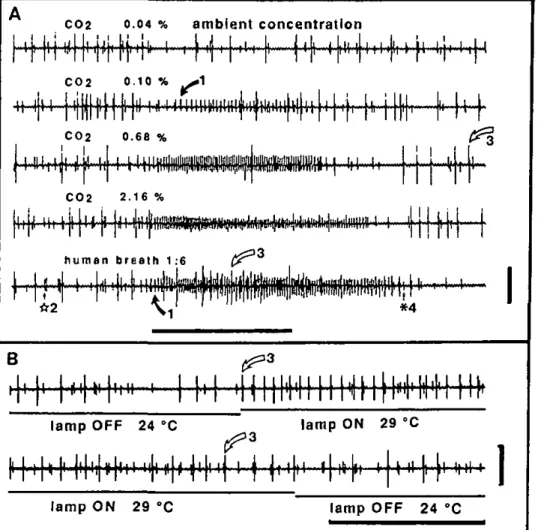
Fig. 2. A Representative responses of a capsular sensillum bearing a CO1
-excited receptor of a male A.
variega-tion (small biphasic spike I with bold arrow) to increasing CO2
concentra-tions and diluted human breath. Stimuli were injected into the stim-ulus-delivery-tube 25 cm from its out-let. so that the stimulus onset at the preparation was not very sharp, and cell frequencies increased gradually within the first 200 ms of the re-sponse. The preparation was main-tained in a humidified air stream at ca. 0.04% CO2. Four spike types are
indicated (see text and Fig. 3); num-bering of spike types same for Figs. 2 and 3. Spike 4 (asterisk) is a sulfide-receptor (according to Steullet and Guerin 1992). Spike 3 (white arrow) is a cell inhibited by increasing COj levels, but activated by breath. Its response seemed to be associated more with temperature changes as in-dicated by its response to turning off and on the microscope lamp (B). Thus, a T° increase activated it, whereas a corresponding T° decrease slightly diminished its activity. T° changes were measured with a therm-istor put at the place of the prepara-tion. In A, horizontal bar, 1 s stim-ulation; vertical bar, 1 mV. In B, horizontal bar, i s; vertical bar,
RESULTS - CHAPTER 3-1.
27
670 P. Steullet and P.M. Guerin: Perception or breath components by Ambtyomma I
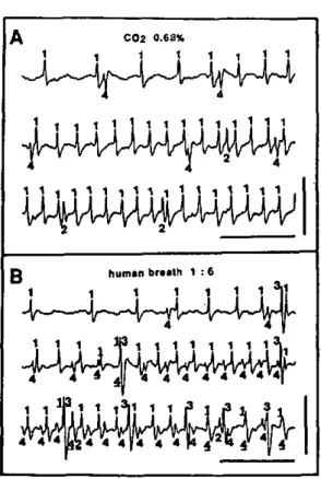
A C02 0.68%
' i i t 1 1 1 1
Ï 1 1 1 1 1 1 1 1 1. * .1 1 1 )
U^UiU^AlUUV
180
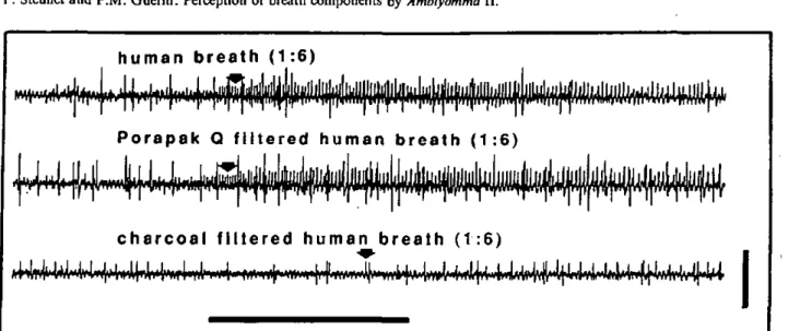
Fig. 3A, B. Detailed sections from the recordings illustrated in Fig. 2 to show how the 4 spike categories were discriminated visu-ally. A Recording from the first 750 ms of stimulation with 0.68% CO2, and B recording from the first 750 ms of stimulation with
human breath diluted 1:6 in air (ca. 0.6% CO2). Spike 1, activated
either by breath or CO2, is the COj-excited receptor. Spike 2 is
another cell with a persistently low firing rate. Spike 3 may be a thermoreceptor, responding to a slight increase in T0 during
stim-ulation with human breath. Spike 4. which changed its sign during stimulation with breath, is a sulfide-reccptor responding to H2S
(described in Steullet and Guerin 1992). Spike numbers underlined are overlapping events. Numbering of spike types as in Fig. 2. Horizontal bars 100 ms; vertical bars 1 mV
o.I ! CO2 concentration %
10
Fig. 4. Dose-response curve of the C02-excited receptor of male
A. variegatum established from the first 160 ms of the response
(phasic part). Preparations were maintained in a humidified air stream at ca. 0.04% CO2 into which CO2 stimuli were injected 3 cm
from the outlet of the tube to the preparation. Data (mean ± SD) have been obtained with 4 C02-excited receptors, all from different
males. Abscissa: estimated concentrations of CO2 arriving at
preparations
Figure 4 shows the dose-response relation established with C02-excited receptors from 4 different males which were stimulated with increasing CO2 concentrations in-jected 3 cm from the outlet of the stimulus-delivery-tube
(sharp stimulus onset). The response magnitude was de-termined from the first 160 ms of stimulation (phasic portion). The possibility of overlapping spikes was also considered in the determination of the response intensity. The C02-excited receptor responded to a concentration range covering some 2 to 3 log orders of magnitude. At ambient concentration of ca. 0.04%, activity was weak at a mean of 4.4 impulses/s, and gradually increased with higher concentrations up to about 140 impulses/s for 5% CO2. The relation between the CO2 dose and the tonic
Fig. SA-C. Amblyomma variegatum C02-excited receptor response
un-der continuous and repetitive 1 s stimulation: A 0.7%, B 1.2%, and C 1.8% CO2. Horizontal bars :
stim-ulus duration. The preparation was maintained in a humidified air stream at 0.04% CO2 into which
stimuli were injected. These re-sponse profiles were obtained by frequency to voltage conversion of the AC signal after sorting spikes of the C02-cxcitcd receptor from
others with a window discriminator (see Materials and methods). Re-sponses were reproducible