Faculté de génie
Département de génie mécanique
Caractérisation d’échaffaudages explantés en
β -TCP après résorption in-vivo en utilisant
des techniques d’analyse d’images raffinées
corrélées avec de la micro tomodensitométrie
à rayons-X
Accurate characterization of in vivo bone deposition and β -TCP
scaffold resorption using X-ray micro-computed tomography and
image processing
Thèse de doctorat
Specialité : génie mécanique
Ahmed SWEEDY
Jury: Gamal BAROUD (directeur)
François GITZHOFER (rapporteur)
Marc BOHNER
Stephan BECKER
Peter JARZEM
T he reason of what I become today.
To my sons, Omar and Seif,
T he source of joy and happiness in my
life.
To my dear wife,
L’utilisation de substituts osseux synthétiques est de plus en plus courante dans les procédés de réparation osseuse. Les biomatériaux de phosphate de calcium (CaP) sont les substituts dont la composition chimique se rapproche le plus de celle des os. Outre leur composition chimique, les propriétés géométriques des substituts osseux et, partant, leur macro et microporosité, ont une incidence sur leur efficacité et leur performance in vivo. Un grand nombre d’études se sont penchées sur le rôle des macro et microstructures des substituts dans le processus de guérison des os. Cependant, le rôle que la structure des substituts joue dans le processus de guérison reste à démontrer et est encore nébuleux en raison, entre autres, de la quasi inexistence de méthodes de caractérisation précises des phases des biopsies d’explants.
La présente thèse comportait par conséquent deux objectifs généraux. Le premier consistait à élaborer, à appliquer et à valider des méthodes novatrices de traitement de l’image utilisant des ensembles de données de microtomographie par rayons X après implantation afin de réaliser avec précision la caractérisation des phases des biopsies d’explants de substituts osseux de mo-dèles in vivo. Une importante difficulté de l’analyse des ensembles de données de microtomo-graphie par rayons X après implantation provient du fait que les résidus osseux et céramiques des images microtomographiques présentent une densité similaire ; il devient par conséquent ardu de différencier les résidus céramiques du tissu osseux dans le matériel prélevé par biopsie. Le second objectif, tel que détaillé ci-dessous, concerne une méthode pour analyser avec précision le dépôt osseux microscopique dans les parois des substituts céramiques.
Ainsi, le premier objectif impliquait la combinaison d’algorithmes de traitement d’images pour procéder à une étude précise des phases relevées dans les biopsies d’explants, et ainsi mieux comprendre le lien qui existe entre le processus de guérison et la structure du substitut. Plus précisément, l’alignement géométrique en 3D du substitut avant implantation sur les résidus du substitut a fourni des données sur la densité des particules osseuses par rapport à celle des résidus céramiques ; cela a donc permis de différencier avec encore plus de précision les phases relevées de la biopsie. Les résultats algorithmiques ont été entièrement validés au moyen de la théorie des matrices similaires et de la comparaison des résultats algorithmiques de cinq images choisies au hasard comprenant au total 556 800 pixels avec ceux obtenus manuellement par un scientifique expert en traitement de l’image. La validation a fait ressortir une concordance à 94 pour cent. Les clichés histologiques ont en outre confirmé les résultats de validation. Le nouvel algorithme en 3D permet donc d’analyser globalement et localement des effets macroscopiques comme la néoformation osseuse et la résorption céramique du processus de guérison. Globalement, ces effets sont analysés pour tout le substitut ; localement, ces effets peuvent être analysés pour chacune des distributions de pores du substitut. Cette approche novatrice aide ainsi à intégrer la conception structurelle du substitut osseux au processus de guérison. Les méthodes et les résultats relatifs à cet objectif sont expliqués plus en détail au chapitre 3.
Le second objectif de la présente thèse impliquait l’élaboration d’une nouvelle catégorie d’al-gorithmes servant à analyser avec précision des effets microscopiques comme la néoformation osseuse dans les pores microscopiques du substitut céramique, permettant ainsi l’étude des effets
microscopiques du processus de guérison. Plus particulièrement, les structures matérielles ob-servées dans les images à haute définition en 2 D de l’histologie et des techniques MEB étaient alignées géométriquement sur la structure en 3D de l’ensemble des données de microtomogra-phie par rayons X du même substitut avant et après implantation. Par conséquent, les ensembles de données de microtomographie par rayons X en 3D avant implantation ont été utilisés pour définir la référence géométrique de la céramique qui se résorbe, ce qui par conséquent a per-mis d’effectuer une analyse précise des phases matérielles des clichés à haute résolution en 2D des coupes d’évaluation histologiques. Plus précisément, une fois que les images produites au moyen des techniques d’imagerie multimodale ont été combinées et alignées, l’information cou-leur provenant de l’histologie et la vacou-leur de gris obtenue au moyen des images MEB ont été utilisées pour analyser les images histologiques ; celles-ci possédaient une résolution moyenne de 1,2 microns, ce qui a rendu possible l’étude des effets microscopiques à l’aide de clichés histologiques en 2D. Les méthodes et les résultats relatifs au second objectif sont au chapitre 4. Toujours au regard du second objectif, les nouveaux algorithmes ont servi à l’analyse des effets microscopiques et macroscopiques pour deux groupes de substituts β -TCP dont la taille des pores s’accroissait progressivement (diamètre de pore moyen = 510 et 1220 microns), implan-tés dans un modèle ovin pendant 6 semaines. Trois échantillons prélevés dans chaque groupe ont servi à étudier la néoformation osseuse et la résorption céramique. Des taux très élevés de néoformation osseuse et de résorption céramique ont été mesurés chez le groupe de substituts dont la taille des pores était la plus petite. Outre le taux plus élevé de néoformation osseuse, le groupe dont la taille des pores était plus petite présentait spécifiquement un important effet de surface ; en effet, le ratio de conversion de la surface de céramique résorbée en tissu osseux était beaucoup plus élevé que chez les deux autres groupes. Des études plus approfondies devront être menées afin d’analyser les effets de surface et les interactions entre le substitut osseux et le processus de guérison osseuse.
Les méthodes et les algorithmes connexes de la présente thèse ont fourni un moyen novateur et original d’évaluer les effets microscopiques et macroscopiques du processus de guérison. Tout comme d’autres méthodes, les nouvelles méthodes mises au point sont limitées. Celles-ci, exigeantes en termes de calculs, ont nécessité la présence d’un nombre suffisant d’éléments géométriques dans le substitut explanté afin d’aligner ces éléments sur ceux de la structure avant implantation.
Mots-clés : analyze multimodal, Dépôt osseux, enregistrement d’image, échafaudage ré-sorption, greffe osseuse, microtomographie, phosphate tricalcique, segmentation d’image
Synthetic bone substitutes are being increasingly used in bone repair procedures. Calcium-phosphate (CaP) substitute biomaterials have the closest chemical composition to bone. Besides the chemical composition, the geometrical properties and hence the macro and micro-porosity of the bone substitutes affect their in vivo effectiveness and performance. A large number of studies have investigated the role of the substitute’s macro- and micro-structure in the bone healing process. The role that the substitute’s structure plays in the healing process, however, remains debatable and unclear because of, among other reasons, the lack of accurate characterization methods of the phases in the explanted biopsies.
This thesis accordingly had two overall objectives. The first was to develop, implement and val-idate novel image-processing methods for using the post-implantation mCT datasets to help ac-curately characterize the phases in bone substitute biopsies explanted from in vivo models. One important difficulty in analyzing the post-implantation mCT datasets is that bone and ceramic remnants in the mCT images appear to have similar density; differentiating between ceramic remnants and bone tissue in the biopsies therefore becomes difficult. The second objective, as detailed below, related to a method to accurately analyze the microscopic bone deposition into the bone substitute’s ceramic wall.
Accordingly, undertaking the first objective involved developing image-based algorithms to ac-curately study the phases found in the explanted biopsies and thus better understand the rela-tionship of the healing process and the substitute’s structure. Specifically, the 3D geometric alignment of the pre-implantation substitute with the substitute’s remnants provided access to data on the density characteristics of the bone versus the ceramic remnants; this accordingly al-lowed a more accurate differentiation of the phases found in the biopsy. The algorithmic results were thoroughly validated, using the similarity matrix theory and by comparing the algorithmic results of five randomly selected images — comprising a total of 556,800 pixels — with those obtained by a skilled image-processing scientist. The validation showed a 94-percent agree-ment. Histology photographs further confirmed the validation results. Accordingly, the new 3D algorithm helps to globally and locally analyze macroscopic effects such as bone deposition and ceramic resorption of the healing process. Globally, these effects are analyzed for the entire substitute, and locally, these effects can be studied for each pore of the substitute. The novel approach thus helps relate the bone substitute’s structural design to the healing process. The methods and the results related to this objective are detailed in Chapter 3.
The second objective of the present thesis entailed developing a new class of algorithms to ac-curately analyze the microscopic effects such as the bone deposition in the microscopic porosity of the ceramic substitute, thereby allowing the study of microscopic effects of the healing pro-cess. Specifically, the material structures seen in the high-resolution 2D images of the histology and SEM techniques were geometrically aligned with the 3D structure in the mCT dataset of the same substitute before and after implantation. Accordingly, the 3D pre-implantation mCT datasets were used to define the geometric reference of the resorbing ceramic, and therefore allowed an accurate analysis to be made of the material phases in the high-resolution 2D
tographs of the histology evaluation cuts. Specifically, once the images from the multimodal imaging techniques were combined and aligned, the color information from histology and the grey value information from SEM images were used to analyze the histology images; the latter had an average resolution of 1.2 microns and this made studying the microscopic effects using the 2D histology photographs possible. The methods and the results related to the second ob-jective are in Chapter 4. Still in line with the second obob-jective, the new algorithms were used to analyze the microscopic and macroscopic effects of two β -TCP substitute groups of incre-mentally increasing pore size (mean pore diameter = 510 and 1220 microns), implanted in an ovine model for 6 weeks. Three samples were selected per group to investigate the microscopic and macroscopic bone deposition and ceramic resorption. Significantly higher bone deposition and ceramic resorption were measured in the substitute group with the smaller pore size. In addition to the higher micro-bone deposition, the smaller pore-size group specifically featured an important surface effect; namely, the conversion ratio of resorbed surface ceramic into bone tissue was significantly higher compared to the two other groups. Further studies are still re-quired to investigate the surface effects and the related interactions between the bone-substitute design and the bone-healing process.
The present thesis’ methods and related algorithms provided novel and original means to evalu-ate microscopic and macroscopic effects of the healing process. Like other methods, the newly developed ones have limitations. The methods are computationally demanding, and required that a sufficient number of geometric features be present in the explanted substitute so as to align these features with those of the pre-implantation structure.
Keywords: Bone deposition, bone substitutes, image segmentation, image registration, micro-computed tomography, multimodal analysis, scaffold resorption, tricalcium phosphate
First of all, I would like to express my sincere gratitude and appreciation to Professor Gamal BAROUD, my Ph.D. supervising director, for his unconditional support throughout my Ph.D. studies and for his patience, motivation, enthusiasm, and his immense knowledge which he so generously made available to me. His guidance and openness were a guiding light throughout all of my research and article and thesis writing. I feel so fortunate to have had him as my Supervising Director. Not only was he a great supervisor, he was an inspiring mentor who enlightened me for my long-term career.
Beside my supervisor, I am deeply thankful and grateful to Professor Marc BOHNER for the many fruitful discussions we have had together and from which I benefited greatly because of his great knowledge in the field of bone augmentation and repair. It is an honor to have had the opportunity to work with him. He was available to discuss and provide me with very valuable and critical comments that helped bring me to a higher scientific level.
I very much want to thank Professor François GITZHOFER for his availability to discuss and his very useful comments. He was unconditionally supportive and encouraging when it was mostly needed. I appreciate him acting as reporter and jury member for my Ph.D. thesis. I would also like to thank Professor Stephan BECKER and Professor Peter JARZEM, who are pioneers in the field of bone substitute research, for accepting to be members of my Ph.D. Evaluation Committee. It is a great honor for me to have them act in this role.
I would like to thank the Natural Sciences and Engineering Research Council of Canada (NSERC) and the Canada Research Chair Program (CRC) for supporting my Ph.D. research. I am par-ticularly very grateful to the Robert Mathys Foundation (RMS) for the support received for my thesis and for continuously providing me with the unique experimental data. I am also grateful for Professor Harry van LENTHE for helping provide essential datasets for my Ph.D. research. My gratitude extends to Brian DRISCOLL, Denis IMBEAULT and Jason GAUTHIER for pro-viding technical support for my research. Many thanks also to all the administrative members of the Mechanical Engineering Department’s office, especially Sylvie PERRON. I thank my former colleague in the Biomechanics Laboratory, Dr. Juan ZHANG, for the discussions and encouragement.
Last but not least, I want to express my heartfelt gratitude to my family for having lovingly and unconditionally supported me throughout my study years. I would like to thank my parents for the unconditional love, support and encouragement throughout my entire life. A special thank you goes to my in-laws for their support and encouragement. Thank you to my loving wife for supporting and encouraging me unconditionally throughout the length of my Ph.D. studies and to my sons Omar and Seifeldin for their patience.
1 INTRODUCTION 1
1.1 Background and significance . . . 2
1.2 Motivation . . . 3
1.3 Problem statement . . . 4
1.4 Objectives of the PhD research project . . . 5
1.5 Organization of the PhD thesis . . . 6
2 LITERATURE REVIEW AND METHODS 9 2.1 Bone defects and healing process . . . 10
2.2 Bone grafting and substitutes . . . 10
2.3 Synthetic bone substitutes . . . 11
2.4 Bone deposition in calcium phosphate ceramics . . . 12
2.4.1 Kinetics of bone deposition inside scaffolds . . . 12
2.4.2 Bone deposition penetration level . . . 17
2.4.3 Influence of scaffold geometrical properties on bone deposition at macro-porous level . . . 17
2.4.4 Influence of scaffold geometrical properties on bone deposition at mi-croporous level . . . 20
2.4.5 Tissue reaction to bone scaffold . . . 21
2.4.6 Biomechanical evaluation of bone scaffold postimplantation . . . 21
2.5 Resorption of calcium phosphate ceramic substitutes in bone . . . 22
2.6 Characterization of calcium phosphate bone substitutes . . . 24
2.6.1 Microcomputed tomography . . . 25
2.6.2 Histology evaluation . . . 26
2.7 Image processing methods for characterizing calcium phosphate bone substitutes 27 2.7.1 Image enhancement . . . 27
2.7.2 Image segmentation methods . . . 31
2.7.3 Active contours . . . 35
2.7.4 Image registration methods . . . 38
2.7.5 Morphometrical characterization parameters and methods . . . 42
3 A NOVEL METHOD FOR SEGMENTING AND ALIGNING THE PRE- AND POST-IMPLANTATION SCAFFOLDS OF RESORBABLE CALCIUM-PHOSPHATE BONE SUBSTITUTES 45 3.1 Abstract . . . 47
3.2 Introduction . . . 47
3.3 Materials and methods . . . 49
3.3.1 Scaffold fabrication, in-vivo model, and mCT scanning . . . 49
3.3.2 Segmentation and alignment algorithm . . . 50
3.3.3 Validation of the segmentation algorithm . . . 56
3.4 Results . . . 57
3.5 Discussion . . . 60
3.6 Conclusion . . . 64
3.7 References . . . 64
4 MULTIMODAL ANALYSIS OF IN VIVO RESORBABLE CaP BONE SUBSTI-TUTES BY COMBINING HISTOLOGY, SEM AND MICROCOMPUTED TO-MOGRAPHY DATA 69 4.1 Abstract . . . 71
4.2 Introduction . . . 71
4.3 Materials and methods . . . 74
4.3.1 Scaffolds, animals and surgical procedures . . . 74
4.3.2 Micro-computed tomography, TBH and SEM imaging . . . 74
4.3.3 The segmentation and alignment algorithms . . . 75
4.3.4 TBH histomorphometry . . . 78 4.3.5 Statistics . . . 78 4.4 Results . . . 78 4.5 Discussion . . . 86 4.6 Conclusion . . . 91 4.7 References . . . 91 5 CONCLUSION 97 LIST OF REFERENCES 103
2.1 Structure of the discrete computational scheme [Perona and Malik, 1990] . . . 30
2.2 g(·)functions versus image gradient . . . 32
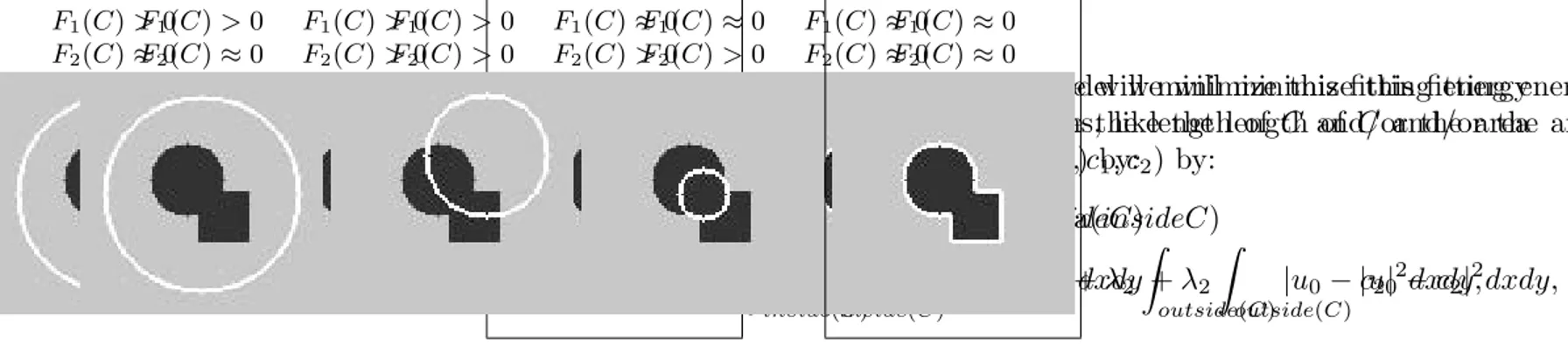
2.3 Illustration of different curve positions [Chan and Vese, 2001] . . . 37
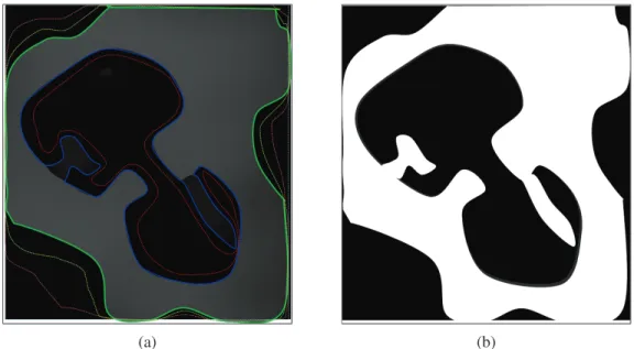
2.4 An illustration of active contours convergence on a schematic of post-implantation mCT of β -TCP scaffold . . . 38
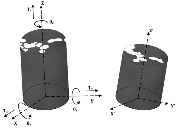
2.5 A schematic example of pre- and post-implantation mCT datasets of resorbable CaP scaffold . . . 41
2.6 An example of SURF features detection for schematics of β -TCP mCT pre- and post-implantation . . . 42
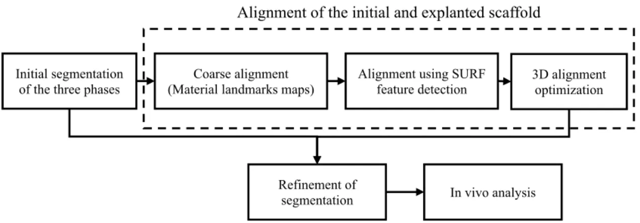
3.1 Block diagram of the presented algorithm . . . 51
3.2 The sigmoidal weighting functions of the grey-value and distance transitions . . 52
3.3 Schematic of the initial and explanted scaffold . . . 53
3.4 An exemplary alignment and segmentation results . . . 58
3.5 Volume fraction / z-direction projection maps of ceramics . . . 59
3.6 Refined segmentation characteristic histograms, intensity image and result for one exemplary section . . . 60
3.7 Comparing the results of the present and Otsu’s method to the histology section and to the corresponding same mCT slice . . . 61
3.8 Validation results of one randomly selected quarter . . . 62
4.1 An exemplary multimodal data from one samples . . . 77
4.2 Segmentation result of the histology section from one sample . . . 80
4.3 Implantation area subdivided into eight elliptical rings from center toward pe-ripheries. . . 81
4.4 Mean and standard error of phases surface area fractions in the total implantation site . . . 82
4.5 Regional surface area fraction of different phases and different pore sizes . . . 83
4.6 Mean and standard error of normalized surface area fractions of ceramic related phases . . . 86
4.7 Mean and standard deviation of thickness of each phase for different pore sizes in the total implantation site . . . 87
4.8 Mean and standard error of the thickness of different phases and different pore sizes in rings R1:R8 . . . 88
4.9 High resolution BSE-SEM image of a histology section. . . 89
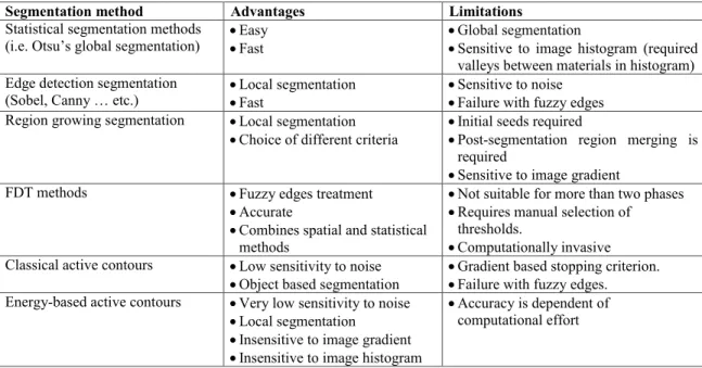
2.1 Summarized comparison between common segmentation methods applied to mCT images . . . 35
3.1 Similarity matrix . . . 61
4.1 Surface area fractions of phases in the total defect and regionally subdivided into rings . . . 84 4.2 Mean and standard deviation of thickness of phases in the total implantation site. 85
Acronym Definition
BCP Biphasic calcium phosphate
BMO Bone marrow-derived osteoprogenitor cells BMPs Bone morphogenetic proteins
BMSC Bone marrow stromal cells
BV Bone volume
CaP Calcium phosphate
DSC Dice similarity coefficient EED Edge enhancing diffusion FDT Fuzzy distance transform
HA Hydroxyapatite
mCT Microcomputed tomography
MSC mesenchymal stem cells
NBV New bone volume fraction PMMA Polymethyl methacrylate POF Pore occupation fraction SBS Synthetic bone substitutes SEM Scanning electron microscopy SIFT scale invariant feature transform SNR Signal to noise ratio
SURF speed-up robust features SSD Sum of squared differences TBH Toluidine blue stained histology
TCP Tricalcium phosphate
THB Threshold of bone
THC Threshold of ceramic remnants
TV Total volume
INTRODUCTION
1.1
Background and significance
In non-critical size defect, the healing process takes place in three consecutive phases; phase of inflammation, phase of osteogenic repair tissue and phase of remodelling [Nather, 2005]. De-spite the three phases are common in bone tissue repair, critical size defect requires orthopaedic intervention, such as bone grafting, to assist the bone to complete the healing process [Spicer et al., 2012]. Such defects are commonly caused by trauma, degradation or diseases usually as-sociated with aging. Since the Dutch surgeon Job van Meekren had made the first documented attempt at tissue engineering by filling bony defect in a soldier’s cranium with a piece of dog skull in 1670 [Nather, 2005], skeletal defects have been replaced by bone grafts. Bone grafting became an accepted technique since the early 20th century. Despite autologous bone grafting re-mains the gold standard in bone transplantation, they are associated with long-term side effects including donor site numbness and pain [Schwartz et al., 2009; Silber et al., 2003]. This re-sulted in decreased ability to work in 10% of the patients, difficulty to walk in 16%, recreational activities limitation in 18% and 19% of these patients suffered pain that affected their ability to perform household chores [Schwartz et al., 2009]. Another similar technique to autograft is allograft (graft harvested from another donor or subject). While allografting eliminates the prob-lems associated with harvest site, it presents other complications associated with transplantation between different persons including increased risk of infection by disease transmission [Nather, 2005; Yaszemski et al., 1996] and immunological reaction that may require the use of immuno-suppressant drugs [LeGeros, 2008; Lemons et al., 1988]. Therefore, the need of synthetic bone substitutes (SBS) emerged as a replacement of autografs and allografts.
By the development of better understanding of the immune system during the 1960s and 1970s, the first generation of synthetic materials for use inside the human body was developed [Hench and Polak, 2002; Nather, 2005]. In terms of bone substitutes, Hench and his colleagues dis-covered the bioactive glass by the end of 1960s and it was followed by two other generations reaching the third-generation bioactive glasses, composites, hybrid materials and macroporous foams that are designed to activate genes that stimulate regeneration of living tissues [Hench, 2006; Hench and Polak, 2002; Will et al., 2011]. The calcium-phosphate (CaP) biomaterial, be-cause of its similar chemical composition to bone, was also considered for treating bone defects as early as 1920, yet the CaP related research surged in the 1970s and CaP materials were used in dental and orthopedic applications [Habraken et al., 2016]. CaP materials showed some attrac-tive features as a bone substitute material including biocompatibility, bioactivity and osteocon-ductivity, in addition to resorbability in the case of β -tricalcium phosphate (β -TCP). Since then, many studies have been conducted to optimize the bone substitutes in terms of material com-position and geometry to achieve the optimum scaffold design that promotes bone decom-position
and scaffold resorption in an integrated pattern that allows the bone substitute to be completely replaced by bone while supports the load bearing functionality of the skeletal structure.
Due to ethical issues, the answer to the question of otimum scaffold design usually involves conducting in vivo experiments to investigate the influence of the substitutes design on their performance and the biological processes taking place inside the defect site. The importance of in vivo characterization of tissue regeneration in bioscaffolds arises from the importance of better understanding of the bone healing process [Gauthier et al., 2005; Papadimitropoulos et al., 2007; van Lenthe et al., 2007; von Doernberg et al., 2006]. Failure to achieve enough tissue regeneration for functional support leads to mechanical failure though scaffold properties are adequate for temporary load bearing [Feng et al., 2012; Hollister et al., 2005].
In vivo studies are always associated with large image data analysis to characterize different phases in the explanted samples or biopsies. Such data could be in the form of 2D images (i.e. histology and SEM) or 3D images (i.e. micro-computed tomography). The accurate evaluation of these data is a necessary step in the process of data analysis, leading to accurate conclusions and better understanding of the biological processes in bone repair using SBS.
1.2
Motivation
With annual estimation of two million patients are subjected to the bone grafting procedure for repairing critical size bone defects [Bohner, 2010], bone is ranked as the second transplanted and replaced material in human body [Zorzi, 2012]. Hence, scientific attention to bone grafts development arises to find the optimum scaffold for assisting the healing process of defects that are commonly caused by trauma, degradation or diseases which are usually associated with aging [Nather, 2005; Zorzi, 2012]. Bone graft substitutes market is not only one of the high demanding markets with over one billion dollars revenue but it has also one of the high annual market growth rate of about 15% [Bohner, 2010]. In addition to the demanding market, the socioeconomic impact of the critical size bone defects induces a high demand for research and development of better understanding of the properties of synthetic bone substitutes and their influence on bone regeneration as well as the healing process being exerted in order to find the optimum scaffold material and micro-structural design. The optimum bone scaffold is the scaffold which is resorbed completely and synchronously replaced by bone in a pattern that allows the scaffold of being replaced without affecting its mechanical support functions [Carlier et al., 2011].
Next to the material composition, the geometrical design and structure of the bone substitutes was found to influence the bone deposition and the substitute resorption [Gauthier et al., 2005;
Papadimitropoulos et al., 2007; van Lenthe et al., 2007; von Doernberg et al., 2006]. For in-stance, there is an agreement in literature on introducing macro-porosity (pore size > 50 microns) to bone substitutes to help cellular migration and vascularization [LeGeros, 2008; Lemons et al., 1988]. However, the optimum pore size that yields the best performance of bone substitutes is still under debate [von Doernberg et al., 2006].
Furthermore, recent findings showed that introducing microporosity (pore size < 50 microns) to non-resorbable hydroxyapatite (HA) bone substitutes improves their performance and results in better osseointegration [Polak et al., 2011; Scaglione et al., 2012]. This was explained by Scaglione et al. [Scaglione et al., 2012] as the microporosity in non-resorbable HA substitutes acts as the structure to which new deposited collagen fibers attaches. However, the role of mi-croporosity in resorbable β -TCP bone substitutes is still unclear and not sufficiently understood.
The applied appraoch to understand the biological processes in the resorbable β -TCP substitutes is by characterizing the substitute structure before its implantation in terms of the morphometri-cal parameters (e.g. porosity, pore size, wall thickness, etc), then characterize the existing phases found after in vivo implantation, and relate these findings to the pre-implantation morphometri-cal parameters [Bashoor-Zadeh et al., 2010; Lapczyna et al., 2014; von Doernberg et al., 2006]. This approach however did not allow to accurately characterize the ceramic resorption nor it allowed to study the 3D biological processes in reference to the pre-implantation geometric structure. Until now, a method that combines the pre-implantation substitute geometry to the post-implantation biopsy to localize resorbed material and links it to bone deposition to enable analysis of the interactions between bone deposition and substitute resorption at local level does not exist.
1.3
Problem statement
Several imaging techniques, providing different classes of images and leading to insight infor-mation about biological interactions taking place inside the resorbable implant, exist to better understand the bone repair process. These techniques has been used separately by the scientists to examine different hypotheses as shown in the detailed literature review presented in chap-ter 2. A systematic methodological integration of these classes of images is still required for an objective systematic evaluation of the scientific effects related to the osseointegration of the SBS. Specifically, this methodological integration can lead to more accurate segmentation and characterization of the phases in the SBS explanted biopsies and inherently to more accurate conclusions related to the bone repair process.
Microcomputed tomography, an image-based technique, allows examining the microstructure of radiopaque bone substitutes (i.e. bone substitutes that are opaque to the X-ray either because of their material composition or an added radiopaque agent) at the micro-scale and accordingly it has been recently deployed in examining the structure of porous substitutes used in the bone repair process. In vivo animal models, e.g. rabbits and sheep models, are used to examine the effectiveness of bone substitutes in the bone repair process. Specifically, these substitutes are implanted in critical-size bone defects and explanted systematically after predetermined implan-tation periods. The explanted biopsies are then cleaned from soft tissue and often embedded in polymeric resin and sectioned carefully for analysis such as histology, SEM and energy-dispersive X-ray spectroscopy (EDX). These techniques and methods have been very useful over the last century. However, they are labour intensive, often destructive and have limited accuracy in terms of characterizing the material phases and resorption in the biopsies.
This thesis attempts to introduce new methods and image based algorithms, combining images of different modalities and techniques including histology, SEM and mCT, to analyze post-implantation biopsies. Specifically, aligning the reference structure of the pre-post-implantation with the biopsy ceramic remnants structure provided access to accurate evaluation of the phases in the biopsy. Also, advanced multimodal analysis, combining histology, SEM and pre-implantation mCT data images provided access to a detailed description of the resorption process, micro- and macro-scales effects of the healing process.
1.4
Objectives of the PhD research project
The general objectives of this thesis is to formulate, develop, implement, verify and validate new imaging-based methods to study the phases found in biopsies explanted from experimental models used to study the effectiveness of porous bone substitutes for the repair of critical bone defects. Broadly, the thesis has two well-defined general objectives.
The first general objective of this thesis, related to developing a method to understand the biolog-ical processes at the 3D macroscale (i.e. structures and features of size larger than 50 microns), is subdivided into two sub-objectives. The first sub-objectives is to develop and implement a segmentation method to accurately isolate the three phases present of the explanted ceramic bone substitute scaffolds, i.e. bone, ceramic, and soft tissue. The second sub-objectives is to localize the resorption in the post-implantation mCT of the explanted biopsies of resorbing CaP bone substitutes.
The second general objective, related to developing a method to understand the microscopic ef-fects at the microscale (i.e. structures and features of size less than 50 microns), is subdivided
into two sub-objectives. The first sub-objective is to introduce a new method to characterize the material phases found in toluidine blue histology (TBH) images of explanted biopsies of resorbable bone substitutes. The second sub-objective is to demonstrate the utility of the devel-oped method by analyzing two groups of β -TCP substitutes of two different macropore sizes, explanted from an ovine model after implantation for six weeks.
These two general objectives are the subjects of two scientific papers included in the thesis and submitted for publication.
1.5
Organization of the PhD thesis
This thesis comprises five chapters, references, lists of acronyms, figures and tables. The con-tents of the chapters are described as follows.
Chapter 2 "literature review and methods", presents a review of the previous work related to the optimization of synthetic bone substitutes and the characterization of the bone substitute biopsies. In addition, this chapters reviews the methods used in the characterization of bone substitute biopsies, including imaging techniques (e.g. histology, SEM and mCT) as well as available image processing methods (e.g. denoising, segmentation, registration, etc). Further details of the selected methods (e.g. active contours) were reviewed and explained.
Chapter 3: This chapter presents the first article addressing the first general objective of this thesis. The article is entitled "A novel method for segmenting and aligning the pre- and post-implantation scaffolds of resorbable calcium-phosphate bone substitutes". A brief literature review, challenges, objectives and the novel method are described. In addition, an exemplary application of the method to one sample and the validation results of the methods are shown. The methods and results are discussed in the discussion section. Finally, the conclusions and references sections of this article are included in this chapter.
Chapter 4: This chapter presents the second article addressing the second general objective of this thesis. The article is entitled "Multimodal analysis of in vivo resorbable CaP bone substitute by combining histology, SEM and Microcomputed tomography data". The newly developed image-processing methods, including the segmentation and the alignment methods, are described in details in the materials and methods section. The utility of the novel method was demonstrated by characterizing quantitatively six samples of two different macropore sizes without pairing, explanted from an ovine model after implantation for six weeks. The novel method and the findings of the characterization process are discussed in light of the literature
and previous work. Finally, the conclusions and references of this article are included in this chapter.
Chapter 5: This chapter includes the summary of the results, contributions, and conclusions of the thesis.
References: In the references, the bibliography used in non-article chapters and sections are included.
LITERATURE REVIEW AND METHODS
2.1
Bone defects and healing process
Bone in its two forms, cortical and trabecular, integrate together to satisfy several functions including reserve of minerals, blood production and mechanical functionalities [Weiner and Wagner, 1998; Zhao et al., 2012; Zorzi, 2012]. Both types of blood cells, red and white, are produced by the hematopoietic marrow located in trabecular bone [Zhao et al., 2012]. Mechan-ical functionalities of bone include protection of vital organs such as brain, providing body with rigid structure and movement. Therefore, the last function is the main promotion for bone graft-ing to assist the healgraft-ing process to take place at the defect site [Bohner, 2010; LeGeros, 2008; Weiner and Wagner, 1998]. Based on cause, bone defects can be classified into traumatic or pathological which could be a result of osteoporosis, cyst or cancer [Nather, 2005; Spicer et al., 2012]. Critical size defect is defined as the tissue defect that will not completely heal over the natural lifetime of human or animal [Spicer et al., 2012].
2.2
Bone grafting and substitutes
Bone grafting could be defined as a surgical procedure for replacing missing bone in bone de-fects that can pose a significant health risk or fail to heal properly by itself [Habraken et al., 2016]. Bone grafts may be used to fill critical-size defects [Bohner, 2010; LeGeros, 2008]. Bone grafting is classified into autograft, allograft, xenografting and synthetic bone grafting [Bohner, 2010].
Autograft procedures involves moving bone from one site to another site within the same patient [Bohner, 2010; Nather, 2005]. The need for bone into grafting procedures triggered the formu-lation of bone banking and resulted in technique involves implanting bone from other persons, which is allografting [Nather, 2005; Zorzi, 2012]. The high demanding and low availability of living bone donation triggered the searching for new source for grafting, the synthetic bone grafts [Bohner, 2010]. Moreover, autograft and allograft are associated with problems includ-ing pain and lack of donation sites in the case of autograft and disease transmission in the of allograft procedures [Bohner, 2010; Nather, 2005].
Due to the lack of bone resources required for allograft and problems resulting from autograft and allograft procedures, there is an increase in the use of synthetic substitutes including poly-mers synthetic ceramics such as bioactive glasses, calcium sulfates, calcium phosphates, calcium carbonate [Bohner, 2010; LeGeros, 2008]. Based on the ability to stimulate bone growth, syn-thetic substitutes could be classified into bioinert such as metal substitutes-the materials that do not stimulate bone and consequently do not bond with bone and results in weak interface
between bone and material, and bioactive such as natural polymers and calcium phosphate, the material that stimulate bone tissue formation and results in strong bone material bond and inter-face [LeGeros, 2008; Parikh, 2002].
2.3
Synthetic bone substitutes
Synthetic bone grafts includes polymers, bioactive glasses, ceramics and metals [Nather, 2005; Zorzi, 2012]. However, synthetic bone grafts should present biological properties that enable implant integration and healing of the defect as well as mechanical properties for structure sup-port of bone at defect [Barrère et al., 2006; LeGeros, 2008].
Synthetic polymers such as Polymethyl methacrylate (PMMA) have been widely used for treat-ing bone defects, augmentation and coattreat-ing of metal implants [Sta´nczyk and van Rietbergen, 2004; Zorzi, 2012]. Despite the long-term clinical experience and high immediate stability, PMMA is associated with polymerization heat which results in necrosis, pain and in some cases nerve injuries [Zorzi, 2012]. Hence, synthetic ceramics such as hydroxyapatite (HA) or trical-cium phosphate (TCP) are used for filling bone defects resulting from injuries [Gaasbeek et al., 2005] or diseases such as cysts or cancer metastases [Wang et al., 2013] as well as for oral implants pre-fixation augmentation [Zerbo et al., 2005].
Synthetic ceramics such as calcium phosphates (CaP) biomaterials has similar composition to bone minerals which are calcium phosphate in the form of carbonate apatite [Barrère et al., 2006; LeGeros, 2008; Nather, 2005]. It also exhibits other properties that enable bone growth including bioactivity, biodegradability, osteoconductivity and osteoinductivity [LeGeros, 2008; Podaropoulos et al., 2009; Scaglione et al., 2012; van der Pol et al., 2010].
Addition of osteoinductive properties to CaP materials enhancing their application to bone repair and regeneration; could be done by the incorporation of calcium scaffolds with growth factors such as bone morphogenetic proteins (BMPs) [LeGeros, 2008; Polak et al., 2011; Warren et al., 2004]. For the limitations of BMPs in clinical use, other methods for osteoinductivity addition are used such incorporation of bone marrow stromal cells (BMSC) [Komlev et al., 2006; Pa-padimitropoulos et al., 2007], mesenchymal stem cells (MSC) [Warren et al., 2004], platelet rich plasma (PRP) [Becker et al., 2008] and the design of material with the appropriate geomet-rical properties including microporosity, macroporosity and concavities that favours entrapment of circulating growth factors and osteoprogenitor cells inducing neo-bone formation [LeGeros, 2008; Polak et al., 2011].
Due to the clinical limitations of the BMPs including their carcinogenic effect and the high price [Becker et al., 2008; Laine et al., 2013], alternative solutions such as the PRP [Becker et al., 2008], BMSC [Komlev et al., 2006; Papadimitropoulos et al., 2007], MSC [Warren et al., 2004] emerged. Furthemore, the microporosity and the microstructuring of the ceramic stubti-tute [Polak et al., 2011] were demonstrated to improve the bone deposition and to accerlate the healing process. The type, density and pattern of the neo-bone formation could be controlled by the geometrical properties of the synthetic bone substitutes on macroscale [Feng et al., 2012; Scaglione et al., 2012] and on microscale [Barrère et al., 2006; Hing et al., 2005; Polak et al., 2011].
2.4
Bone deposition in calcium phosphate ceramics
2.4.1
Kinetics of bone deposition inside scaffolds
Many experiments has been executed in order to measure the rate of bone growth but most of them were done in animal model ranging from mice [Komlev et al., 2010, 2006; Mastrogia-como et al., 2006; Papadimitropoulos et al., 2007; Scaglione et al., 2012] rabbit [Feng et al., 2012; Gauthier et al., 2005; Lu et al., 2002, 1998; van Lenthe et al., 2007], sheep [van der Pol et al., 2010; von Doernberg et al., 2006] and dog models [Draenert et al., 2013]. It has been observed that rate of bone growth could be affected by some parameters including material type, implantation site, animal models and geometrical properties of the implant.
One of the parameters that influence the bone deposition rate is the animal model in which the experiment is executed. As it is well-known that the metabolism in small animals is much higher than in large animals, it can be noticed that the larger the animal the slower the bone deposition rate. For example, in sheep model β -TCP scaffolds experienced lower bone deposition rate (31.9± 8.0%,38.5 ± 9.8% and 40.5 ±12.3% at 2, 4 and 12 months respectively) [van der Pol et al., 2010]; in comparison with the higher bone regeneration observed in rabbit model (NBV = 35.00± 6.64% and 52.07 ± 7.53% at 12 and 24 weeks respectively in β -TCP) [Lu et al., 1998]. It is shown in the following a review of some studies made to evaluate the bone deposition in different animal models.
After implantation in immunodeficient mice model with scaffolds seeded with bone marrow-derived mesenchymal stem cells (MSC), newly formed bone volume was maximum in Bio-Oss ones, natural bone mineral derived scaffolds, (8.4 ± 0.5 %) followed by the biphasic Skelite scaffolds (synthetic calcium phosphate multiphase biomaterial containing 67% silicon stabilized tricalcium phosphate) after 8 weeks implantation in mice (NBV=7.1 ± 0.5 %), and the lowest
bone volume were observed in the pure HA Engipore scaffolds (NBV=5.2± 0.5 %) [Komlev et al., 2010]. In similar study in mice [Papadimitropoulos et al., 2007], mCT analysis of the postimplantation Si-TCP scaffolds seeded with MSC showed bone deposition increases from 2 (BV/TV = 5.5± 0.3%) to 4 months (BV/TV = 10.3 ± 0.5%) and remained constant in quantity but higher mineralization from 4 to 6 months (BV/TV = 10.2± 0.5%). Mineralization increase was observed by the shift in the new bone peak toward higher absorption rate in the mCT his-togram. Once more, the stoppage of bone deposition was explained by the author according to two hypotheses; poor vascularization and lack of mechanical load which is peculiar in immun-odeficient mice model. Therefore, they conducted new studies using ovine larger animal model to test the second hypothesis [Papadimitropoulos et al., 2007].
The MSC seeded HA and β -TCP scaffolds exhibited increase in the osteocalcin (OCN), which is main indicator to the osteoblastic activity, from first to eighth week postimplantation in the back of male Fischer rats; accompanied with increase in bone formation which was observed as early as first week in close contact with the vasculature [Warren et al., 2004]. Similar results were obtained in β -TCP seeded with bone marrow osteoprogenitor cells implanted in Fischer rats [Dong et al., 2002], osteocalcin was observed to increase from 10ng/ml at second weeks postimplantation to 70ng/ml at week 24 postimplantation. In agreement, bone formation was observed at 2 weeks postimplantation inβ -TCP/BMO scaffolds with osteogenic medium and amount of mature bone increase until 24 weeks [Dong et al., 2002]. Bone formation was close to the surface of the scaffolds without soft tissue formation. Moreover, roughness of the surface increased from week 2 to week 8, with existence of extracellular collagenous matrix as well as rough round osteoblastic cells at the surface. Blood vessels and multinucleated cells, which may contribute to the degradation, existed near the surface of the scaffold at week 24. Bone formed not only inside the scaffolds but also could be found at the outer surface of the scaffold.
In sake of understanding the orientation and pattern of deposited bone inside scaffolds [Scaglione et al., 2012], both HA and HA-Col with 80% porosity seeded with bone marrow cells was im-planted in mice. At one month postimplantation, almost all pores of the scaffolds were colo-nized with osteoprogenitor cells that had layered new tissue over the pores surface. Moreover, mature osteoblasts were lining the deposited bone matrix within pores of HA while cells were completely embedded in the woven bone matrix in HA-Col. At two-month postimplantation time point, the amount of the newly formed bone increased significantly inside the pores with elongated lining cells found covering the bone matrix. Moreover blood vessels were well rep-resented within the interconnected pores. At six months after implantation, bone inside HA scaffolds showed the typical structure of an ossicle with a marrow cavity filled with
hematopoi-etic cells, adipocytes and formation of functionally active vascular structures, surrounded by concentric lamellae of bone matrix.
Postimplantation in immunodefficient mice of two different geometry HA scaffolds [Mastro-giacomo et al., 2006]; spongy matrix and foaming, histological analysis exhibited neo-bone formation 2 weeks postimplantation in foaming scaffold whereas bone tissue did not exist in scaffold A at the same period despite BMSC density was higher in spongy matrix scaffold than in foaming ones. At 4 weeks postimplantation, neo-bone formation approached 35% of the total new tissue formed in scaffold B which is close to the value of 36% found at 8 weeks. On the other hand, neo-bone formation exhibited value of 22% of total new tissue formation in spongy matrix scaffold at 4 weeks and this value increase at 8 weeks postimplantation in vivo to be 37%. The faster bone formation rate in foaming scaffold proposes that neo-bone formation is affected by factors related to the implant (e.g. surface area available for bone growth and resorbability of the material) and others related to physiological equilibrium. The authors suggest that the coex-istence of bone matrix, blood vessels, marrow and undifferentiated mesenchymal cells in steric but dynamic equilibrium would result in reaching neo-bone formation saturation level. This saturation level could be exceeded using resorbable material, i.e. β -TCP, which allows bone to grow in a manner proportional to resorption of the material [Mastrogiacomo et al., 2006].
New bone formation was higher in β -TCP than HA scaffolds with better integration into host bone and higher penetration levels [Lu et al., 1998]. Moreover, they have observed higher bone deposition at cortical site than in trabecular site. Postimplantation in rabbits, Higher bone regeneration was observed in cortical site (i.e. NBV = 35.00 ± 6.64% and 52.07 ± 7.53% at 12 and 24 weeks respectively in β -TCP) than in trabecular site (i.e. NBV = 22.39± 3.10% and 31.97± 9.34% at 12 and 24 weeks respectively in β -TCP) which is higher than medullar site (i.e. NBV = 14.64± 1.51% and 4.86 ± 4.84% at 12 and 24 weeks respectively in β -TCP) [Lu et al., 1998]. Similar results were obtained by Feng and colleagues after 12 weeks implantation of β -TCP scaffolds of different geometry in large segmental defect of rabbit model, new bone volume was measured in the porous orthogonal rods (BV/TV=37±3) and spoke-like implant structures (i.e. cylindrical substitute with void space formed by orthogonally intersecting hollow tubes (BV/TV=34.8±3) [Feng et al., 2012]. Comparable results obtained when two different granule size biphasic calcium phosphate (BCP); the fist material with 80-200 µm granules size and 200-500 µm granules size for the second one, were implanted for 6 weeks in 10 mature rabbit femur [Gauthier et al., 2005]. According to the SEM histological analysis, neo-bone developed in close contact with BCP particles without any fibrous interface. According to quantitative mCT, The volume fraction of neo-bone formed inside defects with smaller granules was 38.5±5.5% whilst in the one with larger granules it was 24.8±3.6% [Gauthier et al., 2005].
Once more in rabbit model for both HA and β -TCP exhibiting the same porosity of 50%, macro-pore size of 100-300 µm and interconnection size of 30-100 µm, histomorphometric analysis showed that both HA and β -TCP scaffolds exhibited neo-bone formation with few lacunae and many osteoblasts and a few osteoclasts at 3 weeks after implantation. At the period 8-24 weeks, β -TCP scaffolds exhibited more bone ingrowth in the whole scaffold from peripheries to the center accompanied with numerous osteoblasts and many osteoid tissues whereas HA scaffolds exhibited bone growth limited at the peripheries of the scaffold [Lu et al., 2002].
After implantation of two HA scaffolds of different porosity; one has a cellular porosity of 55%, whilst the other one has a strut-like porosity of 32.2%, in sheep animal model for 33 days [Jones et al., 2007]. The percentage of the porosity occupied by bone ingrowth is 27.8% for first and 39.8% for second; the overall volumetric bone growth is similar for the two scaffolds. In both specimens, the bone ingrowth appears to be predominately on the periphery of the scaffold with ingrowth concentrated along the pore surfaces. At 12 weeks postimplantation, average pore occupancy fraction of 68% for cellular and 61.5% for strut-like porous HA scaffolds [Jones et al., 2007]. Comparable results observed in HA scaffolds implanted in mini pig mandible model, pore occupancy fraction of 40-50% was observed at 6 weeks and 70-80% at 18 weeks [Hollister et al., 2005].
In another study in sheep model β -TCP has shown better osteoconductive properties than bio-composite scaffolds (i.e. poly lactic acid scaffolds) that has exhibited inflammatory reaction [van der Pol et al., 2010]. Micro-CT evaluation of newly formed bone in the defects showed that bone ingrowth was minimum inside the biocomposite scaffolds at 2 (BV/TV = 1.8±1.1 %) and 4 months (BV/TV = 3.0±1.6 %) while it was intermediate at 12 months (BV/TV = 22.4±12.3 %). Bone concentrated at the peripheries of the scaffolds at two and four months time point in both biocomposite and empty defects while it was observed at the center of the β -TCP scaffolds at 4 months. Histomorphometrical evaluation of the explanted samples showed a fibrous tissue separating newly formed bone from the scaffold surface in biocomposite scaf-folds with bone densities of 10.2± 3.7%, 12.6 ± 3.4% and 29.4 ± 4.8% at two, four and eight months respectively. Macrophages and foreign body cells were observed at 2 and 4 months and disappeared at 12 months. The soft tissue existence was a result of the inflammatory reaction which accompanies the release of lactic acid from the polymeric scaffold. This inflammatory reaction was not observed in β -TCP scaffolds which experienced higher bone deposition (31.9 ± 8.0%, 38.5 ± 9.8% and 40.5 ± 12.3% at 2, 4 and 12 months respectively) with osteoclastic activity observed till 4 months point [van der Pol et al., 2010].
Adding microporosity to the scaffolds could result in better osteoconductive properties, bone growth and fast healing time similar to the use of BMPs which has many clinical restrictions
[Polak et al., 2011]. In scaffolds with microporosity (MP) implanted in pigs, bone was uni-formly distributed with respect to radial distance from the centers whilst in scaffold with no microporosity (NMP) bone concentrated near the perimeter of the scaffolds. In addition, micro-porosity had a significant influence on bone fraction while BMP did not, i.e., the BF was greater for samples with microporosity in comparison to the ones with NMP with BFs of 38.5±1.6% and 33.7±1.6, respectively. On the other hand, the BMP and no-BMP had statistically similar BFs of 34.8 ± 1.3% and 37.5 ± 1.6%, respectively. With respect to time, both MP and BMP samples reached stability in terms of BF by 3 weeks, and accordingly healing time of 6 weeks which is two times faster than the ones with neither MP nor BMP. Furthermore, scaffolds with BMP as well as the ones with microporosity experienced uniform radial bone distribution [Polak et al., 2011].
Dog animal model was also used for testing the effectiveness of the bone substitutes. Hydrox-yapatite and β -TCP scaffolds of 600-µm pore size were implanted in tibial defects of German Sheppard dogs [Draenert et al., 2013]. In this study, complete healing was exhibited in β -TCP after 6 weeks with bone deposition covering completely the scaffold surface and in direct contact pattern with it, revealing osseointegration with scaffold. The cancellous bone was completely reconstructed after 4 months in β -TCP scaffolds. On the other hand, HA implants exhibited fully mineralized woven bone growth at depth limited to two pores from the scaffold peripheries at 6 weeks postimplantation. Osseo-integration in HA scaffolds continued at 2 months postim-plantation, exhibiting ordered lamellar bone growth in scaffold pores and ceramic integration in the hosting bone as building block in the hosting cancellous structure. Noteworthy, micropores (even smaller than 5 µm) exhibited bone ingrowth. At 4 months postimplantation, HA scaffolds showed strong bone support using compact bone near cortical bone and in the outer pores of the scaffold in addition to fully mineralized mature lamellar covering the surface of the scaffold bordering pores and acting as bone reinforcement of the scaffolds. At 15 months postimplanta-tion, ceramic was further reinforced with lamellar bone with normal bone marrow appearance. Moreover phagocytised ceramic particles were observed at this period [Draenert et al., 2013]. In another study, β -TCP granules of size 250-500 µm, were implanted in iliac dog defect of 10 mm and resulted in bone surface fraction of 40.31±5.42% in β -TCP group after 4-month implantation [Podaropoulos et al., 2009].
Unlike animal models, biopsies in human cannot be taken unless it does not affect the healing process which means it can be taken after the osteotomy at 6-24 months postimplantation [Gaas-beek et al., 2005] or at reoperation for disease recurrence at 7 and 10 months postimplantation [Wang et al., 2013]. Several facts could influence bone deposition and healing process in human
which do not exist in animal models such as smoking which result in poorer bone deposition [Vandeweghe and De Bruyn, 2011].
2.4.2
Bone deposition penetration level
For porous scaffolds, neo-bone deposition started and concentrated near the periphery of the scaffold at early time of implantation [Feng et al., 2012; Hollister et al., 2005; Jones et al., 2007; Lu et al., 2002; Polak et al., 2011; Scaglione et al., 2012; von Doernberg et al., 2006]. In some studies, bone continued to concentrate at the peripheries while bone fraction at the center of the scaffold was very low even long time post implantation. In HA scaffolds, at 63 days pore occupancy fraction at the center was 10% while it reached 45% at the peripheries while at 84 days values over 60% pore occupation fraction (POF) was observed at the center [Jones et al., 2007]. In another study [Hollister et al., 2005], average pore occupancy fraction of 40-50% was observed with POF (10-16%) at innermost in pore sizes greater than 600 µm and (4-8%) innermost pores of 400 µm pore size scaffolds. Bone ingrowth concentrated at the peripheries of porous HA scaffolds at 8-24 weeks of implantation [Lu et al., 2002]. Conversely, in highly resorbable scaffolds β -TCP scaffolds bone deposition extended to the center area of the scaffolds at 8 weeks [Lu et al., 1998], 12 weeks [von Doernberg et al., 2006] and only tubular scaffolds [Feng et al., 2012]. For better bone penetration and uniform distribution with respect to the distance from peripheries, addition of microporosity exhibited perfectly uniform radial bone distribution (RBD=0.51) in porous HA scaffolds [Polak et al., 2011].
2.4.3
Influence of scaffold geometrical properties on bone
deposi-tion at macroporous level
Bone deposition inside scaffolds could be characterized using geometrical parameters of the scaffold that could control the type, density and pattern of the neo-bone formation [Feng et al., 2012; Scaglione et al., 2012]. As bone deposition takes place at the surface of the implanted scaf-fold [Dong et al., 2002; Jones et al., 2007; Komlev et al., 2010; LeGeros, 2008; Lu et al., 2002; Scaglione et al., 2012], targeted bone regeneration was controlled by driving collagen fibers assembly via the specific biomaterial design [Scaglione et al., 2012]. The use of an ordered and confined geometry of hydroxyapatite (HA) foams resulted in collagen fibers concentration within pores and formed compact lamellar bone [Lu et al., 2002; Scaglione et al., 2012]. Con-versely, the use of nanofibrous collagen-based sponges (HA-Col) resulted in a nematic phase distribution of new collagen fibers and formed woven isotropic bone [Scaglione et al., 2012].
The interpretation of the trabecular patterns of bone was observed inside β -TCP scaffolds using radiological [van der Pol et al., 2010; von Doernberg et al., 2006] and histological [Feng et al., 2012] analyses. Likewise biocomposite (PLA/β -TCP) [van der Pol et al., 2010], in CaP bone cement (CPC) [Lu et al., 2002] and in injectable calcium phosphate bone substitute [Gauthier et al., 1999, 2005], trabecular bone growth was observed in direct contact with the substitute granules. However, woven bone was observed at the implant peripheries in early stage, 2 weeks in CPC [Lu et al., 2002] and 3 weeks in HA and β -TCP scaffolds [Lu et al., 1998], of bone formation. In large segmental defects, porous β -TCP scaffolds experienced bone deposition at the peripheries with soft tissues at the center of the scaffolds while bone deposition in tubular scaffolds approached the center of scaffold with restoration of the marrow cavity achieving recanalization [Feng et al., 2012].
Pore size could affect the volume [Hollister et al., 2005] and pattern [Hing et al., 2005] of deposited bone inside the scaffold. In larger pore size HA scaffolds bone exhibited deposition in the form of islands in the middle of pores while in smaller pore HA scaffolds bone was deposited on the surface of the material [Hing et al., 2005]. Moreover significantly higher bone deposition volume was observed in the first 3 weeks in larger pores size scaffolds [Hing et al., 2005]. In addition mineral apposition rate was higher in larger pore size scaffolds [Hing et al., 2005]. Despite no statistical significant difference was noted in total bone fill based on either pore size or pore geometry, larger pores tends to favour bone ingrowth [Hollister et al., 2005]. Higher POF (10-16%) at innermost in pore sizes greater than 600 µm at 6 weeks was observed, whereas in pore size of 400 µm innermost pores POF was 4-8% [Hollister et al., 2005].
In addition to influencing the pattern of deposited bone inside the scaffolds as shown previously, pore shape could influence bone deposition volume inside scaffolds [Jones et al., 2007; Mastro-giacomo et al., 2006]. While both scaffolds exhibited similar overall bone volume deposition at early time point, cellular HA scaffolds exhibited higher bone volume than strut-like porous HA ones at late time point. An early time point (33 days) and for two scaffolds of different geomet-rical porosity, one has a cellular porosity of 55% and mean pore radius of 136 µm, whilst the other one has a strut-like porosity of 32.2% and mean pore radius of 126 µm. The percentage of the porosity occupied by bone ingrowth is 27.8% for first and 39.8% for second; the overall vol-umetric bone growth is similar for the two scaffolds. In both specimens, the bone ingrowth ap-pears to be predominately on the periphery of the scaffold with ingrowth concentrated along the pore surfaces. At late time point (84 days) cellular scaffolds exhibited average pore occupancy fraction (POF) of 68% while strut-like porous scaffolds exhibited POF of 61.5% [Jones et al., 2007]. Histological analysis exhibited neo-bone formation 2 weeks postimplantation in foaming HA scaffolds whereas bone tissue did not exist in spongy matrix scaffold at the same period
de-spite BMSC density was higher in the spongy matrix than in foaming scaffolds [Mastrogiacomo et al., 2006]. According to author [Mastrogiacomo et al., 2006], the faster and more efficient bone formation in foaming scaffolds (i.e. scaffolds in the form of interconnected spheres) could be a result of the "filter effect" of the scaffold (i.e. scaffolds of small pore and interconnection sizes could result in higher cell density closer to the peripheries of the scaffold). Also, the appro-priate choice of granule size could result in better bone deposition and healing process [Gauthier et al., 2005]. According to the SEM histological analysis, neo-bone developed in close contact with BCP particles without any fibrous interface. According to quantitative mCT, The volume fraction of neo-bone formed inside defects with smaller granules (80-200 µm) was 38.5±5.5% whilst in the one with larger granules (200-500 µm) it was 24.8±3.6% [Gauthier et al., 2005].
Not only deposited bone volume is influenced by scaffold geometry but also vascularization inside the scaffolds is affected by the geometrical properties such as pore interconnection size [Mastrogiacomo et al., 2006] and scaffolds structure [Feng et al., 2012]. Vascularization was related to the interconnection size in both scaffolds [Mastrogiacomo et al., 2006]. In spongy ma-trix scaffolds, which exhibited higher inter connection size, higher number of large blood vessels was found (mean diameter: 91±38 µm) whilst large blood vessels were never observed running through several consecutive pores in foaming scaffolds (mean diameter: 61±18 µm), the one with lower interconnection size, yet the vascularization was well represented [Mastrogiacomo et al., 2006].
Scaffolds geometry can affect the vascularization inside the scaffolds and consequently the bone deposition [Feng et al., 2012]. Vascularization in TCP was analyzed using ECT at 2, 4, 8 and 12 weeks after surgery [Feng et al., 2012]. The uptake ratio in defect region increased with time achieving steady state gradually 4 weeks after surgery. Both tubular and porous scaffolds showed comparable uptake ratio (i.e.The uptake ratio of99mTc− MDP(T /NT ) between the implant site and the contralateral radius) at week 2 while tubular scaffolds showed higher perfusion at week 4 than porous ones. Twelve weeks post surgery, tubular scaffolds reached uptake ratio of 4.0±0.3 which is higher than porous groups (2.77±0.32) [Feng et al., 2012]. A proposed explanation for this phenomenon could be porous scaffolds with its structure are exposed to the surrounding environment which sustains inflammatory reaction accompanied with traumatic condition of segmental bone. This inflammatory reaction is acknowledged for possibility of inhibiting the process of vascularization [Feng et al., 2012; Rücker et al., 2006; Sung et al., 2004].
2.4.4
Influence of scaffold geometrical properties on bone
deposi-tion at microporous level
Addition of osteoinductive properties could increase the bone deposition, decreases healing time and extends bone deposition toward the center of implant resulting in uniform bone distribution inside the scaffold, at early time and (6 weeks) in HA scaffolds [Polak et al., 2011]. There are two methods that can be used to add osteoinductive properties to the CaP materials to further enahnce their application in the bone repair: (1) incorporating the CaP scaffolds with growth factors such as BMPs; and (2) designing the CaP scaffolds with the appropriate geometrical properties including microporosity, macroporosity and concavities that favour entrapment of cir-culating growth factors and osteoprogenitor cells inducing neo-bone formation [LeGeros, 2008; Polak et al., 2011]. Surface characteristics of the scaffolds affect bone deposition pattern as well as healing time of the defect [Polak et al., 2011; Scaglione et al., 2012]. Microporosity is one of the properties that have been proven to play crucial role in bone deposition. It has been shown that scaffolds with microporosity have faster healing time (6 weeks) than ones without micro-porosity (12-24 weeks) [Polak et al., 2011]. Also, According to the analysis of bone fraction and radial bone distribution, the microporosity appeared to ensure that a given bone volume was distributed more uniformly. Concluding that the effect of including microporosity in a scaffold could be increasing bone fraction as compared to a scaffold without microporosity and promot-ing a more uniform bone distribution [Polak et al., 2011]. osteoblasts recognize and attach to the surface of the scaffolds depositing collagen fibers infiltrating its micropores [Scaglione et al., 2012].
Another method to add osteoinductive properties is the use of osteoprogenitor stem cells [Dong et al., 2002; Warren et al., 2004] or bone marrow stromal cells [Komlev et al., 2006; Papadim-itropoulos et al., 2007]. Bone deposition was observed in HA and β -TCP scaffolds seeded with mesenchymal stem cells as early as 1 week in one study [Warren et al., 2004] and 2 weeks in an-other study [Dong et al., 2002]. In the control group with non-osteogenic medium, many blood vessels were observed at 2 weeks postimplantation [Dong et al., 2002]. However, soft tissue formed inside the scaffolds without bone formation even at 8 weeks postimplantation. Con-versely, bone formation was observed at 2 weeks postimplantation in β -TCP/BMO scaffolds with osteogenic medium and amount of mature bone increase until 24 weeks. Bone formation was close to the surface of the scaffolds without soft tissue formation. Moreover, roughness of the surface increased from week 2 to week 8, with existence of extracellular collagenous matrix as well as rough round osteoblastic cells at the surface. Blood vessels and multinucleated cells, which may contribute to the degradation, existed near the surface of the scaffold at week 24.
Bone formed not only inside the scaffolds but also could be found at the outer surface of the block [Dong et al., 2002].
The clinical use of the BMPs is limited because of their potential complications (Laine et al., 2013). In addition, the BMPs have high price compared to PRP [Becker et al., 2008]. Also, MSCs and BMSCs could result in post-operative complication in addition to the difficulty of extracting them in sufficient numbers [Zorzi, 2012]. Therefore, microporosity seems to be an efficient and safe method to add osteoinductive properties to CaP bone substitutes. Yet, the mechanisms, by which microporosity results in better osseointegration, are still not sufficiently understood.
2.4.5
Tissue reaction to bone scaffold
Tissue reacts differently to implants depending on the implantation site; highest bone deposi-tion was observed at cortical bone followed by cancellous bone and lowest at medullar cavity. Biodegradation of the scaffolds was in reverse to the bone deposition; highest in medullar cavity, higher in cancellous and lowest in cortical bone. Furthermore, correlation between bone deposi-tion rate and degradadeposi-tion rate of the material was observed in β -TCP scaffolds [Lu et al., 1998]. Higher bone regeneration was observed in cortical site (i.e. NBV = 35.00± 6.64% and 52.07 ± 7.53% at 12 and 24 weeks respectively in β -TCP) than in trabecular site (i.e. NBV = 22.39 ± 3.10% and 31.97 ± 9.34% at 12 and 24 weeks respectively in β -TCP) which is higher than medullar site (i.e. NBV = 14.64± 1.51% and 4.86 ± 4.84% at 12 and 24 weeks respectively in β -TCP) [Lu et al., 1998].
2.4.6
Biomechanical evaluation of bone scaffold postimplantation
One of the main functions of the implants is to bear mechanical loading resulting from daily life. Measurement of the mechanical strength of scaffolds before and postimplantation is cru-cial for evaluation of implant success [Dong et al., 2002; Feng et al., 2012; Gauthier et al., 2005]. Tubular scaffolds exhibited higher compressive strength in both pre and post implanta-tion in rabbit (8.1±0.44 MPa native and 49.89±2.62 MPa harvested) compared to porous ones (3.24±0.48 MPa native and 37.12±2.13 MPa harvested) [Feng et al., 2012]. Furthermore, bony callus formation in the interspace between scaffolds and hosting bone was very important to support the scaffold and lack of it resulted in failure of scaffolds at the compressive strength test [Feng et al., 2012]. Yield strength of the sample with smaller ceramic granules (80-200 µ m) was 26.8±4.5MPa versus 18±5.4MPa for the one with larger ceramic granules (200-500 µ m) which are higher than non-implanted rabbit femur (2.8±1.6MPa) [Gauthier et al., 2005].
![Figure 2.1 Structure of the discrete computational scheme [Perona and Malik, 1990]](https://thumb-eu.123doks.com/thumbv2/123doknet/2600137.57341/50.918.310.631.161.474/figure-structure-discrete-computational-scheme-perona-malik.webp)