Growth Factor Activation of ErbB2/ErbB3 Signaling Pathways
Regulate the Activity of Estrogen Receptors (ER)
par
Mélanie Sanchez
Département de Biochimie Faculté de Médecine
Thèse présentée à la Faculté des Études Supérieures en vue de l’obtention du grade de Philosophiæ Doctor (Ph.D.)
En Biochimie
Avril, 2010
Faculté des études supérieures
Cette thèse intitulée:
Growth Factor Activation of ErbB2/ErbB3 Signaling Pathways
Regulate the Activity of Estrogen Receptors (ER)
Présentée par : Mélanie Sanchez
évaluée par un jury composé des personnes suivantes :
Muriel Aubry, président-rapporteur André Tremblay, directeur de recherche
Stéphane Roy, membre du jury Julie Carrier, examinateur externe
Résumé
La signalisation par l’estrogène a longtemps été considérée comme jouant un rôle critique dans le développement et la progression des cancers hormono-dépendants tel que le cancer du sein. Deux tiers des cancers du sein expriment le récepteur des estrogènes (ER) qui constitue un élément indiscutable dans cette pathologie. L’acquisition d’une résistance endocrinienne est cependant devenue un obstacle majeur au traitement des cancers hormono-dépendants devenus hormono-indépendants.
L’émergence de cancers hormono-indépendants est décrite comme étant causée par une variété de voies telles que l’activation de ER en absence d’estrogène, l’hypersensibilité du récepteur aux faibles concentrations plasmique d’estrogène ainsi que l’activation de ER par des modulateurs sélectifs. De plus, l’activité du ER est fortement influencée par l’environnement cellulaire tel que l’activation de voie de signalisation des facteurs de croissances, la disponibilité de protéines co-régulatrices et des séquences promotrices ciblées. Présentement, les études ont principalement considéré le rôle de ERα, cependant avec la découverte de ERβ, notre compréhension de la diversité des mécanismes potentiels impliquant des réponses ER-dépendantes s’est grandement améliorée. L’activation hormono-indépendante de ER est retrouvée dans les tumeurs mammaires estrogène-dépendantes. L’activation des voies des kinases entraîne le développement d’un phénotype tumoral résistant aux traitements actuels. Nos connaissances concernant les voies impliquées dans l’activation de la signalisation du ER ainsi que le rôle des évènements de phosphorylation affectant l’activité du ER sont restreintes. ERα est considéré comme le sous-type dominant et corrèle avec la plupart des facteurs de pronostic dans le cancer du sein. Par contre, le rôle de ERβ reste imprécis. Les résultats présentés dans cette thèse ont pour objectif de mieux comprendre l’implication de ERβ dans la prolifération cellulaire par l’étude du comportement de ERβ et ERα suite à l’activation des voies de signalisation par les facteurs de croissance.
Nous démontrons que l’activation des récepteurs de surfaces de la famille ErbB, spécifiquement ErbB2/ErbB3, inhibe l’activité transcriptionnelle de ERβ, malgré la présence du coactivateur CBP, tout en activant ERα. De plus, l’inhibition de ERβ est attribuée à un résidu sérine situé dans la région charnière, absente dans chez ERα. Des études supplémentaires de ErbB2/ErbB3 ont révélé qu’ils activent la voie PI3K/Akt ciblant à son tour la sérine de la région charnière de ERβ. En effet, cette phosphorylation de ERβ par PI3K/Akt induit une augmentation de l’ubiquitination du récepteur qui promeut sa dégradation par le système ubiquitine-protéasome. Cette dégradation est spécifique pour ERβ. De façon intéressante, la dégradation par le protéasome requiert la présence du coactivateur CBP normalement requis pour l’activité transcriptionnelle des récepteurs nucléaires. Malgré que l’activation de la voie PI3K/Akt corrèle avec une diminution de l’expression des gènes sous le contrôle de ERβ, une augmentation de la prolifération des cellules cancéreuses est observée. L’inhibition de la dégradation de ERβ réduit cette prolifération excessive causée par le traitement avec Hrgβ1, un ligand de ErbB3.
Un nombre croissant d’évidences indique que les voies de signalisations des facteurs de croissance peuvent sélectivement réguler l’activité transcriptionnelle des sous-types de ER. De plus le ratio de ERα/ERβ dans les cancers du sein devient un outil de diagnostique populaire afin de déterminer la sévérité d’une tumeur. En conclusion, la caractérisation moléculaire du couplage entre la signalisation des facteurs de croissance et la fonction des ERs permettra le développement de nouveaux traitements permettant de limiter l’apparition de cellules tumorales résistantes aux thérapies endocriniennes actuelles.
Mots-clés : Cancers hormone-dépendant; Récepteurs aux Estrogènes, ER; Facteurs de
croissances; Récepteur de tyrosine kinase, RTK; ErbB; CBP/p300; Phosphorylation; Ubiquitination; Mdm2; PI3K/Akt
Abstract
It has long been appreciated that estrogenic signaling plays a critical role in the development of hormone-dependent cancers such as breast cancer. Two-thirds of breast cancers express estrogen receptor (ER) which has been demonstrated to play an irrefutable role in tumour development and progression. However the acquisition of endocrine resistance has become a major obstacle in the treatment of hormone-dependent cancers that have acquired a hormone-independent state.
Hormone-independent cancers emerge from an array of pathways involving ER activation in the absence of estrogen, hypersensitivity of ER to low serum levels of estrogen and activation by estrogen antagonists. The activity of ER is critically influenced by the cellular environment such as growth factor signaling pathways, availability of coregulatory proteins and the promoter sequence of target genes. The mechanisms studied have mostly considered the role of ERα, however with the discovery of the second subtype, ERβ, the understanding on the diversity of potential mechanisms involving ER-dependent responses have improved. Hormonal-independent activation of ER can occur in estrogen-dependent breast tumours, with concomitant rise in kinase signaling pathways, resulting in the acquisition of a therapeutic resistant phenotype in treated women. Our knowledge is relatively limited on which pathways trigger ER signaling and how these phosphorylation-coupled events affect ER activity. ERα is considered the dominant subtype and correlates with most of the prognostic factors in breast cancers. Conversely the role of ERβ remains unclear. The results presented in this thesis were carried out with the objective of gaining a better understanding of ERβ’s role in cellular proliferation by examining the behavior of ERβ and ERα during the activation of growth factor signaling pathways by cell-surface receptor-tyrosine kinases.
We demonstrate here that the activation of cell surface receptors of the ErbB family, specifically ErbB2/ErbB3, inhibits the transcriptional activity of ERβ despite the presence of the coactivator CBP, yet activated ERα. Furthermore the inhibition of ERβ was attributed to a specific serine residue located within the hinge region, not present in ERα. Additional studies of ErbB2/ErbB3-initiated signaling revealed that it triggered the activation of the PI3K/Akt pathway which targeted the serine residue within the hinge region of ERβ. In fact, phosphorylation of ERβ by the PI3K/Akt pathway led to an increase in receptor ubiquitination which promoted its degradation by the ubiquitin-proteasome system which was subtype specific. Interestingly, proteasomal degradation required the presence of the coactivator CBP, which is normally involved in assisting nuclear receptor transcriptional activity. Although the activation of the PI3K/Akt pathway correlated with a decrease in the expression of ERβ target genes it led to an increase in the proliferation of breast cancer cells. Inhibiting the degradation of ERβ reduced the enhanced proliferation of breast cancer cells brought about by the treatment of ErbB3’s ligand, Hrgβ1.
Increasing evidence indicates that growth factor signaling pathways can selectively regulate the transcriptional activity of ER subtypes, and the ratio of ERα/ERβ expression in breast tumours is becoming a popular prognostic factor to evaluate the severity of the tumour. Therefore the molecular characterization of the coupling between growth factor signaling and ER function should provide improved therapeutical approaches to overcome or delay the onset of resistance to endocrine therapy in hormone-dependent cancers.
Keywords: Hormone-dependent cancer; Estrogen Receptor, ER; Growth factors; receptor
tyrosine kinase, RTK; ErbB; CBP/p300; phosphorylation; Ubiquitination; Mdm2; PI3K/Akt
TABLE OF CONTENTS
Résumé ... iii
Abstract ... v
List of tables ... xi
List of figures ... xii
Abbreviation List ... xiv
Remerciements/Acknowledgements ... xix
CHAPTER 1: INTRODUCTION ... 1
1 Hormone-Dependent Cancers ... 1
2 Steroid Hormones ... 2
2.1 Estrogen; From Synthesis to Function ... 5
2.1.1 Synthesizing Estrogen ... 6
2.1.2 Importance of Estrogen ... 7
2.2 Steroid hormones- ligands to steroid receptors ... 8
3 Steroid Receptors ... 9
3.1 Evolution of Steroid Receptors ... 10
3.2 The Ancestral Steroid Receptor was an Estrogen Receptor ... 11
4 Estrogen Receptors ... 13
4.1 ERα and ERβ: Discovery of two subtypes ... 13
4.2 ERα and ERβ; Pertinent or redundant? ... 14
4.3 Structure and isoforms of the ERs ... 17
4.3.1 Structure of ERs ... 18
4.3.2 Isoforms/variants of ERα and ERβ ... 20
4.4 Activation of ERs ... 21
4.4.1 Ligand-Dependent Activation of ERs ... 22
5 Cell-Surface Receptors ... 24
5.1 Members of ErbB Clan ... 24
5.2 Activation of ErbB ... 26
5.3 ErbB Intracellular signaling ... 28
5.3.1 Ras/Raf/MAPK pathway ... 29
5.3.2 PI3K/Akt pathway ... 29
5.3.3 Membrane receptor signaling pathways can regulate ERs activity ... 30
5.3.3.1 Impact on ERα ... 32
5.3.3.2 Impact on ERβ ... 35
5.4 GPCR can regulate ERs ... 37
5.5 ERs at the chromatin; a direct (ERE) and indirect (AP-1/ sp/1) relationship with DNA 38 6 Coregulatory complexes... 40
6.1 Coactivators ... 43
6.1.1 SRC-family ... 43
6.1.2 CBP and p300 ... 44
6.1.2.1 CBP and p300- Two of the same?... 45
6.1.2.2 Functional domains ... 46
6.1.2.3 CBP/p300- impact on ER activity ... 48
6.2 Corepressors ... 49
7 Other Estrogen Receptor modifications ... 50
7.1 The Ubiquitin-Proteasome pathway ... 51
7.1.1 E3-Ubiquitin Ligases ... 53 7.1.1.1 MDM2 ... 54 7.2 SUMOylation ... 57 7.3 Methylation ... 57 7.4 Glycosylation ... 58 7.5 Non-Genomic actions of ER ... 59
7.5.1 ERs at the membrane ... 59
7.5.2 GPR30 ... 61
8 Implications of ER action ... 62
8.1 ER subtypes in tumour progression ... 62
8.2 ERβ; Friend or foe ... 62
8.2.1 ERβ, potential oncogene? ... 62
8.2.2 Tumour-suppressor activity? ... 64
8.3 Tackling ER-dependent cancers ... 65
8.3.1 ER interacting compounds ... 66
8.3.1.1 Selective ERs modulators (SERMs) ... 66
8.3.1.2 Selective ER down-regulators (SERDs) ... 67
8.3.1.3 Estrogen agonist-like compounds ... 67
8.3.2 Targeting alternative pathways to control ER ... 71
8.3.2.1 Aromatase Inhibitors (AI) ... 71
8.3.2.2 Signal transduction inhibitors (STIs) ... 72
8.3.2.3 Small-molecule HAT inhibitors ... 73
8.3.2.4 The proteasome as a drug target ... 73
9 Hypothesis and Objectives ... 75
CHAPTER 2 : RESULTS ... 77
1 The Hormonal Response of Estrogen Receptor β is Decreased by the PI3K/Akt Pathway via a Phosphorylation-Dependent Release of CREB-Binding Protein ... 77
2 Mdm2 and CBP cooperate in targeting Estrogen Receptors β proteasomal degradation in response to growth signals ... 122
CHAPTER 3: GENERAL DISCUSSION, PERSPECTIVES AND CONCLUSIONS ... 180
1.1 Activation of ErbB2/ErbB3 in hormone-independent cancers ... 181
1.1.1 Intracellular pathways regulating ERs activity ... 183
1.2 Downregulation of ERβ is intensified by CBP ... 184
1.2.1 The power of switching on and off transcription by CBP ... 184
1.2.2 ER regulation following post-translational regulation ... 186
1.2.3 A Ubiquitin-ligase activity for CBP? ... 188
1.3 Mdm2 and CBP regulate the level of ERβ ... 189
1.3.1 Mdm2 is activated by ErbB2/ErbB3 ... 189
1.3.2 Role of Mdm2 on ERs ... 190
1.3.2.1 Ser-166, a negative regulator of Mdm2 ?... 191
1.3.3 Mdm2 and CBP ... 191
1.4 The hinge region- regulation of transcriptional activity ... 192
1.4.1 The hinge of ERβ ... 194
1.5 Influence of ERβ regulation by Akt and CBP in breast cancer cells ... 195
1.5.1 Hrgβ1 treatment of breast cancer cell lines ... 196
2 Perspectives ... 198
2.1 An anti-proliferative role for the hinge region ... 199
2.1.1 Tumour-suppressor activity ... 200
2.1.2 Reduction in cellular proliferation ... 200
2.1.3 Possible cellular mechanisms at play ... 201
Conclusion ... 202
CHAPTER 4: APPENDIX ... 203
Challenging Estrogen Receptor β with Phosphorylation ... 203
List of tables
List of figures
Figure 1 The adrenal gland ... 3
Figure 2 Human steroid biosynthesis. Adapted from (Ghayee and Auchus, 2007). ... 4
Figure 3 Phylogeny of steroid receptors. The blue circles represent gene duplication events. Adapted from (Thornton, 2001). ... 10
Figure 4 Phylogenetic tree of the evolutionary relationships of ERα and ERβ based on amino acid sequences. Adapted from (Kelley and Thackray, 1999). ... 12
Figure 5 Hormone Response Element- Orientation of Hormone response element and their cognate receptors. ... 16
Figure 6 ERα and ERβ functional domains and sequence homology. Adapted from Sanchez et al. TEM. ... 18
Figure 7 Isoforms of ERs- Schematic representation of the different splicing produtcs from the ERα and ERβ Heldring et al Physiol Rev 87:905-931, 2007. ... 20
Figure 8 Ligand-dependent activation of ERs. ... 22
Figure 9 Four types of ErbB receptors ... 24
Figure 10 ErbB Receptor Activation. ... 27
Figure 11 Intracellular pathways activated by ErbB receptors. ... 31
Figure 12 Schematic representation of experimentally-supported phosphorylation sites on ERα. ... 35
Figure 13 Schematic representation of experimentally-supported phosphorylation sites on ERβ. ... 36
Figure 14 Classical vs non-classical ERβ pathways of target gene expression. ... 39
Figure 15 Mechanism of receptor activation through a change of coregulatory complexes in the presence of estrogen. ... 42
Figure 16 CBP and p300 functional domains. The different domains and the factors known to associate with CBP and p300 are labeled above CBP. aa: amino acids. ... 47
Figure 18 Non-genomic effects of ERs. ... 60 Figure 19 Mechanisms of action of therapeutic agents presently used to treat
hormone-dependent cancers- Adapted from (Ali and Coombes, 2002). ... 72
Figure 20 Summary of signaling pathways described in the introductory chapter and
unraveled by our studies regulating the transcriptional activity (classical and non-classical) and the non-genomic activities of ERα and ERβ following membrane receptor activation, with a particular emphasis on ErbB2 (dark pink)/ErbB3 (light pink) RTK activation. Figure was adapted from (Sanchez et al., 2009)... 182
Abbreviation List
ACTH adrenocorticotrophin hormone ADP adenosine diphosphate AMP adenosine monophosphate
AF Activation Function
AIB1 Amplified in breast cancer AP-1 Activator Protein-1
AR Androgen Receptor
P19Arf P19 Alternate reading frame ATF Activating transcription factor ATP Adenosine Triphosphate BARD BRCA1 associated RING domain BRCA1 Breast Cancer suppressor protein
cAMP cyclic AMP
CARM1 coactivator-associated arginine methyltransferase
CBP CREB Binding Protein
Cdk Cyclin-dependent protein
CREB cAMP-responsiveelement-binding protein
DBD DNA Binding Domain
DFS Disease free survival
DHEA dehydroepiandrosterone DHT dihydroxytestosterone DNA Deoxyribonucleic Acid
DRIP Vitamin D Receptor Interacting Protein E2 Estradiol
E1A viral protein E1A
EGF Epidermal Growth Factor
EGFR Epidermal Growth Factor Receptor
ER Endoplasmic Reticulum
ER Estrogen Receptor
ERE Estrogen-Response Element ERK extra-cellular regulated kinase FGF Fibroblast growth factor
FSH Follicle-stimulating hormone GATA-1 TF binding gata DNA sequence
GH growth hormone
GPCR G Protein-Coupled Receptor
GPER G protein-coupled estrogen receptor
GR Glucocorticoid Receptor
GRIP1 GR Interacting protein-1 HAT Histone acetyl transferase hCG human chorionic gonadotropin HDAC histone deacetylase
HIF Hypoxia-inducible factor Hrgβ1 Heregulin β1
Hsp heat shock protein IGF Insulin Growth Factor IKKα IκB Kinase-α
JNK Jun N-terminal Kinase KDa KiloDaltons
KO Knockout LBD Ligand Binding domain
LH Luteinizing hormone
MAPK mitogen-activated protein kinase Mdm2 Murine Double Minute 2
MR Mineralocorticoid Receptor NCoR Nuclear Corepressor NF-κB Nuclear Factor-Kappa B NHR Nuclear hormone receptor OHT Hydroxy-tamoxifen
Pak1 p21-activated kinase-1 P/CAF p300/CBP Associated Factor P/CIP p300/CBP interacting protein PDGF Platelet –derived growth factor PI3K Phosphatidyl Inositol 3 Kinase PIP2 Phosphatidyl Inositol bisphosphate
Pit1 Pituitary-specific positive transcription factor 1 PKA Protein Kinase A
PKC Protein Kinase C
PML Promyelocytic leukaemia
PR Progesterone Receptor
PRMT Protein arginine methyltransferase SRC Steroid Receptor Coactivator RIP 140 receptor interacting protein 140 RING Really interesting gene
Rpn Proteasome non-ATPase subunit Rpt Regulatory particle ATPase RTK Receptor Tyrosine Kinase
RNA Ribonucleic Acid
SERM selective estrogen receptor modulator
SF Steroidogenic factor
SMAD designation for homologues Sma and Mad
SMRT silencing mediator of retinoid and thyroid hormone SREBP sterol regulatory element-binding protein
SUG1 Proteasome 26S subunit ATPase 5 SUMO small ubiquitin-like modifier
Tal-1 T-cell acute lymphocytic leukemia 1 TFIIB Transcription factor II B
TGF-β transforming growth factor-β TIF2 transcriptional intermediary factor 2 TM transmembrane
TNF-α Tumour Necrosis Factor-α
TRAP220 Thyroid hormone receptor-associated protein TRIP TGF-β receptor interacting protein
To the ones who travelled with me on this journey,
“If we knew what we were looking for, it
would not be called research, would it?”- Albert Einstein
“A desire presupposes the possibility of action to achieve it; action presupposes a goal which is worth achieving". Ayn Rand
Remerciements/Acknowledgements
This has been so far the most challenging, amazing, dramatic, introspective, insightful and incredibly fulfilling adventure I have had the chance of engaging. Nevertheless this journey would have never been as valuable if it wasn’t for a number of influential, inspirational and certainly instructive individuals who have made every moment extremely worthwhile. Therefore this is my occasion to engrave and relate about the ones who made a difference.
First and foremost I would like to thank my supervisor, André, who took a chance and gave me one of the best opportunities one could be given; to do what you want the way you want to do it. But before you get there, you need to learn and no one knows more than your supervisor! The transition from a junior to a senior has taken a few years and André was there from the basics to the obscure. He was there when I needed him, there when I didn’t think I needed him anymore (but still did!) and will always be there. Thank you for the opportunity, thank you for your commitment and most of all, thank you for your guidance, which I have deemed indispensable throughout this expedition.
Next I would like to thank the “ladies”. Since I have started in the lab, few men have trespassed yet a number of women have left their mark. To the ones who started off with me, Cath, Véro and Roby, thank you for your support and your encouragement, I am privileged to have been able to count on you as my friends then and my friends now. To the ones who are seeing me off, I hope to be surrounded one day by such remarkable women as I have been these last few years. Amélie you have been my voice of reason, my chocolate pusher and a dear friend. Amélie, Nath, Karine and Véro the endless discussions on receptors, relationships, westerns blots, food, ChIP assays, life, antibodies and the outings have made lab life far more stimulating than people can probably imagine. Thank you!
To my dearest friends who were there since before it all began and are still there now that my adventure comes to a close. Marie and Isa, you have been more than friends; you have been my family since I arrived to Montreal and I cannot thank you enough for being part of it
all. Thank you for helping me through the challenges, rejoicing through the good times and for the life lessons. It is my privilege to have you both as my sisters.
To mum and dad, two of the most charismatic, determined and influential individuals I know. Thank you for it all. Thank you for your advice, your sacrifices, your unrelenting strength, your dedication, your support and most of all, thank you for making it happen. Mum, your everlasting energy, resourcefulness and guidance are my inspiration. Papa, your tenacity, determination and achievements are my motivation. Steven, although I don’t get to have you in my life as much as I would want to, you’re a great source of insight and your endeavors never cease to amaze me. A mi yayo, que aunque me hubiera gustado que estuvieras aqui, te doy las gracias por todo lo que me has ensenado y con mucho trabajo algun dia espero llegar a ser tan sabio como tu! A mi yaya que con ese amor incondicional, esa dulzura y ese caracter, gracias por todo lo que has hecho! Aux Sanchez et Van Der Knaap, merci de toujours être présent et pour tout le support que vous m’avez apporté, et Dom, tu as été une partenaire autant dans les sports qu’au jour le jour, merci pour tout!.
Thank you Dr. Cheri Deal, Dr. Michelle Brochu and Dr. Guy Van Vliet and everyone on the first floor of the research center at Ste-Justine, for your advice and for being part of my journey,
Finally to David, with whom I have the wonderful opportunity of sharing everything with. Thank you for your love, your insight, your perception, your discussions, your strength and your persistence to forever to push me to be better than I can be. What a privilege to have met you and what a gift to be with you. Thank you for everything and thank you for letting me be part of your adventure!
Merci à tous!
CHAPTER 1: INTRODUCTION
1 Hormone-Dependent Cancers
Steroid-hormone dependent cancers which include those of the breast, ovaries, endometrium and prostate are a leading cause of morbidity and mortality amongst diagnosed patients. Breast cancer is probably the most common form of cancer in women comprising 25% of the total incidence of diagnosed cancer and accounts for approximately 18% of mortality (Parkin and Fernandez, 2006)(Canadian Cancer Society/National Cancer Institute of Canada; Canadian Cancer Statistics, Toronto, Canada 2008) (Parkin et al., 2005). Public health data show that the global burden of breast cancer in women (measured by incidence, mortality and economic costs) is significant and increasing (Mackay J et al., 2006). The rate of breast cancer incidence is greatest in North America with 99.4 per 100,000 persons, closely followed by Western Europe (84.6 per 100,000) and Asia exhibiting the least number of incidences (roughly 25 per 100,000) (Botha et al., 2003).
Approximately 95% of breast cancers, whether in pre- or postmenopausal women, are initially hormone-dependent (Henderson et al., 1988; Soule and McGrath, 1980; Osborne et al., 1985; Lippman et al., 1986), however after a period of time (which may last several years) the tumour can potentially become hormone-insensitive by mechanisms which still need to be clarified. The majority of breast cancers develop during the postmenopausal period, when the ovaries are no longer functional. Despite the low levels of circulating estrogens, the concentration of estrogen metabolites (estradiol, estrone and their sulphates) are several times greater in the affected tissue than those found in the plasma, implying that a specific tumoural biosynthesis and accumulation of these hormones occur (Pasqualini et al., 1996; Chetrite et al., 2000).
The pathways involved in reaching a state of hormone independence is complex and physiologically important, especially when considering that over 30% of all human breast tumours which express both estrogen receptors (ERs) and progesterone receptors (PRs) fail to regress following anti-estrogen treatments (Clark and McGuire, 1988; Gurpide, 1991).
The perception that the interaction between estrogen and ER, by itself, can entirely mediate estrogen-target gene transcription is a great oversimplification. In fact, recent progress in understanding ER function has revealed a complex signaling cross-talk between ERs and other signal transduction pathways, mainly triggered by receptor tyrosine kinases (RTKs) and the expression and activity of coregulatory proteins. At least 70% of breast cancers are determined to be ER-positive breast cancers (Normanno et al., 2005). Therefore knowing that ERs remain present in ligand-resistant tumours it is primordial to fully understand the cellular mechanisms which are elicited during the development of ER ligand-independent regulation.
2 Steroid Hormones
Lipophilic hormones such as steroids (sex-steroids, corticosteroids, mineralocorticoids and ecdysteroids) as well as non-steroids (such as retinoic acid, thyroid hormone and vitamin D3) are molecules implicated in signal transduction in vertebrates and invertebrates (Reichel and Jacob, 1993; Beato, 1989). Steroids are widely dispersed throughout the animal kingdom functioning as regulators of numerous biochemical and physiological processes with their synthesis beginning in the adrenal gland (Figure 1).
Minutes after the hypothalamic adrenocorticotrophic hormone (ACTH) stimulates the adrenal glands, we observe steroid hormone production (Figure 1). The first step is the conversion of cholesterol which is cleaved to pregnolenone, the steroid precursor to all steroidogenesis (Figure 2). Only certain cells in humans are able to convert cholesterol to pregnolenone: Leydig cells, placental trophoblast cells, ovarian theca cells, corpus luteum cells, the adrenal cortex cells and some neuronal cells in the brain. This is a tightly regulated step and it allows for a rapid production of steroids in response to stimuli. Next,
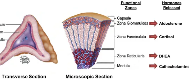
The adrenal gland is made up of the cortex and the medulla. The three functional zones that comprise the adrenal cortex each make distinctive steroids and this zone-specific synthesis parallels their distinct expression of steroidogenic enzymes. The zona glomerulosa (outer zone) produces mineralocorticoids (aldosterone) which monitors salt and water balance. The zona fasciculate (middle layer) makes cortisol regulating carbohydrate metabolism and vascular response to cathecholamines. The innermost layer, the zona reticularis, makes the androgen precursors such as DHEA and its sulfate (DHEA[S]). Neuroendocrine cells which make up the medulla synthesize and secrete catecholamines. Image from illustrator Diantha Tevis-2006.
Figure 1 The adrenal gland
Aldosterone Cortisol DHEA Cathecholamines Hormones Released Functional Zones
Transverse Section Microscopic Section
pregnolenone is converted into the various intermediates and active steroid hormones. Few organs are capable of making steroids from cholesterol however many can transform circulating steroids, such as adrenal dehydroepiandrosterone DHEA (Figure 2), which is converted to testosterone in peripheral tissues, a route generating the major source of testosterone in women (Labrie et al., 2003). Hormones can potentially become more powerful or activated in target tissues, such as testosterone, which is converted to its active form, dihydrotestosterone (DHT) in the prostate gland. A general principle in steroidogenesis is that reactions are unidirectional as most are irreversible, and the hydroxysteroid dehydrogenase reactions (HSD) (Figure 2), while reversible, predominantly proceed in one direction. The enzymes necessary for these steps are 3β-HSD2, 17βHSD and the family of P450 enzymes, which include CYP19A1 (aromatase).
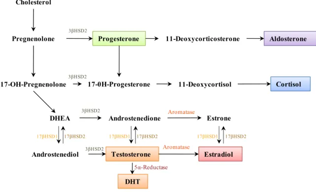
Figure 2 Human steroid biosynthesis. Adapted from (Ghayee and Auchus, 2007). Cholesterol
Pregnenolone Progesterone 11-Deoxycorticosterone Aldosterone
17-OH-Pregnenolone 17-0H-Progesterone 11-Deoxycortisol Cortisol
DHEA Androstenedione Estrone
Estradiol Testosterone Androstenediol 3βHSD2 3βHSD2 3βHSD2 3βHSD2 17βHSD1 17βHSD2 17βHSD1 17βHSD2 17βHSD1 17βHSD2 Aromatase Aromatase DHT 5α-Reductase
The testis and ovaries have little capacity to synthesize aldosterone and cortisol. Alternatively, steroid metabolism in these organs is focused on making androgens and estrogens, while the corpus luteum of the ovary produces progesterone. The testis efficiently completes the biosynthesis of testosterone and can export this potent androgen whereas the ovaries synthesize primarily androstenedione to convert it into estrogens as well as variable amounts of testosterone (Figure 2). Steroidogenesis in the ovary is compartmentalized in a cell-specific manner, where the theca cells primarily produce androstenedione and the granulose cells complete the synthesis of estradiol (E2). In adrenals and gonads, ACTH or Luteinizing hormone (LH) mobilize cholesterol into the steroidogenic pathways in bursts or pulse. Follicle-stimulating hormone (FSH) is critical in promoting granulose cell development and estradiol synthesis during ovulation. In the placenta, steroid production is less pulsatile and dependent on chorionic gonadotropin (hCG) early in gestation and later is mostly unregulated.
2.1 Estrogen; From Synthesis to Function
Steroid hormones are among the most powerful and enduring signaling molecules in the body. When transported via the circulation, steroids travel great distances from the site of synthesis in an endocrine organ to a distant target organ. Alternatively, steroids can act as local autocrine or paracrine signals that impact only the microenvironment, including in the brain. The half-life of steroids is several-fold greater than that of other blood-borne signaling molecules, such as insulin, which disappear within minutes to hours. Estradiol is the final end product of 6 enzymatic conversions from its precursor cholesterol (Figure 2), and it is the most potent steroid, being active at concentrations in the femtomolar range. The critical p450 enzyme aromatase is the rate-limiting step in estradiol synthesis from androgen precursors and is a nodal point of regulation (as discussed in chapter 8).
2.1.1
Synthesizing Estrogen
Estradiol production in the ovary is dependent on the action of 17βHSD. In ovarian granulose cells of developing follicles in cycling humans and rodents, 17βHSD1 converts estrone to estradiol. Upon ovulation, follicles luteinize and transform into corpora lutea which continue to secrete estradiol at high concentrations although, during luteinization, 17βHSD1 expression declines rapidly in the ovary. The expression of 17βHSD1 is mostly abundant in the granulose cells and the syncytiotrophoblasts of the placenta (Sawetawan et al., 1994; Fournet-Dulguerov et al., 1987) and expressed at lower levels in the breast (Miettinen et al., 1999). In the ovary, 17βHSD1 is primarily induced by FSH acting through the cAMP-dependent protein Kinase A (PKA) signaling pathway (Kaminski et al., 1997).
In addition to the source of active steroid hormones derived from the circulation, there are numerous tissues such as epithelial cells of human breast and endometrium that express 17βHSD, 3βHSD and aromatase, having therefore the ability to synthesize active steroid hormones from circulating steroid precursors. The expression of these enzymes in target tissues is very important especially in humans where the adrenal glands will secrete high levels of DHEA. These steroids serve as substrates in peripheral tissues for their eventual conversion to testosterone by one of the isoforms of 17βHSD or to estrone or E2 by aromatase. In these tissues 17βHSD1 catalyzes the conversion of estrone to the more potent form E2 (Figure 2) (Penning, 1997).
The peripheral expression of aromatase is critical, especially in men and postmenopausal women. A major site of peripheral expression of aromatase is in the adipose tissue of men and women (Kamat et al., 2002; Simpson et al., 2002). The conversion of androgens to estrogens in adipose tissue increases with age in postmenopausal women and in elderly men (Kamat et al., 2002; Simpson et al., 2002). The primary site of expression within adipose tissue is in the stromal mesenchymal cells (Simpson et al., 2002). Aromatase is also expressed in osteoblasts and chondrocytes in
males and females (Sasano et al., 1997). Aromatase can also be observed in the brain being primarily expressed in the hypothalamus of male and females as well as in other areas such as the retina (Kamat et al., 2002; Simpson et al., 2002).
2.1.2 Importance of Estrogen
Estrogen has widespread physiological actions, targeting both genomic and non-genomic mechanisms. Estrogen is a key regulator of growth, differentiation and biological function in a wide array of target tissues, including the male and female reproductive tracts, mammary gland, skeletal, cardiovascular and central nervous systems. In breast tissue, estrogens stimulate the growth and differentiation of the ductal epithelium, induce mitotic activity of ductal cylindric cells and stimulate growth of connective tissues (Porter, 1974). Estrogens can also exert histamine-like effects on the microcirculation of the breast (Soderqvist et al., 1993). Estrogens are also thought to have neuroprotective actions such as synaptic and dendritic remodeling (Naftolin et al., 1990) as well as glial activation in brain tissue from adult rats (Garcia-Segura et al., 1999). In neurons of the hippocampus, an area involved in memory, estrogens increase the density of N-methyl-D-aspartate receptors and increase neuronal sensitivity to input mediated by these receptors (Woolley et al., 1997). Estrogens can reduce the generation of β-amyloid peptides, which build up in the brains of patients with Alzheimer's disease and observed in cultured human neuroblastoma cells (Green and Simpkins, 2000).
Estrogens are also known to cause short-term vasodilation by increasing the formation and release of nitric oxide (NO) and prostacyclin in endothelial cells (Kim et al., 1999). A protective role of estrogens against atherosclerosis was suggested by the findings that estrogen treatment reduced the progression of coronary-artery atherosclerosis in oophorectimized monkeys (Clarkson et al., 1996). Furthermore, estrogens can directly inhibit the functions of osteoclasts which express regulators of bone resorption, reducing the risk of fracture in women with osteoporosis (Lufkin et al., 1992; Lindsay et al., 1980).
It has been recently proposed that estrogen plays a role in insulin resistance leading to glucose intolerance and type II diabetes when pancreatic β-cells cannot meet the requirement for insulin (Godsland, 2005). A study showed that treatment of E2 was able to protect β-cells from oxidative injury in mice resulted in protection from proinflammatory cytokine-mediated β-cell death (Le et al., 2006; Contreras et al., 2002). E2 can also exert anti-inflammatory actions in different tissues and animal models (Vegeto et al., 2003; Zancan et al., 1999), through the inhibition of inducible nitric oxide synthase (iNOS) synthesis.
One of the most important and notable effects of estrogens is a very potent mitogenic action in hormone sensitive tissues such as the breast (Evans, 1988; Kumar et al., 1987; Weisz et al., 1990) and the uterus (Quarmby and Korach, 1984; Martin et al., 1973). Prolonged exposure of target tissues or cells to estrogens has long been considered an important etiological factor for the induction of estrogen-associated cancers in experimental animals (Nandi et al., 1995) as well as in humans (Nandi et al., 1995; Ziel and Finkle, 1975; Mack et al., 1976; Pike et al., 1993; McDonald et al., 1977; Grady and Ernster, 1997; Feigelson and Henderson, 1996; Jick et al., 1979).
2.2 Steroid hormones- ligands to steroid receptors
Steroid hormones have long been known as essential metabolic regulators, but the cloning of their respective hormone receptors was an indispensable prerequisite to understand the molecular basis of hormone action transposed into a transcriptional process. In vertebrates, the nuclear receptor superfamily contains intracellular receptor proteins targets (a group of structurally related receptors) with specific affinity to estrogens, androgens, progestins, glucocorticoids, mineralcorticoids and thyroid hormones. (Evans, 1988; Mangelsdorf et al., 1995; Beato et al., 1995; Glass, 1994).
Jensen and coll. in the early 60’s (Jensen and Jacobson, 1962) laid the groundwork demonstrating the presence of a binding protein that would mediate the biological effects of E2 in the uterus. This has paved the way twenty four years later to the cloning of the estrogen receptor, presently known as ERα (Greene et al., 1986; Green et al., 1986). With the cloning of the other steroid receptors such as glucocorticoid receptor (GR), progesterone receptor (PR) and androgen receptor (AR), a considerable progress has been accomplished over the last two decades in understanding the mechanisms of steroid hormone action. In addition, the identification of a growing number of interacting factors recruited to steroid receptors in order to facilitate transcriptional processes in response to hormone stimuli has helped in developing a comprehensive model of cofactor assembly and exchange to mediate target gene expression. However, given the complexity of these various regulatory aspects involved in steroid receptor functions that have emerged from these studies, it became evident that strategies developed to efficiently counteract deregulated functions associated to receptors had to consider the model’s intricate network.
3 Steroid Receptors
The neodarwinian theory of evolution describes that new functions emerge as the phenotypic outcome due to the natural selection of random mutations. Complex organs and functions are believed to be generated from a gradual selective process of elaboration and optimization (Dawkins R, 1986). Vertebrate steroid hormones and the intracellular protein receptor that mediate their effects elegantly illustrate this theory.
3.1 Evolution of Steroid Receptors
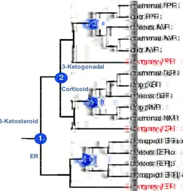
The receptor phylogeny suggests that two serial duplication of an ancestral steroid receptor occurred during the interval when vertebrates were evolving from invertebrates. In the ancestral vertebrate, the first duplication event created an estrogen receptor (ER) and a 3-ketosteroid receptor, whereas the second duplication came from the latter gene to produce a corticoid receptor and a receptor for 3-ketogonadal steroids (androgens, progestins or both) yielding three steroid receptors (Figure 3). At some point in time within the gnathostome lineage (jawed vertebrates comprised of fishes, amphibia, reptilia, aves and mammalia), each of these three receptors duplicated a second time to yield six steroid receptors currently expressed in jawed vertebrates: the ER to create ERα and ERβ, the
Figure 3 Phylogeny of steroid receptors. The blue circles represent gene duplication
events. Adapted from (Thornton, 2001).
ER
Corticoid
3-Ketosteroid
corticoid receptor giving the GR and mineralocorticoid receptor (MR) and the 3-ketogonadal receptor to create the AR and the PR (Figure 3). Although the timing of these events has remained unclear due to divergent hypotheses, gene mapping data supports serial duplications as the mechanism by which steroid receptors have diversified.
The fundamental role of steroids in regulating vertebrate development, reproduction, growth and homeostasis (DeGroot LJ, 1995; Gilbert SF, 1994; Evans, 1988) suggests that these steroid receptor duplications provided vertebrates a selective advantage over other organisms, which would be important in vertebrate competition with the diverse multicellular organisms present in early Cambrian, 540 to 480 million years ago, as well as for vertebrate survival during global extinctions that occurred about 440 and 370 million years ago (Raup, 1994). Nuclear receptors are an excellent example of how gene duplications and divergence can generate a protein family that responds to diverse signals to regulate a wide variety of physiological processes.
3.2 The Ancestral Steroid Receptor was an Estrogen Receptor
An ancestral protein is likely to have resembled in sequence and function to a descendent gene which evolved more slowly after the duplication event, compared to one with a more rapid evolutionary rate. Relative rate tests based on amino acid distances and reconstruction of branch lengths suggest that the ancestral steroid receptor was a functional estrogen receptor, the sequence of which was conserved among descendent ERs.The reconstructed ancestral receptor is 71% identical to the human ERα however radically different to the PR, AR, GR and MR. This result indicates that the ancestral steroid receptor activated genes with estrogen-response palindromes (AGGTCA-figure 5) and bound estrogens. In the synthesis of estradiol and estrogens, progesterone and testosterone are synthesized as intermediates. These steroids, and the enzymes that produce them, would therefore have been present during the period when only one receptor for
estrogen existed. After gene duplications of the ancestral estrogen receptor gene and followed by considerable sequence divergence, receptors emerged to give these intermediate compounds novel signaling functions.
Redundant receptors created by gene duplication could then diverge in sequence from their ancestors and evolve affinity for these steroids, creating signaling functions for what were once intermediates. The expansion of the steroid receptor family by gene duplication and ligand application allowed a greater specificity in hormone control over physiological functions. Estrogen regulation appears to be the most ancient of all modes of steroid/receptor control which is supported by the apparent role of estrogen in branchiostome and echinoderm reproduction (Fang et al., 1994; Hines et al., 1992). Comparisons of the evolution of steroid receptors indicate that land animals show a slow sequence divergence. The fish estrogen and glucocorticoid receptors have about 70 and 60% sequence identity respectively, to their human orthologs. This puts these steroid
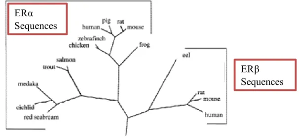
Figure 4 Phylogenetic tree of the evolutionary relationships of ERα and ERβ based on
amino acid sequences. Adapted from (Kelley and Thackray, 1999).
ERβ
Sequences
ERα
receptors in the class of slowly changing proteins (Doolittle et al., 1996). The discovery of a second form of ER (Kuiper et al., 1996; Mosselman et al., 1996) termed ERβ, generated questions about the biological importance of this newly discovered gene. Previous sequence alignments have shown that ERβ sequences share common elements that are distinct from ERα sequences (Pettersson et al., 1997) supporting the idea that the ERβ sequences belong to a separate monophyletic clade (a group composed of a single ancestor and all its descendents) with respect to ERα and have evolved in parallel (Naylor and Brown, 1998) (Figure 4). The fact that the ERβ gene is widespread among chordates (animals which are either vertebrates or one of several closely related invertebrates) and comprises a separate genetic lineage dating back at least 450 million years argues that this gene performs distinct biological functions that have been maintained by natural selection for this long period of time.
4 Estrogen Receptors
In the 1950s, Jensen and Jacobson (Jensen and Jacobson, 1962; Jensen EV and Jacobson HI, 1960) used tritium labeled E2 to demonstrate that it was specifically retained by estrogen target tissues which led them to hypothesize that a receptor must exist for this molecule.
4.1 ERα and ERβ: Discovery of two subtypes
In the next decade an ER was identified by Toft and Gorski (Toft and Gorski, 1966) and isolated from several mammalian species, including rat and human (Toft and Gorski, 1966; Jensen et al., 1968). However, it was not until the mid 1980s that the first ER, now called ERα, was cloned by two groups of investigators (Green et al., 1986; Greene et al., 1986; Walter et al., 1985). In the mid 1990s, a second ER, called ERβ, was identified from a library scan of rat prostate cDNA library (Kuiper et al., 1996) and subsequently cloned from several species including the mouse, human, and fish (Kuiper et al., 1996; Mosselman
et al., 1996; Tremblay et al., 1997) which meant that the biological properties associated to ER signaling in terms of subtype selectivity, ligand specificity, and tissue distribution had to be reviewed (Giguère et al., 1998; Gustafsson, 1999).
At first, a human ERβ with 477 amino acids was reported (Mosselman et al., 1996). A few months later, Enmark et al. (Enmark et al., 1997) identified an ERβ mRNA species of 485 amino acids, and it was hypothesized to reflect full-length ERβ. The following year, Ogawa et al. (Ogawa et al., 1998b) reported the cloning of an additional ERβ species consisting of 530 amino acids, which is now considered to represent full-length ERβ. A few months later, Moore et al. (Moore et al., 1998) also identified the same 530-amino acid sequence as the full-length ERβ in addition to various isoforms. Similarly to ERα, ERβ expression has also been associated with cancers of the breast (Dotzlaw et al., 1997; Dotzlaw et al., 1999; Speirs et al., 1999a; Speirs et al., 1999b), colon (Foley et al., 2000; Campbell-Thompson et al., 2001), and ovarian tissues (Pujol et al., 1998; Rutherford et al., 2000).
4.2 ERα and ERβ; Pertinent or redundant?
Although in vitro studies have demonstrated redundancy in the roles of these two receptors, tissue localization has revealed distinct expression patterns for each receptor suggesting that each ER subtype might perform specific functions. ERα is expressed in a variety of tissues classically associated with estrogenic activity including the uterus, ovaries (theca cells), cervix, vagina, breast, bone and several additional target organs such as in the prostate (stroma) and brain but to a lesser degree in bladder, liver and thymus. ERβ is predominantly expressed in normal colon, prostate (epithelium), ovary (granulosa cells), bone marrow and brain, with smaller amounts reported in uterus, bladder, lung and testis (Kuiper et al., 1997; Shughrue et al., 1998; Veeneman, 2005) and in the spleen, hypothalamus, and thymus (Couse et al., 1997). ER tissue expression is also tied to
developmental stage, specifically in both uterus and pituitary where ERβ is expressed during development but is later replaced by ERα (Shupnik, 2002; Nishihara et al., 2000).
The development of KO models has helped us to unmask unidentified estrogen signaling systems as well as those that are independent of either ERα or ERβ. Studies with ERα and ERβ knockout (KO) mice have revealed a role for ERs signaling in bone formation, male and female sexual maturation, fertility, cardiovascular and angiogenesis effects, and behavior (Bocchinfuso and Korach, 1997; Couse et al., 1995; Eddy et al., 1996; Hess et al., 1997; Korach et al., 1996; Krege et al., 1998; Lindberg et al., 2003; Ogawa et al., 1996; Ogawa et al., 1997; Ogawa et al., 1998a; Ogawa et al., 1998d; Rubanyi, 2000; Schomberg et al., 1999; Windahl et al., 2001; Windahl et al., 2002). Both sexes of the αERKO are infertile. In the female αERKO mice, estrogen insensitivity leads to hypoplasia in the reproductive tract with enlarged cystic and haemorrhagic follicles in the ovaries. Also, lack of pubertal mammary gland development and excess adipose tissue was observed in females (Couse and Korach, 1999). In αERKO males, testicular degeneration and epididymal dysfunction are the major phenotypes. Conversely, male βERKO mice are fertile and show no obvious phenotype. However female βERKO mice exhibit inefficient ovarian function and subfertility due to a block in the last step of follicular development. This can be overcome when animals are treated with FSH and LH. A recent report described an abnormal vascular function resulting in hypertension of βERKO mice (Zhu et al., 2002). The generation of mice that do not express either receptor isoform (DERKO) has provided additional information on the role of these two receptors in regulating physiological and behavioural processes. The adult ovarian phenotype is masculinised, coinciding with a reduction in oocyte number. In addition the ovaries do present structures that resemble testicular seminiferous tubules (Couse and Korach, 2001). Absence of both receptors leads to a significant drop in sexual and aggressive behaviour (Ogawa et al., 2000; Simpson and Davis, 2000; Zhu et al., 2002).
The difference in tissue distribution between ERα and ERβ can only partly explain the tissue specific effects of estrogens. Tissue specificity may actually be attributable to the type of dimers formed by these two receptors and their interaction with accessory proteins. Several groups have reported that ERα and ERβ can form functional heterodimers (Cowley et al., 1997; Ogawa et al., 1998b; Pettersson et al., 1997; Tremblay et al., 1999b).
Moreover, the ERα/ERβ heterodimer appeared to form preferentially over each homodimer when both receptors are expressed, and was shown to bind to the consensus estrogen response element (ERE) sequence (Figure 5) with an affinity comparable to that of the ERα homodimer and greater than the ERβ homodimer (Tremblay et al., 1999b; Cowley
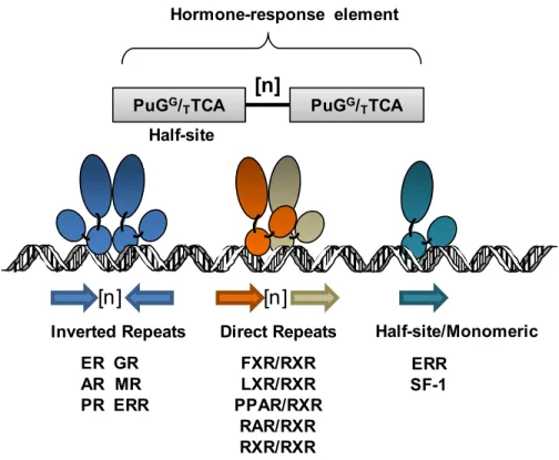
Figure 5 Hormone Response Element- Orientation of Hormone response
element and their cognate receptors.
Hormone-response element ER GR AR MR PR ERR FXR/RXR LXR/RXR PPAR/RXR RAR/RXR RXR/RXR Inverted Repeats Direct Repeats
PuGG/
TTCA PuGG/TTCA [n] [n] [n] Half-site ERR SF-1 Half-site/Monomeric
et al., 1997). Consequently, profiles in gene expression may diverge upon ER dimerization properties, and therefore the ratio of different receptor types in tissues may be an important determinant of a biological response.
4.3 Structure and isoforms of the ERs
ERα and ERβ are each encoded by unique genes localized on chromosome 6 and 14 in humans respectively (Enmark et al., 1997; Tremblay et al., 1997; Giguère et al., 1998). Both ER subtypes share the distinctive modular structure of functional domains characteristic of the superfamily of nuclear hormone receptors (Kumar et al., 1987; Evans, 1988). Nuclear receptors have been clustered into 6 subclasses based on sequence comparison and phylogenetic analysis (Laudet, 1997) and a unified nomenclature was proposed thereafter (Laudet et al., 1999). Members include receptors for estrogen (ER), glucocorticoids (GR), progesterone (PR) and androgens (AR), as well as the orphan estrogen-related receptors (ERRs), which are contained within the NR3 subclass, reflecting their apparent abilities to bind to response elements organized as inverted repeats (Figure 5) (Beato et al., 1995). Receptors that share the heterodimerization partner retinoid X receptor (RXR) bind response elements organized as direct repeats, such as for retinoic acids (RAR), prostaglandins and fatty acids (PPAR), thyroid hormones (TR), vitamin D (VDR) and oxysterols (LXR), and are mostly found in the NR1 subclass (Truss and Beato, 1993; Glass, 1994; Mangelsdorf et al., 1995). Members of NR2 subclass are able to bind as homodimers on direct repeat elements including RXR, HNF4 and COUP-TF. Subclasses NR4-6 are comprised of orphan nuclear receptors for which no specific ligand has yet been identified (Laudet, 1997).
4.3.1 Structure of ERs
There are six functional domains that characterize ERα and ERβ termed A through F (Figure 6). These consist of a highly conserved (Umesono and Evans, 1989) (97%) DNA-binding domain (C) containing two Zn2+-finger motifs necessary for dimerization and specific binding to genomic response elements (Figure 5), a globular C-terminal region (EF) relatively well conserved (60%) (Warnmark et al., 2003) that contains the LBD and an activation function (AF-2) that mediates receptor trans-activation. The AF-2 domain is characterized by a highly conserved amphipathic α-helix (H12), essential for ligand dependent activation of transcription and interaction coregulatory proteins (McKenna et al., 1999; Heery et al., 1997). The low rate of change and the conservation of critical residues within the DBD and the LBD imply that there has been strong selective pressure to maintain these functions in both ERα and ERβ. In addition, the amino acids that make
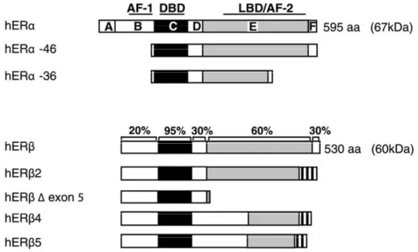
Figure 6 ERα and ERβ functional domains and sequence homology. Adapted from
Sanchez et al. TEM.
ER
β
ER
α
< 15%
97%
60%
DNA Hinge
Ligand
Modulator
AF-1
DBD
AF-2
direct contacts with the DNA in the crystal structure of the DBD are completely conserved (Schwabe et al., 1993). Also the amino acids of the ligand binding cavity, identified in the crystal structure of the LBD, involved in the direct and indirect hydrophobic interactions with the ligand (Brzozowski et al., 1997), are conserved with only a few changes within the clade.
The LBD region is preceded by a flexible hinge region (D) that was previously describe to possess signals for nuclear localization and the binding of chaperones such as heat shock proteins (hsp), which provide the receptors proper folding and a means to interact with protein trafficking systems. However, over the past few years, studies have demonstrated the hinge region to play a much more extensive role in the regulation of both receptor isoforms, through different post-translational modifications (Sanchez et al., 2007; Giordano et al., 2010; Berry et al., 2008; Herynk et al., 2009).
The N-terminal (AB) domain (Figure 6) is the region that differs dramatically between both ER subtypes with a feeble 15% homology. Although the N-terminal appears to be relatively unconstrained compared with the DBD and the LBD, it plays an important role in the transactivation of gene expression. Experiments have shown that transcriptional activation functions (AF) in the N-terminal domain (AF-1) and the LBD are both required for full receptor activity (Kumar et al., 1987; Tzukerman et al., 1994). From the structure-function analysis presently available, the apparent differences between AF-1 and the AF-2 in conformation suggest that the two activation functions have evolved different approaches to achieve transcriptional activation (Warnmark et al., 2003). The N-terminal region of ERs contains serine residues which have been implicated in cross-talk with various cell signaling pathways (Tremblay et al., 1997; Weigel, 1996; McInerney and Katzenellenbogen, 1996). The serine phosphorylation sites in the N-terminal domain of ERα and ERβ are not conserved, suggesting that ERα and ERβ may be regulated differently by cell signaling pathways. Indeed the AF-1 ligand-independent domain which controls the
recruitment of coregulators can be both similar and unique from those employed by the AF-2 (McKenna et al., 1999; Webb et al., 1998). Finally the F terminal domain comprised of the last 30-45 amino acids (depending on the subtype) has approximately 18% homology (Gustafsson, 1999) and appears to regulate the conformation of ERs in order to control the transcriptional response to its ligand (Yang et al., 2008).
4.3.2 Isoforms/variants of ERα and ERβ
From the eight total exons that code for ERα, detection of up to five different isoforms/variants have been discovered in humans from alternative splicing, intronic exons and exonic duplications (Hirata et al., 2003). The full-length ERα is defined as being 595 aa (hERα-66 (66KDa)), however shorter transcripts have been observed to be expressed in
Figure 7 Isoforms of ERs- Schematic representation of the different splicing produtcs
different cell lines, such as hERα-36 (36 KDa) and hERα-46 (46KDa) (Flouriot et al., 2000; Wang et al., 2005). hERα-36 lacks both AF domains but contains sites that could be myristoylated suggesting that it would have the potential of anchoring itself in the plasma membrane. hERα-46 (46KDa) lacks the AF-1 but still manages to show antagonizing activity on the proliferative effects of the full length hERα-67 in MCF-7 cells (Penot et al., 2005) (Figure 7).
As for ERβ, five different variants (ERβ1-5) have been cloned (Figure 7) and examined. ERβ1 is considered the full-length receptor and is the only variant to contain fully functioning helices 11 and 12 (Wurtz et al., 1996) and therefore capable of interacting with ligands and recruiting coregulatory complexes (Henttu et al., 1997). Few studies have looked at the preference in dimerization partners between ERα and ERβ, however in gel-shift assays, ERβ4 and ERβ5 heterodimerised with ERα (Poola et al., 2005) which affected the response to estrogen signaling, similar to the heterodimerization of ERβ2 (ERβcx) with ERα. ERβcx is the only variant to possibly exhibit clinical relevance. Although this protein does not respond to any particular ligand, it has a dominant negative effect on ERα transcriptional activity (Ogawa et al., 1998c; Zhao et al., 2007).
4.4 Activation of ERs
Full transcriptional activity of a nuclear receptor is accomplished not only by the synergism between its AFs but also relies on a number of events. The transcriptional potential of each AF is dependent on external determinants such as cell type, posttranslational modifications, promoter context and interaction with coregulatory complexes (Berry et al., 1990; Aronica and Katzenellenbogen, 1993; Hadzopoulou-Cladaras et al., 1997; Tzukerman et al., 1994; McInerney et al., 1998; Pham et al., 1992; Cenni and Picard, 1999).
4.4.1 Ligand-Dependent Activation of ERs
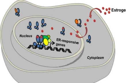
ERs, in absence of estrogen, are attached to receptor-associated proteins which function as chaperones (Figure 8) stabilizing the receptor in an inactivated state by masking the DNA binding domain (Couse et al., 1999). Following the binding of E2, an activating conformational change is generated within the ERs promoting dimerization and high affinity binding to specific DNA response elements found within the regulatory regions of target genes (refer to figure 5). ERα and ERβ have been shown to interact with identical DNA response elements and exhibit a similar binding affinity profile for naturally occurring estrogens such as E2 when assayed in vitro. Both ERs recognize a distinct palindromic sequence, normally specific to the type of nuclear receptor. In the case of ERs it is an inverted repeat sequence separated by three nucleotides; AGGTCAnnnTGACCT (Parker et al., 1993) (Figure 5). However, only a small number of estrogen-responsive
Figure 8 Ligand-dependent activation of ERs.
Estrogen Coactivator Complex Estrogen Receptor Legend: Cytoplasm Nucleus Chaperone
genes contain the consensus sequence. Several of the genes identified having a functional ERE sequence not only consist of one or more changes from the consensus but are made up of multiple copies of imperfect EREs (Driscoll et al., 1998). Depending on the cell and promoter context, the different combinations of DNA-bound ERs exert either a positive or negative effect on the expression of downstream target genes (Wood et al., 1998).
4.4.2 Ligand-Independent Activation of ERs
The responsiveness of steroid receptors to cell signaling pathways in the absence of their hormone can be different. ERs are quite responsive to cell signaling pathways. In fact, endogenously expressing ER-positive cells maintained in phenol-red free, charcoal-stripped serum used to minimize steroids, frequently display a considerable transcriptional activity in the absence of estrogen (Smith et al., 1993). Although ERs belong to a family of ligand-activated receptors, they are also phosphoproteins and their activity can equally be regulated by phosphorylation of specific sites which can occur as part of both the induced activity (Ali et al., 1993; Arnold et al., 1994; Kato et al., 1995) and/or ligand-independent activity (Arnold et al., 1995b; Bunone et al., 1996; Tremblay et al., 1999a; Tremblay et al., 1998). Studies from other transcription factors such as CREB and PR, have shown that phosphorylation can play roles in nuclear translocation, DNA binding, interaction with other proteins and trans-activation (Hill and Treisman, 1995; Denner et al., 1990a). Polypeptide growth factors can activate ERs and increase the expression of ER target genes (Smith, 1998). Phosphorylation occurs predominantly at specific serine/threonine or tyrosine residues and is catalyzed by enzymes such as mitogen-activated protein kinases (MAPK) (Shao and Lazar, 1999). MAPKs are composed of several serine-threonine kinases that are activated in response to various cell-growth signals and transduce extracellular signals to intracellular targets by way of membrane receptors.
Activation of ERs by signaling pathways (section 5.3) in the absence of E2 was first identified in the early 1990s (Ignar-Trowbridge et al., 1992). Ovariectomized mice were
treated with epidermal growth factor (EGF), promoting the translocation of ER towards the nucleus and stimulating its activity. Furthermore, EGF antibodies were administered to ovariectomized mice 3 days prior to hormone treatment resulting in a marked decrease of uterine DNA synthesis (Ignar-Trowbridge et al., 1995), leading to believe that EGF had a role in the proliferative effects of estrogen in reproductive tissues. Further assessment of the cell-surface receptor tyrosine kinases (RTKs) demonstrated their role in the recruitment of multiple signal transduction cascades that act to increase theactivation of MAPK Erk1/2, PKB/Akt, Jnk, p38 and protein kinase C (PKCα and δ), key elements inthe regulation of cell proliferation and survival signals (Bonni et al., 1999; Campbell et al., 2001; Gibson et al., 1999; Stambolic et al., 1999) (Amit et al., 2007). Chapter 5 will focus on the detailed description of the different signaling pathways regulating the activity of ERs.
5 Cell-Surface Receptors
It is now widely documented that the activation of growth factor (GF) signaling cascades through asupply of GF ligands via up-regulation and increased activation of their target growth factor membrane receptors and their recruited downstream signaling elements, can promotehormone-like responses.
5.1 Members of ErbB Clan
Figure 9 Four types of ErbB receptors and their ligands.
? EGF TGFα HB-EGF EPR HRG-1 HRG-2 NRG-1-4 HB-EGF EPR
Growth factors and their receptors play a fundamental role in the communication between outside the cell surface and the inside compartments (Schlessinger and Lemmon, 2006; Scaltriti and Baselga, 2006). The human epidermal growth factor family (ErbB/HER) is comprised of four closely related receptors (Figure 9); epidermal growth factor receptor (EGFR, HER1, ErbB1), human EGFR-2 (HER2, ErbB2), ErbB3 and ErbB4. They are transmembrane oncoproteins containing an extracellular ligand binding domain and an intracellular receptor tyrosine kinase (RTK) domain sharing 40-45% homology to one another. This family of proteins has an important role in tumour processes including angiogenesis and metastasis and is associated with a poor prognosis in many human malignancies due to their overexpression or constitutive activity (Salomon et al., 1995; Hemming et al., 1992). Although all the aforementioned receptors share a strong homology within their TK domains, they are quite distinct in their extracellular N-terminal and cytoplasmic C-terminal domain (Klapper et al., 2000).
The significance of ErbBs in normal development was, as with ERs, obtained from knockout-generated mice. Null mutations in individual ErbB loci are lethal demonstrating that ErbB receptors play a pivotal role in regulating vertebrate embryogenesis and development. ErbB1 KOs are lethal at the embryonic and up to perinatal stages as mice develop abnormalities in the brain, lungs, gastrointestinal tract and the skin (Threadgill et al., 1995; Sibilia and Wagner, 1995; Sibilia et al., 1998; Miettinen et al., 1995). ErbB2 and ErbB4 KOs are lethal at the stage of midgestation due to malformations of the heart (Gassmann et al., 1995; Lee et al., 1995). ErbB3 KO mice are embryonically lethal due to malformations of the heart valves in addition to neural crest defect and lack of Schwann cell precursors (Riethmacher et al., 1997; Erickson et al., 1997).
5.2 Activation of ErbB
Several ligands bind to the ErbB receptors (Figure 9). Members of the EGF superfamily include epidermal growth factor (EGF) (Todaro et al., 1980), transforming growth factor-α (TGF- α) (Shoyab et al., 1988) and amphiregulin, which specifically bind to ErbB1 (EGFR). Heparin-binding EGF and epiregulin (EPR) bind to both EGFR and ErbB4 (Figure 10) (Toyoda et al., 1995). Neuregulins 1 and 2 (also known as heregulins (HRG) or neu differentiating factor) bind to both ErbB3 and ErbB4 (Falls, 2003). Binding of GF to ErbBs induces receptor dimerization and activation of intracellular protein tyrosine kinase with subsequent initiation of numerous downstream signaling events (Figure 10) (Press and Lenz, 2007). All ErbB ligands exist initially as membrane-anchored precursors that require proteolysis to liberate them as soluble mature ligands (Massague and Pandiella, 1993; Harris et al., 2003b).
In the absence of ligand, ErbB1, ErbB3 and ErbB4 are monomeric and can be partially or completely inhibited (Schlessinger, 2002; Ferguson et al., 2003; Bouyain et al., 2005). This inhibition is caused by the extracellular portion autoinhibiting the ligand surface due to its conformation status. Binding of a ligand leads to an alteration within the extracellular domain which creates a ligand-binding pocket and protrusion of a dimerization area. This change aids in receptor oligomerisation and the formation of homo- and heterodimers (Hynes and Lane, 2005; Citri et al., 2003; Leahy, 2004). Dimerization brings the intracellular kinase domains of the two receptors close together encouraging transphosphorylation of tyrosine kinase residues in the cytoplasmic tail of one receptor by the kinase domain of the adjacent receptor (Figure 10) (Jorissen et al., 2003; Schlessinger, 1988). Unlike its family counterparts, ErbB2 has not yet had a ligand identified to regulate its activity (Yarden and Sliwkowski, 2001) (Klapper et al., 1999; Citri et al., 2003).
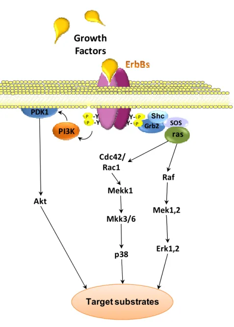
Figure 10 ErbB Receptor Activation.
Receptor Auto/trans phosphorylation
Intracellular signal
Dimerisation following ligand binding
EGF P P P P HRG ? EGF TGFα HB-EGF EPR HRG-1 HRG-2 NRG-1-4 HB-EGF EPR
ErbB1 ErbB2 ErbB3 ErbB4
Plasma Membrane Intracellular
ErbB2 is the preferred heterodimerising partner of ligand-bound ErbB3 but can also bind ErbB1 and ErbB4 (Karunagaran et al., 1996), (Graus-Porta et al., 1997). Dimers containing ErbB2 are known to enhance and prolong the signaling of several ErbB ligands, and this may be due to the reduced dissociation of the receptor complex (Karunagaran et al., 1996) in addition to the reduction in the rate of internalization of the complex leading to recycling rather than degradation (Holbro et al., 2003). ErbB3 harbors a substitution in crucial residues of the C-terminal intracellular domain rendering its kinase activity dead (Guy et al., 1994) therefore ErbB3 homodimers are inactive. This forces the receptor to heterodimerize with other ErbBs to become phosphorylated and trigger an intracellular signal (Kim et al., 1998). In addition, ErbB3 contains seven copies of the Tyr-X-X-Met motif in its c-terminal motif recognized by phosphatidylinositol 3-Kinase (PI3K) which leads to the activation of the Akt pathway (Prigent and Gullick, 1994; Songyang et al., 1993). ErbB3’s preferred dimerizing partner is ErbB2, in fact, the ErbB2-ErbB3 heterodimer is the most prevalent receptor complex and one of the most potent signaling pathways that regulate cell growth and transformation (Pinkas-Kramarski et al., 1996; Karunagaran et al., 1996).
5.3 ErbB Intracellular signaling
Autophosphorylation of the C-terminal tyrosine residues serving as docking sites for cytoplasmic signaling proteins contain Src-homology (SH-2) and phosphotyrosine-binding (PTB) domains (Olayioye et al., 2000; Yarden and Sliwkowski, 2001). Each ErbB receptor exhibits a phosphotyrosine profile that allows for binding of enzymes such as Src, phospholipase Cγ, and the p85 regulatory subunit of PI-3K, or adapter molecules such as Shc and Grb2 linking ErbB activity to many downstream effectors (Figure 11) (Olayioye et al., 2000; Hynes and Lane, 2005). Although the Ras-Raf-MAPK and PI3K pathways are the major signaling pathways by the ErbB family, each dimeric receptor complex can activate different combinations of these signaling cascades, resulting in a wide range of signaling events (Figure 11).