O
pen
A
rchive
T
OULOUSE
A
rchive
O
uverte (
OATAO
)
OATAO is an open access repository that collects the work of Toulouse researchers and
makes it freely available over the web where possible.
This is an author-deposited version published in :
http://oatao.univ-toulouse.fr/
Eprints ID : 16788
To link to this article : DOI : 10.1080/02772248.2015.1133818
URL :
http://dx.doi.org/10.1080/02772248.2015.1133818
To cite this version :
Mouchet, Florence and Gancet, Christian and
Flahaut, Emmanuel and Pinelli, Eric and Boutonnet, Jean–Charles
and Gauthier, Laury International standardized procedures forin
vivoevaluation of multi-walled carbon nanotube toxicity in water.
(2016) Toxicological & Environmental Chemistry, vol. 98 (n° 8).
pp. 829-847. ISSN 0277-2248
Any correspondence concerning this service should be sent to the repository
administrator:
staff-oatao@listes-diff.inp-toulouse.fr
International standardized procedures for in vivo evaluation of
multi-walled carbon nanotube toxicity in water
Florence Moucheta,b,c, Christian Gancetc,d, Emmanuel Flahautc,e, Eric Pinellia,b,c, Jean!Charles Boutonnetc,fand Laury Gauthiera,b,c
a
EcoLab (Laboratoire d’Ecologie Fonctionnelle et Environnement), INP, UPS, Universit!e de Toulouse, Tolosan, France;bEcoLab, CNRS, Tolosan, France;c
Laboratoire Commun NAUTILE (CNRS!UPS!INPT!ARKEMA France), Laboratoires EcoLab/CIRIMAT/GRL;d
Laboratoire d’Ecotoxicologie, D!epartement Analyse,
Groupement de Recherches de Lacq, ARKEMA France, Lacq, France;eCentre Inter-Universitaire de Recherche
et d’Ing!enierie des Mat!eriaux ! CIRIMAT, UMR UPS INPT CNRS 5085, Toulouse, France;f
Direction S!ecurit!e Environnement Produits, ARKEMA France, Colombes, France
ABSTRACT
The classical approach in ecotoxicological evaluation of chemical substances consists of conducting standardized bioassays on organism models. In this work, the potential impact of industrial multi-walled carbon nanotubes was investigated by ecotoxicological standardized procedures using aquatic organisms of different trophic levels, namely bacteria, green algae, invertebrates, fish, and amphibians. The results indicated (1) inhibition of growth in amphibians at 50 mg L¡1and higher, and (2) no effects on daphnia
and fish up to 100 mg L¡1. With the exception of algae (for which Fe deficiency is measured), it seems that the observed toxicity may be due to physiological effects in relation to the ingestion of carbon nanotubes not necessarily related to their intrinsic effects.
KEYWORDS Multi-walled carbon nanotubes; ecotoxicity; standardized bioassays; bacteria; algae; fish; amphibians Introduction
The publication of Iijima (1991) generated unprecedented interest in the world of carbon nanostructures and led to an exponential growth in research on carbon nanotechnology. Carbon nanotubes (CNTs) can be described as graphene sheets rolled up to form cylinders that are closed at both ends. There are two main types, i.e. single-walled CNTs (SWNTs) and multi-walled CNTs (MWNTs). They have remarkable physical, i.e. mechanical, electric, and thermal and chemical properties (inertness, stability), making them a material of choice for polymer composites, electromagnetic shields, super capacitors, gas including hydrogen storage devices, batteries, structural composites, or medical applications (Eklund et al. 2007), especially of MWNTs for biomedical engineering, used in biosensors, as vehicles for drug delivery, and in gene therapy (Kostarelos, Bianco, and Prato2009).
Most likely, during production and use, some quantities will get into the environment, especially the aquatic compartment. Even if toxicological data are available, obtained
CONTACT Florence Mouchet florence.mouchet@ensat.fr http://dx.doi.org/10.1080/02772248.2015.1133818
most often with in vitro systems (Guadagnini et al.2013) and with animal models (Van der Zande et al.2011), nevertheless ecotoxicological exposure and effect data are necessary for understanding the potential hazards these new carbon-based materials may pose for the environment. As new substances, CNTs require registration under the Toxic Substan-ces Control Act in the USA and in the European Union according to REACh (Registra-tion, Evalua(Registra-tion, Authorization and Restriction of Chemicals) regulations (EU 2008). Some (eco)toxicological and environmental properties of SWNTs and MWNTs are listed in the report ENV/JM/MONO 13/REV (2008), but more is required for the proper evalu-ation of the potential ecotoxicity of CNTs. Aquatic ecotoxicity assessment of CNTs is a challenge since tests have been developed for water-soluble chemical compounds. Never-theless, standard environmental hazard assessment is generally appropriate for nanoeco-toxicological research (Crane et al.2008), especially using the test battery concept (Kahru et al.2008; Blaise et al. 2008) in order to accumulate knowledge about their ecotoxicity (Kahru and Dubourguier2011) toward a wider range of biological species providing valu-able insight into likely exposure scenarios (Zhao and Liu2012).
The aim of the present work is to contribute to the ecotoxicological assessment of the potential impact of MWNTs as an example of industrial CNTs in aquatic organisms belonging to different trophic levels by carrying out ecotoxicological standardized proce-dures. The selected species were decomposers (bacteria), primary producers (photosyn-thetic green algae, Pseudokirchneriella subcapitata), primary consumers (invertebrates Daphnia magna), and secondary consumers (vertebrate fish and amphibians, Danio rerio and Xenopus laevis).
Materials and methods
MWNTs preparation of suspensions
MWNT (Graphistrength® C100, Arkema, Colombes, France) suspensions in ultrapure water were prepared by sonication for 10 min at 45 kHz, 80 W (USC 300T, VWR, Fonte-nay sous Bois, France) just before each bioassay. The physical characteristics and trans-mission electron microscopic observations of the MWNTs were previously described by Mouchet et al. (2010).Figure 1displays scanning electron micrographs of MWNTs.
Biological bioassays
Activated sludge respiration inhibition test, OECD (1984) Guideline 209
The inoculum was activated sludge of a small biological domestic wastewater treatment plant (Abidos, France). MWNTs were studied at 500 and 5000 mg L¡1. Dissolved oxygen concen-trations were determined with an oxygen electrode (Stirrox G, WTW, Weilheim, Germany) and meter (OXI 538, WTW). The inhibitory effect was expressed as percentage of the mean respiration rate of two controls, calculated from the recorder trace as mg O2L
¡1h¡1over a period of 10 min. The inhibition was expressed as percentage relative to the mean of the res-piration rates in two controls: % inhibition D [1 ¡ (2Rs/(Rc1CRc2))] £ 100, where Rsis the oxygen consumption rate at the tested concentration of test substance, and Rc1 and Rc2 are the oxygen consumption rates for controls 1 and 2. The sensitivity of the test system and the method were evaluated with 3,5-dichlorophenol.
Algal growth inhibition test (P. subcapitata), OECD (2006) Guideline 201
P. subcapitata (CCAP 278/4 stock) was obtained from the Culture Centre of Algae and Protozoa (Ambleside, UK). The cell density (measured fluorescence, Cytofluor 2350, Millipore, Molsheim, France) for the preliminary test was 1.21 £ 106cells mL¡1, and for the definitive test, 1.09 £ 106cells mL¡1. Algae were exposed under static conditions over Figure 1.Scanning electron microscopy observations of MWNTs, raw MWNTs from the same sample are observable in balls at different magnifications: (a) 100 X, (b) 3000 X and (c) 20,000 X.
a time period of 72 h to MWNTs dispersed in water (EN ISO 8692,2004) at (1) 100, 50, 10, 5, 1, and 0 mg L¡1in the preliminary test, and (2) 1000, 500, 230, 105, 48, 22, 10, and 0 mg L¡1for the definitive test. The MWNT concentrations resulting in 0% and 100% of the uninhibited cell growth rate, and growth rate inhibition causing a 50% reduction in biomass (EbC50) within 72 h (EbC50!72 h) and in growth rate (ErC50!72 h) were esti-mated. The sensitivity of the test system and the method were evaluated by performing an algal growth inhibition test on K2Cr2O7 (Sigma, Lyon, France). The growth inhibition data were analyzed using an Excel sheet to calculate the effective concentration (EC50 value) and the 95% confidence interval. Probit analysis was used to calculate the 24-, 48-, and 72-h EC50 values. The no-observed effect concentration (NOEC), the highest tested concentration at which no significant inhibition of growth is observed relative to the con-trol, was estimated by Dunnett’s test. Values of pH (345 pH meter, Mettler Toledo, Viro-flay, France) and dissolved O2(OXI 538 oxymeter, WTW) were measured.
Analytic complementary experiments have been carried out to study the ecotoxicologi-cal response of algae in relation to a potential deficiency of ionic metallic species, with the well-known property of CNTs to adsorb ionic species (Li et al.2009; Stafiej and Pyrzynska 2007). B, Mn, Fe, Co, Ni, Cu, Zn, and Mo were measured in algal media (water dilution) with and without MWNTs after filtration (0.45 mm to remove most of MWNTs) by ICP!MS (ICP!MS 7500, Agilent, Les Ulis, France) at the end of the experiment. Detec-tion limits were 1 mg L¡1. Metal traces were measured in water dilution alone, with and without Fe. The algal growth inhibition test was carried out with and without Fe to check the effect of iron deficiency.
D. magna acute immobilization test, OECD (2004) Guideline 202
D. magna Straus (Cladocera, Crustacea), clone 5 and clone A, were from stock breeding in the laboratory reared in Volvic® water added of 0.1 mL L¡1 B12solution (1 mg L
¡1,
Alfa Aesar, Kandel, Germany), 0.1 mL L¡1 Na2SeO3¢5H2O solution (6.7 mg L¡1, Sigma), 1 mL L¡1 solution of Ca(NO3)2¢4H2O (208 g L
¡1, Sigma) and MgCl
2¢6H2O
(28 g L¡1, Sigma), and unicellular green freshwater algae (P. subcapitata and Chlorella vulgaris). A stock suspension at 100 mg L¡1was used to realize dilutions of 100, 50, 10, 5, 1, and 0.1 mg L¡1of MWNTs in water (EN ISO 6341,1996) for the preliminary test. In the definitive test, based on the results of the preliminary test, a limit test was per-formed at 100 mg L¡1. Five D. magna aged from 6 to 24 h were added to each test flask. Two preliminary and four definitive test replicates were prepared for each concentra-tion. As controls, two preliminary tests and four definitive test flasks without MWNTs were prepared under the same conditions. After 24-h incubation (definitive test), mobile D. magna were counted and flasks were placed back for continued incubation. At 48 h, mobile D. magna were counted again (preliminary and definitive tests). The sensitivity of the test system and the method were evaluated every month by perform-ing an inhibition test with K2Cr2O7 (Sigma). At 24 h and at the end of the 48-h test period, the actual concentrations inhibiting the mobility of daphnids by 50%, i.e. EC50!24 h and EC50!48 h, were estimated. The NOEC was estimated when possible. Dissolved O2 (OXI 538 oxymeter, WTW) and pH (345 pH meter, Mettler Toledo) were measured at the highest concentration and in the control at the beginning and at all concentrations, and in the control at the end of the test.
D. magna reproduction test, OECD (2008) Guideline 211
Daphnia were exposed to MWNTs in a semi-static test from 5 to 100 mg L¡1. Exposure water was prepared with Volvic® water complemented as follows: 0.1 mL L¡1B12solution (1 mg L¡1, Alfa Aesar, Kandel, Germany), 0.1 mL L¡1 Na2SeO3¢5H2O solution (6.7 mg L¡1, Sigma), 1 mL L¡1solution of Ca(NO3)2¢4H2O (208 g L¡1, Sigma), and MgCl2¢6H2O (28 g L¡1, Sigma). The test was performed with one Daphnia per vessel and with 10 repli-cates for each concentration. Ten control flasks without MWNTs were prepared under the same conditions. The positive control was with K2Cr2O7(Sigma). For each exposure concentration, the percentage of inhibition of reproduction was recorded after 21 days. The results of the acute toxicity test were used to define the concentration range for the reproduction test. The MWNT concentrations resulting in 0% and 100% inhibition of reproduction were determined by observation, and EC50 was estimated by calculation using the HILL model (an Excel® macro REGTOX http://www.normalesup.org/ »vindimian/fr_download.html). The lowest observable effect concentration (LOEC) and NOEC were determined using Dunnett’s test.
Fish acute toxicity test (D. rerio), OECD (1992) Guideline 203
The organisms used for the test were D. rerio (Teleostei, Cyprinidae), batch n" 10/Br/01/1 supplied by Aquatrade (Saint Forgeux, France). The sensitivity of the biological reagent was checked at least once for each new batch of fish by determining the lethal concentra-tion at 24 h (LC50!24 h) of K2Cr2O7. Fish were exposed under static conditions to 1, 35, 50, and 100 mg L¡1of MWNTs dispersed in water (EN ISO 7346,1998) for the prelimi-nary test and to 100 mg L¡1for the definitive test. Two replicate test chambers were main-tained for each treatment and each control group. For the range-finding test, MWNT suspensions were directly prepared for each concentration, and for the definitive test by weighing the respective amounts of MWNTs into 100 mL water and under adjustment to the 5 L in the test tanks. In both tests, the fish were considered dead if no reaction was observed when no respiratory movement was observed upon stimulation of their caudal peduncle. Visible anomalies were noted, as were any sublethal effects such as loss of bal-ance, altered pigmentation, changes in swimming behavior, or respiratory malfunction. The dead fish were counted and removed from the aquaria. At 24, 48, and 72 h and at the end of the 96-h test period, the concentrations killing 50% of the fish, i.e. LC50!24, !48, !72 and !96 h, were estimated. Dissolved O2 (OXI 538 oxymeter, WTW) and pH (345 pH meter, Mettler Toledo) were monitored.
Amphibian (X. laevis) bioassays
Eggs were obtained from the Ecolab laboratory. The procedure for rearing of X. laevis and breeding until they reached the development stage appropriate for experimentation ! stage 50 of the development table of Nieuwkoop and Faber (Nieuwkoop and Faber1956) ! is described by Mouchet et al. (2008). MWNT dilutions were made in 20 mL of ultra-pure water in glass tubes, and then sonicated (Bioblock 89863, Fisher Scientific, Illkirch, France) for 5 min before their transfer to the exposure media. Exposure was in reconstituted water (RW), i.e. distilled tap water to which nutritive salts were added as described in ISO 21427!1 (ISO2006). The negative control condition (NC) was RW alone.
The first exposure was under static conditions for 96 h. Larvae were exposed in tripli-cate groups of 10 animals per flask containing either RW or test media at 10, 50, 100, and 500 mg L¡1MWNTs in RW. Each day, the number of dead larvae was counted and the lethal concentration at which mortality occurred for 50% of the animals (LC50) was calcu-lated. The sensitivity of the test system and the method were evaluated using CdCl2 (Sigma).
The second type of exposure was performed for 12 d according to ISO 21427!1 (ISO 2006) for the amphibian micronucleus test (MNT) with a daily renewal of the exposure medium. For the positive control (PC), cyclophosphamide (Sigma) in RW at 20 mg L¡1 was used. Xenopus larvae were exposed to 0.05, 0.1, 0.5, 1, 5, 10, 25, and 50 mg L¡1 of MWNTs in RW. Larvae were exposed in groups of 20 animals in dishes containing either control media (NC and PC) or test media (0.1, 1, 10, and 50 mg L¡1 of raw MWNTs in RW). Acute toxicity (mortality) of larvae exposed to MWNTs was examined for 12 d by visual inspection and counting. Chronic toxicity, i.e. growth inhibition, was evaluated by measuring the size of each surviving larva (n D 20) at the beginning of exposure (t0) and at the end of the exposure at day 12 (t12). The measurements and sta-tistical analyses were performed according to Mouchet et al. (2011) using a Krus-kall!Wallis test followed by Dunn’s test to isolate the group(s) that differ(s) from others, using a multiple comparison procedure with unpaired data versus the NC group (a < 0.05). Graphic representations are proposed based on the growth rate calculated as mentioned in Mouchet et al. (2011).
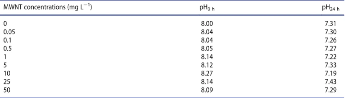
The MNT was performed according to ISO 21427!1 (ISO2006). At the end of 12 d of exposure ! stage 54 (Nieuwkoop and Faber1956) ! larvae are anaesthetized by immersion in a MS222 solution (0.2 g L¡1, Sigma) and a blood sample was obtained from each larva by cardiac puncture. The number of erythrocytes containing one micronucleus (MN) or more (micronucleated erythrocytes) was determined under a microscope in a total of 1000 erythrocytes per larva. The statistical method was described in Mouchet et al. (2008). Val-ues of pH (345 pH meter, Mettler Toledo) were measured three times during the 12 days of exposure just before the renewal of the exposure medium (pH24 h) and just after (pH0 h). Al, Fe, and Mo were measured in RW with and without MWNTs after filtration (0.45 mm to remove most of MWNTs) by ICP!MS (ICP!MS 7500, Agilent).
Results
Activated sludge respiration inhibition test
The method was applied with respect to the following criteria: (1) the difference in respi-ration rates between the two controls was below 15% (Table 1) and (2) EC50of the control
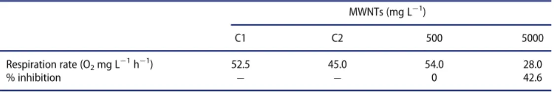
Table 1.Results of respiration and inhibition rate of microorganisms of activated sludge after 3 h in the presence of MWNTs. MWNTs (mg L¡1) C1 C2 500 5000 Respiration rate (O2mg L¡1h¡1) 52.5 45.0 54.0 28.0 % inhibition ! ! 0 42.6 C1, C2: control 1, 2
test with the reference 3,5-dichlorophenol was between the validity specified range of 5 and 30 mg L¡1(14 mg L¡1).
MWNTs did not affect the respiration rate of activated sludge in the conditions of the test up to a concentration of 500 mg L¡1 at 54 and 28 mg O2 mg L
¡1 h¡1,
respectively, for 500 and 5000 mg L¡1 of MWNTs. Inhibition percentage was 0% and 42.6% for 500 and 5000 mg L¡1, respectively. EC50 (3 h) was, therefore, higher than 5000 mg L¡1.
Algal growth inhibition test (P. subcapitata)
The study was performed in compliance with the following quality criteria: (1) biomass in the control cultures increased exponentially by a factor of 102 higher than 16 within the 72-hour test period which corresponds to a specific growth rate of 0.92 d¡1; (2) the mean coefficient of variation for section-by-section specific growth rates (days 0!1, 1!2, and 2!3, for 72-h tests) in the control cultures did not exceed 35%; and (3) the coefficient of variation of average specific growth rates during the whole test period in replicate control cultures did not exceed 7%.
In both tests, inhibition percentage of cell growth and growth rate increase with the increase in MWNT concentrations (Table 2). Total inhibition was observed to 500 mg L¡1of MWNTs for the cell growth and to 1000 mg L¡1for the growth rate. The MWNTs concentration causing a 50% reduction in cell growth (EbC50) was estimated at 34 (23!47) mg L¡1, and the growth rate (E
rC50) was estimated at 120 (87!160) mg L ¡1. The NOEC was also estimated at 10 mg L¡1for the growth rate inhibition and less than 10 mg L¡1for the cell growth. It has to be emphasized that the endpoint used for regulatory pur-poses is the growth rate and not the cell growth (biomass increase). It was observed that the majority of MWNT particles did not remain in suspension between the beginning and the end of the tests but gathered at the lower part of each flask. Microscopic observa-tions confirmed that the algae appeared normal at the end of the test: The normal shape of P. subcapitata algae is a crescent-shaped cell with an average length of 5!10 mm. An increase in the pH was globally observed in both tests for a given concentration between the beginning and the end of the exposure in accordance with classical measures with Table 2.Average percentage inhibition of cell growth (IAi) and growth rate (Imi) of the freshwater algae Pseudokirchneriella subcapitataexposed to MWNTs for 72 h: (a) preliminary test; (b) definitive test.
Nominal concentration of MWNTs (mg L¡1) I Ai(%) Imi(%) (a) 0 0 0 1 0 0 5 0 0 10 0 1 50 27 7 100 54 13 (b) 0 0 0 10 13 2 22 26 6 48 54 14 105 90 44 230 99 73 500 100 82 1000 100 102
algae (Table 3). This may be associated with consumption of the dissolved CO2due to the growth of algae. Above 230 mg L¡1of MWNTs, pH becomes stable during 72 h. Evolu-tion of dissolved O2concentration during 72 h is not significant.
Table 4highlights the decrease of Fe concentrations under MWNT exposure. 11.65 mg kg¡1 of Fe was measured in the presence of Fe and the absence of MWNTs, whereas no Fe was measured in the presence of both Fe and MWNTs. Zn concentra-tions also decreased in less proportion from 3.4 mg L¡1 in the presence of Fe and the absence of MWNTs. Other elements were not affected by the treatment. The results of cell growth and growth rate inhibition without Fe check the effect of iron deficiency and demonstrate iron absorption by MWNTs. Indeed, both inhibition rates lead to 88.88% and to 57.22% in Fe-deprivation medium, whereas there is no inhibition of growth in presence of Fe.
D. magna acute immobilization test
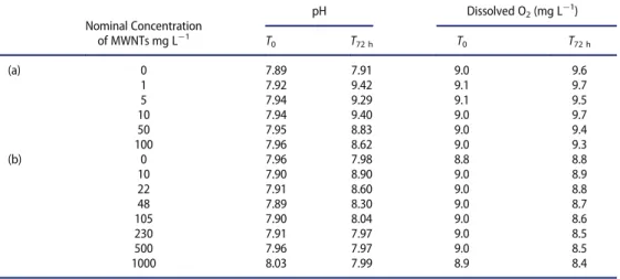
The study was performed in compliance with the following quality criteria: (1) the immobilization in the control did not exceed 10% at the end of the test, (2) daphnids in the control were not trapped at the surface of the water, and (3) the dissolved oxy-gen concentration remained above 3 mg L¡1 over the test period. No immobilization Table 3.Measured pH and O2concentrations in the preliminary test (a) and in the definitive test (b) of the exposure of the freshwater algae Pseudokirchneriella subcapitata to MWNTs.
Nominal Concentration pH Dissolved O2(mg L¡1) of MWNTs mg L¡1 T 0 T72 h T0 T72 h (a) 0 7.89 7.91 9.0 9.6 1 7.92 9.42 9.1 9.7 5 7.94 9.29 9.1 9.5 10 7.94 9.40 9.0 9.7 50 7.95 8.83 9.0 9.4 100 7.96 8.62 9.0 9.3 (b) 0 7.96 7.98 8.8 8.8 10 7.90 8.90 9.0 8.9 22 7.91 8.60 9.0 8.8 48 7.89 8.30 9.0 8.7 105 7.90 8.04 9.0 8.6 230 7.91 7.97 9.0 8.5 500 7.96 7.97 9.0 8.5 1000 8.03 7.99 8.9 8.4
Table 4.Measured metal species in mg kg¡1using ICP!MS in alga medium in presence or absence of Fe and MWNTs.
B Mn Fe Co Ni Cu Zn Mo
Alga medium ¡Fe ¡MWNTs 53.2 110.4 <1 <1 <1 <1 1.6 2.7 Alga medium CFe ¡MWNTs 39.5 122.8 11.6 <1 <1 <1 3.4 3.3 Alga medium CFe CMWNTs 43.8 94.8 <1 <1 <1 <1 <1 3.2 ICP!MS: Inductively coupled plasma!mass spectrometry.
B: Boron ! Mn: Manganese ! Fe: Iron ! Co: Cobalt ! Ni: Nickel ! Cu: Copper ! Zn: Zinc ! Mo: Molybdenum. Measured values correspond to mean value from two replicates.
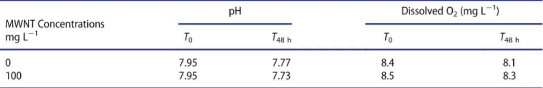
effect is observed, regardless of the MWNT concentration in both tests, at 24 and 48 h. After the 24-h and 48-h test periods, the actual concentrations inhibiting the mobility of daphnids, i.e. EC50!24 h and EC50!48 h, were estimated to be both higher than 100 mg L¡1. Neither pH nor concentrations of dissolved O2were impacted in the pres-ence of 100 mg L¡1of MWNTs during 48 h (Table 5). The appearance of the test sus-pensions was visually checked at the beginning and at the end of the test: as flasks were continuously maintained under axial rotation by use of a cylindrical roller device, MWNTs remained in suspension.
D. magna reproduction test
The study was performed in compliance with the quality criteria: (1) the mortality in the controls (parent females) did not exceed 20% at the end of the test, and (2) the average cumulative number of living young produced per surviving parent female was higher than 60 in the controls at the end of the test. The percentage of inhibition of the reproduc-tion was dose dependent (Table 6). Inhibition percentage ranged from 0.30 (to 10 mg L¡1 of MWNTs) to 21.06% (at 100 mg L¡1of MWNTs). The EC50value was 317.75 mg L¡1, and LOEC and NOEC were 100 and 47 mg L¡1, respectively. After filling and between each renewal, there was sedimentation of the MWNTs at the bottom of the flasks.
Fish acute toxicity test (D. rerio)
The study was performed in compliance with the following quality criteria: (1) the mortal-ity in the control did not exceed 10% at the end of the test; (2) the concentration of dis-solved oxygen in the test vessels remained above 60% of the air saturation value at the end of the test; (3) the pH did not vary by more than 1 unit. The results indicated no mortality effect regardless of the MWNTs concentration in both tests and irrespective of exposure time, 24, 48, 72 or 96 h (Table 7). LC50were then higher than 100 mg L¡1at each time. pH (Table 7) and saturation in oxygen (Table 8) were stable both in the preliminary and Table 5.Dissolved O2and pH measured at the beginning (T0) and at the end (T48 h) of the exposure of Daphnia magnato MWNTs for the definitive test of immobilization.
MWNT Concentrations pH Dissolved O2(mg L¡1) mg L¡1 T 0 T48 h T0 T48 h 0 7.95 7.77 8.4 8.1 100 7.95 7.73 8.5 8.3
Table 6.Results of the Daphnia magna reproduction test. Percentage of inhibition of reproduction measurement after 21 days of exposure.
MWNT concentrations (mg L¡1) 0 5 10 22 47 100
Mean 193.70 197.67 193.11 186.40 169.70 152.90
Standard deviation 21.78 64.40 61.74 15.60 27.08 46.73
definitive tests. Thanks to the stirring device, it was observed that many of the MWNT particles remained in suspension within each tank.
Amphibian (X. laevis) bioassays
No mortality was observed until 72 h of exposure whatever the MWNT concentration. Very low mortality was observed from 50 mg L¡1at 96 h of exposure and was not signifi-cant compared to the negative control (0 mg L¡1). EC50was then estimated to be higher than 500 mg L¡1.
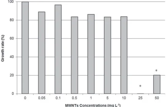
Results of Xenopus exposure for 12 days in semi-static conditions indicated 20% mor-tality at 50 mg L¡1 of MWNTs. No mortality was observed at lower concentrations. Growth inhibition results (Figure 2) were significantly evidenced in larvae exposed to 25 (no growth of larvae) and 50 mg L¡1of MWNTs (four times less). MN induction in Xeno-pus larvae after 12 days of exposure to the referent genotoxic CP (Figure 3) was significant Table 7.Measured pH in (a) preliminary test at the beginning (0 h) of the experiment and at the end of exposure (96 h) and in (b) definitive test, each having a total exposure time of 24 h in fish experiment.
pH (a) MWNT concentrations (mg L¡1) 0 h 96 h 0 7.81 7.81 1 7.76 7.81 35 7.83 7.83 50 7.86 7.87 100 7.85 7.87 pH (b) MWNT concentrations (mg L¡1) 0 h 24 h 48 h 72 h 96 h 0 7.76 7.77 7.46 7.78 7.83 100 7.82 7.75 7.76 7.80 7.85
Table 8.Measured O2concentrations (a) in the preliminary test at the beginning of the experiment (0 h) and at the end of exposure (96 h) and (b) in the definitive test.
Dissolved O2(mg L¡1) (a) MWNT concentrations (mg L¡1) 0 h 96 h 100 8.0 9.2 50 8.3 9.3 35 8.3 9.2 1 8.1 9.1 0 8.2 9.1 Dissolved O2(mg L¡1) (b) MWNT concentrations (mg L¡1) 0 h 24 h 48 h 72 h 96 h 100 95 99 96 97 98 100 95 99 95 98 98 0 95 98 93 98 98 0 95 98 93 97 97
Figure 2.Growth inhibition measurement of Xenopus larvae after 12 days of semi-static exposure to MWNTs.
Note:#indicates a significant lower length compared to the control (0 mg L¡1). Growth rate is calcu-lated as a percentage based on the length measurement of larvae at the beginning of the exposure and at the end.
Figure 3.Micronucleus induction measurement (median § IC 95%) in erythrocytes of Xenopus larvae after 12 days of semi-static exposure according to the concentration of MWNTs.
Note:#indicates a genotoxic condition compared to the negative control NC (0 mg L¡1of MWNTs).PC: Positive Control, Cyclophosphamide, genotoxic of reference to 20 mg L¡1.MNE"/oo: Micronucleated erythrocytes.
compared to the NC. This result validates the bioassay. The results of MN induction in larvae exposed to MWNTs indicated no genotoxicity compared to the NC group. Median values were distributed without MWNTs’ dose!effect relation. pH values (Table 9) were slightly lower after 24 h of exposure (pH24 h) compared to just after the renewal of the exposure medium (pH0 h). It may be in relation to the excretion process of larvae for 24 h and the acidification of the exposure media. There was no pH modification in relation to MWNT concentrations. Metals were dosed in water exposure in the presence or absence of 50 mg L¡1of MWNTs, without larvae, after 24 h of contact (Table 10). 12.8 § 0.6 mg L¡1 of Mo and 131.5 § 5.4 mg L¡1 of Al were measured in medium in the presence of MWNTs, whereas concentration of Fe was under the quantification limit (<10 mg L¡1).
Discussion
The aim of the present work is not to compare the biological effects between different bio-logical models but to contribute to a better understanding of the ecotoxicity of CNTs and their environmental exposure assessment to provide valuable insight into likely exposure scenarios at different levels of the trophic chain. Synthetic results of the biological effects in organisms after MWNT exposure are presented inTable 11. The results are as follows: no toxicity in activated sludge (bacteria) at 500 mg L¡1 of MWNTs, no acute toxicity in fish and daphnia up to 100 mg L¡1of MWNTs, inhibition of growth in amphibian larvae at 25 mg L¡1of MWNTs, and a notable effect at high concentrations of MWNTs with the growth inhibition test in algae (EC50D120 mg L
¡1and NOEC D 10 mg L¡1).
Observed toxicity in this present work is in accordance with much of the data pub-lished in the literature relative to the potential toxicity of raw CNTs in aquatic organisms. Table 9.Measured pH values during the 12 days of exposure of Xenopus larvae to different concentra-tions of MWNTs. pH0 hwas measured just after the renewal of the exposure medium. pH24 hwas mea-sured just before the renewal.
MWNT concentrations (mg L¡1) pH0 h pH24 h 0 8.00 7.31 0.05 8.04 7.30 0.1 8.04 7.26 0.5 8.05 7.27 1 8.14 7.22 5 8.12 7.33 10 8.27 7.19 25 8.14 7.43 50 8.09 7.29
Table 10.Measured metal species (Al, Fe, Mo) in mg L¡1using ICP!AES in amphibian mediums in the presence or absence of 50 mg L¡1of MWNT after 24 h of contact.
Fe Mo Al
!MWNTs <10 mg L¡1 <10 mg L¡1 <50 mg L¡1
CMWNTs <10 mg L¡1 12.80 § 0.58 mg L¡1 131.50 § 5.36 mg L¡1 Fe: Iron ! Mo: Molybdenum ! Al: aluminum.
Calculated values correspond to mean value from 4 replicates (§ standard error). Quantification limit (QL) is 10 mg L¡1for Fe and Mo, and 50 mg L¡1for Al.
Table 11. Synthetic results of the biologic effects in organisms after exposure to MWNTs. Bi oassay Orga nism Biolog ical endpo int Expo sure time Measur e EC 50 mg L ! 1 NOEC mg L ! 1 LOEC mg L ! 1 OEC D 209 Bac teria activate d slud ge In hibition perc entage 3 h 0% at 500 mg L ¡ 1 42.6 % at 5000 mg L ¡ 1 > 5000 OEC D 201 Fres h algae Pseu dokirchneriella subcapit ata Ce ll growth ! biom ass 72 h 34 < 10 Gr owth rate ! ce ll multiplication 72 h 120 10 OEC D 202 Daph nid Mob ility 24 h 48 h > 100 > 100 OEC D 211 D aphnia magna Rep roduction 21 days 317.7 5 47 100 ISO 21 427 ! 1 Amp hibia n Xenopus laevi s Mor tality 96 h > 500 Mor tality 12 days 20% at 50 mg L ¡ 1 Gr owth inhi bition 12 days Signi fi cant inhi bition from 25 mg L ¡ 1 Mic ronuclei (MN) in duction 12 days No signi fi cant MN indu ction
An LOEC of 10 mg L¡1was observed in daphnia (Roberts et al. 2007), in marine copepod (Templeton et al.2006) and in amphibians (Mouchet et al.2008). Zhu and collaborators (Zhu et al.2009) calculated EC50to 8.72 and 1.30 mg L¡1for immobilization of daphnia exposed to raw SWNTs and MWNTs, respectively, and to 22.57 and 2.42 mg L¡1for mor-tality exposed to raw SWNTs and MWNTs, respectively. Kennedy and collaborators (Kennedy et al.2008) calculated EC50for mortality after 48 h of raw MWNTs exposure in daphnia at 50.9 mg L¡1. No toxicity was evidenced for hydra and crustaceans up to 100 mg L¡1 of raw SWNTs (Blaise et al. 2008), whereas toxicity to algae exposed to SWNTs was observed at 10 mg L¡1. Cheng, Flahaut, and Cheng (2007) observed hatching delay in fish eggs exposed to 120 mg L¡1of raw SWNTs and 240 mg L¡1of raw DWNTs. Neither mortality nor growth inhibition was observed in urodele amphibian larvae up to 1 g L¡1of raw DWNTs (Mouchet et al.2007), whereas mortality was observed at 50 mg L¡1 and growth inhibition from 10 mg L¡1 in anuran amphibian larvae exposed to raw DWNTs (Mouchet et al.2008, 2011) as well as growth inhibition at 50 mg L¡1 for raw MWNTs exposure (Mouchet et al. 2010). Kahru and Dubourguier (2011) calculated on the basis of 34 median values a L(E)C50 between 1 and 10 mg L
¡1 for SWNTs and MWNTs (L(E)C50 derived from 77 individual values. The majority of the published results indicate that exposure to CNTs generally leads to biological disorder at different levels but usually above unrealistic concentrations of 10 mg L¡1. In surface water in Europe, for the simulation results of the predicted environmental concentrations, Gott-schalk et al. (2009) indicated lower CNTs concentrations, with 0.004 ng L¡1 (most fre-quent value) and 0.0035 ng L¡1as the range of the lower quantile and 0.021 g L¡1as the upper quantile. Nevertheless, it could be hypothesized that CNT concentrations accumu-late into the environment over time.
Toxicity obtained from algae in the present work appears to be in relation to the effect of Fe deficiency on algal growth. Indeed, algal growth experiments in the absence of iron indicated 88.8% of cell growth inhibition (biomass) and 57.2% of growth rate inhibition (cell multiplication) compared to the absence of inhibition when iron was present. More-over, among the 8 micro-nutrients that are present in the algal culture medium (boron, manganese, iron, cobalt, nickel, copper, zinc, and molybdenum), iron is strongly removed by the MWNTs from the filtrated culture medium (<1 mg kg¡1). When compared to the initial algal medium in which iron concentration is 11.65 mg kg¡1, this leads to suspect iron adsorption by MWNTs. Iron ion adsorption has already been demonstrated in chem-ical studies using different types of CNTs (Li et al.2009). In contrast to the present growth inhibition of P. subcapitata due to iron deprivation, Schwab et al. (2011) indicated that observed inhibition of C. vulgaris and P. subcapitata is in relation to light masking by CNTs, which can adhere to algal surfaces and hence restrict light accessibility to the cells, resulting in the inhibition of growth. Long et al. (2012) indicated that their MWNTs sig-nificantly inhibited the algal growth of Chlorella sp. with a negligible contribution of metal catalyst residues in the MWNTs and nutrient elements adsorbed by MWNTs. These authors hypothesize that the toxicity of algae could mainly be explained by the combined effects of oxidative stress, agglomerations and physicals interactions, and shading effects, with the quantitative contributions from these mechanisms depending on the MWNT size and concentration. In any case, comparison of results between these different works must be limited because of the diversity of studied CNTs. Nevertheless, Verneuil et al. (2014) indicated that only direct exposure to 50 mg L¡1 of MWNTs (the same type of
MWNTs as in the present study) led to growth inhibition of Nitzschia palea after 48 h and suggested that EPS (extracellular polymeric substances) provide considerable protection against MWNTs, without alteration of the photosynthesis.
Concerning the absence of genotoxicity in erythrocytes of Xenopus larvae, the present results are in agreement with the previous ones obtained on amphibian larvae in the same conditions of exposure to MWNTs (Mouchet et al.2010) and DWNTs (Mouchet et al. 2007, 2008). The majority of the time, if acute and chronic toxicities are generally observed after CNTs exposure of different biological models, genotoxicity, especially via micronucleus induction mechanism, is not demonstrated. Kim et al. (2011) indeed obtained no genotoxicity of raw MWNTs according to OECD test guidelines 471 (bacte-rial reverse mutation test), 473 (in vitro chromosome aberration test with and without S9), and 474 (in vivo micronuclei test). Di Sotto et al. (2009) and Szendi and Varga (2008) also reported that MWNTs had no mutagenic effect in bacteria systems. In the same way, Wirnitzer et al. (2008) indicated no genotoxicity of raw MWNTs testing for chromosome aberrations in V79 cells and for gene mutations in the Salmonella microsome test. Never-theless, genotoxic effects may be produced either by direct interaction of particles with genetic material or by secondary damage from particle-induced reactive oxygen species. In this context, some authors demonstrated oxidative stress by MWNTs (Reddy et al. 2010; Srivastava et al.2011), and, for example, in Xenopus larvae after MWNT exposure (Saria et al.2014).
Exposure media for the different organisms would play a role in the observed toxicity of the present work. Nevertheless, characterizations of the MWNT suspension in exposure medium do not appear essential to place it in relation with biological effects because they are observed at very high and unrealistic concentrations. The effects obtained in the pres-ent work are globally weakly marked, probably in relation with the MWNTs’ limited bio-availability in the water column for organisms because of CNTs’ sedimentation at the bottom of containers. As displayed inFigure 1, raw MWNTs appeared as large rather spherical agglomerates of bundles without free or isolated nanotubes. Observed effects in organisms may be in connection with exposure to these agglomerates inducing respiratory and/or intestinal clogging in relation with their absorption and not necessarily related to the intrinsic effects of CNTs (Mouchet et al.2010 and2011; Petersen et al.2011). This result is also in accordance with the observation of CNTs in the guts of aquatic organisms such as Lumbriculus variegatus (Petersen, Huang, and Weber 2008), Arenicola marina (Galloway et al.2010), D. magna (Zhu et al.2009), Hyalella azteca, Leptocheirus plumulo-sus, and Ceriodaphnia dubia (Kennedy et al. 2009). Li and Huang (2011) describe the ingestion of CNTs in C. dubia followed by excretion in exposure media. Many of these studies tend to highlight that ingested CNTs by organisms may enter the ecological pyra-mid via their move up through the food chain. Moreover, excreted CNTs may contribute to maintaining a pressure contamination of CNTs in media.
Nevertheless, a few mg L¡1 of Mo and Al (and no Fe) were measured in the water medium of amphibian exposure after 24 h containing the higher concentration of MWNTs (50 mg L¡1). It would suggest that they may contribute, especially Al, to the tox-icity observed in amphibians to high concentrations of MWNTs, although no genotoxtox-icity was observed. This result encourages us to investigate the potential release of metal impu-rities at lower concentrations and in function of time in the different water exposures.
Conclusion
The present knowledge concerning the ecotoxic effects of CNTs is rather limited and deserves to be documented more extensively. First, the ecotoxicological hazard assessment needs approaches and measurement tools using standardized test methods. Then, adapta-tion of well-known protocols is necessary. This work is thus a contribuadapta-tion to the assess-ment of the potential ecotoxicity of CNTs within the aquatic compartassess-ment; it could be helpful for regulatory purposes. The results indicate that MWNT effects are weakly marked and expressed at unrealistic nominal concentrations (approximately 10 mg L¡1), in relation with probable MWNT ingestion. Considering their increasing use in commer-cial products, this study emphasizes the need to further study their ecotoxicity and high-lights that assessing the risks of CNTs requires a better understanding of their toxicity, bioavailability, and behavior in relation with their intrinsic physicochemical properties.
Acknowledgments
The authors acknowledge Annie Perrault and Floriane Bourdiol for their technical help. Thanks to the Groupement de Recherche de Lacq (ARKEMA) for MWNTs suspensions and technical help. We are also indebted to Michel Lagoin (Universit!e Paul Sabatier, Toulouse, France) for reviewing the English version of the manuscript and to the language services of Elsevier.
Disclosure statement
Part of this research has been developed in the framework of the French collaborative laboratory NAUTILE (Nanotubes et Ecotoxicologie), which is the first public/private joint laboratory dedi-cated to the study of the ecotoxicological impact of CNTs in the aquatic environment. The frame-work agreement was signed by (1) Arkema France, (2) Centre National de la Recherche Scientifique (CNRS), (3) Institut National Polytechnique de Toulouse (INPT), and (4) Universit!e Paul Sabatier (UPS) of Toulouse in September 2010. The authors declare no competing financial interests or con-flict of interest.
References
Blaise, C., F. Gagn!e, J.F. F!erard, and P. Eullafroy.2008. “Ecotoxicity of Selected Nano-materials to Aquatic Organisms.” Environmental Toxicology 23 (5): 591!598.
Cheng, J., E. Flahaut, and S.H. Cheng.2007. “Effect of Carbon Nanotubes on Developing Zebrafish (Danio rerio) Embryos.” Environmental Toxicology and Chemistry 26 (4): 708!716.
Crane, M., R.D. Handy, J. Garrod, and R. Owen.2008. “Ecotoxicity Test Methods and Environmen-tal Hazard Assessment for Engineered Nanoparticles.” Ecotoxicology 17: 421!437.
Di Sotto, A., M. Chiaretti, G.A. Carru, S. Bellucci, and G. Mazzanti.2009.“Multi-walled Carbon Nanotubes: Lack of Mutagenic Activity in the Bacterial Reverse Mutation Assay.” Toxicology Letters 184 (3): 192!197.
Eklund, P., P. Ajayan, R. Blackmon, A.J. Hart, J. Kong, B. Pradhan, A. Rao, and A. Rinzler.2007. International Assessment of Research and Development of Carbon Nanotube Manufacturing and Applications. Baltimore, MD: World Technology Evaluation Center.http://www.wtec.org/cnm/
CNM_final_report.pdf.
EN ISO 6341. 1996. “Water Quality ! Determination of the Inhibition of Mobility of Daphnia magna Straus (Cladocera, Crustacea) ! Acute Toxicity Test.” Geneva: International Organiza-tion for StandardizaOrganiza-tion.
EN ISO 7346.1998. “Water Quality ! Determination of the Acute Lethal Toxicity of Substances to a Freshwater Fish [Brachydanio rerio Hamilton!Buchanan (Teleostei, Cyprinidae)]. Part 1: Static Method. Part 2: Semi-static Method. Part 3: Flow-Through Method.” Geneva: Interna-tional Organization for Standardization.
EN ISO 8692.2004. “Water Quality ! Fresh Water Algal Growth Inhibition Test with Unicellular Green Algae.” Commission Regulation (EC) No 440/2008 of 30 May 2008. Geneva: Interna-tional Organization for Standardization.
ENV/JM/MONO 13/REV. 2008. “Environment Directorate ! Joint Meeting of the Chemicals
Committee and the Working Party on Chemicals, Pesticides, and Biotechnologies Series on the Safety Manufactured Nanomaterials.” No. 6. List of Manufactured Nanomaterials and List of Endpoints for Phase One of the OECD Testing Programme JT03248749. Unclassified, 07!Jul!2008. 1!13. Paris: Organization for Economic Co-operation and Development (OECD).
EU 2008. “Regulation EC No 1272/2008 of the European Parliament and of the Council of 16
December 2008 on Classification, Labelling and Packaging of Substances and Mixtures, Amend-ing and RepealAmend-ing Directives 67/548/EEC and 1999/45/EC, and AmendAmend-ing Regulation (EC) No 1907/2006.” Official Journal of the European Union L353: 1355.
Galloway, T., C. Lewis, I. Dolciotti, B.D. Johnston, J. Moger, and F. Regoli.2010. “Sublethal Toxicity of Nano-titanium Dioxide and Carbon Nanotubes in a Sediment Dwelling Marine Polychaete.” Environmental Pollution 158: 1748!1755.
Gottschalk, F., T. Sonderer, R. Scholz, and B. Nowack.2009. “Modeled Environmental Concentra-tions of Engineered Nanomaterials (TiO2, ZnO, Ag, CNT, Fullerenes) for Different Regions.” Environmental Science and Technology 43: 9216!9222.
Guadagnini, R., B. Halamoda Kenzaoui, L. Cartwright, G. Pojana, Z. Magdolenova, D. Bilanicova, M. Saunders, et al.2013. “Toxicity Screenings of Nanomaterials: Challenges Due to Interference with Assay Processes and Components of Classic In Vitro Tests.” Nanotoxicology (Supplement 1) 9: 13!24. doi:10.3109/17435390.2013.829590
Iijima, S.1991. “Helical Microtubules of Graphitic Carbon.” Nature 354: 56!58.
ISO.2006. ISO International Standard. Water Quality ! Evaluation of Genotoxicity by Measure-ment of the Induction of Micronuclei ! Part 1: Evaluation of Genotoxicity Using Amphibian Larvae. ISO 21427!1, ICS: 13.060.70. Geneva: International Organization for Standardization. Kahru, A., H.C. Dubourguier, I. Blinova, A. Ivask, and K. Kasemets.2008. “Biotests and Biosensors
for Ecotoxicology of Metal Oxide Nanoparticles: A Mini Review.” Sensors 8: 5153!517. Kahru, A., and H.C. Dubourguier.2011. “From Ecotoxicology to Nanoecotoxicology.” Toxicology
269 (2!3): 105!119.
Kennedy, A.J., J.C. Gunter, M.A. Chappell, J.D. Goss, M.S. Hull, R.A. Kirgan, and J.A.A. Steevens.
2009. “Influence of Nanotube Preparation in Aquatic Bioassays.” Environmental Toxicology and Chemistry 28 (9): 1930!1938.
Kennedy, A.J., M.S. Hull, J.A. Steevens, K.M. Dontsova, M.A. Chappell, J.C. Gunter, and C.A. Weiss.2008. “Factors Influencing the Partitioning and Toxicity of Nanotubes in the Aquatic Environment.” Environmental Toxicology and Chemistry 27 (9): 1932!1941.
Kim, J.S., K. Lee, Y.H. Lee, H.S. Cho, K.H. Kim, K.H. Choi, S.H. Lee, K.S. Song, C.S. Kang, and I.J.
Yu. 2011. “Aspect Ratio Has No Effect on Genotoxicity of Multi-wall Carbon Nanotubes.”
Archives of Toxicology 85 (7): 775!86.
Kostarelos, K., A. Bianco, and M. Prato.2009. “Promises, Facts and Challenges for Carbon Nano-tubes in Imaging and Therapeutics.” Nature Nanotechnology 4 (10): 627!633.
Li, H.B., L.K. Pan, Y. Zhang, and Z. Sun.2009. “Ferric Ion Adsorption and Electrode Sorption by Carbon Nanotubes and Nanofibres Films.” Water Science and Technology 59 (8): 1657!1663. Li, M., and C.P. Huang.2011. “The Responses of Ceriodaphnia dubia Toward Multi-walled Carbon
Nanotubes: Effect of Physical!Chemical Treatment.” Carbon 49: 1672!1679.
Long, Z., J. Ji, K. Yang, D. Lin, and F. Wu.2012. “Systematic and Quantitative Investigation of the Mechanism of Carbon Nanotubes’ Toxicity Toward Algae.” Environmental Science and Technol-ogy 46 (15): 8458!8466.
Mouchet, F., P. Landois, V. Datsyuk, P. Puech, E. Pinelli, E. Flahaut, and L. Gauthier.2011. “Use of the International Amphibian Micronucleus Standardized Procedure (ISO 21427!1) for In Vivo Evaluation of Double-Walled Carbon Nanotubes Toxicity and Genotoxicity in Water.” Environ-mental Toxicology 26 (2): 136!145.
Mouchet, F., P. Landois, E. Flahaut, E. Pinelli, and L. Gauthier.2007. “Assessment of the Potential In Vivo Ecotoxicity of Double-Walled Carbon Nanotubes (DWNTs) in Water, Using the Amphibian Ambystoma mexicanum.” Nanotoxicology 1 (2): 149!156.
Mouchet, F., P. Landois, P. Puech, E. Pinelli, E. Flahaut, and L. Gauthier.2010. “CNTs Ecotoxicity in Amphibians: Assessment of Multi-walled Carbon Nanotubes (MWNT) and Comparison with Double-Walled Carbon Nanotubes (DWNT).” Special Focus Environmental Toxicity of Nanoparticles. Nanomedicine 5 (6): 963!974.
Mouchet, F., P. Landois, E. Sarrem!ejean, G. Bernard, P. Puech, E. Pinelli, E. Flahaut, and L. Gauth-ier.2008. “Characterisation and In Vivo Ecotoxicity Evaluation of Double-Wall Carbon Nano-tubes in Larvae of the Amphibian Xenopus laevis.” Aquatic Toxicology 87: 127!137.
Nieuwkoop, P.D., and J. Faber. 1956. Normal Tables of Xenopus Laevis (Daudin). Amsterdam: North-Holland.
OECD (Organisation for Economic Co-operation and Development).1984. “Activated Sludge, Res-piration Inhibition Test.” Guideline N"209 for the Testing of Chemicals, Adopted April 4, 1984. Paris: Organisation for Economic Co-operation and Development.
OECD (Organisation for Economic Co-operation and Development).1992. “Fish, Acute Toxicity Test.” Guideline N"203 for the Testing of Chemicals, Adopted 17 July 1992. Paris: Organisation for Economic Co-operation and Development.
OECD (Organisation for Economic Co-operation and Development).2004. “Daphnia sp., Acute
Immobilisation Test.” Guideline 202 for the Testing of Chemicals, Adopted 13 April 2004. Paris: Organisation for Economic Co-operation and Development.
OECD (Organisation for Economic Co-operation and Development).2006. “Freshwater Alga and Cyanobacteria, Growth Inhibition Test.” Guideline 201 for the Testing of Chemicals, Adopted 23 March 2006. Paris: Organisation for Economic Co-operation and Development.
OECD (Organisation for Economic Co-operation and Development). 2008. “Daphnia magna,
Reproduction Test.” Guideline 211 for the Testing of Chemicals, Adopted: 3 October 2008. Paris: Organisation for Economic Co-operation and Development.
Petersen, E.J., Q. Huang, and W.J. Weber.2008. “Ecological Uptake and Depuration of Carbon Nanotubes by Lumbriculus variegates.” Environmental Health Perspectives 116 (4): 496!500. Petersen, E.J., L. Zhang, N. Mattison, D. O’Carroll, A.J. Whelton, N. Uddin, T. Nguyen, et al.2011.
“Potential Release Pathways, Environmental Fate, and Ecological Risks of Carbon Nanotubes.” Environmental Science and Technology 45: 9837!9856.
Reddy, A.R., Y.N. Reddy, D.R. Krishna, and V. Himabindu.2010. “Multi-wall Carbon Nanotubes Induce Oxidative Stress and Cytotoxicity in Human Embryonic Kidney (HEK293) Cells.” Toxi-cology 272 (1!3): 11!6.
Roberts, A.P., A.S. Mount, B. Seda, J. Souther, R. Qiao, S. Lin, P.C Ke, A.M. Rao, and S.J. Klaine.
2007. “In Vivo Biomodification of Lipid Coated Carbon Nanotubes by Daphnia Magna.” Envi-ronmental Science and Technology 41: 3025!3029.
Saria, R., F. Mouchet, A. Perrault, E. Flahaut, C. Laplanche, J.C. Boutonnet, E. Pinelli, and L. Gauth-ier.2014. “Evaluation of Oxidative Stress and DNA Damage in Xenopus laevis Tadpoles Exposed to Multi-walled Carbon Nanotubes.” Ecotoxicology and Environmental Safety. 06/2014; 107C: 22!29. doi:http://dx.doi.org/10.1016/j.ecoenv.2014.05.010.
Schwab, F., T.D. Bucheli, L.P. Lukhele, A. Magrez, B. Nowack, L. Sigg, and K. Knauer.2011. “Are Carbon Nanotube Effects on Green Algae Caused by Shading and Agglomeration?” Environ-mental Science and Technology 45: 6136!6144.
Srivastava, R.K., A.B. Pant, M.P. Kashyap, V. Kumar, M. Lohani, L. Jonas, and Q. Rahman.2011. “Multi-walled Carbon Nanotubes Induce Oxidative Stress and Apoptosis in Human Lung Can-cer Cell Line!A549.” Nanotoxicology 5 (2): 195!207.
Stafiej, A., and K. Pyrzynska.2007. “Adsorption of Heavy Metal Ions with Carbon Nanotubes.” Sep-aration and Purification Technology 58(1): 49!52. doi:10.1016/j.seppur.2007.07.008.
Szendi, K., and C. Varga.2008. “Lack of Genotoxicity of Carbon Nanotubes in a Pilot Study.” Anti-cancer Research 28: 349!352.
Templeton, R.C., P. Lee Ferguson, K.M. Washburn, W.A. Scrivens, and G.T. Chandler.2006. “Life-Cycle Effects of Single-Walled Carbon Nanotubes (SWNTs) on an Estuarine Meiobenthic Cope-pod.” Environmental Science and Technology 40: 7387!7393.
Van der Zande, M., R. Junker, X.F. Walboomers, and J.A. Jansen.2011. “Carbon Nanotubes in Ani-mal Models: A Systematic Review on Toxic Potential.” Tissue Engineering Part B Rev 17 (1): 57!69.
Verneuil, L., J. Silvestre, F. Mouchet, E. Flahaut, J.C. Boutonnet, F. Bourdiol, T. Bortalomiol, D. Baque, L. Gauthier, and E. Pinelli.2014. “Multi-walled Carbon Nanotubes, Natural Organic Matter and the Benthic Diatom Nitzschia palea: ”A Sticky Story“.” Nanotoxicology. [Epub ahead of print] (doi:10.3109/17435390.2014.918202).
Wirnitzer, U., B. Herbold, M. Voetz, and J. Ragot.2008. “Studies on the In Vitro Genotoxicity of Baytubes®, Agglomerates of Engineered Multi-walled Carbon Nanotubes (MWCNT). Toxicity of Engineered Nanomaterials.” Toxicological Letters 186 (3!8): 160!165.
Zhao, X., and R. Liu.2012. “Recent Progress and Perspectives on the Toxicity of Carbon Nanotubes at Organism, Organ, Cell, and Biomacromolecule Levels.” Environment International 40: 244!255.
Zhu, X., L. Zhu, C. Yongsheng, and T. Shengyan.2009. “Acute Toxicities of Six Manufactured Nanomaterial Suspensions to Daphnia magna.” Journal of Nanoparticle Research 11 (1): 67!75.