Université de Montréal
Moduler la costimulation des lymphocytes T pour la
prévention du rejet de l'allogreffe rénale chez les primates
non humains / Modulating T Cell Costimulation to Prevent
Renal Allograft Rejection in Nonhuman Primates
par Lijun Song
Département de chirurgie Faculté de médecine
Thèse présentée à la Faculté des études supérieures en vue de l’obtention du grade de Philosophiae Doctor (Ph.D.)
en Sciences Biomédicales
Août, 2016
Université de Montréal
Faculté des études supérieures et postdoctorales
Cette thèse intitulée :
Moduler la costimulation des lymphocytes T pour la prévention du rejet de
l'allogreffe rénale chez les primates non humains / Modulating T Cell
Costimulation to Prevent Renal Allograft Rejection in Nonhuman Primates
Présentée par : Lijun Song
a été évaluée par un jury composé des personnes suivantes :
Dr Richard Bertrand, président-rapporteur Dr Huifang Chen, directeur de recherche
Dr Louis Dumont, codirecteur Dr Édouard Kouassi, membre du jury Dr Stanislaw Stepkowski, examinateur externe Dr Jean-François Cailhier, représentant du doyen de la FES
Résumé
La transplantation d'organes est souvent la meilleure approche thérapeutique pour l'insuffisance organique au stade terminal. Le rejet de greffe est le principal obstacle pour une transplantation réussie, car cette dernière est le plus fréquemment réalisée entre individus génétiquement distincts. Les tissus ou organes transplantés sont généralement reconnus par le système immunitaire comme corps étrangers et sont rapidement détruits. Une série d'approches a été réalisée en clinique pour augmenter l'acceptation de la transplantation d’organes. Les immunosuppresseurs ont un rôle clé dans le combat contre le rejet de greffe. Actuellement, les résultats à court terme de la transplantation d'organes ont été considérablement améliorés avec l'émergence des inhibiteurs de la calcineurine (ICN), mais les résultats à long terme sont encore insatisfaisants. L'une des principales raisons est que les médicaments immunosuppresseurs actuels manquent de spécificité. Ces agents, en particulier ICN, sont considérés comme les facteurs de risque pour la perte tardive du greffon et les décès avec un greffon fonctionnel en raison de la toxicité des médicaments, de la sur-immunosuppression et d'autres effets secondaires. Ainsi, il y a un besoin urgent de rechercher de nouvelles thérapies idéales.
Les lymphocytes T jouent un rôle central dans l'initiation du rejet des allogreffes. La pleine activation des cellules T nécessite au moins deux signaux combinés, mais distincts. En plus du signal généré par l'interaction entre le récepteur des cellules T (TCR) et les complexes CMH-peptides, le second signal, appelé signal de costimulation qui est délivré par
l'engagement du récepteur de costimulation avec son ligand, est essentiel. L'engagement du TCR en l'absence de signaux de costimulation peut donner lieu à l'anergie des cellules T, un état d'absence de réponse immunitaire. Les molécules costimulatrices acquièrent ainsi l'attention en tant que cibles thérapeutiques potentielles. Du fait que ces molécules soient largement limitées à des cellules T et/ou des cellules présentatrices d'antigène, cibler la voie de signalisation de costimulation offre la possibilité de moduler le système immunitaire d'une manière plus sélective par rapport à des agents immunosuppresseurs conventionnels.
À ce jour, de nombreuses molécules costimulatrices ont été identifiées et certaines ont été testées en tant que cibles thérapeutiques dans des modèles de transplantation d'organes. Les axes CD28–CD80/86 et CD40–CD40L sont importants et les deux voies de signalisation de costimulation les mieux connues. Bélatacept est un variant de l'antigène 4 des lymphocytes T cytotoxiques-immunoglobuline G (CTLA4-Ig) qui bloque la voie de signalisation CD28– CD80/86. C'est le seul bloqueur de la costimulation à être approuvé pour utilisation clinique en transplantation d'organes. Par rapport à la thérapie basée sur les ICN pour les receveurs de transplantation rénale, les thérapies à base de bélatacept montrent un taux similaire de survie, une fonction supérieure du greffon et l'amélioration du profil de risque cardiovasculaire. Cependant, bélatacept est également associée à des taux plus élevés de rejet aigu et de syndrome lymphoprolifératif post-greffe (SLPG).
Dans notre étude, l'efficacité d'ASKP1240, un nouvel AcM anti-CD40 complètement humain qui bloque la voie de costimulation CD40–CD40L, a été évaluée sur la prévention du rejet d'allogreffe avec le modèle de transplantation rénale chez le singe cynomolgus. Quand
ASKP1240 a été administré seul, il a réduit l'incidence du rejet aigu et a prolongé la survie de l'allogreffe rénale dépendamment de la dose administrée. L'acceptation de l'allogreffe rénale a été encore améliorée chez des singes qui ont reçus des traitements d'ASKP1240 combiné avec le tacrolimus (dose sub-thérapeutique) ou le mycophénolate mofétil (MMF). Le rejet aigu d'allogreffe a été totalement éliminé chez ces animaux et la médiane de survie du greffon rénal de ces groupes était significativement plus longue que ceux des groupes avec un traitement monothérapie. ASKP1240 a été bien toléré pour un traitement allant jusqu'à 180 jours. Il n'y avait pas d'effets secondaires évidents, y compris les complications thromboémboliques liées au médicament. L'administration d'ASKP1240 n'a pas induite de libération de cytokines.
Ensuite, nous avons étudié les effets d'ASP2409 sur le rejet de l'allogreffe rénale et la survie chez des singes cynomolgus. ASP2409 est une nouvelle CD86-sélective variante de CTLA4-Ig, qui possède une affinité de liaison au CD86 14 fois plus élevée que le bélatacept in vitro et une amélioration de la puissance immunosuppressive. Une haute dose d'ASP2409 en monothérapie a montré des résultats supérieurs dans la réduction de rejet aigu et la prolongation de la survie de l'allogreffe rénale en comparaison avec une faible dose d'ASP2409 en monothérapie. Une faible dose d'ASP2409 en combinaison avec tacrolimus (dose sub-thérapeutique) inhibe complètement le rejet aigu d'allogreffe et prolonge significativement la survie de l'allogreffe rénale par rapport à une monothérapie avec une faible dose d'ASP2409 ou une dose sub-thérapeutique de tacrolimus. La médiane de survie de l'allogreffe des animaux recevant un traitement à base d'une dose élevée d'ASP2409, bélatacept, ou une dose thérapeutique de tacrolimus étaient identiques (> 91 jours). Les
traitements à base d'une dose élevée d'ASP2409 présentaient de meilleurs résultats histopathologiques que le traitement à base de bélatacept. En outre, la fréquence des cellules FoxP3+ dans les allogreffes rénales a été observée plus haute dans les traitements à base
d'ASP2409 et de bélatacept comparés aux traitements à base de tacrolimus. L'étude a également montré que ASP2409 est sans danger pour les animaux pour les doses qui ont été testées. Nous n'avons pas trouvé de graves effets secondaires liés à ASP2409 au cours des 91 jours d'étude.
Collectivement, ces résultats suggèrent que la modulation sélective de la costimulation des cellules T avec des bloqueurs de la costimulation est une stratégie réalisable pour la prévention et le traitement du rejet d'allogreffe. ASKP1240 et ASP2409 sont tous deux des agents immunosuppresseurs prometteurs pour les régimes d'évitement ou de réduction des inhibiteurs de la calcineurine.
Mots-clés: blocage de la costimulation, transplantation rénale, primate non humain, rejet aigu
Abstract
Organ transplantation is often the best therapeutic approach for end-stage organ failure. Graft rejection is the major obstacle to successful transplantation because transplantation is most frequently carried out between genetically distinct individuals. Transplanted tissues or organs are usually recognized by the immune system as foreign and are rapidly destructed without immune interventions. A series of approaches have thus been applied in clinic to inhibit the allogenic immune responses and in turn increase organ transplant acceptance. Immunosuppressive drugs are the key players in the "war" against immune cell-mediated rejection of allogenic transplants. Currently, the use of calcineurin inhibitors (CNIs) has dramatically decreased the risk of acute transplant rejection and improved the short-term outcomes of organ transplantation, but the long-term outcomes are still unsatisfied. One of the main reasons causing unsatisfied long-term outcomes is that current immunosuppressive drugs do not specifically target immune cells that cause transplant rejection. These immunosuppressive agents, especially CNIs, are the risk factors for late graft loss and death with functioning graft (DWFG) due to drug toxicity, over-immunosuppression, and other side-effects. Thus there is an urgent need for seeking novel therapies that can selectively eliminate the alloreactive immune responses while preserving the integrity of the remainder of the host immune system.
It has been known that T cells play a central role in initiating allograft rejection. Full activation of T cells requires at least two collaborative but distinct signals. The first signal is
generated by the interaction between T cell receptor (TCR) and MHC-peptide complex. The second signal, termed costimulatory signal, is delivered through the engagement of costimulatory receptors by their ligands. Importantly, TCR engagement in the absence of costimulatory signals can result in T cell anergy, a state of T cell unresponsiveness. Costimulatory molecules thus gain attention as potential therapeutic targets. Because these molecules are primary expressed on T cells and/or antigen-presenting cells, targeting costimulatory signaling pathway offers the oppertumities to modulate immune system in a more selective way relative to conventional immunosuppressive agents.
To date, numerous costimulatory molecules have been identified and some have been tested as therapeutic targets in organ transplant models. CD28–CD80/86 and CD40–CD40L axis are two important and the most well known costimulatory signaling pathways. Belatacept, a variant of cytotoxic T lymphocyte antigen 4-immunoglobulin G (CTLA4-Ig) that blocks CD28–CD80/86 signaling pathway, is the only costimulation blocker to be approved for clinical use in organ transplantation. Compared to CNI-based regimen for kidney transplant recipients, belatacept-based regimens show similar patient and graft survival rate, superior graft function, and improved cardiovascular risk profile. However, belatacept is also associated with higher rates of acute rejection and posttransplant lymphoproliferative disorder (PTLD). Disruption of CD40–CD40L interaction with anti-CD40L mAbs has also been demonstrated to be a reliable approach for preventing acute rejection and for prolonging allograft survival. Unfortunately, unexpected thromboembolic complications in preclinical studies and clinical trials discontinued the development of anti-CD40L mAbs. The main objective of this thesis is to identify the optimal T cell costimulation blockers that can show
improved safety and non-inferior efficacy in promoting allograft acceptance relative to current costimulatory blocking agents.
Anti-CD40 mAbs are an alternative approach to block CD40–CD40L costimulatory pathway. CD40 is not involved in the stability of platelet thrombi and anti-CD40 mAbs are expected to not cause thromboembolic events. ASKP1240 is a novel fully human anti-CD40 mAb. In this study, the efficacy of ASKP1240 in the prevention of renal allograft rejection was evaluated in cynomolgus monkey kidney transplantation model. When ASKP1240 was administered alone, it dose-dependently reduced the incidence of acute rejection and prolonged renal allograft survival. Renal allograft acceptance was further improved in the monkey which received regiments using ASKP1240 combined with conventional immunosuppressive drugs tacrolimus (sub-therapeutic dose) or mycophenolate mofetil (MMF). Acute allograft rejection was totally eliminated in these animals and the kidney allograft median survival times (MST) of these groups were significantly longer than those of monotherapy groups. ASKP1240 administration did not induce cytokine release. This agent was well tolerated in monkey kidney transplant recipients during the 180-day treatment period. There were no obvious side effects including drug-related thromboembolic complications.
Next, we tested the hypothesis that a CD86-selective variant of CTLA4-Ig might show non-inferior efficacy on the prevention of allograft rejection and prolongation of graft survival in comparison with belatacept. CD86 is the dominating ligand between the two natural ligands for CD28 in alloimmune response. Improvements in CD86 binding affinity of CTLA4-Igs confer increased immunosuppressive potency. A CD86-selective CTLA4-Ig variant may
reach therapeutic levels of CD86 occupancy while occupies substantially less CD80 ligand than non-CD86-selective CTLA4-Igs. Preservation of CD80 signaling may be favoring to retain protective immunity and to improve safety of CTLA4-Ig therapy. This thesis investigated the effects of a novel CD86-selective CTLA4-Ig variant, ASP2409, on renal allograft rejection and survival in cynomolgus monkeys. ASP2409 possesses 14-fold higher in
vitro CD86 binding affinity than belatacept and improved immunosuppressive potency.
Compared to no-treatment control, low-dose (0.3 mg/kg) ASP2409 monotherapy significantly prolonged renal allograft survival (MST = 5 and 26 days, respectively) but did not decrease the incidence of acute rejection (8/8). The rate of acute renal allograft rejection was reduced in the group of high-dose ASP2409 monotherapy (2/7) and, correspondingly, a much longer MST (>91 days) was shown in this group. Combination therapy of low-dose ASP2409 and tacrolimus (sub-therapeutic dose) completely eliminated acute allograft rejection and notably prolonged renal allograft survival (MST >91 days). The MSTs of renal allografts in the animals receiving high-dose ASP2409-, belatacept-, and therapeutic dose tacrolimus-based immunosuppressive regimens were same (>91 days). High-dose ASP2409-based regimens exhibited better histopathological results than belatacept-based regimen. Furthermore, higher frequencies of FoxP3+ Tregs in renal allografts were observed in ASP2409- and
belatacept-based regimens compared to tacrolimus-belatacept-based regimen. ASP2409 was also demonstrated to be safe for animals with the doses to be tested. There were no serious side effects related to ASP2409 to be found in term of 91-day study.
This study demonstrates that ASKP1240 is as effective as anti-CD40L mAbs on inhibition of acute rejection and prolongation of renal allograft survival, and do not cause thromboembolic
complications. Previous studies indicated that CNIs could diminish the effects of anti-CD40L mAbs. In our study, ASKP1240 in combination with tacrolimus or MMF shows improved efficiency. Furthermore, the effects of ASP2409 on promoting renal allograft acceptance are not inferior to belatacept. These results collectively suggest that ASKP1240 and ASP2409 both are the promising immunosuppressive agents for solid organ transplantation.
Keywords: costimulation blockade, kidney transplantation, nonhuman primate, acute
rejection
Table of Contents
Résumé ………..………..……….…….….…...………... i
Abstract………..……….…..…….………...…..…..….…... v
List of Tables………..…...………..…...…... xvi
List of Figures………..…...……..………….……... xvii
List of Abbreviations………..…...….……….……... xviii
Acknowledgements………...….…….…………...………...……… xxiv
Chapter 1: Introduction……….………….….……….…..……... 1
1.1 Rejection – the major cause of organ transplant failure………... 2
1.2 T cells play a central role in allograft rejection……….…....……... 9
1.3 T cell receptor signaling in T cell activation……….…..……... 13
1.4 Costimulatory signals are required for the full activation of T cells……….. 16
1.5 Costimulatory molecules……….………....……... 18
1.5.1 The immunoglobulin superfamily………..……….…..……... 19
1.5.1.1 CD28/CTLA4–CD80/CD86 pathway………..………….. 20
1.5.1.2 ICOS –B7h pathway………...…..……… 24
1.5.1.3 PD-1–PD-L1/PD-L2 pathway……….…..……... 25
1.5.2 The TNF/TNF-R family……….….…..……... 26
1.5.2.1 CD40–CD40L pathway………..….…..……... 27
1.5.2.2 CD27–CD70 pathway……….……….…..…….. 28
1.5.2.3 OX40–OX40L pathway………..….…..…….. 30
1.5.2.4 4-1BB–4-1BBL pathway……….…..……... 32
1.5.3 The T cell immunoglobulin and mucin domain family…….………... 34
1.5.4 Cell adhesion molecules……….….…..……... 36
1.6 Immunosuppressive strategies in organ transplantation ....………...……... 39
1.7 Costimulatory signaling pathways as novel targets for immunosuppression…... 44
1.8 Hypothesis and objectives of this thesis……….………... 46
Chapter 2: Effects of ASKP1240 Combined with Tacrolimus or Mycophenolate Mofetil on Renal Allograft Survival in Cynomolgus Monkeys………... 57
Abstract...……….…..….……….…..….……….….. 60
Introduction...……….…..….……….…..…………... 61
Results……….…..….……….…..….………... 62
Renal allograft survival……….…..….……….…..……... 62
Renal graft function……….…..….……….…..……... 66
Biochemistry……….…..….……….…..………... 66
Hematological determinations……….…..….………... 66
Body weight and clinic symptoms……….…..….………. 68
Pharmacokinetic evaluation……….…..….……….…….. 68
Serum ASKP1240 concentrations……….…..………. 68
Blood concentration of tacrolimus……….…..………... 68
Plasma concentration of mycophenolic acid……….…..………. 69
Anti-ASKP1240 antibody assay……….…..….………... 69
Cytokine Assay……….…..….……….…..………... 71
Histopathology……….…..….……….…..…………... 71
Discussion……….…..….……….…..……….. 73
Materials and Methods……….…..….……….…..…………... 77
Animals……….…..….……….…..….……….. 77
Life supporting kidney transplantation……….…..….……….. 77
Experimental group and treatment……….…..….………... 78
Biochemical and hematological determinations……….…..…………. 78
Pharmacokinetic evaluation……….…..….……….…….. 79
Immunological assays……….…..….……….…..………. 79
Anti-ASKP1240 antibody………….…..….……….…..……….. 79
Cytokine Assay………….…..….……….…..……….. 79
Histopathological determinations……….…..….………... 80
Statistical analysis……….…..….……….…..…………... 80
Acknowledgments……….…..….……….…..……….. 81
Funding Sources……….…..….……….…..….……… 81
Conflict of Interest Statement……….…..….……….……….. 81
Supplemental Digital Content……….…..….……….……….. 82
Chapter 3: ASP2409, a Next-Generation CTLA4-Ig, Versus Belatacept in Renal Allograft Survival in Cynomolgus Monkeys……….…..…………. 90
Abstract……….…..….……….…..….………. 93
Introduction……….…..….……….…..….………... 94
Materials and Methods……….…..….……….…..….……….. 96
Animals……….…..….……….…..….……….. 96
Life supporting kidney transplantation……….…..….………... 96
Experimental group and treatment……….…..….………. 97
Biochemical and hematological determinations……….…..…………. 98
Pharmacokinetic (PK) evaluation……….…..….……….. 98
Anti-ASP2409 antibody……….…..….……….…..…….. 99
Pharmacodynamics (PD) evaluation……….…..….……….. 99
Detection of regulatory T cells by immunohistochemical staining assay in paraffin
embedded sections of renal allografts……….……….. 99
Histopathological determinations……….…..….………... 100
Statistical analysis……….…..….……….…..…………... 100
Results……….…..….……….…..….………... 101
Renal allograft survival……….…..….……….…..…... 101
Renal graft function……….…..….……….…..…………. 103
Biochemistry……….…..….……….…..………... 103
Hematological determinations……….…..….………... 103
Body weight and clinical symptoms……….…..….………... 103
Pharmacokinetic evaluation……….…..….……….…... 105
Serum ASP2409 concentration……….…..….………. 105
Serum belatacept concentration……….…..….……… 105
Blood concentration of tacrolimus……….…..….……… 105
Plasma concentration of mycophenolic acid……….…..………. 106
Anti-ASP2409 antibody assay……….…..….………... 106
Pharmacodynamics evaluation……….…..….………... 106
CD3+FoxP3+ Tregs in renal allografts……….…..….………... 109
Histopathology……….…..….……….…..……… 109
Discussion……….…..….……….…..….………. 112
Acknowledgments……….…..….……….…..….………. 118
Disclosure……….…..….……….…..….………... 118
Chapter 4: Discussion……….…..….……….…..………... 119
4.1 Blockade of the CD40–CD40L costimulatory pathway in organ transplantation…. 120 4.2 Blockade of CD28-mediated T cell costimulation in organ transplantation……... 124
4.3 Costimulation blockade and regulatory T cells……….…..………... 129
4.4 T cell costimulation blockers in combination with other immunosuppressive drugs……….…..….……….…..….………... 133
Concluding remarks……….…..….……….…..………... 141
Chapter 5: Bibliography……….…..….……….…..…………... 144
List of Tables
Table 1.1 Costimulation blockers in organ transplantation ….…..……… 49
Table 2.1 Renal Allograft Survival and Histopathological Evaluation ………. 64
Table S2.1 Mean (SD) serum alanine aminotransferase (U/L) in each group……… 82
Table S2.2 Mean (SD) serum aspartate aminotransferase (U/L) in each group ….…….. 83
Table S2.3 Mean (SD) serum albumin (g/L) in each group …....…..……… 84
Table S2.4 Mean (SD) serum creatine kinase (U/L) in each group………...…..… 85
Table S2.5 Mean (SD) serum potassium (mmol/L) in each group …………...…………. 86
Table S2.6 Mean (SD) serum sodium (mmol/L) in each group………. 87
Table S2.7 Mean (SD) serum chloridion (mmol/L) in each group……… 88
Table S2.8 Mean (SD) serum alkaline phosphatase (U/L) in each group……….. 89
Table 3.1 Renal Allograft Survival and Histopathological Evaluation……….. 102
List of Figures
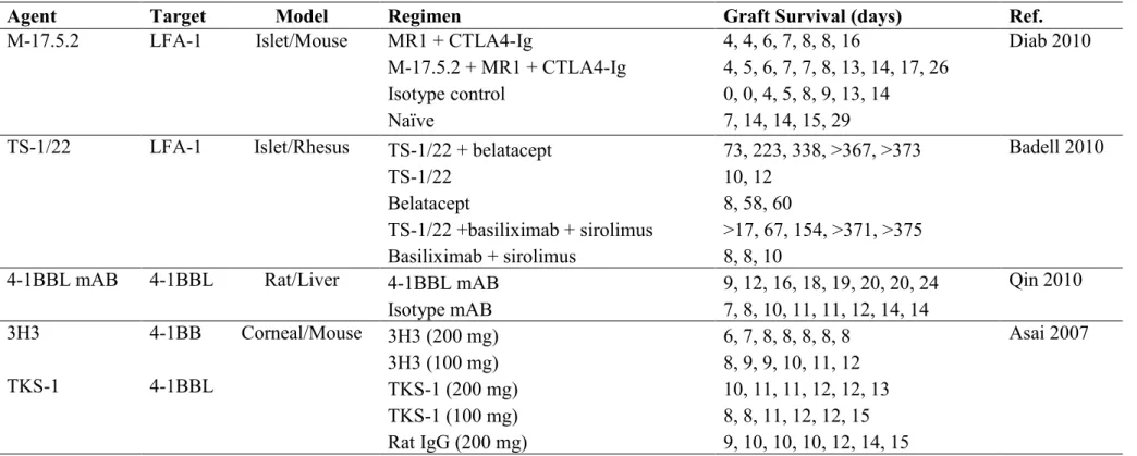
Figure 1.1 Rejection rate and graft survival in kidney transplantation.…………..….…... 5
Figure 1.2 Costimulatory signals are essential for full T cell activation... 18
Figure 1.3 CD40–CD40L pathway plays a role in both cellular and humoral immune responses... 29
Figure 2.1 Renal allograft survival in each group. ……….………….….……….. 65
Figure 2.2 Renal graft function. …………...…. ……….………….….………. 67
Figure 2.3 Pharmacokinetic evaluation. ……….………….….………. 70
Figure 2.4 Proportions of histological types in each group. ……….………….….……… 73
Figure 3.1 Renal graft function and survival. ……….………….….………. 104
Figure 3.2 Pharmacokinetic evaluation. ……….………….….………. 107
Figure 3.3 Pharmacodynamics evaluation. ……….………….….………. 108
Figure 3.4 The infiltration of CD3/FoxP3 double-positive Tregs in renal allografts…… 111
Figure 4.1 The rate of acute renal allograft rejection in ASKP1240-based or ASP2409-based treatment groups. ……….………….….……… 135
Figure 4.2 The acute renal allograft rejection rates and graft survival in low-dose ASP2409 monotherapy, sub-therapeutic dose tacrolimus monotherapy, and low-dose ASP2409 and sub-therapeutic dose tacrolimus combination therapy groups. ……….………….….………... ……….………….…. 137
List of Abbreviations
ACR: Acute cellular rejection
ADCC: Antibody-dependent cell-mediated cytotoxicity AHR: Acute humoral rejection
ALG: Antilymphocyte globulin AP-1: Activation protein-1 APCs: Antigen-presenting cells ATG: Antithymocyte globulin AZA: Azathioprine
BCR: B cell receptor BUN: Blood urea nitrogen CAMs: Cell adhesion molecules CAN: Chronic allograft nephropathy CD: Cluster of differentiation
CDC: Complement dependent cytotoxicity CNIs: Calcineurin inhibitors
CsA: Cyclosporine
CTL: Cytotoxic T-lymphocyte
CTLA4: Cytotoxic T-lymphocyte-associated protein 4
CTLA4-Ig: Cytotoxic T lymphocyte antigen 4-immunoglobulin G CVF: Cobra venom factor
DAG: Diacylglycerol DC: Dendritic cell DD: Death domain
DSAs: Donor-specific antibodies DST: Donor-specific transfusion DTH: Delayed-type hypersensitivity DWFG: Death with functioning graft ECs: Endothelial cells
ECL: Electrochemiluminescent ECM: Extracellular matrix FasL: Fas ligand
FcγRs: Fc gamma receptors
FDA: Food and Drug Administration FoxP3: Forkhead box protein 3
GITR: Glucocorticoid-induced TNFR family-related protein Grb2: Growth factor receptor-bound protein 2
GVHD: Graft-versus-host disease HCT: Hematocrit
HGB: Hemoglobin
HLA: Human Leukocyte Antigens HVEM: Herpes virus entry mediator ICOS: Inducible T cell costimulator IDO: Indoleamine 2, 3-dioxygenase
IFN-γ: Interferon gamma Ig: Immunoglobulin
IgSF: Immunoglobulin superfamily IKK: IκB kinase
ILA: Induced by lymphocyte activation IP3: Inositol trisphosphate
ITAM: Immunoreceptor tyrosine-based activation motif ITIM: Immunoreceptor tyrosine-based inhibition motif iTregs: Induced regulatory T cells
JAK-3: Janus kinase-3
JNK: C-Jun N-terminal kinase LAT: Linker for activation of T cells
LFA-1: Leukocyte function-associated antigen 1 liCTLA4: Ligand-independent CTLA4
LIGHT: Homologous to lymphotoxin, exhibits inducible expression and competes with HSV glycoprotein D for binding to herpesvirus entry mediator, a receptor expressed on T
lymphocytes
mAbs: Monoclonal antibodies
MALT: Mucosa-associated lymphoid tissue MAPK: Mitogen-activated protein kinase MFI: Mean fluorescence intensity
MHC: Major histocompatibility complex miH: Minor histocompatibility antigen
MMF: Mycophenolate mofetil MPA: Mycophenolic acid MST: Median survival time
mTOR: Mammalian target of rapamycin NCAM: Neural cell adhesion molecule NFAT: Nuclear factor of activated T cells
NF-κB: Nuclear factor kappa-light-chain-enhancer of activated B cells NHP: Nonhuman primate
NK: Natural killer
nTregs: Naturally occurring Tregs
PAG: Phosphoprotein associated with glycosphingolipid-enriched microdomains PAS: Periodic Acid-Schiff
PBMCs: Peripheral blood mononuclear cells PD: Pharmacodynamics
PD-1: Programmed death-1
PI3K: Phosphatidylinositol 3-kinase
PIP2: Phosphatidylinositol 4,5-bisphosphate
PK: Pharmacokinetic PKC: Protein kinase C PLCγ1: Phospholipase Cγ1 PLT: Platelets
PODs: Post-transplantation days PtdSer: Phosphatidylserine
PTLD: Post-transplant lymphoproliferative disorder RasGRP: Ras guanyl nucleotide-releasing protein RBC: Red blood cell
S1P: Sphingosine-1-phosphate SCr: Serum creatinine
Sema4A: Semaphorin 4A
SIT: Src homology 2 domain-containing protein tyrosine phosphatase-interacting TRAP SLP-76: SH2 domain-containing leukocyte phosphoprotein of 76 kDa
SNBL: Shin Nippon Biomedical TBI: Total body irradiation TCR: T cell receptor
TFH cell: T follicular helper cell Th cell: Helper T cell
TIM: T cell Ig domain and mucin domain TNF: Tumor necrosis factor
TNFR: Tumor necrosis factor receptor
TNFRSF9: Tumor necrosis factor receptor superfamily member 9 Tr1: Type 1 regulatory T cells
TRAF: TNF receptor associated factor TRAP: Transmembrane adapter protein Tregs: Regulatory T cells
TRIM: T cell receptor interacting molecule TTx: Anti-tetanus toxoid
VCAM-1: Vascular cell adhesion molecule 1 VLA-4: Very late antigen 4
WBC: White blood cell
ZAP-70: Zeta-chain-associated protein kinase 70
Acknowledgements
I would like to acknowledge my profound sense of gratitude and to express my deepest respect to all people who have made this thesis possible. I consider myself fortunate that there are a lot of friends have helped and inspired me during my doctoral study.
I especially want to express my heartfelt gratitude and sincere appreciation to my supervisors Dr. Huifang Chen and Dr. Louis Dumont for their scientific guidance and encouragement throughout the years pursuing my doctoral degree, and their patience and editing skills in the completion of this thesis.
I wish to express my special thanks to my doctoral committee members: Dr. Richard Bertrand, Dr. Édouard Kouassi, Dr. Stanislaw Stepkowski, Dr. Jean-François Cailhier for their profound and carefully aimed comments and suggestions, and constructive criticism on my thesis.
It gives me great pleasure to extend my sincerest thanks to the members of my comité de parrainage: Dr. Muhammad Zafarullah, Dr. Shaoling Zhang, and Dr. Caigan Du. Their advice, supports, valuable suggestions, and positive criticism made it possible for me to achieve my goals during doctoral studies.
I must thank my colleagues in the lab: Dr. Anlun Ma, Dr. Yanxin Hu and Dr. Hao Dun for their excellent support and splendid co-operation. They are always available with helping
hands whenever I need them. We have accomplished a large amount of work in past years. I feel very lucky to have had the opportunity working together with them.
Special thanks are also given to Dr. Guangzhou Zhang, Mr. Jicheng Wang, Ms. Huizhen Liu, Mr. Guanhua Bi, and Mr. Zhiyuan Hou in Laboratory Animals Center of the Academy of Military Medical Sciences for their generous helping, collaboration and technique assistant during my research work. I always enjoyed the time spent with them in Beijing. I also learned a lot from them especially the knowledge about laboratory animals including nonhuman primates.
I would also like to convet my great appreciation to Ms. Lucile Chambily and Mr. Dieudonné-Joël kupa Bilolo for their warm-hearted help in correcting the Frech abstract of my thesis.
Finally, I especially want to express my sincere thanks to my family. Actually, there are not enough words to describe my deep sense of gratitude for their emotional and financial support, encouragement, patience, sacrifice and care. Their unflagging loves are always the greatest sources of inspiration and motivation for pursuing my dreams.
Chapter 1
Introduction
In 1954, organ transplantation from an idea in ancient legend successfully became a realistic therapeutic approach in clinical practice (Merrill 1956). Since then, transplantation of organs including kidney, heart, lung, liver, pancreas etc. gradually become a routine procedure to save the life of the patient with end-stage organ failure. Every year tens of thousands organ transplantations are preformed in the world. To date, organ transplantation is still the only cure for some end-stage organ failure, such as heart and liver failure.
1.1 Rejection – the major cause of organ transplant failure
Organ transplantations are mostly carried out between genetically distinct individuals because the donor who is genetically identical to the recipient is very rare. Although the outcome of transplantation could be affected by many factors including surgical techniques, organ quality, age and race of recipient, post-transplant complications etc., allograft rejection is the most common cause of transplant failure.
The word "rejection" come from the French word "réjection", or directly come from the Latin word "reiectionem". It means "act of throwing back". In the 1940s, through serial studies of skin transplantation, Medawar and his colleagues demonstrated that transplant rejection was an immunologic event (Gibson 1943; Medawar 1944). During and after transplantation, the recipient's immune system recognizes certain components of transplanted cells, tissues, or organs as foreign, and then initiates a cascade of reactions aimed towards the allografts. Transplant injury caused by donor disease, organ harvesting, organ preservation, implantation, and reperfusion triggers a non-specific inflammatory response. Numerous proinflammatory
cytokines, chemokines and adhesion molecules are released during this process (Kim 2008). They promote various inflammatory cells including dendritic cell (DC), monocyte, and lymphocyte to migrate in and out the allograft and lead to antigen specific immune response to eliminate the transplant. Allograft rejection is usually an integrated result that many factors and multiple mechanisms involve in. Both innate and adaptive immunity play a role in this event even though one component of the immune system may dominate a certain type of rejection (Moreau 2013; Farrar 2013).
The antigenic disparity between donors and recipients is the main factor to determine the potency of the alloimmune response (Sánchez-Fueyo 2011). Studies have demonstrated that incompatibility of the surface glycoproteins named major histocompatibility complex (MHC) is the most important cause of rapid graft rejection. MHC is found in all jawed vertebrates. These molecules are encoded by a set of genes and are involved in a lot of physiological processes including immunological and non-immunological activities (Gruen 1997; Kelley 2005). In humans, the MHC is also known as Human Leukocyte Antigens (HLA). The HLA gene group is located on the short arm of chromosome 6 and consists of more than 200 genes (Shiina 2009). Based on gene locations, product functions and structures, these genes are categorized into class I–III three subgroups. HLA Class I molecules are found on most nucleated cells. On contrary, expression of HLA class II molecules are seen on DCs, B cells, and macrophages/monocytes. Molecules of these two groups serve as both stimulus and target in alloimmune response. Genes of HLA class III subgroup encode complements, cytokines, heat shock proteins, and the molecules not related to immune function (Shankarkumar 2004). Besides MHC, minor histocompatibility antigens (miH) that are MHC-bound peptides derived
from various endogenous proteins, can also act as alloantigens. They elicit cytotoxic T cell mediated immune responses and cause allograft rejection in MHC-matched individuals (Dzierzak-Mietla 2012; Dierselhuis 2009).
Rejection occurs in almost all allografts if there are no immunomodulatory therapies to be applied to suppress the immune response against the transplant. Based on its clinical features, allograft rejection is classified into three types: hyperacute rejection, acute rejection, and chronic rejection.
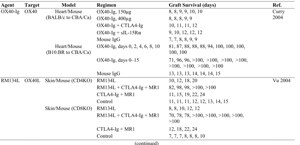
The most common pattern of allograft rejection is acute rejection. According to the literatures of the 1980s and 1990s, acute rejection occurred in 50% to 75% of kidney transplants (Gulanikar 1992), 64% of liver transplants (Wiesner 1998), and 70% to 85% heart transplants (Hunt 1983). The incidence of acute rejection dramatically declined with the advent of calcineurin inhibitors and other new immunosuppressive drugs (Figure 1.1). Organ Procurement and Transplantation Network data showed that the acute rejection rate within one year in kidney transplant recipients was on average approximately 15% in the period from 2001 to 2003 (Djamali 2006).
Acute rejection is a rapid reaction against allograft resulting in the injury of transplant parenchyma and blood vessels. This event can be mediated by T cells (acute cellular rejection, ACR) or antibodies (acute humoral rejection, AHR), or both together (Nankivell 2010). Acute rejection is most likely to happen within the first several posttransplant weeks, but may occur at any time, even many years after transplantation. In the process of acute rejection, the
allograft encounters inflammation, cell injury, and upregulation of MHC class I and II expression. The release of various inflammatory cytokines and infiltration of different cells lead to destruction of parenchymal and endothelial cells (ECs), edema and blood flow decline in tissues. Partial or whole transplanted tissue infarction may occur in the case of destruction of blood vessels (Moreau 2013; Nankivell 2010). Although nowadays the incidence of acute rejection has been significantly decreased, it still contributes to about 12% renal graft loss due to functional failure during the first six months after transplantation (El-Zoghby 2009).
Adapted from Stewart F. Organ Transplantation. 1999 Figure 1.1 Rejection rate and graft survival in kidney transplantation.
80 65 60 45 45 35 25 15 10 40 60 65 85 90 90 96 97 0 10 20 30 40 50 60 70 80 90 100 1960 1965 1970 1975 1980 1985 1990 1995 2000 2005 2010 2015 Per cen t Year
1-year incidence of acute rejection 1-year survival • Radiation • Prednisone • 6-MP • AZA •ATGAM ☆ Cy-A • OKT3 • CsA Emulsion • Tacrolimus • MMF • Dicluzimab • Basiliximab • Thymoglobulin • Sirolimus • Belatacept 5
The principal mechanisms of cell-mediated acute rejection are to evoke cluster of differentiation (CD) 4+ T helper (Th) cell-mediated delayed-type hypersensitivity (DTH)
reactions and CD8+ cytotoxic T-lymphocyte (CTL)-mediated cytotoxicity (Rocha 2003;
Wood 2012). To date various CD4+ T cell subsets including Th1, Th2, Th9, Th17, Th22, T
follicular helper (TFH) cells, and regulatory T cells (Tregs) have been identified. Each subset exhibits distinct cytokine production patterns and effector functions. Traditionally Th1 cells such as interferon gamma (IFN-γ)- and tumor necrosis factor (TNF)-secreting Th1 cells are the common CD4+ Th cell subsets that cause DTH. They secrete proinflammatory cytokines
such as IFN-γ, TNF-α, and IL-1. These proinflammatory cytokines help to recruit and activate non-antigen-specific cells including monocytes and macrophages. These activated cells release proteolytic enzymes, nitric oxide, and other soluble factors that cause nonspecific local inflammation, and affect vascular tone and permeability, as well as enhance chemotaxis to result in cellular infiltrate, edema, and tissue damage. In turn, these non-antigen-specific activities promote the further activation and differentiation of antigen-specific T cells. Recently CD4+ Th17 cells are found to promote macrophage and endothelial activation, and
to cause inflammatory tissue damage and graft rejection (Yuan 2008; Heidt 2010). Suppression of DTH leading to long term allograft acceptance from another angle demonstrates that DTH plays important role in allograft rejection (VanBuskirk 1998).
CD8+ CTLs are the major effector cells in the cell-mediated cytotoxicity. Activation and
differentiation of a naïve CD8+ T cell (CTL precursor) into a functional CTL occur within
secondary lymphoid tissue. CTL precursors are activated by the interaction of the TCR and CD8+ molecules with MHC class I molecules expressed on donor APCs. CD4+ Th cells
provide help during the process via cell-to-cell contact or secretion of IL-2/IFN-γ cytokines (Ridge 1998). Activated CTL precursors differentiate into functional CTLs with up-regulating expression of IL-2 receptor, and forming lytic granules which contain perforins, granzymes, serglycin, calreticulin, and granulysin etc. CTLs then migrate into the allograft and identify target cells by specific interactions between MHC class I on the target cell and TCR as well as CD8 on the CTL. The contents within granules are released into the immunological synapse after cell-cell contact (Rocha 2003). Target cell apoptosis is induced via perforin/granzyme cell death pathway, or via the Fas/FasL pathway.
AHR is less common than ACR. The incidence of AHR in kidney transplant recipients is about 5–7% (Takemoto 2008). This type of rejection is mediated by donor-specific antibodies (DSAs) that may either be pre-existed or de novo generated after transplantation. The prognosis of AHR is poorer than ACR because conventional immunosuppressive therapy is typically not effective enough for AHR. The vasculature of graft is the primary target in this type of rejection. The binding of DSAs to alloantigens (mainly MHC molecules) on the graft vascular ECs triggers activation of both the classical complement and coagulation cascades, and results in endothelial injury, intravascular thrombosis, necrosis of smaller vessels, edema and haemorrhage, and eventually destruction of the transplanted organ (Puttarajappa 2012; Lucas 2011). DSAs may also cause cell injury via antibody-dependent cell-mediated cytotoxicity (ADCC).
Hyperacute rejection is the most rapid and aggressive form of allograft rejection, and fortunately it is extremely rare nowadays due to pretransplantation cross-matching is routinely
applied as a prophylaxis. This type of rejection is mediated by high levels of preformed alloantibodies against donor antigens, particularly MHC class I or ABO blood group antigens. The vascular ECs are generally the main targets. Preexisting alloantibodies bind to endothelial antigens in transplanted organs, and lead to activation of complement and coagulation cascades. Both of them promote extensive intravascular thrombosis and irreversible tissue injury in the graft. Transplanted organ failure may occur within hours or even minutes (Colvin 2006).
Chronic rejection is a slow and progressive process over months to years after transplantation. It usually develops insidiously and is the major cause of later graft loss. The pathologic features of chronic rejection in vascularized grafts include intimal thickening (caused by edema and cell infiltration), smooth muscle cell proliferation in the medial layer, and disruption of elastic lamina etc. These changes result in vascular occlusion and interstitial fibrosis in kidney and heart allograft (Gloor 2006; Tan 2007), bronchiolitis obliterans in lung allograft (Belperio 2009), and vanishing bile duct syndrome in liver allograft (Inomata 2001). The pathogenesis of chronic rejection is not fully understood. Multiple factors including DSAs, the degree of HLA mismatch, previous acute rejection episodes, immunosuppressive drug toxicity, ischaemia/reperfusion injury, infection, and hyperlipidaemia are associated with the development of chronic rejection. It implies that the pathogenesis of chronic rejection may involve both immune and nonimmune factors (Costello 2013). For this reason, in kidney transplant recipients, chronic allograft rejection was replaced by the term chronic allograft nephropathy (CAN). Recently the more descriptive phrase interstitial fibrosis and tubular atrophy (IFTA) took the place of CAN to define the level and severity of chronic renal
allograft damage. IFTA can be found in recipients with good allograft function and is closely associated with late renal transplant failure (Fletcher 2009; Shimizu 2016).
1.2 T cells play a central role in allograft rejection
T cells are essential in the event of allograft rejection. This notion was demonstrated by the experimental studies that fully mismatched transplants were not rejected in the animals lacking T cells, and adoptive transfer of normal T cells could restore allograft rejection in those animals (Pennycuik 1971; Manning 1973; Whitby 1990). Naïve T cells recognize foreign histocompatibility antigens particularly MHC is a key step to initiate allorejection. There are three different ways for immune recognition known as direct, indirect, and semi-direct allorecognition (Afzali 2008).
As soon as the recirculation is established in the transplanted organ, recipient APCs enter the allograft where they encounter foreign antigen derived from donor and become activated. The donor-derived DCs in the transplant may also be activated as a result of cell injury incurred during the transplant procedure. Those activated APCs (either donor-derived or recipient-derived) then migrate to the recipient’s draining lymph nodes. Meanwhile, the soluble MHC proteins that come from transplant may be bound, processed, and presented by B cells. Naïve T cells circulating in the blood enter the lymph node via the afferent lymphatic vessels. They encounter and interact with APCs at the T cell zones in the lymph node paracortex, where allorecognition initially takes place. In the case of direct allorecognition, recipient T cells recognize intact foreign class I and class II MHC molecules expressed on donor-derived
APCs via interaction between TCR and MHC-peptide complexes. Donor-derived DCs are irreplaceable in this type of antigen recognition (Lechler 1982). This process induces a vigorous polyclonal T cell response; as a result, both CD4+ and CD8+ T cells that target donor
antigens are generated. Direct allorecognition plays a significant role in acute allograft rejection (van Besouw 2005).
Indirect allorecognition is similar to the way of T cell activation in response to pathogens. The allogeneic antigens from donor (e.g. cell membrane fragments, soluble MHC and miH molecules) are captured by recipient APCs (DCs and B cells) and are degraded into peptides within cells. These peptides bind to newly synthesized recipient MHC class II molecules, and then are transported to the cell surface. They integrate into the cell membrane of recipient APCs in the form of MHC-peptide complexes. CD4+ T cells recognize those peptide
fragments via interactions between TCRs and MHC-peptide complexes, and the process of cell activation is then initiated. Beside of CD4+ T cells, CD8+ T cells can also be activated by
indirect antigen recognition pathway (Carbone 1990; Zinkernagel 2002). Some studies have indicated that indirect allorecognition is involved in the induction and progression of chronic transplant rejection (Liu 1996; Vella 1997; Womer 2001).
The third mechanism of antigen recognition is semi-direct allorecognition. This type of allorecognition pathway is proposed based on a well-recognized biological process that immunological cells such as DCs have the capacity to transfer membrane components between them via cell-to-cell contact or the exosomal route (Smyth 2008). Semi-direct allorecognition occurs when entire MHC peptide complexes released from donor DCs and
ECs are captured by recipient APCs (Bedford 1999; Herrera 2004). This kind of recipient APCs can then present donor peptide antigens either to CD4+ T cells in the form of self-MHC
II peptide complexes or to CD8+ T cells via captured donor MHC I molecule. They possess
the ability that same APC activates both CD4+ T help cell via indirect antigen recognition
pathway and CD8+ cytotoxic T cell via direct antigen recognition pathway, even
simultaneously (Afzali 2008).
Allorecognition triggers further molecular interactions between T cells and APCs to extend the contact time. Naïve T cells that fail to make prolonged contact with specific antigen then leave the lymph node and return to the circulation. T cells that encounter antigen-specific interaction with APCs remain in the lymph node for 3–4 days. With the help of costimulatory molecules and other molecules, they become fully activated and undergo proliferation and differentiation.
In adaptive immune response mediated allograft rejection, DTH, CTL-mediated cytotoxicity, and antibody-mediated responses are three principal mechanisms that cause destruction of transplanted tissues and organs. The CD4+ T cells orchestrate all three processes. Upon
activation, with stimulation of cytokines secreted activated T cells, naïve CD4+ T cells
differentiate into various Th cell subsets. Th cells secrete a range of cytokines and chemokines including IL-2, IL-4, IL-5, IFN-γ, and TNF-α etc. These cytokines and chemokines attract and activate corresponding effector cells, and lead to various immune responses. Th 1 and Th 17 involve in DTH (Cher 1987; Ishii 2010). The proinflammatory cytokines produced by them cause inflammatory damage of allografts.
In CTL-mediated cytotoxicity, although the effector cell is MHC class I restricted CD8+ T
cell, CD4+ T cell is required for this immune response process. The differentiation and
expansion of CD8+ T cells are CD4+ T cell dependent (Behrens 2004). CD8+ T cell and CD4+
T cell need to be activated by same APC (Bennett 1997). APC is firstly licensed via interaction with CD4+ T cell, with the help of IL-2 produced by CD4+ T cell, the same APC
engages with CD8+ T cell and trigger cell activation and proliferation. Another model to
generate functional effector CTLs is that CD4+ T cell, CD8+ T cell and APC form a three-cell
cluster. Activated CTLs leave the secondary lymphoid organs and migrate to the graft where they attack donor cells and destruct transplanted organs. In addition, CD4+ T cells promote
CD8+ T cell priming by amplifying DC production of IL-15 via CD40–CD40L interactions
(Greyer 2016).
Antibody-mediated responses are also T cell dependent. T cells involve in various steps of B cell immune response including activation, differentiation, and isotype switching. Naïve B cells in the circulation can only survive for a limited time unless they encounter their antigen. The engagement of B cell receptor (BCR) with antigen can trigger the activation of B cells. But, in the case of T cell absence, their interaction induces only low-level B cell proliferation. As a result of that, few antibodies are produced, or even there are no specific antibody productions in some time. The participation of CD4+ Th cells can remarkably enhance B cell
functions (Reinitz 1990; Noelle 1991). When T cells and B cells encounter each other, CD4+
Th cells recognize the processed donor MHC peptides through the interaction between TCRs on the CD4+ cell membranes and MHC class II molecules on B cells. With the help of
costimulatory molecules, coreceptors and adhesion molecules, CD4+ Th cells become
activated. Activated CD4+ Th cells release various types of cytokines. Those cytokines
promote B cell proliferation, differentiation into plasma cells, and early isotype switching. Plasma cells produce specific alloantibodies primarily targeted ECs. The interaction between alloantibodies and ECs lining the donor vasculature activates both complement and coagulation cascades, and induces ADCC resulting in cell and organ damage (Wasowska 2010).
1.3 T cell receptor signaling in T cell activation
In the context of transplantation, the binding of the TCR variable regions to the MHC-peptide complex is an essential for generating signals to initiate T cell activation. The TCR is a heterodimer composed of two (either α/β or γ/δ) transmembrane polypeptide chains. The cytoplasmic domains of the TCR α and β chain are very short and there are no any specific protein-binding motifs. To possess the capability of transmembrane signal transduction, therefore, the TCR assembles with CD3 molecules to form TCR-CD3 complex (Weiss 1994). The CD3 is a multimeric protein complex and is composed of four (γ, δ, ε and ζ) different chains. The intracellular domains of all CD3 chains contain a characteristic sequence motif for tyrosine phosphorylation, known as immunoreceptor tyrosine-based activation motif (ITAM). The ITAM is required to transduce receptor-mediated antigen recognition signals into the cytoplasm of T cells and to induce a series of intracellular signaling cascades (Flaswinkel 1995; Guy 2009). In addition to the CD3 complex, coreceptor CD4 or CD8 is also involved in the antigen recognition process. CD4 and CD8 bind to nonpolymorphic
regions of MHC class II and class I molecules, respectively, to stabilize the T cell–APC immunological synapse and to promote effective signal transduction (Barber 1989).
Following TCR engagement, activation signals that are transmitted into the cell by CD3 trigger a phosphorylation cascade. PCR signaling is initiated by the activations of two Src-related protein tyrosine kinases (PTKs), Fyn that associates with the cytoplasmic tails of the CD3 complex and lymphocyte protein tyrosine kinase (Lck) that noncovalently associates with the cytoplasmic domains of the CD4 and CD8 coreceptors (Veillette 1988). Inactivated Lck has a closed configuration. Upon activation, Lck changes its conformation from the closed shape to an open structure by CD45-mediated dephosphorylation, and possesses active kinase function. CD3 is one of the substrates of Lck. Lck is recruited to CD3 complex to induce the tyrosine phosphorylation of the ITAMs (Barber 1989). This creates a docking site to facilitate another tyrosine kinase protein zeta-chain associated protein kinase 70 (ZAP-70) bind to phosphorylated CD3ζ-chain ITAMs and being activated. The activation of ZAP-70 further amplifies the response by phosphorylating tyrosine residues on various adapter molecules including linker for activation of T cells (LAT), SH2 domain-containing leukocyte phosphoprotein of 76 kDa (SLP-76), phospholipase Cγ1 (PLCγ1), T cell receptor interacting molecule (TRIM) etc (Williams 1999; Zhang 1998; Bruyns 1998). These activated adapter molecules serve as docking sites for a number of cellular enzymes and lead to the activation of multiple downstream signaling pathways. PLCγ1 hydrolyzes the plasma membrane phospholipid phosphatidylinositol 4,5-bisphosphate (PIP2) to generate inositol trisphosphate
(IP3) and diacylglycerol (DAG). IP3 induces the calcium release from calcium pool within the
endoplasmic reticulum and the activation of plasma membrane calcium channel. The
serine/threonine phosphatase calcineurin is then activated along with the increase of intracellular calcium level. Calcineurin dephosphorylates nuclear factor of activated T cells (NFAT). NFAT is then able to translocate to the nucleus and execute its function as transcription factor. DAG is an activator of various signaling molecules including protein kinase C (PKC), Ras guanyl nucleotide-releasing protein (RasGRP). The activations of PKC and RasGRP initiate nuclear factor kappa-light-chain-enhancer of activated B cells (NF-κB) and mitogen-activated protein kinase (MAPK) cascades. These lead to the generation of transcription factors including activation protein-1 (AP-1), NF-κB, and NFAT etc. These transcription factors enter the nucleus to modulate transcription of various genes that are involved in cell cycle initiation, proliferation, and differentiation (Smith-Garvin 2009; Zhong 2008).
Various factors are involved in the regulation of TCR signaling. The costimulatory molecule CD28 engagement by CD80/CD86 provides an initial adhesion capacity to allow T cell approaching APC that is an essential for T cell activation. With CD28 signals, naïve T cells become more sensitive to the stimulation of alloantigen because the TCR number required for activation is decreased. The activation of ZAP-70 is also affected by CD28 signals. Furthermore, CD28 signals allow T cell to enter cell cycle G 1 phase to achieve clonal expansion, and to activate transcription of the IL-2 gene by the induction of the NFAT (Appleman 2002; Boomer 2010). CD45 is also a critical regulator of TCR signaling because it induces the dephosphorylation of the tyrosine kinases including Lck and Fyn (Saunders 2010). Other factors that modulate TCR signaling include actin cytoskeleton (Beemiller 2013).
TCR signaling is also controlled by negative regulators to prevent the hyperactivation of T cells. Activated T cells may upregulate the expression of a costimulatory molecule CTLA4. CTLA4 shares same ligands with CD28 but possesses 10–20 fold higher affinity. Hence, the ligands (CD80/CD86) preferentially bind to CTLA4 and CD28 signals are antagonized. CTLA4 also execute it negative regulation via inhibition of the phosphorylation of TCR (Gough 2005). A transmembrane adapter protein (TRAP), Src homology 2 domain-containing protein tyrosine phosphatase-interacting TRAP (SIT), is a critical negative regulator of TCR signaling (Simeoni 2005). And another TRAP, phosphoprotein associated with glycosphingolipid-enriched microdomains (PAG), negatively regulates Ras activation by altering Src kinase activity (Brdicka 2000; Simeoni 2008).
1.4 Costimulatory signals are required for the full activation of T
cells
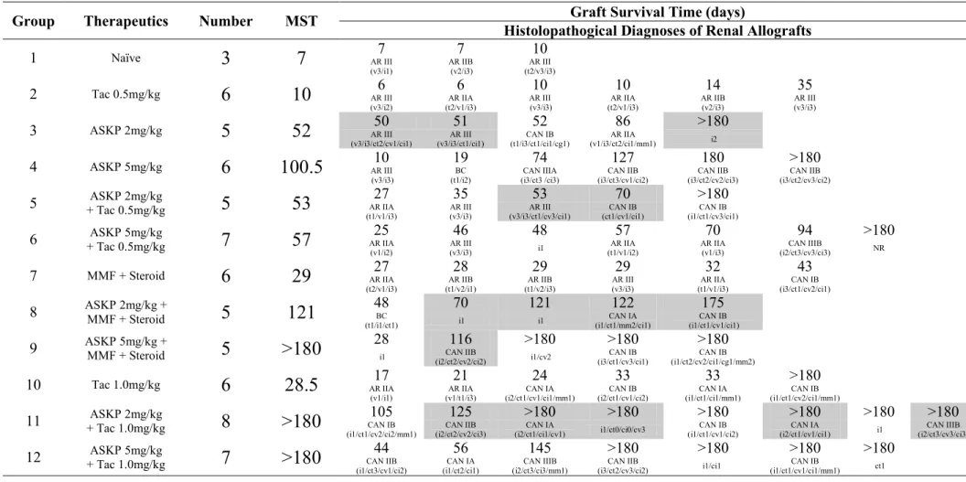
T cells are the crucial mediators and controllers in the alloimmune response induced by foreign histoincompatible alloantigens. Full activation of T cell is a result of cooperation of multiple signals (Durrbach 2010). In 1987, Helen Quill and Ron Schwartz described a phenomenon termed T cell anergy. When functional APCs were absent, purified MHC/peptide complexes that were incorporated into artificial planar lipid membranes could induce high-affinity interaction between TCR and MHC/peptide complexes, but the T cells presented a state of proliferative nonresponsiveness rather than activation (Quill 1987). It was hypothesized that, besides the signal provided by the TCR engagement with MHC/peptide complexes, accessory signals that emanated from the interaction between functional APCs
and T cells were required for T cell to be fully activated. Results from subsequent studies provided concrete evidences to support this notion (Turka 1990; Koulova 1991; Gimmi 1993). The two-signal model of T cell activation was then recognized (Bernard 2002). In the process of T cell activation, TCR engagement with MHC/peptide complexes generates Signal 1 that is antigen-specific. This signal is transmitted into the cell via CD3 complex and triggers various early activation events including the activation calcium-dependent calcineurin protein, tyrosine phosphorylation, and inositol metabolism. The binding of TCR with MHC/peptide complexes is quite brief (Lanzavecchia 2000). The intrinsic affinity between them is very low and some coreceptors and adhesion molecules which stabilize the engagement between TCR and MHC/peptide complexes may take part in the process to enhance the activity. Signal 1 alone is insufficient to lead to T cell being efficiently activated. An additional signal termed costimulatory signal (Signal 2) is essential for the full activation of T cells. Costimulatory signal is induced by the interaction between T cell costimulatory receptors and their corresponding ligands that are expressed by the functional APCs. This signal is non-antigen-specific. In the absence of costimulatory signal, T cells cannot acquire the capacity of cell proliferation and become effector cells (Jain 1995; Serfling 1995). Costimulatory signals play an important role in the enhancement of tyrosine kinase activity. If there is no costimulatory signal, ZAP-70 is unable to achieve the critical threshold to activate downstream molecules. As a result of that, the entire intracellular phosphorylation cascade will be terminated. Consequently, the fates of T cells are either anergy or apoptosis (Figure 1.2). The exact molecular mechanisms of T cell anergy are still unclear. Studies indicated that both immune signal transduction pathways and the ubiquitin proteasome system are associated with clonal anergy (Appleman 2003; Safford 2005; Zheng 2008; Fathman 2007).
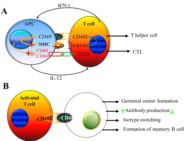
Figure 1.2 Costimulatory signals are essential for full T cell activation. The combined actions of Signal 1
and Signal 2 result in T cells to become fully activated (A). In the absence of Signal 2, T cells exhibit a state of proliferative unresponsiveness rather than activation (B).
1.5 Costimulatory molecules
CD40 and CD28 are the two best characterized costimulatory molecules. They were identified in 1985 and 1986, respectively. Since then, numerous molecules that are involved in costimulatory signal transduction have been discovered. These molecules form a large group known as second signal family. Among them, some molecules provide enhancement signals to promote T cell activation, survival, and/or differentiation. These molecules are named positive costimulatory receptors and ligands. In contrast to them, some other molecules produce signals to antagonize TCR signaling resulting in termination of T cell activation.
APC T cell TCR/CD3 MHC Signal 1 Signal 2 CD80 CD86 CD28 NF-κB + MAP-K Calcineurin Proliferation & Cytokine Secretion T Cell Differentiation APC T cell TCR/CD3 MHC Signal 1 CD80 CD86 CD28 Anergy Apoptosis Ag Ag
A
B
18They are referred as negative costimulatory receptors and ligands. Negative costimulatory signaling pathways play important roles in the maintenance of peripheral T cell tolerance and reducing inflammation after infection. However, the activations of naïve T cells will not be affected because naïve T cells do not express negative costimulatory molecules. The negative costimulatory molecules are expressed by effector T cells, and the expression of these molecules will be up-regulated at the end of an immune response. Apart from basing on the functional properties of costimulatory receptors mentioned above, costimulatory molecules can also be sorted according to their structural properties. Basing on their molecular structures, costimulatory molecules are classified into four distinct groups i.e. immunoglobulin (Ig) superfamily, tumor necrosis factor receptor (TNF-R) family, cell adhesion molecules, and T cell immunoglobulin and mucin domain (TIM) family.
1.5.1 The immunoglobulin superfamily
Costimulatory molecules in the Ig superfamily all possess a characteristic domain referred to Ig domain in their extracellular region. Ig domain contains a sandwich-like structure known as Ig fold which is formed by two sheets of antiparallel beta strands (Chattopadhyay 2009). Members in this superfamily play a central role in both immune activation and immune regulation. Some members of this superfamily promote (costimulatory) T cell activation and differentiation, whereas other members exhibit opposing effects (coinhibitory) on the activation of T cells. Generally the transductions of stimulatory and inhibitory signals are mediated by two loosely conserved motifs in their cytoplasmic region termed ITAM and immunoreceptor tyrosine-based inhibition motifs (ITIM), respectively (Sharpe 2002).
However, in some circumstances, ITAM may propagate inhibitory signals and ITIM may transmit activation signals (Blank 2009; Waterman 2010; Barrow 2006). In addition, some molecules of Ig superfamily such as CD28 also play an important role in the homeostasis and function of a population of Tregs (Sansom 2006).
1.5.1.1 CD28/CTLA4–CD80/CD86 pathway
The CD28 molecule is a 44 kDa homodimeric transmembrane glycoprotein which consists of 202 amino acids. The human CD28 gene is localized on chromosome 2q33, and the mouse CD28 gene maps to chromosome 1 at band C (Lafage-Pochitaloff 1990; Howard 1991). The extracellular region (134 amino acids in length) of CD28 receptor comprises a single disulphide-linked V-like domain. A hexapeptide motif (MYPPPY) is essential for the interaction between CD28 and its ligands (Peach 1994; Boćko 2002). Forty one amino acids constitute the intracellular region of the CD28 receptor. This region contains four tyrosine residues that can be inducibly phosphorylated. They provide the binding sites for phosphatidylinositol 3-kinase (PI3K) and growth factor receptor-bound protein 2 (Grb2), and
involve the regulation of downstream signaling cascade (Zhou 1993; Schneider 1995; Truitt 1994; Cai 1995).
In humans, CD28 is constitutively expressed on the surface of all naïve T cells, 80–95% CD4+ T cells, and approximately 50% of CD8+ T cells. In contrast, CD28 is expressed by all
CD4+ and CD8+ mouse T cells (June 1994; Hutchcroft 1994; Paterson 2009). CD28 expression has also been found on γδ T cells (Testi 1989), some human plasmablasts and plasma cells (Pellat-Deceunynck 1994; Kozbor 1987), and human fetal peripheral blood
natural killer (NK) cells (Nagler 1989). The expression of CD28 in humans is down-regulated with age and in chronic disease states, but it is not the case in mice.
CD80 (B7-1) and CD86 (B7-2) are two known ligands for CD28. CD80 is a 60 kDa type I transmembrane monomeric glycoprotein that is constituted by 262 amino acids. CD86 is also a glycoprotein with molecular weight of 70 kDa. It shares structural homology to CD80 (Freeman 1989; Schwartz 1992). The genes encoding CD80 and CD86 are localized to the same region in human chromosome 3q13.3-3q21 and 3q13-3q23 respectively (Freeman 1992; Fernández-Ruiz 1995). Both CD80 and CD86 molecules contain a single IgV-like domain and a single IgC2-like domain within their extracellular region. The amino-acid sequence analysis indicates that the SQDXXXELY motif in the immunoglobulin C-like domain is a putative CD28-binding sequence (Freeman 1991; Engel 1994; Fargeas 1995).
CD80 is expressed on activated APCs including B cells, macrophages and DCs. Activated T cells and FoxP3+ Tregs also express CD80. In contrast, CD86 is constitutively expressed on
APCs, and the level of CD86 expression will be upregulated upon stimulation by inflammatory cytokines. In addition, CD86 is also expressed on activated T cells (Azuma 1993). Some studies indicated that CD86 probably acted as the major initial ligand for CD28, and CD80 was the preferential ligand for CD28 because CD80 possessed approximately 10-fold higher binding affinity compared to CD86 and appeared to be a more potent costimulus in terms of T cell activation (Fields 1998; Olsson 1998). However, another in vitro study indicated that CD86 was the dominated ligand to induce naïve T cells to become IL-4 producers (Freeman 1995).
When the TCR is properly engaged, CD28 costimulatory signal produced by the interaction between CD28 and its B7 ligands can result in a dramatic augmentation of the expression of genes induced by TCR signaling alone. The cooperative effects between TCR and CD28 signals will lead to full activation of T cells (Figure 1.2). CD28 signaling pathway involves numerous activities. It participates in the regulation of glucose metabolism (Frauwirth 2002), promotes the production of IL-2 (June 1989), mediates entry of T cells into the cell cycle (Appleman 2000), increases the generation of cytokines and chemokines (Thompson 1989), controls Treg cell homeostasis (Salomon 2000), and reinforces resistance to apoptosis (Sperling 1996). CD28 costimulation can lower the threshold of activation by decreasing the number of TCRs required for T cell activation (Viola 1996). Together with CD28 signal can also promote cytoskeleton reorganization to the TCR contact site (Viola 1999), these might partially explain how CD28 exerts its costimulatory effects.
Cytotoxic T-lymphocyte-associated protein 4 (CTLA4, CD152) is a T cell surface molecule with high sequence and structural homology to CD28. It is a glycoprotein constituted of 223 amino acids. The genes for CTLA4 and CD28 are located on the same chromosomal region and share the same overall intron/exon organization. The human CTLA4 gene contains four exons. The exon 1 consists of the leader peptide sequence, and exon 2 to 4 encodes the ligand binding site, the transmembrane region, and the cytoplasmic tail respectively (Ling 1999). Between CTLA4 and CD28, there is about 20% identity at the gene structure level and about 30% identity at the amino acid level (Howard 1991, Greenwald 2005). Similar to CD28, the extracellular domain of CTLA4 also contains a hexapeptide motif MYPPPY which is required for ligand binding.