Upper gastrointestinal tract bleeding management : Belgian guidelines for adults
and children
I. Colle1, A. Wilmer2, O. Le Moine3, R. Debruyne4, J. Delwaide5, E. Dhondt6, E. Macken7, A. Penaloza8, H. Piessevaux9, X. Stéphenne10, S. Van Biervliet4, P.-F. Laterre11
(1) Department of Hepatology and Gastroenterology, Ghent University Hospital, Ghent, Belgium ; (2) Department of Medical Intensive Care Unit, Leuven University Hospital Gasthuisberg, Leuven, Belgium ; (3) Department of Hepatology and Gastroenterology, Université Libre de Bruxelles ULB Erasme, Anderlecht, Belgium ; (4) Department of Pediatric Hepatology and Gastroenterology, Ghent University Hospital, Ghent, Belgium ; (5) Department of Hepatology and Gastroenterology, Liège University Hospital, Liège, Belgium ; (6) Department of Emergency and Disaster Medicine, Universitair Ziekenhuis Brussel, Brussels, Belgium ; (7) Department of Hepatology and Gastroenterology, Antwerp University Hospital, Antwerp, Belgium ; (8) Emergency Department, Catholic University Hospital of Brussels, UCL, Brussels, Belgium ; (9) Department of Hepatology and Gastroenterology, Catholic University Hospital of Brussels, UCL, Brussels, Belgium ; (10) Department of Pediatric Hepatology and Gastroenterology, Catholic University Hospital of Brussels, UCL, Brussels, Belgium ; (11) Department of Intensive Care, Catholic University Hospital of Brussels, UCL, Brussels, Belgium.
Abstract
Upper gastrointestinal bleeding (UGIB) remains a common dis-ease affecting 100 to 170 per 100 000 adults per year and causing thereby a significant burden to healthcare resources. Despite the improvements in the management of this disorder, the associated mortality ranges from 5 to 14%.
Since the general management of UGIB is not uniform, the main objective of this work is to provide guidelines for the care of adults and children presenting with bleeding caused by gastro-duodenal ulcer or variceal rupture.
Methods : In the absence of evidence-based recommendations, these guidelines were proposed after expert opinions reconciliation and graded accordingly. They are based on the published literature up to September 2010 and graded according to the class of evidence.
Results : The current guidelines for the management of UGIB include recommendations for the diagnostic process, general sup-portive care, pharmacological therapy aiming at bleeding control, specific and endoscopic treatment of acute bleeding and follow-up for both gastro-duodenal ulcers and portal hypertension-induced bleeding. (Acta gastro enterol. belg., 2011, 74, 45-66).
Key words : upper gastrointestinal bleeding, varices, ulcers, treatment, guidelines, endoscopy.
Introduction
Introduction
Upper gastrointestinal bleeding (UGIB) is a common disorder with an important burden on healthcare resources. Its incidence in Europe is around 1-2/1000 inhabitants and UGIB is still associated with a mortality rate of 5-14%. Gastro-duodenal ulcers and other erosive lesions account for most of the cases (80%). Portal hypertension represents about 10 to 15% of UGIB episodes with 75% related to oesophageal variceal bleeding and 20% to gastric variceal bleed-ing (1). Upper gastrointestinal endoscopy is the standard of care in the diagnosis and in the first-line treatment of UGIB. It reduces recurrent bleeding, need for surgery and mortality (2). However, management of UGIB involves many other aspects of care including resuscita-tion, transfusion, medical therapy, radiological interven-tions or surgery.
There is considerable variability in the management of these different aspects of the care of UGIB between different European countries and between hospitals with-in the same country (3,4). In addition, various specialists including general practitioners, emergency physicians, gastroenterologists and hepatologists, intensivists, radio -logists and surgeons may be involved in the management of UGIB augmenting the potential for variability and a less than optimal care. We therefore considered that interdisciplinary guidelines for the management of UGIB based on current standards for Evidence Based Medicine (EBM) would provide an opportunity for clinicians to improve the management of their patients with UGIB. We do not believe that guidelines are mandatory tenets appropriate for all patients. Clinical guidelines should constitute a canon or principle tailored to the individual patient (5). In addition, EBM might provide quality indicators for the assessment of care to bleeding patients at a local, national or international level as recently published (6).
Processes and methods to define the Belgian guidelines
The current guidelines have been based on the published literature up to September 2010 and graded according to the class of evidence (Table 1). Where EBM recommen-dations were lacking, guidelines were proposed on reconciled expert opinions and commented in the manuscript.
—————————
Correspondence to : Prof Dr. Isabelle Colle, Hepatology and Gastroenterology Department, 1K12 IE, Ghent University Hospital, De Pintelaan 185, 9000 Gent. E-mail : [email protected]
Submission date : 27/12/2010 Acceptance date : 29/12/2010
Resuscitation
Any patient with tachycardia above 100/min or a sys-tolic blood pressure < 100 mmHg or an increased lactate should be considered for volume replacement. Analogous to patients presenting with severe sepsis or septic shock, the target for volume loading is a mean arterial blood pressure (MAP) > 65 mmHg and/or a sys-tolic blood pressure > 100 mmHg (5-D) (7). In patients with a tentative diagnosis of non-variceal bleeding 2 litres of crystalloids followed by colloids constitute a reasonable initial approach. In patients with a strong sus-picion of variceal bleeding due to liver cirrhosis, albumin 5% is deemed to be the preferred volume expander (5-D). Once hemodynamic targets have been reached and in the absence of contra-indications due to cardiac or renal co-morbidity, volume replacement at 30 mL/kg per 24 h is appropriate.
In patients with persistent hypotension (MAP < 65 mmHg) for more than 30 minutes in spite of vigor-ous fluid loading, temporary normalization of MAP with a vasopressor can minimize or prevent subsequent organ dysfunction. The vasopressor of choice in this setting is noradrenalin (5-D). In case of variceal bleeding, terlipressin can be a valid alternative (5-D).
Blood transfusion
Based on a large randomised trial in critically ill patients, transfusion of packed red blood cells above 8 g/dL con-fers no benefit for outcome. In patients with a history of coronary or cerebrovascular diseases the transfusion trig-ger is higher and set at 9-10 g/dL (1-B).
Intubation and sedation
It is well known that endoscopy performed in optimal conditions is associated with a better control of bleeding, shorter intensive care unit (ICU) and hospital stay (1b-A). Unfortunately, there is a complete absence of General management of UGIB before endoscopy
Establishing a tentative diagnosis
Endoscopy is the gold standard for diagnosing the cause of UGIB. Until the time of endoscopic investiga-tion, the history of the patient (if necessary and if avail-able through next of kin) and the clinical presentation will help to establish a tentative diagnosis. A tentative diagnosis of variceal bleeding will lead to a different management strategy than for non-variceal bleeding (5-D). Any physician having the initial contact with a patient presenting with signs of UGIB should certainly attempt to answer at least the following four questions at the very beginning of the approach to the patient : 1) Does the patient have a history of aortic aneurysm
with or without surgery ? If the answer is yes and the patient is hemodynamically stable, the initial investi-gation should not be endoscopy but contrast CT scan. If the patient is unstable, prompt surgical expert advice must be sought for with the option of immedi-ate surgery because bleeding from an aorto-enteric fistula is highly probable (5-D).
2) Is the history or clinical examination suggestive of bleeding related to portal hypertension ? If the answer is yes, vasoactive medication must be started immediately (1-A).
3) Does the clinical examination suggest impaired vital organ function (tachycardia, hypotension, tachypnea, altered consciousness, cold extremities) ? If the answer is yes, the patient should be placed in a mon-itored environment immediately, contact should be established with an emergency physician or intensive care specialist and the initial lab investigation should include lactate measurement and blood typing and cross-matching (5-D).
4) Is the patient taking anticoagulant or antiplatelet therapy ? Vide infra.
Table 1. — Level of evidence and grade of recommendations
RCT : randomized controlled clinical trial.
Level of Evidence Grading Criteria Grade of Recommendation
1a Systematic review of RCT’s including meta-analysis A
1b Individual RCT with narrow confidence interval A
1c All or none RCT’s B
2a Systematic review of cohort studies B
2b Individual cohort study and low quality RCT B
2c Outcome research study C
3a Systematic review of case-control studies C
3b Individual case-control studies C
4 Case series, poor quality cohort and case control studies C
randomized trials and a paucity of trials in general that have been looking at whether or not prophylactic intuba-tion might improve outcome in patients with UGIB (8-10). The existing literature is exclusively based on retro-spective studies and the data suggest that prophylactic intubation in both variceal and non-variceal bleeding does not change the incidence of cardiovascular events, aspiration pneumonia or mortality (4-C) (8-11). However, common sense supports that endotracheal intubation should be performed before endoscopy in patients with ongoing hematemesis, hemodynamic insta-bility in spite of volume loading, agitation with the absence of co-operation or Glasgow Coma Scale (GCS) < 8 (5-D) (12). An additional indication for intubation is the patient that becomes agitated or suffers from renewed hematemesis during the endoscopic procedure (5-D).
What is a high-risk patient ? Who should be transferred to an ICU ?
Currently, 2 scores are widely used for assessing patient’s risk of adverse outcome (need for transfusion, rebleeding or death). The Rockall score is the oldest pub-lished and integrates endoscopic variables to predict the risk of mortality or of recurrent bleeding (Table 2) (13,14).
However, until endoscopy several decisions will have to be made which depend very much on whether or not the patient has a high risk profile. A modified admission Rockall score was developed without including endo-scopic variables, but it is less powerful as predictor. More recently, the Glasgow Blatchford bleeding score (GBS) was described which includes simple admission variables (15) (Table 3). The GBS has been validated in different countries and is more powerful than both the admission and full Rockall scores in predicting the need for transfusions, endoscopic treatment, surgery or death (16). Moreover, it allows to safely select patients with very low risk (GBS = 0), to be managed as outpa-tients (17). A patient should be classified as high risk in the presence of a GBS ≥ 8 (2-B). These patients should be transferred to an ICU as soon as possible. Other
patients that may clearly benefit from ICU care are those with persistent bleeding in spite of endoscopic therapy and those who have witnessed massive aspiration of blood (5-D).
Vasoactive medication before endoscopy
Vasoactive medication for suspected variceal bleeding (oesophageal and gastric varices)
The aim of using vasoactive drugs is to cause splanch-nic vasoconstriction, thereby decreasing portal pressure and thus stopping or reducing bleeding from varices. Vasoactive medications in the setting of variceal bleed-ing are clearly indicated as they stop the bleedbleed-ing in 75-80% of patients and because they reduce the risk for recurrent bleeding (1a-A). Vasoactive drugs should be started as soon as possible before diagnostic endoscopy, when variceal hemorrhage is suspected. This can be done during transfer to or at arrival at the hospital (1b-A) (18-20). Although optimal duration of the vasoactive drug treatment is not well established, most centres maintain the treatment for 5 days, because this is the period during which the risk of rebleeding is the highest. The recent Baveno V guidelines of May 2010 state that the vasoac-tive therapy should be continued for 5 days (5-D) (20) ; while the Baveno IV guidelines stated 2 to 5 days (1a-A) (18,20,21).
The choice of vasoactive drug should be based on local availability and efficacy. In the US, only octreotide is available, however evidence for using octreotide in variceal bleeding is very low because of tachyphylax-is (12). In Belgium and Europe octreotide, somatostatin, vapreotide, vasopressin and terlipressin are available. Vasopressin has too many side effects and is not used anymore in variceal bleeding. Terlipressin is a potent splanchnic vasoconstrictor with fewer side effects than vasopressin. Somatostatin has also few side effects and can also be used adjunctively to endoscopic therapy.
Either terlipressin or somatostatin should be used in combination with endoscopic treatment, because combination therapy increases the control of bleeding by another 12% and lowers the risk for rebleeding by
Table 2. — Rockall score (13,14)
The Rockall risk scoring system identifies patients at risk of adverse outcome following acute UGIB. The total score is calculated by simple addi-tion of the scores for the different variables. A score < 3 carries a good prognosis ; a total score > 8 carries a high mortality risk.
Variable Score 0 Score 1 Score 2 Score 3
Age (years) < 60 60-79 > 80
Shock No shock Heart rate > 100/min Systolic blood pressure < 100 mmHg
Comorbidity No major Chronic heart failure, ischemic heart
dis-ease, major comorbidity
Renal failure, liver failure, metastatic cancer
Diagnosis Mallory Weiss All other diagnoses GI malignancy
bolus can be repeated up to 3 times within 1 hour. In patients with active bleeding at the time of endoscopy, the infusion regimen can be increased up to 12 mg/24 h (= 500 µg/h). Side effects are rare and there are no major contraindications (21,25,28).
When comparing somatostatin with terlipressin, they have the same bleeding control, risk for rebleeding and mortality rate are found to be similar (21).
Vasoactive medications for non-variceal bleeding In 1997 a meta-analysis of 14 trials that included 1829 patients treated with somatostatin or octreotide compared with histamine-2-receptor antagonists or placebo, concluded that somatostatin may reduce the risk for continued bleeding from acutely bleeding ulcer dis-ease. The authors suggested that large clinical trials were needed in patients carefully stratified by source, severity and type of bleeding were needed. In 2000, a prospective study with 48 patients showed that somatostatin was no more effective than ranitidine. No further evidence has been published in peer-reviewed Journals since then sup-porting the routine use of somatostatin in peptic upper GI bleeding. Furthermore no study with the current gold standard of acid suppression (i.e. proton pump inhibitors - PPIs) has been conducted in this setting (29,30).
Therefore, there is insufficient evidence to recom-mend somatostatin or any other vasoactive medication in the routine treatment of suspected non-variceal UGIB (2b-C).
PPI before endoscopy
PPI in non-variceal bleeding
In patients with overt signs of UGIB but without per-sistent hypovolemic shock, an IV bolus of 80 mg omeprazole, followed by 8 mg/hour for 72 hours, admin-istered after hemodynamic stabilization and before endoscopy, was reported to be effective in accelerating the resolution of signs of bleeding from ulcers and in reducing the need for therapeutic endoscopy. A recent Cochrane meta-analysis confirmed these findings (31) (1a-A). Another 2010 meta-analysis showed that there is no evidence that continuous high dose PPI (8 mg/h for 72 h) has a better outcome than fractionated low dose (2 × 40 mg/d) PPI for active ulcer bleeding (32). Therefore the panel recommends using 2 × 40 mg/d, the first dose being given before endoscopy as soon as a tentative diagnosis of non-PHT related bleeding is made (5-D). In low risk patients tolerating oral medication, the strategy of giving oral PPI before and after endoscopy, with endo-scopic hemostasis for those with no major stigmata of recent hemorrhage, is likely to be the most cost-effec-tive (31-38).
PPI in variceal bleeding
There is no evidence for the use of PPI in the setting of active variceal bleeding or after endoscopic therapy (Baveno V) (20).
another 12% (20,22,23). There are no RCT’s comparing both drugs combined with endoscopic therapy. However, terlipressin versus placebo has shown improved survival in one small RCT and in a meta-analysis (21,24-26).
Terlipressin improves the rate of bleeding control (within 48 h : 75-80% ; within 5 days 67%) and survival. The dose of terlipressin is an initial intravenous (IV) bolus : < 50 kg = 1 mg ; 50-70 kg = 1.5 mg ; > 70 kg = 2 mg followed by an IV bolus of 1-2 mg every 4 h for 48 h, followed by 1 mg every 4 h till day 5. Contraindications for the use of terlipressin include coro-nary insufficiency, peripheral vascular disease, pregnan-cy, heart rhythm disease, severe bronchial asthma, uncontrolled arterial hypertension and epilepsy. The risk for side effects appears to be less when given in contin-uous IV infusion (e.g. 4-6 mg/24 h) at least for hepatore-nal syndrome (27).
Somatostatin treatment starts with a continuous IV infusion of 6 mg/24 h (= 250 µg/h) immediately fol-lowed by a bolus of 250 µg IV within 5 minutes. The
Table 3. — The Glasgow-Blatchford bleeding score
(GBS) (15)
Low risk if Score is “0”
High risk is any score > 0 needing medical intervention, transfusion, endoscopy or surgery
ICU admission is indicated if GBS ≥ 8 Score is "0"only if all the following are present :
1. Hemoglobin level > 12.9 g/dL (men) or > 11.9 g/dL (women) 2. Systolic blood pressure > 109 mm Hg
3. Pulse < 100/minute
4. Blood urea nitrogen level < 39 mg/dL 5. No melena or syncope
6. No past or present liver disease or heart failure.
BLOOD UREA (mg/dL) SCORE VALUE
39-48 2
49-60 3
61-150 4
> 150 6
HEMOGLOBIN FOR MEN (g/dL)
12-12,9 1
10-11,9 3
< 10 6
HEMOGLOBIN FOR WOMEN (g/dL)
10-11,9 1
< 10 6
SYSTOLIC BLOOD PRESSURE (mmHg)
100-109 1 90-99 2 < 90 3 OTHER MARKERS Pulse ≥ 100/min 1 Melena 1 Syncope 2 Hepatic disease 2 Cardiac failure 2 TOTAL score
Antifibrinolytic medication
Seven double-blind randomized trials on tranexamic acid vs. placebo have been published to date, most dating from the seventies and eighties. Only one of these trials includes endoscopic treatments and proton pump inhibitors, i.e. the present standard of care. The trial did not find any significant effect of tranexamic acid on clin-ical outcome measures. There is no indication for the use of antifibrinolytics in the treatment of variceal or non-variceal UGIB (1a-A) (39).
Antimicrobial prophylaxis
In the setting of UGIB, only patients with known or suspected cirrhosis should receive antimicrobial prophy-laxis. The initial work-up of these patients should include an active search for infection (blood, ascites and urine cultures) (1a-A). Proposed first line antibiotics are
amoxicillin-clavulanic acid, third generation
cephalosporins or quinolones according to local resist-ance patterns. In patients receiving maintenresist-ance therapy with quinolones as prophylaxis for spontaneous bacteri-al peritonitis, third generation cephbacteri-alosporins are the first choice. Prophylactic antibiotherapy should not be con-tinued for more than 7 days (18,20).
Coagulation disorders, should anti-Vit K medication and/or anti-platelet medication be discontinued in bleed-ing ?
Antiplatelet drugs and anticoagulants are commonly used for the treatment and the prophylaxis in vascular diseases. Endoscopic therapy in patients with an acute bleeding while using antiplatelet agents (acetyl salicylic acid, clopidogrel, ticlopidine, dual antiplatelet therapy, glycoprotein IIb/IIIa inhibitors…) is both warranted and safe (40,41). Stopping these agents prior to endoscopy may pose unnecessary atherothrombotic risks and there-fore needs careful evaluation. On the other side, in patients with clinically significant UGIB, continued administration of antiplatelet drugs or anticoagulants is known to be associated with a high risk of persistent bleeding.
There is no clear recommendation about either how long these drugs should be discontinued, or whether the reversal of anticoagulation should be partial or complete. The individual decision must be based on the balance between the risk of thromboembolism and that of contin-uing bleeding. The same balance should be considered to determine the timing of reinstitution of these treat-ments (40-44).
The American Society of Gastroenterology (ASGE) proposed a classification of high or low risk for throm-boembolic event, thereby providing a tool to appreciate this balance (Table 4) (40,41).
In non-cirrhotic patients there is good evidence that anti-Vitamin K therapy (AVK therapy), targeted at an INR of 2.5 (range 2-3) is associated with a lower risk of bleeding than therapy targeted at an INR > 3. In patients with UGIB correction of the INR < 2.5 is advised (4-C). Correction to an INR of 1.5 to 2.5 allows successful endoscopic diagnosis and therapy (45).
For patients with serious bleeding and elevated INR (regardless of the magnitude of the elevation), the American College of Chest Physicians recommends to hold the AVK and to give 10 mg vitamin K by slow IV infusion. A supplement of fresh frozen plasma (FFP) or prothrombin complex concentrate (PCC) is also proposed depending on the urgency of the situation but certainly in a life threatening bleeding (2C) (41).
The timing for resumption of anticoagulation should be individualized taking into account the risk for throm-boembolic event (see Table 4). In patients with high risk for thromboembolic event and for rebleeding, the use of unfractionated heparin (UFH) could be considered due to the advantage of a short half-life.
For non-cirrhotic patients using antiplatelets drugs, there are no clear recommendations. Some suggest with-holding these medications until hemostasis is achieved, however, due to the long half life of clopidrogel (7 days), stopping the drug will not have any impact on the short term, so endoscopy and treatment are safe under antiplatelet therapy (5-D). In the absence of randomized controlled trials (RCT’s) the panel suggests that platelets transfusion could be considered if the platelet count is
Table 4. — Condition risk for thromboembolic events (ASGE standards of practice committee for the management of antithrombotic agents for endoscopic procedures) (40)
Higher-risk condition Low-risk condition
Atrial fibrillation associated with valvular heart disease, prosthetic valves, active congestive heart failure, left ventricular ejection fraction < 35%, a history of a thromboembolic event, hypertension, diabetes mellitus, or age > 75 y
Uncomplicated or paroxysmal nonvalvular atrial fibrillation
Mechanical valve in the mitral position Bioprosthetic valve
Mechanical valve in any position and previous thromboembolic event Mechanical valve in the aortic position
Recently (< 1 y) placed coronary stent Deep vein thrombosis
Acute coronary syndrome
(80%) but rebleeding after balloon deflation is high (50%). The complication rate (aspiration, migration, necrosis and perforation of oesophagus) is high and asso-ciated with high mortality. The gastric balloon should be deflated within 24 h to avoid necrosis of the stomach (in case of simultaneous inflation of an oesophageal balloon this one should be deflated every 4 hours).
Linton tamponade (600 mL) can be used for oesophageal and gastric varices (52). Sengstaken Blakemore tamponade can only be used for oesophageal varices (5-D).
Recently a RCT showed that a covered oesophageal stent can be placed for oesophageal varices, however this is not recommended yet and should be confirmed in other studies (12,20,21,53).
Endoscopic and other specific treatment modal-ities for acute UGIB
Optimal timing for endoscopy ?
The optimal timing for endoscopy for non-PHT relat-ed blerelat-eding is within the 24 h of patient’s presentation (1a ;A) (54). This allows for safe and prompt discharge of patients classified as low risk, it improves patient out-comes for high risk patients and reduces the use of resources for patients at low or high risk. No evidence exists for any clinical benefit of performing endoscopy within 12 hours of presentation (reduction of rebleeding rate or survival improvement) as this may lead to increased use of endoscopic therapy, which may not be necessary. This correlates to studies showing that performing emergency endoscopy in bad conditions (without adequate human and material resources) is associated with increased recurrent bleeding, need for surgery and hospital stay (2b-B) (55). Besides this statis-tical evidence, each case should indeed be individually discussed, according to patient’s clinical situation and maybe the GBS, as suggested before.
If a variceal bleeding is suspected, endoscopy should be performed as soon as possible but within 12 hours (5-D) (18-20).
Hemodynamically unstable and high risk patients should be transferred to ICU for conditioning and performing endoscopy, after intubation, in proper and safe conditions.
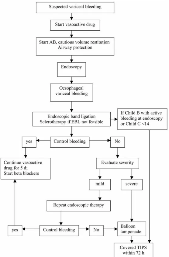
Endoscopic treatment for non-variceal bleeding (Fig. 1)
Ulcers
Endoscopic treatment should be performed at the time of diagnosis by an experienced endoscopist. Lesions that are actively bleeding (Forrest Ia, b) and non-bleeding visible vessels (Forrest IIa) should be treated endoscopi-cally. Clots (Forrest IIb) should be removed by washing, using a basket, snare or a cap (50) until the ulcer base is visible for adequate treatment of the underlying ves-sel (56,57,58) as the risk of rebleeding after endoscopic treatment from a Forrest IIb ulcer is the same as for a lower than 30,000/µL in case of ongoing bleeding
(5-D) (40,44). The timing for resumption of antiplatelet medication should be individualized taking into account the risk for thromboembolic event (Table 4).
In cirrhotic patients, PT/INR is not a reliable indicator of the coagulation status (Baveno V). On the basis of currently available data, in these patients no definite rec-ommendations can be made regarding management of coagulopathy and thrombocytopenia (5-D) (18,20). There is certainly no evidence for administration of coagulation factors prior to endoscopy (5-D). Over-transfusion with fresh frozen plasma (FFP) and platelets cause an increase in portal pressure and may be associat-ed with sustainassociat-ed bleassociat-eding (2b-B). In the absence of RCT’s the panel suggests that platelets transfusion could be considered if the platelet count is lower than 30,000/µL in case of ongoing bleeding (5-D).
In the patient suffering from an acute coronary syn-drome or recently having had placed a vascular stent, no strong recommendations concerning antiplatelet drugs can be made. A cardiologist should always be consulted before stopping any antiplatelet agent, but drugs must be continued in many cases (5-D).
Stomach cleansing before endoscopy
In the case of UGIB, a clean stomach or duodenum during endoscopy is highly desirable. Naso-gastric lavage is associated with complications and fails to be efficient in half of the cases. A recent pharmacologic method for cleansing the stomach has gained a 1a-A evi-dence for its use before endoscopy after demonstration of effectiveness (46-48) and cost / effectiveness (49) : the IV administration of 250 mg erythromycin over 5 min, 20 min before endoscopy, dramatically increases the rate of empty stomach, decreases the time of endoscopy and the need for a second look. In this setting erythromycin acts as a motilin agonist which induces potent gastric contractions and evacuation of gastric blood clots.
In case of failure to clean the stomach, large operative channel endoscopes or other tricks may be used like an attachable distal cap that can facilitate the removal of blood clots during endoscopic treatment (50).
Feeding and nasogastric tube
Although there are no studies, there are probably no contra-indications to place a nasogastric tube after band ligation or treatment of non-PHT related bleeding unless other recommendation of the endoscopist. Oral feeding can start when the patient is hemodynamically stable and conscious (5-D).
When is tamponade indicated ?
Balloon tamponade is indicated for uncontrolled bleeding from oesophageal/gastric varices for which a definitive therapy is planned within 24 h (1-B) (51). Balloon tamponade is effective in bleeding control
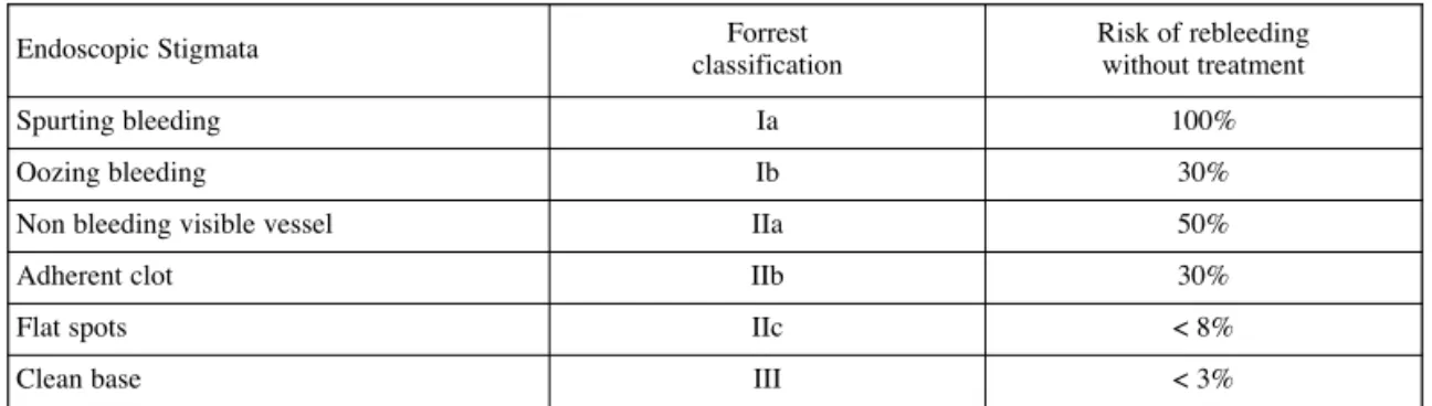
Forrest IIa. Ulcers with a clean or black base do not need endoscopic treatment (1a-A). The Forrest classification is described in Table 5.
Hemostasis can be performed using different inde-pendent or combined methods.
a. Injection therapy with saline is widely applica-ble, easy, inexpensive and can be used when front view of the bleeding point cannot be maintained. Addition of epinephrine (1:10,000 dilution) or a sclerosant offers no advantage (59). Injection therapy should only be considered as a temporary measure because its effect is
only transient. It might allow clearing the endoscopic field (60,56,61,62) (1a-A). This method is not to be considered as a definite treatment but as an adjunct to more efficient and long-lasting hemostatic procedures.
b. Thermal coagulation and mechanical clipping are more effective and sustained than injection therapy alone in terms of bleeding control, rebleeding and need for surgery (61,62).
Vascular ectasia
Argon Plasma Coagulation (APC) is useful for the treat-ment of a vascular ectasia (63).
Abbreviations : GBS : Glasgow-Blatchford bleeding score.
surgery (OR 0,56 CI : 0,45-0,70) but not mortality. A beneficial impact on mortality was only found in the sub-group of patients with active bleeding or non bleeding visible vessel who received haemostatic therapy and high dose IV PPIs for 72h (OR 0,57 CI 0,34-0,96). Lower doses of PPIs either IV or oral had no impact on mortality in this subgroup. As far as costs are considered for this strategy, high-dose IV PPI for high risk patients after therapeutic endoscopy appear more effective and less costly in Europe (68). However, the question of lower IV PPIs dose is still open (32).
Failure to control non-variceal bleeding : when should embolisation or surgery be considered ?
In patients in whom non-variceal bleeding cannot be controlled with a second endoscopic attempt, angio-graphic embolisation or surgery becomes the next thera-peutic option. Where available, arteriography is an alter-native option to surgery. This procedure can often obvi-ate the need for surgery and is particularly useful for those unfit for surgery. Visualization of the bleeding ves-sel requires active bleeding at the time of arteriography at a rate of approximately 2 ml/min. Even though bleed-ing rate is not constant, angiography has a high probabil-ity of detecting an actively bleeding blood vessel if a patient is requiring more than 2 units of red blood packed cells per 4 hours to maintain a hemoglobin > 7 g/dL. The technical success of angiographic embolisation is above 90%, but prolonged clinical success is less at 65 to 70%. Early rebleeding will occur in approximately 20 to 30% of cases. The procedure has a low rate of major compli-cations. In high-risk patients (APACHE II score > 16) mortality has been reported to be 23% versus 50% in patients treated with surgery. Transcatheter arteriography and intervention is the best treatment method for bleeding occurring into the biliary tree or pancreatic duct (69-72). In the USA the use of surgery to control bleeding has decreased from 13.1% in 1993 to 9.7% in 2006. There has also been a significant decrease in the use of defini-tive surgery (vagotomy or resection). Surgery has a lower incidence of rebleeding than angiographic emboli-sation (72).
In conclusion, angiographic embolisation or surgery is indicated when arterial bleeding cannot be controlled Mallory-Weiss syndrome
Mallory-Weiss syndrome heals spontaneously in many cases, endoscopic therapy is occasionally neces-sary. When endoscopic treatment is needed, the same rules apply as for ulcer bleeding (61). The use of a duo-denoscope may be very helpful in this setting (5-D). Tumour
Endoscopic haemostasis for bleeding related to an UGI tumour is often a temporary measure. Permanent haemostasis is difficult in many cases due to diffuse bleeding and the unique solution is surgical resection if possible (64).
Dieulafoy’s lesion
Bleeding from Dieulafoy’s lesions (mainly located in the stomach and duodenum) can be controlled by endo-scopic therapeutic procedures. Hemoclipping is more effective than injection therapy (65). A safe alternative is injection plus heater probe coagulation (66). Band liga-tion was successful in case series but there is a risk of perforation and delayed bleeding (contraindicated in the duodenum). Argon plasma coagulation has been used in a case series (67). Tattooing of the lesion with India Ink is helpful for locating the lesion in case of endoscopic retreatment or surgical wedge excision.
PPI continuation or administration after therapeutic endoscopy
In a large RCT - after therapeutic endoscopy - high-dose intravenous omeprazole (80 mg bolus IV, followed by 8 mg/hour for 72 hours) was shown to significantly decrease the rate of rebleeding by approximately 5 to 10% (35). The need for surgery was reduced non-signif-icantly by approximately 3%. Mortality was significant-ly reduced onsignificant-ly in patients with active bleeding or non-bleeding visible vessel. Similar results were obtained with esomeprazole (after therapeutic endoscopy : 80 mg bolus IV, followed by 8 mg/hour for 72 hours). An updated meta-analysis of RCT’s which included 5792 patients (5), disclosed that PPI therapy after endoscopy (whether therapeutic or not) dramatically reduced the rebleeding rate (OR 0,45 CI : 0,36-0,57) and
Table 5. — Forrest classification for the risk of rebleeding
Endoscopic Stigmata classificationForrest Risk of rebleedingwithout treatment
Spurting bleeding Ia 100%
Oozing bleeding Ib 30%
Non bleeding visible vessel IIa 50%
Adherent clot IIb 30%
Flat spots IIc < 8%
with endoscopy. In high risk patients, angiographic embolisation is preferred over surgery because of lower mortality (3-C).
Endoscopic treatment for PHT related bleeding
Oesophageal variceal bleeding (Fig. 2)
The diagnosis of an acute variceal bleeding is made upon the following criteria : active bleeding from a varix
(oozing, spurting) ; presence of a “white nipple” fibrin clot overlying a varix (signature of variceal rupture) ; a clot on a varix ; the presence of varices without other potential source of bleeding and fresh blood in the stom-ach (12,21,73).
Endoscopic treatment should be performed at the time of diagnostic endoscopy (within 12 h after admis-sion) by a skilled endoscopist and should always be used in association with vasoactive treatment, preferably Abbreviations : AB : antibiotic ; EBL : endoscopic band ligation ; TIPSS : transjugular intrahepatic portosystemic shunt.
Endoscopic treatment for gastric variceal bleeding (Fig. 3)
Fundal varices account for 1-3% of variceal bleeds. The risk of gastric variceal bleeding is lower than oesophageal variceal bleeding, however the bleeding is more severe and mortality is higher (21). There are different types of gastric varices as shown in Figure 4.
Gastro-oesophageal varices type 1 are an extension of oesophageal varices along the lesser curvature and should be treated as oesophageal varices (18,52). started before endoscopy (1aA) (18,20). Both sclero
-therapy and endoscopic band ligation (EBL) have shown to be effective in controlling oesophageal variceal bleed-ing. The Baveno IV and V guidelines are conclusive in their advice : endoscopic band ligation is preferred as this endoscopic therapy for oesophageal variceal bleed-ing is better for bleedbleed-ing and rebleedbleed-ing control and has less adverse events compared to sclerotherapy. Sclerotherapy can be accepted if EBL is not feasible, technically difficult or not available (1b-A) (12,18-21,23,51,74,75).
Abbreviations : BRTO : Balloon retrograde transvenous obliteration ; PHT : portal hypertension ; TIPSS : transjugular intrahepatic portosystemic shunt.
Endoscopic band ligation or tissue adhesives can be used (Baveno V) (20).
There are limited data for gastric variceal bleeding (from isolated gastric varices type 1 and 2 and gastro-oesophageal varices type 2) but the guidelines are quite uniform : variceal obliteration with a tissue adhesive (e.g. N butyl-cyanoacrylate, isobutyl 2 cyanoacrylate) is recommended and is more effective than EBL or scle-rotherapy (1b-A) (18,20,51). Obliteration with a tissue adhesive has a better bleeding and rebleeding control, should be performed by an expert in endoscopy, can be difficult during active bleeding and complications (emboli) can occur (21,52).
If obliteration is not possible, transjugular intra hepat-ic portosystemhepat-ic stent shunt (TIPSS) should be consid-ered (1-B). Tamponade with a Linton balloon can be per-formed as a bridge to definite therapy (21,52). A Linton tamponade with 600 mL inflation is more effective than a Sengstaken Blakemore tamponade (52).
A second endoscopic attempt to stop gastric variceal bleeding is not indicated providing glue was used during the first session (21,52).
Treatment for portal hypertensive gastropathy
Portal hypertensive gastropathy (PHG) is supposed to cause less than 3% of acute bleedings. There are no RCT’s assessing treatment for this cause of bleeding. Treatment is hypothetically based on decreasing gastric blood perfu-sion with vaso-active agents such as terlipressin or somatostatin (5-D) (52). Chronic bleeding from PHG can be treated with beta-blockers or TIPSS (52). Bleeding from PHT related jejunopathy should also be considered and assessed by capsule endoscopy (76,77).
Variceal bleeding from non cirrhotic portal hypertension or extrahepatic portal hypertension
Little literature about the treatment of variceal bleed-ing from non-cirrhotic portal hypertension is available. However the same endoscopic therapy for acute bleeding
is advised as in cirrhotic variceal bleeding (2b-B). For secondary prophylaxis, endoscopic therapy is effective (2b-B). Decompressive procedures should be considered for patients with endoscopic failure (5-D) (18,20). The place of TIPSS and Surgery in PHT related UGIB (Figure 1 and 2)
WHEN SHOULD COVEREDTIPSS BE CONSIDERED?
Polytetrafluoroethylene (PRFE)-covered TIPSS is extremely effective in controlling variceal (oesophageal and gastric) bleeding (control rates approaches 95%) and has a low risk of obliteration compared to non-covered stents. The goal of TIPSS placement is to reduce hepatic venous pressure gradient below 12 mmHg (3a-C) (78).
TIPSS is preferred above surgical shunts (3b-C) (78) and should be placed by an experienced interventional radiologist or a specially trained physician (5-D) (20,78). Sometimes retrograde embolisation of gastric varices may be required despite adequate decompression of por-tal venous system (3a-C) (78). TIPSS may decrease liver function capacity and cause encephalopathy (21). EARLYTIPSS PLACEMENT
Two studies from Monescillo (79) and Garcia Pagan (80) show that high risk patients for rebleeding, have a survival benefit when early PTFE-covered TIPSS (within 72 h of admission) is placed compared to stan-dard therapy (combined vasoactive treatment and band ligation) (1 year survival of 86% vs. 60%, respectively). Moreover, early TIPSS had a much lower rate of failure to control bleeding and of early rebleeding (3%) compared to combined vasoactive treatment and band ligation (50%) (80).
Based on these 2 studies, the recent Baveno V consen-sus meeting states that early covered TIPSS placement (within 72 h of admission) should be considered in patients at high risk for treatment failure. This is in patients with Child B cirrhosis and active bleeding at endoscopy and in Child C patients with < 14 points, after
Fig. 4. — Different types of gastric varices :
GOV1 : gastro-oesophageal varices type 1 is an extension of oesophageal varices along the lesser curvature GOV2 : gastro-oesophageal varices type 2 is an extension of oesophageal varices along the fundus IGV1 : isolated gastric varices in the fundus
or death. Failure to control bleeding implies a change in treatment options. TIPSS is the preferred strategy and should be placed within 24 hours.
Second-look endoscopy
The definition of a second-look endoscopy (also called scheduled repeat endoscopy) is an endoscopy scheduled within 24-72 h after the index bleed, without any clinical sign of rebleeding. However, the perform-ance of second-look endoscopy remains controversial. In the published studies there are important differences in recruitment criteria, in definitions of recurrent bleeding, and important heterogeneity in the endoscopic therapies used, which all contribute to confound the effects of overall efficacy. In 3 RCT’s either endoscopic injections or fibrin-glue in second endoscopy was used. Only one of these studies showed a marginal benefit in the reduc-tion of recurrent bleeding with second-look endoscopy, and none showed any benefit on mortality or need for surgery. In 2 RCT’s (both with small sample size) using thermal coagulation, a significant reduction in recurrent bleeding was obtained, but no effect on mortali-ty (81,82). Most of the beneficial effects of a second-look endoscopy were published when injection therapy was used alone and when post endoscopy PPIs were not used. Therefore, currently, combination of endoscopic treatments and PPI administration really question the usefulness of a second look endoscopy (83). Based on the published data, the panel does not recommend rou-tine second-look endoscopy, independent of the endo-scopic technique used to stop the bleeding (1a-A).
However, a clinically meaningful and probably cost-effective strategy is to select high-risk patients for sec-ond-look endoscopy and retreatment, especially if the initial treatment is questionable (5-D). High-risk patients can be selected on the basis of the Glasgow Blatchford bleeding or Rockall score > 6 (1a-B) (see Table 2 and 3) (13-15).
Recurrent bleeding
Non-variceal rebleeding
Definition
Rebleeding is defined as clinical evidence of recurrent bleeding after a successful initial treatment. Risk factors associated with rebleeding are a past history of peptic ulcer bleeding, a history of smoking, presentation with hematemesis, hypovolemic shock before endoscopy, ulcer size > 2 cm and type of ulcer (84). The GBS and the Rockall scores are good indicators of rebleeding (Table 2 and 3). The use of post endoscopy PPIs dramat-ically reduces the rate of rebleeding from upper gastroin-testinal lesions (see above).
Re-endoscopy with retreatment is mandatory, inde-pendently of the endoscopic technique used to stop the index bleed or the origin of bleeding (1a-A).
medical and endoscopic treatment is installed (20). Reserving TIPSS as a rescue therapy after failure allows the patient sometimes to deteriorate to a point where survival is poor.
TIPSS AS SALVAGE THERAPY:
PTFE-covered TIPSS should be used as a salvage therapy in the following indications :
– if oesophageal and gastric variceal bleeding is not controlled with endoscopic and medical therapy – when oesophageal variceal bleeding recurs despite 2
endoscopic attempts and full medical therapy (1-C) (12,51)
– if gastric variceal rebleeding occurs after a single failure endoscopy or if obliteration is not avail-able (12,51,52).
TIPSS FOR OTHER INDICATIONS
TIPSS can also be recommended for recurrent bleeding from portal hypertensive gastropathy despite beta-blockade (4-C) (78). However, TIPSS is not useful for the treatment of bleeding from gastric antral vascular ectasia (GAVE) (“water melon stomach”) (78).
SHUNT SURGERY FOR BLEEDING FROM PHT DUE TO INTRA -HEPATIC DISEASE(CIRRHOSIS MOSTLY)
Shunt surgery is extremely effective in controlling variceal bleeding (control rates approaches 95%), but due to worsening of liver function, mortality remains high. For this reason TIPSS is preferred above surgical shunts (3b-C) (78).
The ideal patient for surgical therapy is one with well preserved liver function who fails emergent endoscopic treatment and has no complications from the bleeding or endoscopy. The choice of surgery is usually dependent on the training and expertise of the surgeon.
SHUNT SURGERY FOR BLEEDING FROM PHT DUE TO PORTAL VEIN THROMBOSIS
Decompressive surgery or interventional radiological procedures should be considered for patients with portal vein thrombosis and failure of endoscopic therapy for bleeding (5-D). (Baveno 5) (20). The Sugiura procedure (oesophageal devascularisation) may be used in patients who are not candidates for a shunt operation such as those with extensive portal vein throm-bosis with extension into the splenic and superior mesen-teric veins.
Mesenteric-left portal vein bypass (“Rex bypass”) is preferred in managing bleeding from pediatric patients with chronic extrahepatic portal vein thrombosis, if feasible (2b-B) (Baveno V) (20).
Failure to control PHT related bleeding
Definition of failure to control bleeding was defined in the Baveno guidelines IV and V (18,20) as fresh hematemesis ≥ 2 hours after the start of a drug treatment or therapeutic endoscopy ; a 3 g/dL drop in hemoglobin (≅ 9% hematocrit drop) if no transfusion is administered
Failure to control non-variceal rebleeding
The same rules apply as for failure during the index bleed (see above).
Variceal rebleeding
Definitions
Significant rebleeding is defined by Baveno IV and V guidelines (18,20) as hematemesis or melena or a decrease of 3 g/dL hemoglobin if no transfusion is given after a successful initial treatment after the index bleed.
Failure to control bleeding implicates a change in treatment options.
Endoscopic retreatment and vasoactive medication (Figure 2 and 3)
If variceal bleeding is mild and the patient is stable, a second endoscopic therapy might be attempted for oesophageal varices. For gastric varices only 1 endo-scopic attempt is allowed.
If it fails, or when bleeding is severe, the patient should be offered a derivative treatment (such as TIPSS described above) (85), before his clinical status further deteriorates.
Most patients have already received vasoactive med-ication for the treatment of the acute variceal bleeding and to prevent rebleeding. There are no studies showing that changes in vasoactive medications should be consid-ered. However, the panel suggests, that increasing the dose to maximum levels allowed, could be beneficial (5-D).
Is there a place for balloon tamponade or oesophageal covered stents ? See B.14.
BALLOON TAMPONADE
Balloon tamponade is indicated for uncontrolled bleeding from oesophageal/gastric varices for whom a definitive therapy is planned within 24 h (1-B) (51). It is thus mostly used as a bridge to TIPSS or surgery. Balloon tamponade is effective in bleeding control (80%) but rebleeding after balloon deflation is high (50%). Complication rate (aspiration, migration, necro-sis and perforation of the oesophagus) is high and asso-ciated with high mortality. The balloon should be released within 24 h to avoid necrosis of stomach/ oesophagus. Linton tamponade (600 mL) can be used for oesophageal and gastric varices (52). Sengstaken Blakemore can only be used for oesophageal varices (5-D).
OESOPHAGEAL COVERED STENTS
A recent report suggests that oesophageal covered stents might achieve hemostasis in most patients with refractory oesophageal variceal bleeding, with the advantage over tamponade of less severe complications despite longer periods of treatment (12,20,21,53). RCT are ongoing. No statement concerning this treatment has
been made during the Baveno V consensus meeting suggesting that further evaluation is needed (20).
This treatment is contra-indicated in case of suspect-ed or obvious blesuspect-eding from gastric varices.
Upper GI bleeding follow up
Follow up of non-variceal bleeding patient
When should the patient be discharged from the ICU to the ward ?
Patients with an active bleeding or visible vessel can be safely discharged from the ICU to the ward after 1 day, if the patient is stable with no signs of rebleeding and if there is no other underlying pathology necessitat-ing a longer stay in the ICU (5-D) (86).
When should the patient be discharged home ?
Patients without coexisting acute illnesses or bleeding disorders with clean-base ulcers can be discharged in the first day of admission (within 24 hours) (87). Patients with a Glasgow Blatchford score = 0 may even be managed as outpatients with elective endoscopy schedul -ed (16).
The length of hospital stay is often influenced by additional factors. As most of the rebleedings occur with-in 72 hours, 4 days could be an optimal hospital stay for patients with ulcers Forrest Ia-b or IIa and no other pathology. Other patients might be discharged after 3 days of hospitalization. Patients with a clean ulcer may be managed as outpatients (84).
Long term therapy PPI TREATMENT
In case of a bleeding peptic ulcer, treatment with an oral PPI once a day is recommended for 6-8 weeks after treatment of the initial bleeding and achievement of hemostasis. Eliminating precipitating factors such as
Helicobacter pylori (HP) and non steroidal
anti-inflam-matory drugs (NSAID's) is very important. Treatment of HP infection is more effective than anti-secretory thera-py in preventing recurrent bleeding from peptic ulcers. Confirmation of infection (tests outside of the bleeding episode) and of eradication is required after HP treat-ment.
In case of concomitant acetyl salicylic acid or NSAID use, especially in high risk patients or patients with a his-tory of NSAID-related gastric ulcer bleeding, co-therapy with a PPI is recommended to prevent ulcer recurrence on the long-term. In case of gastric ulcer, biopsies must be obtained to exclude underlying neoplasia.
After determining the cause of bleeding by endoscopy, specific therapy can be initiated. No studies are available comparing different dosages and duration of PPI treatment after endoscopic hemostasis of a bleed-ing ulcer, but the panel suggests that, if there is no rebleeding within 24 hours, the patient can be switched to an oral PPI 40 mg once a day for 1 to 2 months.
or minimal encephalopathy (5-D). The patient should have received a check up with ultrasound, Doppler ultra-sound of the hepatic vessels and serum alpha fetoprotein level.
Secondary prophylaxis for variceal bleeding ?
Combination of beta-blockade with EBL is the preferred treatment (I-A) (20).
Non-selective beta-blockers should be given in a maximal tolerated dose, until the heart rate is between 50-60 beats per minute (1a-A) (18,20).
Endoscopic band ligation should be repeated every 1-2 weeks (inquiry during the VthBaveno meeting revealed that most centres performed EBL every 2 to 4 weeks) until eradication (2 to 4 sessions needed mostly), then repeat endoscopy 1 to 3 months later, and then every 6 to 12 months (1-C) (12,51).
The Baveno V (2010) guidelines for secondary prophylaxis for variceal bleeding (20) are as follows :
– In patients with cirrhosis
The preferred therapy is beta-blockers + endoscopic band ligation (1a-A). Hemodynamic response assess-ment to drug therapy provides prognostic information about rebleeding risk and survival. Nitrates in combina-tion with beta-blockers may improve hemodynamic response in non responders to beta-blockers alone (5-D).
– In patients with cirrhosis who are unable or unwill-ing to be treated with endoscopic band ligation :
Beta-blockers + nitrates may be the preferred option.
– In patients who have contraindications or intoler-ance for beta-blockers (Baveno V = IV) :
Band ligation is the preferred treatment for prevention of rebleeding (5-D) (20).
– In patients who fail endoscopic and pharmacologi-cal treatment for prevention of rebleeding :
Covered TIPSS is effective and is the preferred option. Surgical shunts (distal splenorenal shunt or 8 mm H-graft) are effective for patients with Child A/B cirrho-sis (2b-B) ; it is an alternative if TIPSS is not available. Transplantation provides good long-term outcomes in Child C cirrhosis and should be considered (2b-B). TIPSS can be used as a bridge to transplantation (4-C).
– Patients who bled from gastro-oesophageal varices type 1 (GOV1) :
Can be treated with tissue adhesive, beta-blockers or band ligation (2b-B).
– In patients who bled from gastro-oesophageal varices type 2 (GOV2) or isolated varices :
Tissue adhesive (1b-A) or TIPSS (2b-B) are recom-mended.
– In patients who bled from portal hypertensive gastro pathy (Baveno IV = V)
Beta-blockers should be used for prevention of recur-rent bleeding (1b-A)
– In patients in whom beta-blockers are contraindi-cated or failed and who cannot be managed by non-shunt therapy :
ERADICATION OFHP
Documentation of HP is advocated. Histology is pre-ferred. HP should be eradicated in peptic ulcers showing positive results for this bacteria (88,89).
Triple therapy (PPI twice a day, clarithromycin 500 mg twice daily and amoxicillin 1000 mg twice daily for 10-14 days, or PPI twice daily in combination with clarithromycin 500 mg twice daily and metronida-zole 500 mg twice daily (penicillin allergic patients) can be used. Confirmation of HP eradication is re -quired (88,89).
RESUMPTION OF ANTICOAGULANTS AND ANTIPLATELET DRUGS
Resumption of acetyl salicylic acid with concurrent PPI is superior to switching to clopidogrel alone for prevention of gastrointestinal bleeding in patients with acetylsalicylic acid-related disease (90). Available evidence suggests that treatment with a PPI is a useful precaution in patients with a recent history of gastro -intestinal ulceration or bleeding that have to start with aspirin and/or clopidogrel (91,92).
Acetylsalicylic acid, as a prophylaxis of thromboem-bolic events, should be restarted as soon as possible, as withdrawal is associated with a 3-fold higher risk for major adverse cardiac events (93). According to the liter-ature acetylsalicylic acid should be ideally resumed 1 or 2 days after endoscopy.
Recent studies have shown possible interaction between PPI’s and clopidogrel. However, concomitant use of pantoprazole or esomeprazole did not have this effect and may be appropriate PPI’s to be used in patients in whom there is a clear indication for clopido-grel (94,95). Clinical trials have shown that PPI’s in stan-dard doses significantly reduce the incidence of ulcers associated with the use of NSAID’s (96).
Follow up of variceal bleeding patient
When should the patient with variceal bleeding be discharged from the ICU to the ward ?
Patients with variceal bleeding should always be managed at the ICU until the patient is stable.
The patient can be discharged when he is extubated ; has a good saturation with less than 5 L/min of oxygen ; is hemodynamically stable without vasopressors ; has a stable kidney function and has no signs of ongoing bleeding (5-D).
When should the patient with variceal bleeding be discharged to home ?
The highest risk of rebleeding is within 5 days, so the patients should receive pharmacological treatment for 5 days (20). Beta-blockade should be started around day 4 to have overlap with the vasoactive medication (target heart rate is 50-60/min) and when no evidence for acute variceal bleeding within 24 h exists (5-D).
The patient should be hemodynamically stable ; having a stable kidney function ; no active infections ; no
TIPSS or surgical shunts should be considered (4-C) (Baveno IV = V)
Antibiotic (AB) prophylaxis after hospital discharge for variceal bleeding
In patients who have a variceal bleeding, AB prophy-laxis should be started on admission as described above but should be given short term, mostly 7 days (20). When there is a confirmed infection (spontaneous bacte-rial peritonitis excluded) AB’s should be given as usual-ly until the infection is resolved.
When there was a spontaneous bacterial peritonitis present, secondary prophylaxis (oral norfloxacine 400 mg 1/d) should be given life long or until transplan-tation or until ascites resolves completely.
Long term follow-up of the cirrhotic patient
The cirrhotic patient should be screened for the devel-opment of hepatocellular carcinoma every 6 months with ultrasound and determination of serum alpha fetoprotein level.
An overview of the general management of non-variceal and non-variceal bleeding is proposed in Table 6 and 7.
Children’s specific recommendations for the management of UGIB
Introduction
UGIB in children is uncommon but potentially life threatening. Due to its rarity, literature data on pediatric UGIB are scarce. Only one population based pediatric survey (in children from 2 months to 16 years of age) is available which was conducted in France (97). According to this study, the incidence of UGIB in chil-dren is 1-2/10,000 per year. The incidence of clinically significant UGIB in children admitted to the ICU has been reported to be 0.4-1.6% (98). In the French popula-tion study, the source of bleeding was an oesophageal lesion in 40%, a gastric lesion in 38% and varices in 5%. Upper gastrointestinal endoscopy was normal in 17% (97). In centres specialised in pediatric liver disease the proportion of variceal bleeding might be higher. The most frequent cause of life threatening UGIB, however, is variceal bleeding which in children, in contrast to adults, most often is related to extrahepatic portal vein obstruction (EHPVO).
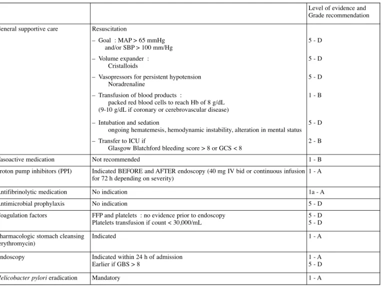
Table 6. — Management of NON-variceal bleeding
Level of evidence and Grade recommendation General supportive care Resuscitation
– Goal : MAP > 65 mmHg and/or SBP > 100 mm/Hg 5 - D – Volume expander : Cristalloids 5 - D
– Vasopressors for persistent hypotension Noradrenaline
5 - D
– Transfusion of blood products :
packed red blood cells to reach Hb of 8 g/dL (9-10 g/dL if coronary or cerebrovascular disease)
1 - B
– Intubation and sedation
ongoing hematemesis, hemodynamic instability, alteration in mental status 5 - D
– Transfer to ICU if
Glasgow Blatchford bleeding score > 8 or GCS < 8
2 - B
Vasoactive medication Not recommended 1 - B
Proton pump inhibitors (PPI) Indicated BEFORE and AFTER endoscopy (40 mg IV bid or continuous infusion for 72 h depending on severity)
1 - A
Antifibrinolytic medication No indication 1a - A
Antimicrobial prophylaxis No indication 5 - D
Coagulation factors FFP and platelets : no evidence prior to endoscopy Platelets transfusion if count < 30,000/mL
5 - D 5 - D Pharmacologic stomach cleansing
(erythromycin)
Indicated 1 - A
Endoscopy Indicated within 24 h of admission
Earlier if GBS > 8
1 - A 5 - D
blood pressure may initially be maintained due to impor-tant vasoconstriction in hypovolemic children. Hypotension and delayed capillary refill indicate the necessity of immediate intervention as these are signs of severe hypovolaemia (> 25% blood loss) and they pre-cede collapse (Table 8).
In case of hypovolemic shock we refer to the official European Pediatric Life Support guidelines for the management of hypovolemic shock in children.
Airway protection is always necessary in case of hemodynamic instability, neurological deterioration (agitation, GCS < 8, deterioration of the level of con-sciousness), respiratory distress and ongoing bleeding. It is also recommended when dealing with infants, in case of signs of severe bleeding (> 20% loss of blood volume), recurrent vomiting and need for invasive procedures (e.g. central venous access, endoscopy).
In a hemodynamically stable child, as in adults, blood volume reconstitution should be done cautiously and As UGIB in children is rare, RCT’s on the
manage-ment of UGIB are lacking. Many treatmanage-ment strategies are deducted from the management of UGIB in adults and similar evidence-based approaches do not exist as it comes to pediatric UGIB. In the following paragraphs, some specificities and differences in the approach to UGIB in children compared to adults will be highlight-ed.
General Management
Initial management and resuscitation
The initial treatment focuses on maintenance of the airway and provision of adequate oxygenation and ven-tilation followed by cardiovascular resuscitation. The first step is the immediate differentiation between a hemodynamically stable child and a child in hypov-olemic shock. In children age-adjusted tachycardia is the most sensitive indicator of severe blood loss, whereas
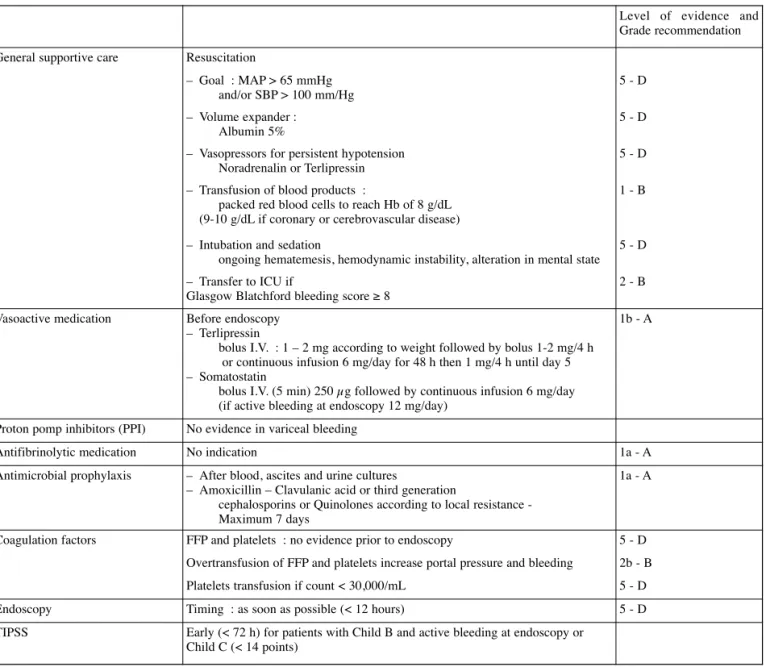
Table 7. — Management of variceal bleeding
Level of evidence and Grade recommendation General supportive care Resuscitation
– Goal : MAP > 65 mmHg and/or SBP > 100 mm/Hg 5 - D – Volume expander : Albumin 5% 5 - D
– Vasopressors for persistent hypotension Noradrenalin or Terlipressin
5 - D
– Transfusion of blood products :
packed red blood cells to reach Hb of 8 g/dL (9-10 g/dL if coronary or cerebrovascular disease)
1 - B
– Intubation and sedation
ongoing hematemesis, hemodynamic instability, alteration in mental state 5 - D
– Transfer to ICU if
Glasgow Blatchford bleeding score ≥ 8
2 - B
Vasoactive medication Before endoscopy – Terlipressin
bolus I.V. : 1 – 2 mg according to weight followed by bolus 1-2 mg/4 h or continuous infusion 6 mg/day for 48 h then 1 mg/4 h until day 5 – Somatostatin
bolus I.V. (5 min) 250 µg followed by continuous infusion 6 mg/day (if active bleeding at endoscopy 12 mg/day)
1b - A
Proton pomp inhibitors (PPI) No evidence in variceal bleeding
Antifibrinolytic medication No indication 1a - A
Antimicrobial prophylaxis – After blood, ascites and urine cultures
– Amoxicillin – Clavulanic acid or third generation
cephalosporins or Quinolones according to local resistance -Maximum 7 days
1a - A
Coagulation factors FFP and platelets : no evidence prior to endoscopy 5 - D
Overtransfusion of FFP and platelets increase portal pressure and bleeding 2b - B
Platelets transfusion if count < 30,000/mL 5 - D
Endoscopy Timing : as soon as possible (< 12 hours) 5 - D
TIPSS Early (< 72 h) for patients with Child B and active bleeding at endoscopy or Child C (< 14 points)
conservatively to maintain hemodynamic stability but to avoid overcorrection which can maintain the bleeding. The amount of blood transfused should be adjusted to the age and weight of the child aiming for a hemoglobin (Hb) concentration of 8 g/dL which can be calculated using the following formula : (target Hb concentration – observed Hb concentration) × 3 × weight of the child in kg.
Cholestatic and/or breastfed infants are at risk for hemorrhagic disease of the newborn due to vitamin K deficiency. The latter should always be suspected in infants presenting with UGIB and prompt administration of IV vitamin K is required.
The cause of severe life threatening UGIB in children is most frequently bleeding from varices but on rare occasions aorto-oesophageal fistula, Dieulafoy lesion, or ulcers can also be the cause.
Two major categories of UGIB in children should be differentiated :
1. Mucosal erosion or ulceration : ask for use of NSAID (97), abdominal pain, foreign body inges-tion, vomiting, stress factors.
2. Variceal bleeding : look for any clinical signs of liver disease or a neonatal history of umbilical catheters. An extrahepatic portal vein obstruction, however, often first presents with a severe bleeding episode in an otherwise perfectly healthy child without any pre-vious history of note.
Some limited blood tests can be helpful in the differ-ential diagnosis and the assessment of the severity of the UGIB : Hb and platelet counts, clotting and liver func-tion tests.
Hospitalisation
Referral to a tertiary care centre where a multidiscipli-nary team of pediatric intensivists, pediatric gastroen-terologists and pediatric surgeons is available is indicat-ed in the following circumstances : important UGIB with drop in Hb level, (suspicion of) variceal bleeding and recurrent bleeding. Since early endoscopy is indicated, transfer should be organized as soon as possible once the child has been stabilised.
Initial medical treatment
The empiric use of acid-suppressive medication in children is justified based on the predominance of peptic causes of UGIB. In children, clear evidence is lacking (only uncontrolled trials and case reports are available) regarding a possible superiority of PPI’s compared to H2-blockers (99).
PPI's : In children most data are available for the old-est molecule : omeprazole. The clearance of omeprazole in children is influenced by age with a slower metabo-lism in infants below 10 days of age, an increased metab-olism between the age of 1 and 6 year and comparable metabolism to adults thereafter (40,100,101). Further on, CYP2C19 activity as well as the presence of systemic inflammatory response will alter the clearance of omeprazole (102). The recommended doses of IV omeprazole vary from 1 mg/kg once daily to 0.5 mg/kg twice daily to 40 mg/1.73 m² in order to maintain the gastric pH above 4 (99,103,104).
H2-blockers : The dose of ranitidine needed to main-tain pH above 4 in children is age dependent. Preterm infants need 0.5 mg/kg IV twice daily, whereas term infants need 1.5 mg/kg IV three times a day and children need 1.5 mg/kg IV four times a day (105,106).
There are no pediatric data on the duration of IV acid-suppressive therapy.
Vaso-active drugs : Based on adult data and because the use of vaso-active medication has been proven to be safe in children, it is recommended to use vaso-active drugs as soon as possible before diagnostic endoscopy in children suspected of variceal bleeding (107). However, there is a lack of controlled trials in children.
In Belgium, in pediatric practice, the most frequently used vaso-active drug is somatostatin (starting bolus
3.5 µg/kg IV over 5 minutes, followed by
3.5 µg/kg/h) (98). In analogy with adults, based on the Baveno V guidelines, treatment duration of 5 days is empirically recommended.
Endoscopy
Indication for endoscopy
An endoscopy can only be performed in a hemo -dynamically stable child, under general anesthesia with size adapted material by an experienced endoscopist.
Table 8. — Normal vital signs, heart rate (HR), respiratory
rate (RR), systolic and diastolic blood pressure according to children’s age
Diastolic blood pressure = 0.5 to 0.66 × systolic blood pressure. AGE HR at rest (/min) RR at rest (/min)
Newborn 120-160 30-60 1-6 months 120-150 30-50 7-12 months 110-140 25-40 1-3 months 90-130 20-30 4-5 years 85-120 20-25 6-12 years 70-100 16-22 13-18 years 60-80 12-18
AGE Systolic blood pressure (mmHg)
Normal Lower limit
0-1 months > 60 50
1-12 months > 80 70
1-10 years 90 + [2 × age in years] 70 + [2 x age in years]