Université de Montréal
Beyond hairballs: Depicting complexity of a
kinase-phosphatase network in the budding yeast
par Diala Abd-Rabbo
Département de biochimie et médecine moléculaire Faculté de médecine
Thèse présentée en vue de l’obtention du grade de Philosophiæ Doctor (Ph.D.)
en Bio-informatique
Janvier, 2017
Résumé
Les kinases et les phosphatases (KP) représentent la plus grande famille des enzymes dans la cellule. Elles régulent les unes les autres ainsi que 60 % du protéome, formant des réseaux complexes kinase-phosphatase (KP-Net) jouant un rôle essentiel dans la signalisation cellulaire. Ces réseaux caractérisés d’une organisation de type commandes-exécutions possèdent généralement une structure hiérarchique. Malgré les nombreuse études effectuées sur le réseau KP-Net chez la levure, la structure hiérarchique ainsi que les principes fonctionnels sont toujours peux connu pour ce réseau. Dans ce contexte, le but de cette thèse consistait à effectuer une analyse d’intégration des données provenant de différentes sources avec la structure hiérarchique d’un réseau KP-Net de haute qualité chez la levure, S.
cerevisiae, afin de générer des hypothèses concernant les principes fonctionnels de chaque
couche de la hiérarchie du réseau KP-Net.
En se basant sur une curation de données d’interactions effectuée dans la présente et dans d’autres études, le plus grand et authentique réseau KP-Net reconnu jusqu’à ce jour chez la levure a été assemblé dans cette étude. En évaluant le niveau hiérarchique du KP-Net en utilisant la métrique de la centralisation globale et en élucidant sa structure hiérarchique en utilisant l'algorithme vertex-sort (VS), nous avons trouvé que le réseau KP-Net possède une structure hiérarchique ayant la forme d’un sablier, formée de trois niveaux disjoints (supérieur, central et inférieur). En effet, le niveau supérieur du réseau, contenant un nombre élevé de KPs, était enrichi par des KPs associées à la régulation des signaux cellulaire; le niveau central, formé d’un nombre limité de KPs fortement connectées les unes aux autres, était enrichi en KPs impliquées dans la régulation du cycle cellulaire; et le niveau inférieur, composé d’un nombre important de KPs, était enrichi en KPs impliquées dans des processus cellulaires diversifiés.
En superposant une grande multitude de propriétés biologiques des KPs sur le réseau KP-Net, le niveau supérieur était enrichi en phosphatases alors que le niveau inférieur en était appauvri, suggérant que les phosphatases seraient moins régulées par phosphorylation et déphosphorylation que les kinases. De plus, le niveau central était enrichi en KPs représentant des « bottlenecks », participant à plus d’une voie de signalisation, codées par des gènes
essentiels et en KPs qui étaient les plus strictement régulées dans l’espace et dans le temps. Ceci implique que les KPs qui jouent un rôle essentiel dans le réseau KP-Net devraient être étroitement contrôlées. En outre, cette étude a montré que les protéines des KPs classées au niveau supérieur du réseau sont exprimées à des niveaux d’abondance plus élevés et à un niveau de bruit moins élevé que celles classées au niveau inférieur du réseau, suggérant que l’expression des enzymes à des abondances élevées invariables au niveau supérieur du réseau KP-Net pourrait être importante pour assurer un système robuste de signalisation.
L’étude de l’algorithme VS a montré que le degré des nœuds affecte leur classement dans les différents niveaux d’un réseau hiérarchique sans biaiser les résultats biologiques du réseau étudié. En outre, une analyse de robustesse du réseau KP-Net a montré que les niveaus du réseau KP-Net sont modérément stable dans des réseaux bruités générés par ajout d’arrêtes au réseau KP-Net. Cependant, les niveaux de ces réseaux bruités et de ceux du réseau KP-Net se superposent significativement. De plus, les propriétés topologiques et biologiques du réseau KP-Net étaient retenues dans les réseaux bruités à différents niveaux. Ces résultats indiquant que bien qu’une robustesse partielle de nos résultats ait été observée, ces derniers représentent l’état actuel de nos connaissances des réseaux KP-Nets.
Finalement, l’amélioration des techniques dédiées à l’identification des substrats des KPs aideront davantage à comprendre comment les réseaux KP-Nets fonctionnent. À titre d’exemple, je décris, dans cette thèse, une stratégie que nous avons conçu et qui permet à déterminer les interactions KP-substrats et les sous-unités régulatrices sur lesquelles ces interactions dépendent. Cette stratégie est basée sur la complémentation des fragments de protéines basée sur la cytosine désaminase chez la levure (OyCD PCA). L’OyCD PCA représente un essai in vivo à haut débit qui promet une description plus précise des réseaux KP-Nets complexes. En l’appliquant pour déterminer les substrats de la kinase cycline-dépendante de type 1 (Cdk1, appelée aussi Cdc28) chez la levure et l’implication des cyclines dans la phosphorylation de ces substrats par Cdk1, l’essai OyCD PCA a montré un comportement compensatoire collectif des cyclines pour la majorité des substrats. De plus, cet essai a montré que la tubuline- γ est phosphorylée spécifiquement par Clb3-Cdk1, établissant ainsi le moment pendant lequel cet événement contrôle l'assemblage du fuseau mitotique.
Mots-clés : Réseau des kinases-phosphatases, Saccharomyces cerevisiae, Complexité des
réseaux, Structure hiérarchique des réseaux, Algorithmes de décomposition de réseaux, propriétés topologiques, Intégration de données multi-omiques, Essai de complémentation des fragments de protéine basé sur la cytosine désaminase chez la levure, Kinase cycline-dépendante de type 1 et la tubuline- γ.
Abstract
Kinases and phosphatases (KP) form the largest family of enzymes in living cells. They regulate each other and 60 % of the proteome forming complex kinase-phosphatase networks (KP-Net) essential for cell signaling. Such networks having the command-execution aspect tend to have a hierarchical structure. Despite the extensive study of the KP-Net in the budding yeast, the hierarchical structure as well as the functional principles of this network are still not known. In this context, this thesis aims to perform an integrative analysis of multi-omics data with the hierarchical structure of a bona fide KP-Net in the budding yeast Saccharomyces
cerevisiae, in order to generate hypotheses about the functional principles of each layer in the
KP-Net hierarchy.
Based on a literature curation effort accomplished in this and in other studies, the largest bona fide KP-Net of the S. cerevisiae known to date was assembled in this thesis. By assessing the hierarchical level of the KP-Net using the global reaching centrality and by elucidating the its hierarchical structure using the vertex-sort (VS) algorithm, we found that the KP-Net has a moderate hierarchical structure made of three disjoint layers (top, core and bottom) resembling a bow tie shape. The top layer having a large size was found enriched for signaling regulation; the core layer made of few strongly connected KPs was found enriched mostly for cell cycle regulation; and the bottom layer having a large size was found enriched for diverse biological processes.
On overlaying a wide range of KP biological properties on top of the KP-Net hierarchical structure, the top layer was found enriched for and the bottom layer was found depleted for phosphatases, suggesting that phosphatases are less regulated by phosphorylation and dephosphoryation interactions (PDI) than kinases. Moreover, the core layer was found enriched for KPs representing bottlenecks, pathway-shared components, essential genes and for the most tightly regulated KPs in time and space, implying that KPs playing an essential role in the KP-Net should be firmly controlled. Interestingly, KP proteins in the top layer were found more abundant and less noisy than those of the bottom layer, suggesting that availability of enzymes at invariable protein expression level at the top of the network might be important to ensure a robust signaling.
Analysis of the VS algorithm showed that node degrees affect their classification in the different layers of a network hierarchical structure without biasing biological results of the sorted network. Robustness analysis of the KP-Net showed that KP-Net layers are moderately stable in noisy networks generated by adding edges to the KP-Net. However, layers of these noisy overlap significantly with those of the KP-Net. Moreover, topological and biological properties of the KP-Net were retained in the noisy networks to different levels. These findings indicate that despite the observed partial robustness of our results, they mostly represent our current knowledge about KP-Nets.
Finally, enhancement of techniques dedicated to identify KPs substrates will enhance our understanding about how KP-Nets function. As an example, I describe here a strategy that we devised to help in determining KP-substrate interactions and the regulatory subunits on which these interactions depend. The strategy is based on a protein-fragment complementation assay based on the optimized yeast cytosine deaminase (OyCD PCA). The OyCD PCA represents a large scale in vivo screen that promises a substantial improvement in delineating the complex KP-Nets. We applied the strategy to determine substrates of the cyclin-dependent kinase 1 (Cdk1; also called Cdc28) and cyclins implicated in phosphorylation of these substrates by Cdk1 in S. cerevisiae. The OyCD PCA showed a wide compensatory behavior of cyclins for most of the substrates and the phosphorylation of γ-tubulin specifically by Clb3-Cdk1, thus establishing the timing of the latter event in controlling assembly of the mitotic spindle.
Keywords: Kinase-phosphatase network, Saccharomyces cerevisiae, Network complexity,
Network hierarchical structure, Network decomposition algorithms, Topological properties, Integration of multi-omics data, protein-fragment complementation assay based on the optimized yeast cytosine deaminase, Cyclin-dependent kinase 1 and γ-tubuline.
Table of Contents
RÉSUMÉ --- I
ABSTRACT --- IV
TABLE OF CONTENTS --- VI
LIST OF TABLES --- XII
LIST OF FIGURES --- XIII
LIST OF ABBREVIATIONS --- XV
ACKNOWLEDGMENTS --- XVIII
CHAPTER 1 INTRODUCTION --- 20
1.1 Complexity of living cells --- 20
1.2 Complexity of the kinase-phosphatase networks (KP-Nets) --- 21
1.3 Kinase-phosphatase networks --- 23
1.3.1 Phosphorylation and dephosphorylation --- 23
1.3.2 Kinases and phosphatases --- 24
1.3.2.1 Protein kinases --- 25
1.3.2.2 Protein phosphatases --- 28
1.3.3 Mapping phophorylation networks --- 29
1.3.3.1 In vitro experimental methods --- 29
1.3.3.1.1 In vitro kinase assays using protein microarrays ... 30
1.3.3.1.2 In vitro kinase assays using peptide libraries ... 30
1.3.3.1.3 In vitro kinase assays using analogue-sensitive kinase alleles ... 31
1.3.3.2.1 In vivo phosphorylation technologies based on liquid-chromatography and
mass-spectrometry ... 32
1.3.3.2.2 In vivo Protein-fragment Complementation Assay (PCA) based on the optimized yeast cytosine deaminase ... 33
1.3.4 Protein phosphorylation and protein interaction databases --- 34
1.4 Analyzing biological networks --- 34
1.4.1 Studying biological processes using control theory --- 35
1.4.2 Assessing network topological properties --- 37
1.4.2.1 Local topological properties of networks --- 38
1.4.2.2 Global topological properties of networks --- 39
1.4.2.3 Network logic motifs --- 40
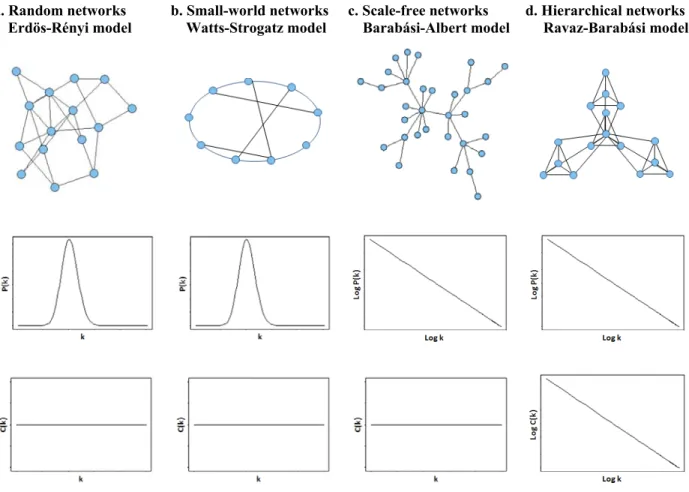
1.4.3 Network models --- 41
1.4.3.1 Erdös-Rényi model - Random networks --- 41
1.4.3.2 Watts-Strogatz model - Small-world networks --- 43
1.4.3.3 Barabási-Albert model - Scale-free networks --- 43
1.4.3.4 Ravaz-Barabási model - Hierarchical networks --- 44
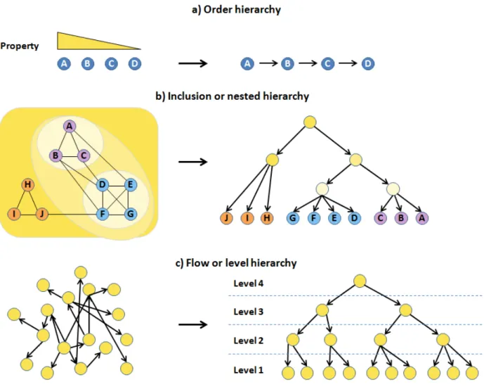
1.4.4 Studying hierarchical structures in networks --- 45
1.4.4.1 Types of network hierarchical structures --- 45
1.4.4.2 Measuring network hierarchical level --- 47
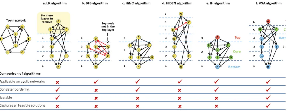
1.4.4.3 Elucidating network hierarchy --- 48
1.5 Correlation between topological properties and biological functions --- 53
1.5.1 Do node topological properties predict node biological functions? --- 53
1.5.2 Does logic motif topology predict logic motif biological functions? --- 54
1.5.3 Does network topology predict network biological functions? --- 55
1.6 Data integration --- 56
1.6.1 Integrating other biological networks with phosphorylation networks --- 57
1.6.2 Integrating biological data with KP-Nets --- 58
1.7 Thesis objectives and organization --- 59
1.7.1 Thesis aim and objectives --- 59
CHAPTER 2 DELINEATING FUNCTIONAL PRINCIPLES OF THE BOW TIE
STRUCTURE OF A KINASE-PHOSPHATASE NETWORK IN THE BUDDING
YEAST --- 61
2.1 Authors’ contributions --- 62
2.2 Abstract --- 63
2.3 Background --- 64
2.4 Results --- 66
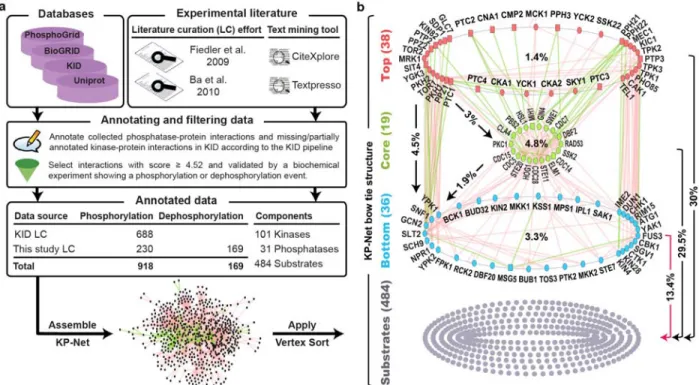
2.4.1 The kinase-phosphatase network (KP-Net) --- 66
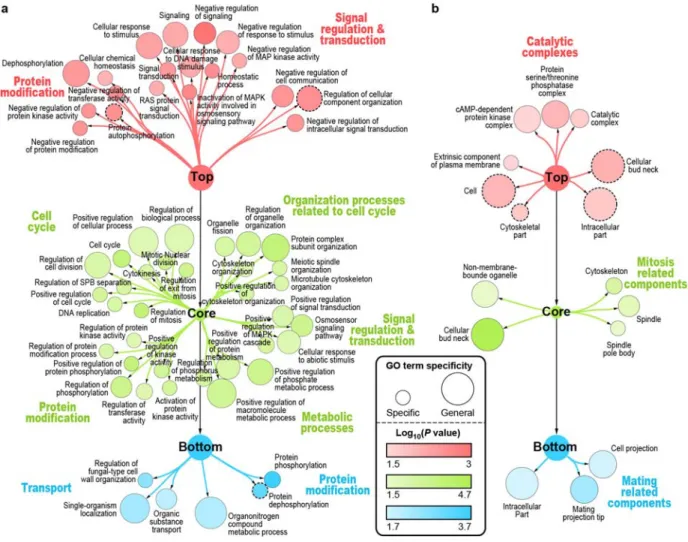
2.4.2 The KP-Net possesses a “corporate” hierarchical structure in the form of a bow tie with a strongly connected core layer --- 67
2.4.3 The three layers of the KP-Net have dissimilar biological roles and subcellular localization --- 69
2.4.4 Phosphatases are less regulated by phosphorylation than kinases --- 71
2.4.5 KP-Net upper levels are the least regulated and KP-Net lower levels are the least to regulate other KPs --- 71
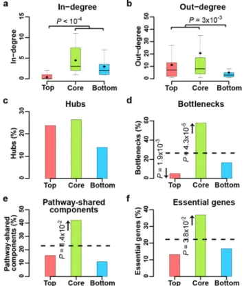
2.4.6 The KP-Net core layer is enriched for essential genes, bottlenecks, and pathway-shared components --- 73
2.4.7 Molecular switches are enriched in KPs in core and bottom layers --- 73
2.4.8 Core layer KPs employ scaffolding to prevent unwanted pathway crosstalk --- 75
2.4.9 Core layer KPs undergo more spatial organization changes than top and bottom layer KPs --- 75
2.4.10Top layer KP proteins are more abundant and less noisy than bottom layer KPs of the KP-Net --- 76
2.4.11The VS algorithm depends on node degree to classify network nodes in three layers --- 77
2.4.12Biological properties of KPs are independent of their in- and out-degrees --- 79
2.4.13Robustness of results and incompleteness of data --- 80
2.4.14Predicting Kinases acting on substrates on high osmolarity stress --- 81
2.4.15Discussions --- 82
2.4.16Conclusions --- 84
2.5 Methods --- 84
2.5.1 Over-representation of various logic motifs in the KP-Net --- 84
2.5.2 Network randomization --- 85
2.5.2.2 Similar degree preserving randomization (SDPR) --- 85
2.5.2.3 Out-degree preserving randomization (ODPR) --- 85
2.5.2.4 Degree non-preserving randomization (DNPR) --- 85
2.5.3 The matching algorithm --- 85
2.5.4 Testing whether the KP-Net GRC is bigger than Erdős–Rényi network GRCs--- 86
2.5.5 Comparing means of node properties in two layers using RT --- 86
2.5.6 Generating subsampled/noisy networks and assessing their layers stability and their overlap with KP-Net layers --- 86
2.5.7 Predicting kinases --- 87
2.6 Acknowledgements --- 87
CHAPTER 3 DISSECTION OF CDK1-CYCLIN COMPLEXES IN VIVO --- 88
3.1 Authors’ contributions --- 89
3.1.1 Quantifying colony growth --- 89
3.1.2 Preys in OyCD PCA are implicated in various biological processes --- 90
3.1.3 Comparison of OyCD PCA data with other datasets --- 91
3.1.3.1 Comparison of first screen of OyCD PCA data with other datasets --- 92
3.1.3.2 Comparison of second screen of OyCD PCA data with other datasets --- 94
3.1.4 Other contributions --- 94
3.2 Abstract --- 95
3.3 Significance --- 95
3.4 Introduction --- 96
3.5 Results --- 98
3.5.1 Identifying Cdk1 complexes in vivo --- 98
3.5.2 Cyclin dependency of Cdk1 complexes --- 100
3.5.3 γ-Tubulin is an in vivo target of Cdk1–Clb3 --- 104
3.5.4 Clb3–Cdk1 preferentially targets γ-tubulin in vitro --- 106
3.5.5 Discussion --- 106
3.6.1 Yeast strains --- 108
3.6.2 Plasmid construction --- 108
3.6.3 Gateway cloning --- 109
3.6.4 OyCD PCA to detect protein–protein interactions with Cdk1 --- 109
3.6.5 Detecting protein–protein interactions in the different cyclin deletion strains --- 110
3.6.6 Analysis of the cyclin deletion strains --- 110
3.6.7 Purification of γ-TUSC substrate and particle analysis --- 111
3.6.8 Cdk1 kinase assays --- 111
3.7 Note added in proof--- 112
3.8 Acknowledgements --- 112
CHAPTER 4 CONCLUSIONS AND FUTURE WORK --- 113
4.1 Conclusions --- 113
4.2 Future directions --- 124
REFERENCES --- 129
APPENDIX 1.METHODS, TABLES AND SUPPLEMENTARY FIGURES OF CHAPTER 2 -- I Supplementary materials --- i
Supplementary methods --- iii
Supplementary tables --- vii
Supplementary figures --- x
APPENDIX 2.SUPPLEMENTARY METHODS, TABLES AND FIGURES OF CHAPTER 3 --- XIX Supplementary methods --- xix
Supplementary tables --- xxi
Supplementary figures --- xxiii
Publications --- xxv Conferences and presentations --- xxvi
List of tables
Table 1.I. Families of protein kinases and their corresponding members. --- 26 Table 1.II. Protein phosphatases families and their corresponding phosphatases members. --- 28 Table 3.I. Top enriched GO terms in the preys that were tested to interact with Cdc28 --- 91 Table 3.II. Significance of the overlap between Cdc28 interactors identified in Ear et al. and in
other datasets. --- 94
Table A1.I. Annotation of dephosphorylation interactions. --- vii Table A1.II. K-Nets and KP-Nets studies. --- viii Table A1.III. Phosphorylation and dephosphorylation interactions used to assemble the KP-Net
(Supplementary file on CD-ROM) ---ix
Table A1.IV.Core layer KPs implicated in decision-making. ---ix Table A1.V. The kinase-substrate interactions that were predicted to be implicated in osmotic
shock in this study (Supplementary file on CD-ROM) ---ix
Table A2.I. List of prey proteins that interact with Cdk1 identified by OyCD PCA --- xxi Table A2.II. False-negative rates (FNR) of the Cdk1 screen --- xxii Table A2.III. Sensitivity and specificity of the Cdk1–protein interaction screen in cyclin deletion
strain backgrounds --- xxii
Table A2.IV.Cdk1 interaction death index for all prey genes (Supplementary file on CD-ROM). --- xxii Table A2.V. Cyclin contingency of 21 Cdk1 interacting proteins (Supplementary file on CD-ROM). -- xxii
List of figures
Figure 1.1. Mechanisms of kinases specificity. --- 27
Figure 1.2. A closed-loop control system.--- 35
Figure 1.3. Different types of network logic motifs. --- 41
Figure 1.4. Network models. --- 42
Figure 1.5. Different types of network hierarchical structures. --- 46
Figure 1.6. Application of the different sorting algorithms on a toy network and a comparison of their performance. --- 52
Figure 2.1. The pipeline used to assemble and to sort the KP-Net, and the KP-Net bow tie structure. --- 67
Figure 2.2. Depleted and enriched biological processes and cellular components in each of the KP-Net layers. --- 70
Figure 2.3. Topological and biological properties of KPs in the different layers of the KP-Net. --- 72
Figure 2.4. Biochemical and spatiotemporal modulators of KPs in the different layers of the KP-Net. --- 74
Figure 2.5. mRNA and protein turnover related properties of KPs in the different layers of the KP-Net. --- 77
Figure 2.6. The VS algorithm depends on node degrees to sort network nodes in three layers. --- 78
Figure 2.7. Stability of KP-Net layers and their overlap with subsampled/noisy network layers. --- 81
Figure 3.1. Dissecting Cdk1 complexes using the OyCD PCA. --- 97
Figure 3.2. Identification of interaction partners of Cdk1. --- 99
Figure 3.3. Cyclin dependence of Cdk1–prey protein interactions. --- 101
Figure 3.4. γ-Tubulin is a Clb3–Cdk1 substrate in vitro. --- 105
Figure A1.1. Distributions of the cumulative degree and of the clustering coefficient of KP-Net nodes follow a power law distribution. ...x
Figure A1.2. Detailed hierarchical structure of the KP-Net. ---xi
Figure A1.3. Percentage of phosphatases in each of the three layers of the KP-Net and distribution of phosphosites among KPs and among the different layers of the KP-Net. --- xii
Figure A1.4. Clustering of the different sets of randomized networks. --- xiii
Figure A1.5. Correlation between KP in-degrees and KP numerical properties. --- xiv
Figure A1.6. In-degree distribution of KPs characterized and not characterized by each of the studied categorical properties. --- xv
Figure A1.8. Out-degree distribution of KPs characterized and not characterized by each of the
studied categorical properties. --- xvii
Figure A1.9. Distribution of the different properties of KPs in each layer of 100 noisy networks. --- xviii Figure A2.1. Optimized yeast Saccharomyces cerevisiae prodrug-converting enzyme cytosine
deaminase (OyCD) protein fragment complementation assay cyclin-dependent
List of abbreviations
5-FC 5-fluorocytosinATP Adenosine triphosphate BFS Breadth-first search Cdk Cyclin-dependent kinase DNPR Degree non-preserving randomization DPR Degree preserving randomization ELM Eukaryotic linear motif database
FBL Feedback loop
FCY1 yCD gene
FFL Feedforward loop
FN False negative
FNR False-negative rate
GO Gene ontology
GRC Global reaching centrality
GST Gluthathione-S-transferase GTP Guanosine triphosphate
HIDEN Hierarchical decomposition of regulatory network HiNO Hierarchical regulatory networks organization
HS Hierarchical score
HSM Hierarchical score maximization
HT Hypergeometric test
HTP High-throughput
IDPR In-degree preserving randomization
KP Kinase and phosphatase
K-Net Kinase phosphorylation network KID Kinase interaction database
KP-Net Kinase-phosphatase network LBM Linear binding motif
LC Liquid chromatography
LTP Low-throughput
MAPK Mitogen-activated protein kinase MS Mass-spectrometry
ODPR Out-degree preserving randomization ORF Open reading frame
OyCD Optimized yeast cytosine deaminase PCA Protein-fragment complementation assay
PDI Phosphorylation and dephosphorylation interaction PPI Protein-protein interaction
PKA Protein kinase A
PPM protein phosphatases manganese or magnesium dependent PPP Phosphoprotein phosphatase
PTM Post-translational modification PTP Protein Tyr-specific phosphatase
RT Randomization test
SCC Strongly connected component
SDPR Similar degree preserving randomization Ser Serine
SIM Single-input module
SPB Spindle pole body STE Sterile
TAP Tandem affinity purification
TF Transcription factor
TF-Net Transcription network Thr Threonine
TP True positive
Tyr Tyrosine VS Vertex-sort
WT Wild type
To my Creator, to the One Who created the immense universe with a lot of secrets, and invites us to unravel them in order to know Him better and to appreciate His gifts. To God, the One and only Creator.
Acknowledgments
Words cannot express my deepest and sincere gratitude for all the blessings that God has given me. It is because of Him first that I was able to realize this research.
I am forever indebted to my advisor, Prof. Stephen Michnick, for his confidence in me providing me the opportunity to pursue a Ph.D. degree in his laboratory. I would like to express my sincere gratitude for his enthusiasm, invaluable advices, guidance, and for being constantly available even during his vacations. I greatly appreciate his great human qualities including but not limited to listening to me, his patience, his exceptional understanding and his constant support and encouragement during difficult moments along this thesis.
I would like also to express my gratitude to the members of the jury of my thesis, Dr. Martine Raymond, Dr. Sébastien Lemieux, Dr. Mathieu Blanchette and Dr. Janos Filep, for having accepted to read and evaluate my work despite their busy agenda, and for their valuable and instructive comments.
My acknowledgements go equally to all my colleagues in the Michnick laboratory, in particular to Jacqueline Kowarzyk, Lara Matta, Emmanuelle Tchekanda, Luz Carrillo, Alessandra Nurrisso, and Durgajini Sivanesan for their support, encouragement, and for the unforgettable moments that we spent together. I would like also to thank Emmanuel Levy and Abdellali Kelil for their pertinent advices and the instructive discussions that I had with them. I would like to say a big thank you for the administrative responsible for the Bioinformatics program, Mrs Elaine Meunier. She provided me with invaluable support and she was always available with her charming smile and affectionate attention.
No words can express my sincere and deepest gratitude to my exceptional mom, Hana, who encouraged me continuously with a lot of affection and who had invested considerable time, energy and effort to help me overcome a major vision deficiency to finally succeed my scholar studies. She tirelessly and with a lot of determination instilled in me great confidence. I owe my grandparents and my uncle so much, Teita Fathiyeh, Jeddo Shafik and uncle Saed for their infinite love, and touching attention. I will never know how to express how much I love them, how much I miss them, and how much I think of them, may their souls rest in peace. An exceptional thank you goes to my special dad, Ahmad, an exceptional example of
an avid reader and a well instructed person who taught me the importance of knowledge; to my step father, Ghassan, a great dentist who taught me how to adopt a systematic and efficient approach in my studies. A huge thank you for the love of my life, Salaheddine, who believed in me and since the first day of our marriage, more specifically four years before the accomplishment of this thesis, he already awarded me the title of Doctor. I would like to thank my sisters, Reem and Noor and my brothers Abboud and Samer, my aunts, in particular, aunt Sana and aunt Sahar for their continuous encouragement, their distinguished attention and their prayers. I would like to infinitely thank my sister and my dearest and closest friend Rahima Ziane, for her friendship, for her precious advices, for her support and for the tons of positive waves she sent me throughout this thesis. Finally, I would like to thank my lovely friends Fati and Dima for being available to listen to me and to always encourage me to continue till the end.
Chapter 1
Introduction
1.1 Complexity of living cells
In nature, living cells are constantly exposed to unstable environmental conditions leading to stress situations such as change in acidity, osmolarity, temperature, availability of nutrient supplies, exposure to radiations, and many other external factors. Interestingly, cells have a remarkable capacity to adapt to new environmental conditions by coordinating their intracellular activities and to correct incidental internal errors (Gasch and Werner-Washburne, 2002). For instance, when DNA damage occurs during DNA replication, the cell cycle stalls and a DNA repair process is launched (Putnam et al., 2009). In case of a successful repair of the DNA damage, both DNA replication and the cell cycle are resumed. Otherwise, the cell is destroyed by launching a programmed cell death called apoptosis. Another example of internal errors occurs when proteins are incorrectly folded. To remedy such situations, the cell attempts to refold these proteins. In case of failure, the cell targets misfolded proteins for degradation (Kriegenburg et al., 2012). These examples and many others show that biological events are well organized in time and space and importantly, are driven by an apparent capacity of the living cells for decision-making.
Notably, the biological events that are triggered, at least in the two examples mentioned above, involve various biochemical mechanisms such as: (1), signaling to sense unusual conditions (DNA damage or protein misfolding in these cases) and to transmit information to other molecules after signal detection; (2), protein-protein interactions (PPI) to form biochemical machineries required for the repair process; (3), post-translational modifications (PTM) including phosphorylation and dephosphorylation interactions (PDI) to regulate individual proteins or the assembled complexes; and (4), metabolic reactions to transform the damage products perceived as cellular toxic to non-toxic products (Chan and Dedon, 2010). Accordingly, living cells can be described as a complex system in which different networks [e.g. signaling network, PPI network, metabolic network, transcription network (TF-Net), ubiquitination network (Venancio et al., 2009) and acetylation network (Choudhary et al., 2009)] interact with each other and affect each other. Complexity in living cells emerges from the interplay between different biological networks on the one hand and on the other hand from the complexity
encountered within each of these networks known usually as complex networks. In fact, there is no single and formal definition for complex networks in the literature. For instance, Barabási and Ablert described complex networks as large networks (having a large number of nodes) that are characterized by topological properties different from those of random networks (Paragraph 1.4.3.1); in particular they contain few nodes that are extremely connected to other nodes (hubs), they expand continuously by adding new nodes, and their new nodes connect preferentially to well-connected nodes (Paragraph 1.4.3.3) (Barabasi and Albert, 1999). Whereas, Steen described a network as complex, if it is so large in size and so interconnected, that it is difficult to understand the behavior of the whole network by examining the behavior of each of its nodes separately (Steen, 2010). This thesis adopts Steen’s definition for complex networks with a minor addition highlighting the importance of the organizational structure of a network. Consequently, in the context of this thesis, a complex network represents a large network formed of extremely interconnected nodes; this interconnectedness hinders the identification of the organizational structure of this network and hinders understanding the behavior of the whole network by observing the individual behavior of its nodes.
1.2 Complexity of the kinase-phosphatase networks (KP-Nets)
Among the various biological networks, the KP-Net is the one that exhibits the most prevalent, immediate and profound effect on the cellular biochemical machinery implicated in a vast range of biological processes (Paragraph 1.3.1). KP-Nets play an essential role in signaling pathways (Graves and Krebs, 1999; Pawson and Scott, 2005). Textbooks usually describe signaling pathways as simple isolated linear cascades of enzymes, particularly of kinases and phosphatases (KP), which are activated in response to a stimulus ultimately to generate a cellular response such as metabolic alterations, gene activation and repression, and PDIs. This cellular response aims at changing the cell behavior to maintain cellular homeostasis. It is now well established that signaling does not occur through linear pathways, but through highly interconnected pathways forming complex signaling networks.
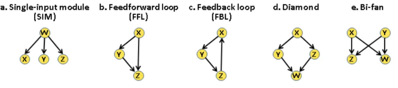
Recent evidence suggests that KP-Nets are extremely complex for different reasons. First, KPs in these networks not only regulate proteins having no enzymatic activity, but also regulate each other, making KPs regulators of regulators or super-regulators (Bodenmiller et al., 2010; Breitkreutz et al., 2010; Fiedler et al., 2009). Second, various types of network building blocks
known as logic motifs (subgraphs) are represented in KP-Nets (Alon, 2007). These over-represented logic motifs have a more complex topology than the simple linear chains corresponding to linear cascades of KPs in KP-Nets (e.g. single-input modules (SIM), feedforward loops (FFL), diamonds and bi-fans, Paragraph 1.4.2.3). Third, results from a recent study showed a high level of complexity associated to a KP-Net in the budding yeast, for which the perturbation of most KPs affect large parts of the KP-Net (Bodenmiller et al., 2010). Finally, another level of complexity in KP-Nets results from the compensatory behaviors of certain KPs. On the knockdown of a particular KP, another KP closely related to the deleted one could partially or completely compensate for the action of the deleted enzyme. In brief, these findings suggest that the KP-Net is an extremely complex network similar to a hairball that cannot be organized in order to delineate its structure and in order to ultimately understand how it functions.
In the following sections, first, I will introduce KP-Nets, by describing: KPs families, the mechanisms guiding the substrates specificity of KPs, the large-scale experiments used to map KP-Nets, strengths and limitations of these experiments, as well as the available databases designed to annotate interactions implicating KPs. Second, I will introduce the tools used to analyze biological networks, by presenting: a general overview about the application of the control theory to study the structure and dynamics of small networks, the network topological properties used to describe networks, the different network models conceived to build networks having predefined properties, and the most used bioinformatics methods applied to determine and elucidate network architectures. Third, I will also touch on studies that aimed at inferring the biological functions of nodes, of logic motifs and of the networks themselves from topological properties of each of these entities. I will also outline the important efforts performed to integrate data within KP-Nets. Finally, given the extensive use of the budding yeast Saccharomyces
cerevisiae as a model organism for studying KP-Nets, a special attention to the literature related
1.3 Kinase-phosphatase networks
1.3.1 Phosphorylation and dephosphorylation
Phosphorylation is a biochemical interaction by which a kinase catalyzes the transfer of a phosphate group from adenosine triphosphate (ATP) or guanosine triphosphate (GTP) principally to serine (Ser), threonine (Thr) or tyrosine (Tyr) residues and less commonly to other residues such as histidine, arginine and lysine of a protein known as the kinase substrate (Ciesla et al., 2011; Ubersax and Ferrell, 2007). Dephosphorylation represents the inverse reaction by which a phosphatase catalyzes the transfer of a phosphate group from an amino acid residue of a phosphoprotein to a water molecule. Hence, the phosphorylation level of a protein residue is affected by the simultaneous activity of KPs on this site. Phosphorylation and dephosphorylation could also target small-molecules such as lipids and carbohydrates (Fiedler et al., 2009), but work in this thesis will focus on protein PDIs.
Phosphorylation is the most prevalent PTM of proteins that affects their function by altering their structural conformation and consequently their enzymatic activity in case they have one, their subcellular localization, their abundance by targeting them for ubiquitination in a phospho-dependent manner and their molecular interactions by shaping their interaction surfaces to create new or disrupt existing PPIs (Hunter, 2000; Seet et al., 2006). This PTM regulates a staggering number of cellular processes ranging from stress response, cell cycle, cell proliferation and death to cellular metabolism. Usually, regulation of biological processes by phosphorylation interaction might necessitate a combined sequence of many PDIs (Graves and Krebs, 1999; Pawson and Scott, 2005).
Generally, a phosphosite is said to be functional if it has a known biological function or if its mutation results in an observable phenotype or change in fitness of an organism (Lasalde et al., 2014; Yoshimi et al., 2012). To date, the function of a limited number of phosphosites has been characterized. Recent studies suggest that most phosphosites are non-functional and that they represent noise due to non-specific phosphorylation of, particularly, abundant proteins encountered by kinases in the crowded environment of the cell (Ba and Moses, 2010; Landry et al., 2009; Levy et al., 2012; Malik et al., 2008). These studies suggest that functional phosphosites could be systematically identified if they meet the following criteria: (1), to occur
in lower abundance proteins; (2), to be on average more conserved across species; and (3), to have high stoichiometry in comparison with non-functional sites (phosphosites having no known function). Although these studies provided elegant approaches to distinguish between functional and non-functional phosphosites, these criteria are not necessarily predictive of phosphosite functionality (Tan et al., 2010; Wu et al., 2011). Moreover, numerous phosphosites appear to have no functional consequences on their mutation, because their mutation would not result in any cellular phenotypic change, or because of the existence of other phosphosites having a redundant role (Lasalde et al., 2014; Yoshimi et al., 2012). Therefore, a safe approach to confidently determine functional phosphosites would be to investigate not only the non-functional consequence of each site separately, but also for all site combinations per protein (Lienhard, 2008). Currently, no screens are considered to assess the functionality of individual and combined phosphosites on a large scale for two reasons. First, the mutation of this staggering number of phosphosites, apart from being tremendously time-consuming, is not feasible given the currently available experimental approaches. Second, it is extremely difficult to find a general experimental assay to determine the functionality of a phosphosite. And yet, advanced site-directed mutagenesis technologies combined with phenotypic high-throughput (HTP) screens could open new avenues for unraveling a considerable number of functional phosphosites in the future.
1.3.2 Kinases and phosphatases
Kinases and phosphatases belong to the class of phosphotransferases, the largest class of enzymes in eukaryotic cells. They are responsible for phosphorylating and dephosphorylating their substrates respectively. Kinases and phosphatases can also act on themselves and on each other (Bodenmiller et al., 2010; Whinston et al., 2013; Wu et al., 2009). They both represent ~2 % of eukaryotic cells proteome. The number of kinases is estimated to 133 in Saccharomyces
cerevisiae, 454 in Caenorhabditis elegans, 251 in Drosophila melanogaster and 518 in Homo sapiens, whereas the number of phosphatases is estimated to 41 in Saccharomyces cerevisiae,
185 in Caenorhabditis elegans, 86 in Drosophila melanogaster and 126 in Homo sapiens (Manning et al., 2002b; Morrison et al., 2000). Recently, KPs were shown to regulate at least 60 % of budding yeast proteins (Yachie et al., 2011). Hence, it is not surprising that aberrant regulation of KPs leads to serious perturbations of the regulatory and signaling networks, a result
that has been clearly observed on the knockdown of most of these enzymes one at a time (Bodenmiller et al., 2010).
1.3.2.1 Protein kinases
Kinases can be subdivided into three groups according to the specificity they exhibit towards the phosphorylated residue: Ser/Thr-specific, Tyr-specific and dual-specificity kinases. Most of the kinases in the budding yeast are Ser/Thr-specific. Only one has been shown to be Tyr-specific (Malathi et al., 1999), a number that could be underestimated (Yachie et al., 2011; Zhu et al., 2000). And seven kinases showed a dual-specificity; they can phosphorylate Ser, Thr or Tyr residues (Hunter and Plowman, 1997).
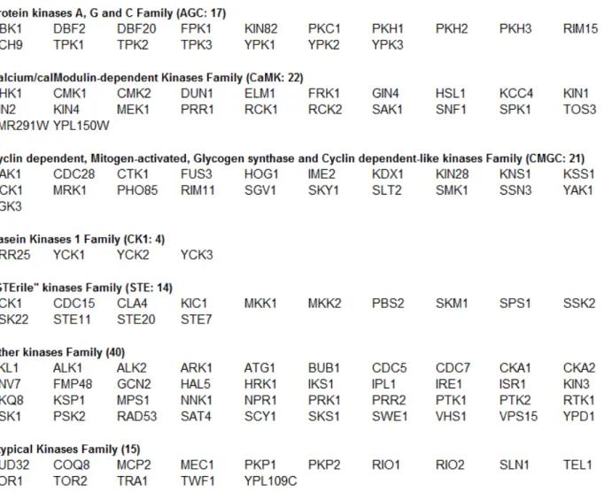
Kinases can also be classified according to the primary sequence similarity of their catalytic domains into seven families: (1), the family that groups protein kinases A, G and C; (2), the calcium/calmodulin-dependent kinase family; (3), the family that groups Cdks, mitogen-activated protein kinases (MAPK), glycogen synthase kinase 3β and Cdk-like kinases; (4), the casein kinase 1 family; (5), the “STErile” family (STE) or kinases of MAPK grouping Ste7, Ste11 and Ste20 subfamilies; (6), the other family grouping kinases that do not share strong similarity with the previously mentioned kinase families; and (7), the atypical family, grouping proteins that lack sequence similarity to the conventional eukaryotic protein kinase catalytic domains, yet they were experimentally shown to have a kinase activity (Hunter and Plowman, 1997) (Table 1.I).
Kinase catalytic domains, made of ~250 amino acids and having the form of a cleft, binds the ATP and the kinase substrate and catalyzes the transfer of a phosphate from the ATP to a Ser (72 %), Thr (~23 %) and rarely to a Tyr (~5 %) residue within the substrate protein sequence in the budding yeast (Yachie et al., 2011). The sequence recognized by a kinase catalytic domain is called the consensus sequence and varies in length between four and eight residues (Ubersax and Ferrell, 2007). Identification of these sequences became possible with the emergence of peptide library screens and other similar technologies (Mok et al., 2010; Olsen et al., 2006; Pinna and Ruzzene, 1996). Although phosphorylation consensus sequences enhance the recognition of a kinase to its substrates, these elements seem to be insufficient to determine protein kinase specificity towards their substrates for at least three reasons. First, different Ser/Thr-specific kinases were found to have similar minimal phosphorylation consensus sequences. For instance,
Cdk and protein kinase A (PKA) both recognize the [S/T]P sequence (Ubersax and Ferrell, 2007). Second, consensus sequences are characterized to be of short length and to have a degenerate nature. Finally, certain authentic phosphorylation sequences do not match identified consensus phosphorylation sequences (Kreegipuu et al., 1998; Ubersax et al., 2003).
Table 1.I. Families of protein kinases and their corresponding members.
Kinases belonging to the different kinase families were determined from (Hunter and Plowman, 1997; Manning et al., 2002a; Thomas et al., 2003).
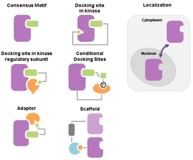
Therefore, living cells possess additional mechanisms to assure kinase specificity towards their substrates (Figure 1.1) (Ubersax and Ferrell, 2007), including: (1), docking site in kinases, more specifically on the substrates binding interaction domains in kinases, increases the local concentration of the substrate around the kinase catalytic domain (Biondi and Nebreda, 2003); (2), docking site in kinase regulatory subunit facilitates targeting the kinase to particular
(Cdk1), also called Cdc28, contain a hydrophobic patch that recognizes an [R/K]XL motif in Cdc28 substrates (Koivomagi et al., 2011; Loog and Morgan, 2005)]; (3), conditional docking sites on substrates target the kinase to specific substrates that has been already phosphorylated [e.g. Plk1 and Gsk3 target substrates that have been already phosphorylated (Elia et al., 2003; Frame et al., 2001)]; (4), localization of kinases or their regulatory partners in different cellular compartments increases the phosphorylation rate towards substrates in the same subcellular localization as the kinase in certain conditions and limits the number of substrates accessible to the kinase in other conditions (Miller and Cross, 2000); and finally (5), adaptors or scaffolds organize the interaction between the kinase and their substrates by recruiting those substrates to the same complex (Bhattacharyya et al., 2006; Pawson and Scott, 1997). Interestingly, conditional docking motifs function as an AND logic gate representing a means by which living cells temporally organize their biochemical events with respect to each other. Similarly, subcellular localization and scaffolding mediate the spatial organization of these biochemical events. In summary, these mechanisms not only enhance kinases specificity towards their substrates, but also constitute the primary platform based on which living cells organize biological events in time and space.
Figure 1.1. Mechanisms of kinases specificity.
The mechanisms used by kinases to enhance their specificity towards their substrates. Adapted from (Bhattacharyya et al., 2006).
1.3.2.2 Protein phosphatases
Similar to kinases, phosphatases can also be subdivided, according to the residues they dephosphorylate, mainly into two groups. The first group corresponds to the Ser/Thr-specific protein phosphatases and consists of two structurally different families: the phosphoprotein phosphatases (PPP) family and the protein phosphatases manganese or magnesium dependent (PPM) family. The second group corresponds to the protein Tyr phosphatases and contains both the protein Tyr-specific phosphatases (PTP) family and the dual-specificity phosphatases family which can dephosphorylate all three phospho-amino acids (Ser, Thr and Tyr) (Barford et al., 1998) (Table 1.II).
Table 1.II. Protein phosphatases families and their corresponding phosphatases members.
Phosphatases belonging to the different phosphatase families were determined from (Breitkreutz et al., 2010; Cherry et al., 2012; Stark, 1996).
Previously, phosphatases were thought of as housekeeping enzymes and phosphate erasers with no major impact on the regulation of cell signaling. Consequently, they were subject of somewhat dismissive attitude from the scientific community in comparison with the extensive attention that was expressed towards kinases. In contrast, it is now well accepted that phosphatases represent true counter-actors of kinases playing a complementary role as critical as that of kinases in controlling signal transduction (Tonks, 2013). Studies based on mathematical modeling and quantitative experimentation of signaling pathways suggest that while kinases
seem to control the amplitude of a signaling response, phosphatases tend to regulate both the amplitude and duration of these responses (Heinrich et al., 2002; Hornberg et al., 2005).
Notably despite the key role phosphatases play in the regulation of cell signaling, a quick comparison of the number of phosphatases (24 Ser/Thr-specific, 8 Tyr-specific and 5 dual-specificity phosphatases, Table 1.II) with that of kinases (125 Ser/Thr-specific, 1 Tyr-specific and 7 dual-specificity phosphatases, Table 1.I) reveals that the number of phosphatases is in general smaller than that of the kinases in eukaryotic cells and in particular, the number of Ser/Thr-specific phosphatases is much smaller than that of Ser/Thr-specific kinases in budding yeast. In addition, phosphatases showed low specificity towards substrates in in vitro assays (Virshup and Shenolikar, 2009). These observations led to suspect that phosphatases represent promiscuous enzymes. However, emerging evidence shows that specificity of phosphatases is enhanced independently of their catalytic domains by a wide range of regulatory subunits associated to catalytic domains of PPPs on one hand and by supplementary domains or conserved sequence motifs in PPMs and PTPs on the other hand (Shi, 2009; Tonks, 2006; Ubersax and Ferrell, 2007; Virshup and Shenolikar, 2009). Similar to kinases, phosphatases has also been observed to exhibit spatial and temporal organization in the budding yeast (Jin et al., 2008; Rossio and Yoshida, 2011).
1.3.3 Mapping phophorylation networks
Over the past decade, significant efforts have been made to map phosphosites and to identify responsible kinases for those phosphorylation events using in vitro and in vivo HTP experiments. As mentioned previously, phosphatases have much less attracted the attention of the scientific community in comparison with kinases and consequently have rarely made the subject of such HTP experiments. Therefore, the following sections will introduce HTP methods used to identify kinases substrates.
1.3.3.1 In vitro experimental methods
In vitro biochemical approaches consist of incubating the kinase of interest with the proteome
exposed differently in the presence of γ32P-ATP. The HTP in vitro approaches that significantly contributed in identifying potential kinase substrates in yeast include in vitro kinase assays using: (1), protein microarrays (Mok et al., 2009; Ptacek et al., 2005); (2), peptide libraries (Mah et al.,
2005; Mok et al., 2010); and (3), analogue-sensitive kinase alleles (Dephoure et al., 2005; Koivomagi et al., 2011; Loog and Morgan, 2005; Ubersax et al., 2003). These in vitro experimental methods made seminal contributions to identify potential substrates of the studied kinases. Each of these methods has its proper strengths and limitations.
1.3.3.1.1 In vitro kinase assays using protein microarrays
Similar to the DNA microarray concept, a large number of purified proteins are spotted on a chip, which is then incubated with a purified kinase in the presence of radiolabeled γ32P-ATP (Ptacek et al., 2005). The chips are then washed, dried and analyzed by autoradiography. Proteins that are phosphorylated in vitro by the kinase of interest could be identified depending on their spatial position on the chip. Kinase assays using protein chips are very practical, since one assay could be used to screen the whole proteome against a specific kinase. But, the main disadvantage of these assays is that proteins are not equally represented on the proteome chip for various reasons: missing clones, misfolded proteins and variable clones expression (Mok et al., 2011).
1.3.3.1.2 In vitro kinase assays using peptide libraries
A huge number of peptides having a fixed length and having a phosphorylation acceptor residue (Ser or Thr) in a predefined central position are generated randomly (Mok et al., 2010). Peptides are then incubated with a purified kinase and radiolabeled ATP in different wells. Subsequently, the aliquots are simultaneously spotted using a pin tool on a membrane. The membrane is then washed, dried and exposed to a phosphor screen, allowing the identification of the phosphorylated peptides. Phosphopeptides could be identified depending on their spatial position on the membrane. Peptides library techniques permitted the mapping of potential consensus phosphorylation motifs targeted by a kinase of interest. However, similar to kinase assays, peptides library techniques also possess drawbacks. Their major drawback is that they considerably depend on the bioinformatics algorithms used to characterize the phosphorylation motifs targeted by the studied kinase.
1.3.3.1.3 In vitro kinase assays using analogue-sensitive kinase alleles
The ATP-binding pocket of a kinase of interest is mutated to accept bulky radiolabeled ATP analogues that are not used by wild type (WT) kinases, producing the so-called analogue-sensitive kinases (Shah et al., 1997; Ubersax et al., 2003). In order to determine whether a protein is phosphorylated by an analogue-sensitive kinase of interest, a library of yeast strains is prepared, in which each strain expresses an open reading frame tagged with a molecular marker such as the gluthathione-S-transferase (GST) tag for protein purification. The addition of radiolabeled γ32P-ATP analogue to the cell lysates of strains of this library containing an analogue-sensitive kinase of interest permits phosphorylation of the analogue-sensitive kinase substrates by radiolabeled ATP. The tagged proteins are then purified and their phosphosites are identified. Kinase assays involving analogue-sensitive kinase alleles succeeded to identify direct potential kinase substrates in an environment having physiological conditions close to those found in living cells; however, their application is limited, because not all kinases could be genetically mutated into the analogue-sensitive version and some analogue-sensitive kinase alleles lose their enzymatic capacity or their specificity towards substrates (Knight et al., 2013). Moreover, it has been shown that some WT kinases could use the analogue bulky form of ATP (Carlson et al., 2011).
Nevertheless, all in vitro experiments share a well-known technical limitation resulting in the generation of high false positive rates. The artificial environment in which these assays are performed lacks the cellular compartmentalization, the temporal expression profiles and the PTM of proteins met in living cells. Hence, a large number of proteins that could never represent genuine substrates of the assayed kinases in natural conditions will be phosphorylated in these experiments lacking the spatial, temporal and PTMs coordination and will be erroneously identified as kinase substrates, a misleading result that increases false positive rates of these approaches. Obviously, absence of natural context is not a problem for in vivo HTP experiments.
1.3.3.2 In vivo experimental methods
1.3.3.2.1 In vivo phosphorylation technologies based on liquid-chromatography and mass-spectrometry
Most of the In vivo large-scale technologies are based on mass-spectrometry (MS) to map phosphosites in cell lysates. Cell extracts are treated with phosphatase inhibitors to protect phosphoproteins from being dephosphorylated by phosphatases in cell lysates and with protease to generate a mixture of small peptides to permit their analysis by MS. Phosphopeptides are then isolated from non-phosphorylated peptides, enriched using different enrichment strategies, introduced into the spectrometer using liquid chromatography (LC), ionized and identified by comparing their fragmentation spectra measured by MS with those in primary sequence databases. In vivo phosphorylation technologies based on Liquid-chromatography and mass-spectrometry (LC-MS/MS) is a large-scale method renowned to detect small phosphoproteins abundance and accurately identify phosphorylated residues as well as the proteins to which they belong.
These technologies were used to quantify phosphosites in cell lysates (Chi et al., 2007; Ficarro et al., 2002) or phosphosites exhibiting changes in their phosphorylation level between WT and KP inhibited or deficient cells, one KP at a time (Bodenmiller et al., 2010; Holt et al., 2009) or between cells under normal and stress conditions. For instance, the latter approach has been used to analyze phosphorylation interactionss under different conditions, such as in cells exposed to alpha factor (Gruhler et al., 2005; Li et al., 2007), to DNA damage agents (Chen et al., 2010; Smolka et al., 2007) or to high osmolarity stress (Kanshin et al., 2015).
Obviously, in vivo screens guarantee that the quantified phosphorylation interactions occurred in their intrinsic environment. Yet, a major drawback of this technique is that the identified proteins showing a decrease in their phosphorylation level on the knockdown or inhibition of a given kinase could be direct substrates of this kinase, substrates of another kinase activated by the kinase of interest, or substrates of a phosphatase inhibited by the kinase of interest.
1.3.3.2.2 In vivo Protein-fragment Complementation Assay (PCA) based on the optimized yeast cytosine deaminase
Recently, a promising in vivo PCA based on the optimized mutant form of the reporter yeast cytosine deaminase (OyCD) has been devised to identify substrates of the cyclin-dependent kinase, Cdc28, and the cyclin regulatory subunit(s) on which the interactions between Cdc28 and its substrates depend in the budding yeast (Ear et al., 2013). The Cdc28 is a cyclin dependent kinase that with its nine cyclins, regulatory subunits, orchestrate regulation of mitotic and meiotic cell cycle events in the budding yeast. Cyclins are classified as G1 phase cyclins (Cln1, Cln2 and Cln3), S phase cyclins (Clb5 and Clb6) and M phase cyclins (Clb1, Clb2, Clb3 and Clb4) depending on the phase in which their expression peaks. They are believed to enhance Cdc28 specificity towards its substrates.
The OyCD PCA is called also the death selection assay, because the enzyme yCD reporter catalyzes the deamination of a yCD-specific prodrug called 5-fluorocytosine (5-FC) to 5-fluorouracil (5-FU). The 5-FU will then be converted by other enzymes in the pyrimidine salvage pathway to 5-FUTP, a toxic compound that causes cell death. This PCA consists of two screens. The first screen is to test the interaction between Cdc28 and a protein of interest using the OyCD PCA and the second screen retests the same interaction but in a strain lacking one of the nine cyclins. The OyCD PCA consists of splitting the yCD into two fragments F[1] and F[2] and fusing one of them to Cdc28 and the other one to the protein of interest. If Cdc28 interacts with this protein, then F[1] and F[2] will be brought into proximity allowing the OyCD reporter enzyme to fold and restore its activity causing cell death in the presence of 5-FC in the first screen. Moreover, if this interaction depends on a given cyclin, so on retesting the same interaction in cells lacking this cyclin, the interaction will not take place, consequently the reporter enzyme will not refold and cells will grow in the second screen.
In contrast to LC-MS/MS, although the OyCD PCA identifies potential direct substrates of Cdc28, it was not conceived to identify amino acids phosphorylated by Cdc28 in its corresponding substrates. However, the OyCD PCA is the first experimental method designed to identify the regulatory subunit or subunits on which the kinase depends to phosphorylate its substrates. Importantly, this PCA could be applied on any KP that depends on regulatory subunit(s) to modify their substrates. Moreover, the OyCD PCA can be performed on the level of
the proteome, because its fragments are compatible with the Gateway cloning system. Finally, this assay is affordable, because it does not require expensive reagents.
1.3.4 Protein phosphorylation and protein interaction databases
Various databases were designed to store phosphorylation data and physical interactions between KPs and other proteins (e.g. regulatory subunits, adaptors and scaffolds) including their substrates. Among those that offered a seminal contribution annotating these events, particularly in the budding yeast, we mention: the PhosphoELM database which provides access to phosphosites detected in in vivo and in vitro experiments (Dinkel et al., 2011); the PhosphoGRID database which contains information about phosphosites validated by in vivo low-throughput (LTP) and HTP experiments and about KPs targeting these phosphopeptides (Stark et al., 2010); the BioGRID database which stores genetic and physical PPI data inferred from HTP and LTP studies (Stark et al., 2006); and the kinase interaction database (KID) which represents the most kinase specialized repository that annotates genetic, physical and phosphorylation interactions of budding yeast kinase with other proteins, validated by LTP and HTP studies and providing details about the experimental techniques used to identify a kinase-protein interaction (Sharifpoor et al., 2011). Also, the KID database annotates kinase-protein interaction based on co-localization, chemical co-fitness and phenotypic correlation studies. Unfortunately, phosphorylation interactions are much more annotated in comparison with dephosphorylation interactions in these databases.
1.4 Analyzing biological networks
Mapping biological networks and designing databases to store their basic components provided a catalogue of KPs, their substrates as well as the PDIs associating these three entities together. Nevertheless, this catalogue of individual components is not sufficient to gain information about how KP-Nets function. We need to understand how these entities are assembled to function together in order to shape the cellular behavior and how changes in one entity of this system may affect other entities.
1.4.1 Studying biological processes using control theory
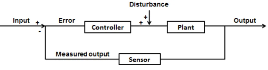
Studying the structure and the dynamic behavior of networks underlying biological processes is possible by benefiting from the principles of control theory, an interdisciplinary branch originating from engineering and mathematics. Control theory aims to study the characteristics and behavior of closed-loop control systems (Nise, 2015). A closed-loop system is a system in the form of a feedback loop (FBL) in which system inputs are determined partially by the system outputs. In its simplest forms, the closed-loop system is composed of a controller, a plant and a sensor (Figure 1.2). The controller drives a plant; the plant is the part of a system to control; and the sensor detects the output of a plant. This system receives an input, generates an output, and can be exposed to disturbance. The difference between the input and the measured output is called the error (Figure 1.2). This error drives the plant to make a correction so that the output will become equal to the input. If this error is zero, then the system does not drive the plant, because the plant is producing the desired output. The system detects the disturbance and corrects for it via the closed-loop, and thus yields the correct output. Various examples in everyday life and in human and other organism physiology represent closed-loop control system (e.g. heating systems, elevators, radar antenna, bacterial chemotaxis, pancreatic secretion of insulin and other hormones, and FBLs in transcription regulatory networks and signaling networks). For instance, in a heating system, the input is a position on a thermostat, the output is the heat, the sensor is a thermostat, the plant is the heater, and the controller is composed of fuel valves and an electrical system to control these valves. As long as a room temperature does not correspond to the desired temperature, the heater will be activated and when the position on the thermostat corresponds to the measured temperature in the room, the heater will stop.
Figure 1.2. A closed-loop control system.
The arrangement of the main components (the controller, the plant, and the sensor) of a control system in a simple closed-loop control system.
Given the conceptual similarities between the engineering and cellular regulatory mechanisms, the tools used to model engineering closed-loops are used to analyze biological networks. FBLs in signaling pathways represent an example of a closed-loop control system. For instance, G-protein coupled receptor signaling in eukaryotic cells represents a FBL responding to a wide range of stimuli (e.g. pheromone α-factor and presence or absence of nutrients in the budding yeast, sense of smell in mice, and hormones and neurotransmitters in human) (Alberts et al., 2002). The protein is composed of three subunits α, β, and γ. In the inactive form of the G-protein, its α-subunit binds GDP. The first step in the activation of the G-protein consists of the binding of a ligand to a transmembrane receptor, causing the α-subunit to bind to the ligand-receptor complex. This binding causes a conformational change in the α-subunit, leading it to release GDP, bind GTP, and to dissociate from the βγ subunits. The GTP-bound α-subunit activates a downstream response, then converts the GTP to GDP, and at the end, the G-protein three subunits are re-associated, closing thus the FBL (Ingalls, 2013).
Each of the described interactions in the G-protein signaling mechanism or in a similar regulatory mechanism can be represented in the form of mathematical formulas such as differential equations (Yi et al., 2003). The terms of these formulas describe the rate of change of the abundance of each component of the model. This approach can be applied to various categories of biological processes such as metabolic, transcription regulatory and signaling pathways (Hynne et al., 2001; Perkins et al., 2006; Yi et al., 2003). Modelling biological processes serves to study different types of the dynamic responses of these processes (graded response, ultrasensitivity (switch-like response), bistability, and multistability) (Szomolay and Shahrezaei, 2012). Moreover, control theory provides a theoretical basis to conceive reverse engineering approaches aiming at determining the structure of regulatory networks (Khammash, 2008; Perkins et al., 2006). Details about the modelling approaches and the nature of the studied responses are outside the scope of this thesis. For a review, refer to (Fischer, 2008; Iglesias, 2013). Although, mathematical modelling yields important details about the structure and dynamic behavior of networks underlying biological processes, it can be computationally expensive. Mathematical modelling, however, can be fruitfully applied to networks composed of a limited number of nodes (in the order of tens). Other approaches are needed to describe the structure and dynamics of larger networks and to help shed light on how these networks function, though limited in detail compared to mathematical modelling.
The subsequent sections introduce how studying organizational structure of biological networks could enhance our understanding about how biological networks work. This involves: (1), assessing networks topological properties (local properties, global properties and logic motifs); (2), classifying networks into categories according to the assessed properties they share with known network models; (3), measuring network hierarchical level; and (4), elucidating their hierarchical structure in case their hierarchical level is perceptible. In the following sections, I will introduce these approaches as well as the four network models that provided valuable insights for understanding and describing diverse real-world networks.
1.4.2 Assessing network topological properties
The charted cellular networks that have been annotated in various databases require a mathematical framework for representation and analysis purposes. The graph representation and graph theory principles constitute a universal and efficient computational tool to study different biological networks. A graph is a set of nodes that are connected by edges. Nodes correspond to molecules such as genes, transcripts, proteins, metabolites or small molecules and edges designate the interactions between these entities. Edges could take the form of arrows or simple links depending on whether node interactions are directed or not, defining directed and undirected networks, respectively. For instance, PPI networks represent undirected networks, because if node u interacts with node v, this reciprocally means that node v interacts with node u. In contrast, KP-Nets represent directed networks, because if node u (de)phosphorylates node v this does not imply that the inverse is also true.
Representing biological networks as a graph composed of a set of nodes and edges permitted the study of biological networks from a topological perspective. Watts and Strogatz were among the first to suggest that networks topology affects networks function, supporting their claims by finding that the infectious diseases spread and information spread are affected by the social network structure and that robustness of power transmission is affected by the power grid topology (Strogatz, 2001; Watts and Strogatz, 1998). From then, topological properties of networks were extensively studied to characterize biological networks in an attempt to infer functional principles of these networks. This discipline encompasses assessing a myriad of metrics that have been proposed to describe biological networks on both local and global levels. As their names indicate, the former category characterizes local network structures, whereas the
latter one describes global network features. This approach also involves identifying the under- and over-representation of logic motifs which represent subgraphs of the network.
1.4.2.1 Local topological properties of networks
In what follows, we define a network G by a set of nodes V and a set of edges E. The number of nodes in G is n=|V| and of edges is m=|E|. Among the local metrics that are commonly studied, we focused on: the node degree, the node clustering coefficient, the shortest path between two nodes and the node betweenness centrality (Kantarci and Labatut, 2014).
The node degree is the number of edges connected to this node. In directed networks, the node degree consists of the sum of its in- and out-degrees (kin and kout), which correspond to the
node in- and out-going edges, respectively.
The clustering coefficient, C, of a given node, called also the node transitivity, measures the probability that neighbors of this node are also neighbors. Neighbors of a node are nodes that are directly connected to the node in question. Formally, the clustering coefficient of a node u is equal to the ratio of the number of triangles actually connected to u and the number of all possible triangles that could be centered on u (Wasserman and Faust, 1994).
( ) = 2 u
u( u− 1)
Where eu is the number of edges actually linking the neighbors of the node u and ku is the degree
of the node u.
The shortest path between two nodes is the minimal set of consecutive edges that should be traversed to go from one node to another. The length of a shortest path represents the number of edges in the shortest path.
The betweenness centrality of a given node quantifies the number of shortest paths going through this node and is calculated using the following formula:
( ) = ( )