ﺔﻴﺒﻌﺸﻟا ﺔﻴﻃاﺮﻘﳝﺪﻟا ﺔﻳﺮﺋاﺰﳉا ﺔﻳرﻮﻬﻤﳉا
ﱄﺎﻌﻟا ﻢﻴﻠﻌﺘﻟا ةرازو
و
ﺚﺤﺒﻟا
ﻲﻤﻠﻌﻟا
DEPARTEMENT OF BIOCHEMISTRY
N°………/SNV/2018THESIS
Presented byAOUACHRIA Sana
For the fulfillment of the requirements for the degree of
DoctoratE of SciEncES
BIOLOGY
Special filed:
BIOcHEmISTrY
TOPIc
Contribution to the phytochemical study and evaluation of the in vitro and
in vivo antioxidant activity of Reichardia picroides L.
Presented publically in: / / 2018
JurY
President: DAHAMNA Saliha Pr. UFA Setif 1 Supervisor:
Co-Supervisor:
BOUMERFEG Sabah BAGHIANI Abderrahmane
Pr. Univ. Bordj bouarreridj Pr. UFA Setif 1 Examiners: LAROUI Saleh KHATEL Bachra ZERARGUI Fatima Pr. Univ. Batna 2 Pr. Univ. Bejaia MCA. UFA Setif 1
Laboratory of applied biochemistry
Université Ferhat Abbas
Sétif 1
Faculté des sciences de la
nature et de la vie
سﺎﺒﻋ تﺎﺣﺮﻓ ﺔﻌﻣﺎﺟ
ﻒﻴﻄﺳ
1
مﻮﻠﻋ ﺔﻴﻠﻛ
ةﺎﻴﳊا و ﺔﻌﻴﺒﻄﻟا
iACKNOWLEDGEMENTS
First of all, I am grateful to Al-mighty ALLAH who give me strength and perseverance, whatever I am today is just because Him (ALLAH subhana-Wa-Taala).
I would like to express my appreciation and grateful thanks to my supervisor Pr. BOUMERFEG Sabah for her advice, guidance, encouragements and support throughout
my doctoral work.
I would deeply like to thank my co-supervisor, Pr. BAGHIANI Abderrahmane for his enriching advices, keen interest, guidance, support and availability throughout my doctoral work.
I would also like to show my gratitude for my committee and their willingness to read my thesis and give critiques and comments in order to make it a better thesis. Thank you to Pr. DAHAMNA Saliha, Pr. LAROUI Saleh, Pr. KHATEL Bachra and Dr. ZERARGUI Fatima.
I want to offer my special thanks to Pr. ARRAR Lekhmissi for his valuable advices, continuous support, assistance, and to all other technical supports. I am also thankful to Pr. CHAREF Noureddine, Pr. KHENNOUF Seddik, Pr. AMIRA Smain, Pr. DAHAMNA Saliha and Dr. AMIRA Fatima for help and provision of research facilities in their laboratories.
My sincere gratitude to Dr. BENBACHA Faycal, Head of anapathology laboratory of Hospitalo-Public Establishment (HPE) of Bordj Bou-Arreridj, for providing all the facilities to realise histological slides and interpreting them. I am also thankful to Dr. TOUABTI, Head of Central laboratory of University Hospital of Setif, for providing the facilities to evaluate biochemical parameters.
My thanks are also extended to Pr. ABU ZARGA Mussa, Faculty of Science, the University of Jordan, Amman, Jordan for the identification and structural elucidation of the isolated compound.
Many thanks send to all my colleagues in laboratory of applied biochemistry, who provided the most amicable and lively working environment.
ﺺﺨﻠﻣ
مﺪﺨﺘﺴﺗ
ﺔﺘﺒﻧ
Reichardia picroides (R. picroides)
،ﺔﻴﺋاﺬﻏ ضاﺮﻏﻷ ﺎﺳﺎﺳأ
ﻞﻤﻌﺘﺴﺗ و
ﰲ ﺮﻜﺴﻟا ﺔﺒﺴﻨﻟ ﺔﻀﻔﺨﻤﻛ �ﺪﻴﻠﻘﺗ
.ﺔﻄﺸﻨﻣ و ﺐﻴﻠﺤﻠﻟ و لﻮﺒﻠﻟ ةرﺪﻣ و مﺪﻟا
ﺎﻨﻤﻠﻋ ﺪﺣ ﻰﻠﻋو
،
ﱃإ ةرﺎﺷﻹا ﻢﺘﺗ ﱂ
ﺎﻬﻃﺎﺸﻧ و ﺔﺘﺒﻨﻟا ﺔﻴﲰ لﻮﺣ رﻮﺤﻤﺘﺗ تﺎﺳارد يأ
ةﺪﺴﻛﻸﻟ دﺎﻀﳌا
.
ﺔﺘﺒﻨﻠﻟ ﻲﺋﺎﻴﻤﻴﻜﻟا ﺐﻴﻛﱰﻟا ﺔﻓﺮﻌﻣ ﱃإ ﺎﻨﺘﺳارد فﺪﺗﻬ
ﻴﻴﻘﺗ و
و �ﱪﳐ ةﺪﺴﻛﻸﻟ دﺎﻀﳌا ﺎﻬﻃﺎﺸﻧ اﺬﻛو ﺎﻬﺘﻴﲰ ﻢ
ﰲ
ﻲﳊا
صﻼﺨﺘﺳا ﰎ .
لﻮﻨﻴﻔﻟا دﺪﻌﺘﻣ
ﺨﺘﺴﳌا ءﺎﻄﻋﻹ مﺪﺨﺘﺴﳌا ﺐﻳﺬﳌا ﺔﻴﺒﻄﻗ ﻰﻠﻋ ادﺎﻤﺘﻋا ﱄوأ ﻞﺼﻔﺑ ﺎﻋﻮﺒﺘﻣ
مﺎﳋا ﺺﻠ
(CrE)و
مرﻮﻓورﻮﻠﻜﻟا ﺺﻠﺨﺘﺴﻣ
(ChE)تﺎﺘﻴﺳأ ﻞﻴﺜﻳﻹا ﺺﻠﺨﺘﺴﻣو
(EAE)ﻲﺋﺎﳌا ﺺﻠﺨﺘﺴﳌا و
(AqE)ﻞﺼﻔﻟا ﺢﲰ .
ﻰﻠﻋ
ﺎﻴﻓﺮﻏﻮﺗﺎﻣوﺮﻛ
ﻟا
دﻮﻤﻌ
ﺎﻜﻴﻠﻴﺴﻟا مﻼﻫ ﻰﻠﻋ
ﻰﻠﻋ لﻮﺼﳊﺑﺎ
12ءﺰﺟ
(F1-F12) ،ﺎﻤﻛ
ﺔﺋﺰﲡ ﻞﻛ ﺖﻌﻀﺧ
ﱃإ
ﻞﻴﻠﺤﺘﻟا
ﻰﻠﻋ
ﺎﻴﻓﺮﻏﻮﺗﺎﻣوﺮﻛ
ﻟا
ﺔﻘﺒﻄ
ﻲﻘﻨﻟا ﺐﻛﺮﳌا لﺰﻋ ﰎ .ﺔﻘﻴﻗﺮﻟا
lutéoline 7-O-β-glucosideﻦﻣ ،نﻮﻓﻼﻔﻟا ﱃإ ﻲﻤﺘﻨﻳ يﺬﻟا ،
F10ﺔﻴﺋﺎﻴﻤﻴﻜﻟا ﻪﺘﻴﻨﺑ ﺪﻳﺪﲢ ﰎو
.يوﻮﻨﻟا ﻲﺴﻴﻃﺎﻨﻐﳌا ﲔﻧﺮﻠﻟ ﻲﻔﻴﻄﻟا سﺎﻴﻘﻟا زﺎﻬﺟ ﺔﻄﺳاﻮﺑ
تﺮﻬﻇأ
ﺞﺋﺎﺘﻧ
ﻲﻤﻜﻟا ﻞﻴﻠﺤﺘﻟا
ﻲﻠﻜﻟا لﻮﻨﻴﻔﻟا ﻦﻣ ﺔﻴﻤﻛ ﻰﻠﻋأ نأ
ﺪﻳﻮﻧﻮﻓﻼﻔﻟاو
ﰲ ﺖﻠﺠﺳ
EAE ) 331.64 ± 3.11غوﺮﻜﻴﻣ
ﻲﻣاﺮﻏ ﺊﻓﺎﻜﻣ
ﻚﻴﻟﺎﻐﻟا ﺾﲪ
ﺺﻠﺨﺘﺴﳌا ﻦﻣ ﻎﻣ /
و
48.14 ± 0.70غوﺮﻜﻴﻣ
ﻲﻣاﺮﻏ ﺊﻓﺎﻜﻣ
ﲔﺘﺳرﺎﻛ
ﺺﻠﺨﺘﺴﳌا ﻦﻣ ﻎﻣ /
ﺐﻴﺗﱰﻟا ﻰﻠﻋ ،
( .دﺎﻀﳌا طﺎﺸﻨﻟا ﺪﻳﺪﲢ ﺔﻴﻐﺑ �ﱪﳐ برﺎﲡ ةﺪﻋ ءاﺮﺟإ ﰎ ﺪﻗو
ـﻟ ةﺪﺴﻛﻸﻟ
CrEﻒﺸﻛ .ﻪﺋاﺰﺟأ و
CrEﻢﻴﻗ ﰲ ةد�ز ﻞﻴﺠﺴﺘﺑ ﻚﻟذو يﻮﻣﺪﻟا لﻼﳓﻼﻟ دﺎﻀﻣ ﱪﺘﻌﻣ ﺪﺟ طﺎﺸﻧ ﻦﻋ ﻩؤاﺰﺟأ و
HT 50ﱵﻟا
ﻦﻣ ﺖﺣواﺮﺗ
76.92 ± 3.59ﺔﻘﻴﻗد
ﺔﺒﺴﻨﻟﺑﺎ
ﺪﻫﺎﺸﻠﻟ
و
188.15 ± 1.91ﺔﻘﻴﻗد
ﺔﺒﺴﻨﻟﺑﺎ
ـﻟ
EAE .نأ ﺪﺟُو
F12ﻚﻠﳝ
طﺎﺸﻨﻟ ﲑﺒﻛ ﺪﳊ وﺎﺴﻣ نﻮﻳﻸﻟ ﻂﺑﺎﻗ طﺎﺸﻧ ىﻮﻗأ
EDTA .ىﺪﺑأ
EAEةرﺪﻘﻟ ﺔﻳوﺎﺴﻣ ﺔﻴﻋﺎﺟرإ ةرﺪﻗ ىﻮﻗأ
Vit C.
ﻞﺠﺳ
F5ﻰﻠﻋأ
ـﻟ ﺔﻴﺣازإ ةرﺪﻗ
2 O 2 Hةرﺪﻗ ﲑﺒﻛ ﺪﳊ قﻮﻔﺗ ﱵﻟاو
BHT.
لﻼﺧ ﲔﺗورﺎﻛﺎﺘﻴﺒﻟا ضﺎﻀﻴﺑﻻ ﻂﻴﺒﺜﺗ ﻞﺠُﺳ
24ﻼﻛ دﻮﺟو ﰲ ﺔﻋﺎﺳ
ﻣ
ﻦ
ChEو
EAEو
F1و
F10و
F11 .رﺬﺟ ﺔﺣازإ رﺎﺒﺘﺧا ﻒﺸﻛ
DPPHنأ
EAEو
ChEو
F12ﺪﺟ ﺔﻴﺣازإ ةرﺪﻗ نﻮﻜﻠﳝ
.ﺔﻴﻟﺎﻋ
ةدﺎﳊا ﺔﻴﻤﺴﻟا ﻢﻴﻴﻘﺗ ﰎ
ناﺮﺌﻔﻟا ﻰﻠﻋ ﺔﺘﺒﻨﻠﻟ
،و
ضاﺮﻋﻷا ﻞﻴﺠﺴﺘﻟ ﻚﻟذ
و ﺔﻴﻤﺴﻠﻟ ﺔﺒﺣﺎﺼﳌا
دﺪﻋ
تﺎﻴﻓﻮﻟا
ةﺪﳌ ت�اﻮﻴﺤﻠﻟ ﺔﻠﻤﺘﶈا
ﲔﻋﻮﺒﺳأ
بﺎﺴﳊ
ﺔﻋﺮﳉا ﻂﺳﻮﺘﻣ
ﺔﺘﻴﻤﳌا
(LD50)ـﻟ
CrE .تﺎﻴﻓو يأ ثوﺪﺣ ﰲ ﺐﺒﺴﺘﺗ ﱂ ةﺎﻄﻌﳌا تﺎﻋﺮﳉا نأ ﺞﺋﺎﺘﻨﻟا تﺮﻬﻇأ
نأ ﺪﺟو ﺎﻤﻛ ،ث�ﻹا و رﻮﻛﺬﻟا ناﺮﺌﻔﻠﻟ مﺎﻌﻟا كﻮﻠﺴﻟا ﰲ تاﲑﻐﺗ وأ
LD50قﻮﻔﻳ
5000ﻎﻣ
ﻎﻛ /
.
ﺔﺳارد ﺺﳜ ﺎﻤﻴﻓو
ﺔﻴﻤﺴﻟا
قﻮﻓ
ةدﺎﳊا
ءﺎﻄﻋإ ﰎ ﺪﻘﻓ ،
CrEتﺎﻋﺮﲜ
250و
500و
1000ةﺪﳌ مﻮﻳ / ﻎﻛ / ﻎﻣ
21.ﺔﻴﻟﺎﺘﺘﻣ ﺎﻣﻮﻳ
ّﲔﺒﺗ و
ﲔﺑ تﺎﻋﺮﳉا نأ
500و
1000ﻰﻠﻜﻟاو ﺪﺒﻜﻟا ﺔﺠﺴﻧأ ﰲ تاﲑﻐﺗ ﺐﺒﺴﺗ ﻎﻛ / ﻊﻣ
.
ﺖﻳﺮﺟأ
ﺔﺳارد
ﻰﻠﻋ
ناﺮﺌﻔﻟا
رﻮﻛﺬﻟا
ﻦﻣ
ﻞﺟأ
ﻢﻴﻴﻘﺗ
ةرﺪﻘﻟا
ةدﺎﻀﳌا
،ةﺪﺴﻛﻸﻟ
ﺚﻴﺣ
ﰎ
ﺎﻌﻣ
ـﺑ ﺎﻬﺘﻠﻣ
CrEتﺎﻋﺮﲜ
250و
500و
1000ةﺪﳌ مﻮﻳ / ﻎﻛ / ﻊﻣ
21ةد�ز ﺞﺋﺎﺘﻨﻟا ﺖﺘﺒﺛأ .ﺔﻴﻟﺎﺘﺘﻣ ﺎﻣﻮﻳ
ىﻮﺘﺴﻣ ﰲ ﺔﻇﻮﺤﻠﻣ
GSHو
ىﻮﺘﺴﻣ ﰲ ﺎﺿﺎﻔﳔا
MDAﻰﻠﻜﻟاو ﺪﺒﻜﻟا ﻦﻣ ﻞﻛ ﰲ
،ﺎﻤﻴﻓ
ﻟ ﻒﻴﻔﻃ ﻦﺴﲢ ﻆﺣﻮﻟ
ةرﺪﻘﻠ
ةدﺎﻀﳌا
ةﺪﺴﻛﻸﻟ
ﳌا ﲑﺛﺄﺘﻠﻟ و ﺎﻣزﻼﺒﻠﻟ
نأ ﺞﺘﻨﺘﺴﻧ ﻪﻴﻠﻋ و .يﻮﻣﺪﻟا لﻼﳓﻼﻟ دﺎﻀ
R. picroidesﻚﻠﲤ
ﺔﻴﻟﺎﻋ ﺪﺟ ةﺪﺴﻛﻸﻟ ةدﺎﻀﻣ ةرﺪﻗ
لﺎﻤﻌﺘﺳا ﺪﻨﻋ ﺔﻨﻣآ و ﺔﻣﺎﺳ ﲑﻏ ﺎ�أ ﱃإ ﺔﻓﺎﺿﻹﺑﺎ ،ﻲﳊا ﰲ وأ �ﱪﳐ اءاﻮﺳ
تﺎﻋﺮﺟ
≥ 250.ﻎﻛ/ﻎﻣ
: ﺢﻴﺗﺎﻔﳌا تﺎﻤﻠﻜﻟا
R. picroides،
،يﺪﺴﻛﺄﺘﻟا دﺎﻬﺟﻹا ،ﺪﻳﻮﻧﻮﻓﻼﻔﻟا ،لﻮﻨﻴﻔﻴﻟﻮﺒﻟا
ةدﺎﳊا ﺔﻴﻤﺴﻟا
،
ﺔﻴﻤﺴﻟا
قﻮﻓ
ةدﺎﳊا
.
iiiABSTRACT
Reichardia picroides (R. picroides) is a species mainly used for daily source; it is used in the
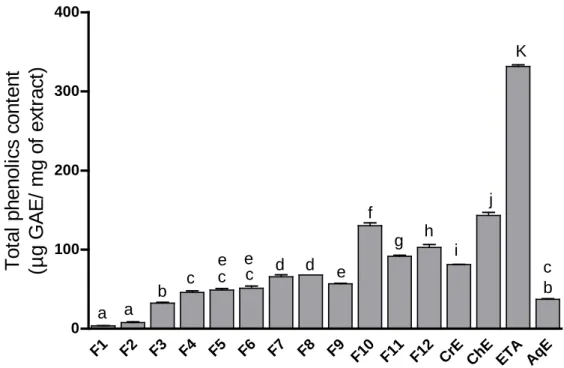
traditional medicine as hypoglycemiant, diuretic, depurative, galactagogue and tonic agent. To our knowledge, there are no studies on the antioxidant effect and the toxicity of this plant. The objective of the present study was, therefore, to evaluate the antioxidant activity and the toxicity of crude extract (CrE) and its fractions in vitro and in vivo. Polyphenols extraction and fractionation from plant material was performed using solvent of different polarity giving the following phases: CrE, chloroform extract (ChE), ethyl acetate extract (EAE) and aqueous extract (AqE). The CrE was then subjected to silica gel column chromatography which results in 12 fractions (F1-F12). Each fraction was subjected to TLC analysis. The pure compound was isolated from F10 and its structure was established by NMR spectroscopy as luteolin 7-O-β-glucoside. Results of polyphenols estimation showed that the highest amount of both total phenols and flavonoids was found in EAE (331.64 ± 3.11 µg gallic acid equivalent (GAE) / mg of extract and 48.14 ± 0.70 µg quercetin equivalent (QE) / mg of extract, respectively). To determine the antioxidant potential of CrE and its fractions, several in vitro assays were carried out. The CrE and its fractions had a very important (p < 0.0001) antihemolytic activity and revealed an extension of HT 50 from 76.92 ± 3.59 min of control to 188.15 ± 1.91 min of EAE. The F12 was found to have the strongest ion chelating activity comparable to that of EDTA. The EAE appeared to have the strongest reducing power closer to that of Vit C. The F5 found to have the strongest H2O2 scavenging effect higher than that of BHT. The bleaching of β-carotene is highly inhibited in the presence of ChE, EAE, F1, F10 and F11.The DPPH scavenging assay revealed that EAE, ChE and F12 possessed a very strong radical scavenging effect. The acute toxicity of CrE was carried out using mice. Signs accompanying toxicity and possible death of animals were monitored for two weeks to ascertain the median lethal dose (LD50) of the CrE. The administrated doses did not resulted mortality or changes in general behaviors. The LD50 was found to be superior to 5000 mg/kg. In subacute toxicity study, the CrE was administered by gavage at the doses of 250, 500 and 1000 mg/kg/day for 21 consecutive days. Daily administration of CrE at doses ranged from 500 to 1000 mg/kg resulted in alteration of liver and kidney tisssues. In vivo approach was performed by administration three doses 250, 500 and 1000 mg/kg to male mice. The analysis of antioxidant potential parameters revealed that the CrE administration increased significantly the level of GSH and decreased the level of MDA in both liver and kidney. The CrE supplementation improved slightly the plasma antioxidant status and the anti-hemolytic protective effect which remained non significant compared to the control group. It can therefore be suggested that R. picroides had potent antioxidant activities both in vitro and in vivo. The use of this extract is safe at doses ≤ 250 mg/kg.
Key words : R. picroides, polyphenols, flavonoids, oxidative stress, acute toxicity, subacute toxicity.
RESUME
Reichardia picroides (R. picroides) est une espèce principalement utilisée à des fins alimentaires, et
traditionnellement comme hypoglycémiante, diurétique, dépurative, galactagogue et tonique. A notre connaissance, aucune étude n’a signalé à la fois sa sécurité et ses activités antioxydantes. L'objectif de la présente étude était donc de cribler les composés phytochimiques de l'extrait brut de la plante (CrE), d'évaluer son profil de sécurité ainsi que d'étudier l'activité antioxydante de CrE et de ses fractions in
vitro et in vivo. L'extraction des polyphénols a été réalisée en se basant sur la polarité du solvant utilisé
pour donner le CrE, l'extrait de chloroforme (ChE), l'extrait d'éthyle d'acétate (EAE) et l'extrait aqueux (AqE). Le CrE a été soumis à une chromatographie sur colonne de gel de silice qui a permis d'obtenir 12 fractions (F1-F12) en suivant leur profil de CCM. Le composé pur a été isolé à partir de F10 et sa structure a été établie par l'utilisation de la spectroscopie RMN. Il s’agit d’un flavone: lutéoline 7-O-glucoside. La teneur en phénols totaux et en flavonoïdes a été quantifiée à l'aide de méthodes colorimétriques. Les résultats ont montré que la plus grande quantité de phénols totaux et de flavonoïdes a été trouvée dans l’EAE (331.64 ± 3.11 μg équivalent d’acide gallique (GAE) / mg d'extrait et 48.14 ± 0.70 μg équivalent de quercétine (QE) / mg d'extrait, respectivement). Pour déterminer le potentiel antioxydant de CrE et de ses fractions, plusieurs essais in vitro ont été réalisés. Le CrE et ses fractions ont eu une activité antihémolytique très importante (p <0.0001) et ont révélé une extension de HT 50 varie de 76.92 ± 3.59 min de contrôle à 188.15 ± 1.91 min d'EAE. La F12 s’est avérée avoir l'activité de chélation des ions la plus forte significativement proche à celle de l’EDTA. L’EAE semble avoir le pouvoir réducteur le plus fort, similaire à celui de la Vit C. La F5 s'est avérée avoir l'effet piégeur de H2O2 le plus fort qui significativement plus élevé que celui du BHT. Le blanchissement du β-carotène est fortement réduit pendant les 24 heures en présence de ChE, EAE, F1, F10 et F11. Le test de piégeage de DPPH a révélé que l'EAE, ChE et F12 possédaient un très fort effet antiradicalaire. La toxicité aiguë de CrE a été réalisée sur les souris. Les signes d'une toxicité accompagnant la mort possible des animaux ont été suivis pendant deux semaines afin de déterminer la dose létale médiane (DL50) de CrE. Les doses administrées n'ont pas provoqué de mortalité ou de changements dans les comportements généraux des souris testés. La DL50 s'est révélée supérieure à 5000 mg / kg. Dans l'étude de toxicité subaiguë, le CrE a été administré par gavage à des doses de 250, 500 et 1000 mg / kg / jour pendant 21 jours consécutifs. L'administration quotidienne de CrE à des doses allant de 500 à 1000 mg / kg pourrait entraîner une altération de l'histologie du foie et des reins. Une approche in vivo a été réalisée par l’administration de trois doses (250, 500 et 1000 mg / kg) à des souris mâles. L'analyse des paramètres du potentiel antioxydant a révélé que l'administration de CrE a augmenté significativement le niveau de GSH et a diminué le taux de MDA dans le foie et les reins. Le statut antioxydant plasmatique et le pouvoir antihémolytique s’est amélioré légèrement par suite à une supplémentation de CrE. En conclusion, on peut suggérer que R. picroides a eu des activités antioxydantes puissantes à la fois in vitro et in vivo. De plus, elle était non toxique en administration aiguë ainsi que l’utilisation de son extrait brut est sûre à des doses ≤ 250 mg / kg.
Mots clés : R. picroides, polyphénols, flavonoids, stress oxidatif, toxicité aiguë, toxicité subaiguë
LIST OF FIGURES
Figure 1. Types of polyphenols……….. 17
Figure 2. Basic skeleton or structure of flavonoids………... 18
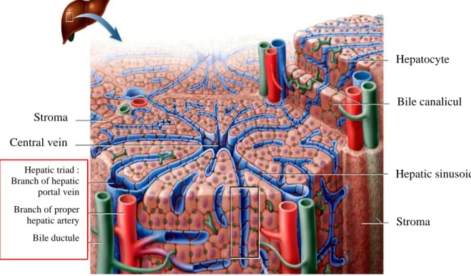
Figure 3. Cross section of a hepatic lobule, illustrating microscopic structure……….. 30
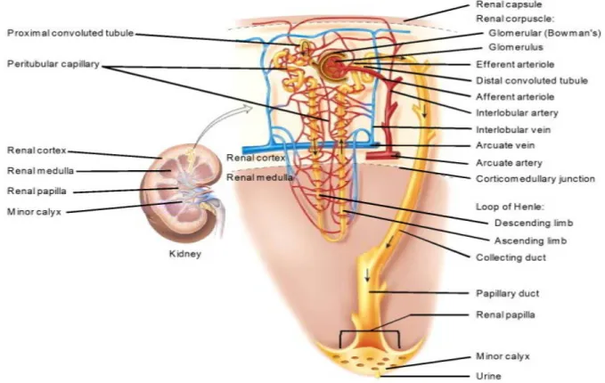
Figure 4a. Anterior view of dissection of right kidney……….. 31
Figure 4b. Cortical nephron and vascular supply………... 32
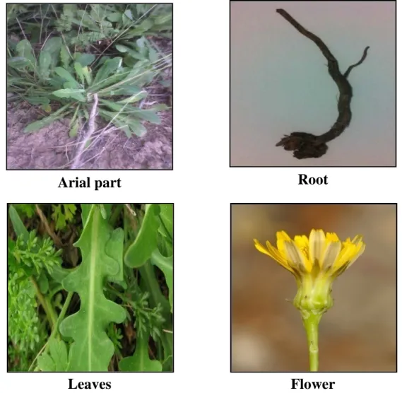
Figure 5. The R. picroides L. Roth plant……….... 35
Figure 6. The R. picroides CrE fractionation... 40
Figure 7. Luteolin 7-O-β-glucoside……… 54
Figure 8. The 1H NMR (DMSO, 500 MHz) of luteolin 7-O-β-glucoside……….. 55
Figure 9. The 13C NMR (DMSO, 125 MHz) of luteolin 7-O-β-glucoside………. 56
Figure 10. The DEPT (DMSO, 500 MHz) of luteolin 7-O-β-glucoside……… 56
Figure 11. Total phenolic content in R. picroides CrE and its fractions……… 58
Figure 12. Flavonoids content in R. picroides CrE and its fractions……….. 59
Figure 13. Kinetics of erythrocytes hemolysis in the presence of CrE, fractions, control and Vit C during 208 min………... 60
Figure 14. The HT 50 values of antihemolytic activity of CrE, fractions, control and Vit C... 61
Figure 15. The EC50 values of ferrous iron chelating activity of CrE, fractions and EDTA… 63 Figure 16. The EC50 values of reducing power of CrE, fractions and Vit C……… 65
Figure 17. The EC50 values of H2O2 scavenging activity of CrE, fractions and BHT……….. 66
Figure 18. Kinetics of β-carotene bleaching in the presence of CrE, ChE, EAE, AqE, water, methanol and BHT during 24h……… 67
Figure 19. Kinetics of β-carotene bleaching in the presence of CrE and its column chromatography fractions, water, methanol and BHT during 24h……….. 68
Figure 20. Percentage values of antioxidant activity (β-carotene bleaching assay) of CrE, fractions and BHT………... 69
Figure 21. The EC50 values of DPPH scavenging activity of R. picroides CrE, fractions, quercetin and BHT..……… 70
Figure 22. Body weight of mice treated orally with R. picroides CrE………... 72
Figure 23. Biochemical parameters of control and mice treated with R. picroides CrE measured during the acute toxicity……….. 74
Figure 24. Histopathological analysis of males’ liver and kidney treated with R. picroides CrE in acute oral toxicity (x 40)……….. 76
Figure 25. Body weight of mice treated orally with R. picroides CrE... 77
Figure26. Biochemical parameters of control and mice treated with R. Picroides CrE and Vit C measured during the subacute oral toxicity……… 79
Figure 27. Histopathological analysis of mice’liver and kidney treated with R. Picroides CrE in subacute oral toxicity (x40)………... 80
LIST OF TABLES
Table 1. Nomenclature of ROS and RNS ………... 5
Table 2. Some diseases implicate oxidative stress………... 12
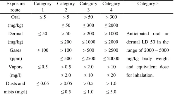
Table 3. Globally harmonized system classification criteria for acute toxicity………... 27
Table 4. Appearance, color and yields of R. picroides CrE and its fractions………. 52
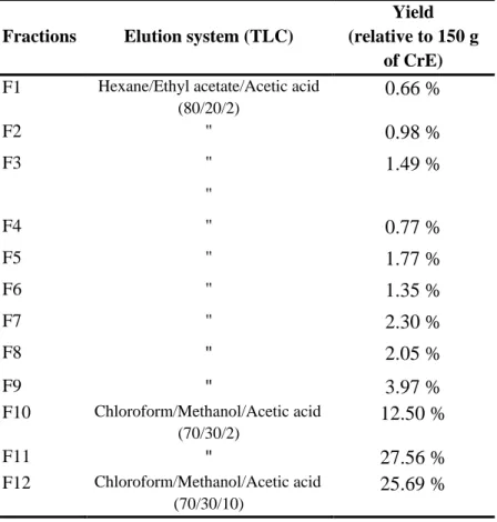
Table 5. Elution system for TLC and yield of each fraction from R. picroides CrE column chromatography………... 54
Table 6. The NMR spectroscopic data of luteolin luteolin-7-O- β-glucoside……… 55
Table 7. Body weight gain (g) of mice treated orally with R. picroides CrE... 72
Table 8. Organs relative weight of mice treated with R. picroides CrE and control group in acute toxicity... 73
Table 9. Organs relative weight of mice treated with R. Picroides CrE and control group in subacute toxiity... 78
Table 10. Estimation of CAT activity (µmole/min/mg of protein), GSH (nmole/g of tissue) and MDA (nmole/g of tissue) in liver and kidney of mice………... 82
LIST OF ABBREVIATIONS
AAPH 2,2'-Azobis (2-amidinopropane) dihydrochloride AlCl3 Aluminium chloride
ALP Alkaline phosphatase ALT Alanine aminotransferase AqE Aqueous extract
AST Aspartate aminotransferase BHT Butylated hydroxytoluene CAT Catalase
ChE Chloroform extract CrE Crude extract
DPPH 1,1-diphenyl-2 picrylhydrazyl
DTNB 5,5 0- dithiobis-(2-nitrobenzoic acid) EAE Ethyle acetate extract
EC 50 Effective concentration of 50% EDTA Ethylenediaminetetraacetic acid eNOS Endothelial nitric oxide synthase GAE Gallic acid equivalent
GPx Glutathione peroxidase GSH Reduced glutathione H2O2 Hydrogen peroxide
HT50 Half-time of 50% hemolysis iNOS Inducible nitric oxide synthase LD50 Lethal dose 50
MDA Malondialdehyde
nNOS Neuronal nitric oxide synthase NOS Nitric oxide synthase
•OH Hydroxyl radical O2•- Superoxide
OECD Organisation for economic co-operation and development ONOO- Peroxynitrite
QE Quercetin equivalent R. picroides Reichardia picroides RNS Reactive nitrogen species ROS Reactive oxygen species SOD Superoxide dismutase TBA Thiobarbituric acid TCA Trichloroacetic acid
TLC Thin layer chromatography Vit C Vitamin C
XO Xanthine oxidase
CONTENTS
Introduction ... 2
Review of literature 1. Oxidative stress ... 5
1.1. Definition ... 5
1.2. Forms of ROS and RNS ... 5
1.3. Sources of ROS and RNS ... 6
1.3.1. Endogenous sources ... 6
1.3.2. Exogenous sources ... 9
1.4. Biological roles of ROS and RNS ...10
1.5. Molecular targets of ROS and RNS ...10
1.5.1. Nucleic acids ...10
1.5.2. Proteins ...11
1.5.3. Lipids ...11
1.6. Pathologic implications of oxidative stress ...11
1.7. Defence system against oxidative stress ...12
1.7.1. Enzymatic antioxidants ...12 1.7.2. Non-enzymatic antioxidants ...13 2. Polyphenols ...16 2.1. Classes of polyphenols ...17 2.1.1. Phenolic acids ...18 2.1.2. Flavonoids ...18 2.1.3. Other phenolics ...19
2.2. Biological activities of polyphenols ...20
2.2.1. Antioxidant activity ...20
2.2.2. Anti-inflammation activity ...21
2.2.3. Antimicrobial activity ...21
2.2.4. Anticancer activity ...22
2.2.5. Cardiovascular protective activity ...22
2.2.6. Neuroprotective activity ...22
2.2.7. Anti-aging activity ...23
2.2.8. Other biological activities ...23
4. Toxicity of plant ...24
4.1. Definition of toxicity ...25
4.2. Assessment of toxicity ...25
4.2.1. Acute toxicity ...26
4.2.2. Subacute toxicity ...28
4.3. Target organ toxicity ...29
4.3.1. Liver toxicity ...29
4.3.2. Kidney toxicity ...31
3. Reichardia picroides ...34
3.1. Taxonomy ...34
3.2. Botanical description ...35
3.3. Traditional use ...36
3.4. Chemical composition ...36
Materials and methods 1. Materials ...38
1.1. Collection of plant ...38
1.2. Experimental animals ...38
1.3. Reagents and apparatus ...38
2. Methods ...39
2.1. Preparation of crude extract and fractions ...39
2.2. Phytochemical characterization ...40
2.2.1. Analytic chromatography method ...40
2.2.2. Determintion of total phenolics contents ...41
2.2.3. Determination of flavonoids contents ...41
2.3. The in vitro evaluation of antioxidant activity ...42
2.3.1. Anti-hemolytic assay ...42
2.3.2. Ion chelating assay ...42
2.3.3. Reducing power assay ...43
2.3.4. Hydrogen peroxide scavenging assay ...43
2.3.5. β- carotene bleaching assay ...44
2.3.6. DPPH scavenging assay ...44
2.4. Acute oral toxicity of crude extract ...45
2.4.1. Observation ...45
2.4.2. Plasma preparation and biochemical analysis ...46
2.4.3. Organs weight ...46
2.4.4. Histological analysis ...46
2.5. Subacute orale toxicity of crude extract and its in vivo antioxidant activity ...46
2.5.1. Animals treatment ...46
2.5.2. Collection of blood and plasma ...47
2.5.3. Oxidative hemolysis inhibition assay ...47
2.5.4. DPPH radical-scavenging activity of plasma ...48
2.5.5. Plasma biochemical analysis ...48
2.5.6. Organs weight, histological analysis and preparation of tissue homogenates ...48
2.5.7. Estimation of CAT activity ...49
2.5.8. Estimation of GSH ...49
2.5.9. Estimation of MDA ...49
2.6. Statistical analysis ...50
Results and discusion
1. Preparation of crude extract and fractions ...52
2. Phytochemical characterization ...53
2.1. Analytic chromatography method ...53
2.1.1. Isolation and identification of compound C1 ...54
2.2. Determintion of total phenolics and flavonoids contents ...57
3. The in vitro evaluation of antioxidant activity ...59
3.1. Anti-hemolytic assay ...59
3.2. Ion chelating assay ...62
3.3. Reducing power assay ...64
3.4. Hydrogen peroxide scavenging assay ...65
3.5. β- carotene bleaching assay ...66
3.6. DPPH scavenging assay ...69
4. Acute oral toxicity of crude extract ...72
4.1. Behavioral observations and mortality patterns ...72
4.2. Body weight evolution ...72
4.3. Relative organ weights ...73
4.4. Biochemical parameters ...73
4.5. Liver and kidneys histopathology ...75
5. Subacute oral toxicity of crude extract and its in vivo antioxidant activity ...77
5.1. Behavioral observations and mortality patterns ...77
5.2. Body weight evolution ...77
5.3. Relative organ weights ...78
5.4. Biochemical parameters ...78
5.5. Liver and kidneys histopathology ...79
5.6. Oxidative hemolysis inhibition assay and DPPH radical-scavenging activity ...81
5.7. Estimation of CAT activity, GSH and MDA ...81
Conclusion ...86
References ...91
Introduction
It is currently hypothesised that many diseases are due to oxidative stress that results from an imbalance between the formation and detoxification of pro-oxidants. Excessive or uncontrolled production of reactive oxygen species (ROS) and reactive nitrogen species (RNS) can cause damage to nucleic acids, proteins and lipids and this is closely associated with human disease pathogenesis such as cancer, autoimmune disorders, rheumatoid arthritis, aging, cardiovascular, and neurodegenerative diseases.
Under physiologic conditions, cellular ROS and RNS accumulation is controlled by a battery of redundant endogenous antioxidant defence systems, both enzymatic and non-enzymatic. To reinforce the spontaneous antioxidant system that may fail under certain conditions, consumption of natural antioxidants from various food supplements and traditional medicines is necessary. Indeed, there is increased interest among phytotherapy researchers to use medicinal plants with antioxidant activity for protection against oxidative stress since they tend to be accessible to everyone, safer and inexpensive.
Reichardia picroides (R. picroides), belongs to Asteraceae family, is one of eight Reichardia
species growing in Algeria. The plant is known for its alimentary purposes. However, R.
picroides leaves are also traditionally known to be used as hypoglycemiant, diuretic,
galactagogue, emollient, depurative of intestine, and tonic. Furthermore, R. picroides roots are used for cough, abdominal pains and kidney problems.
To the best of our knowledge, any work has been carried out on the chemistry, toxicity profile and biological activities of the R. picroides (aerial and root parts). Therefore, it was on this basis that this study aimed to achieve the following objectives:
Phytochemical screening of plant crude extract (CrE) including: extraction, fractionation, purification, characterization of obtained molecule and quantification of phenolic compounds.
Study of antioxidant activity of CrE and its fractions in vitro and in vivo:
• Evaluation of the protective effect against oxidative erythrocytes hemolysis. • Evaluation of transition metals chelating activity.
• Evaluation of reducing power.
• Evaluation of scavenging activity versus hydrogen peroxide (H2O2).
• Evaluation of anti-lipoperoxidation activity using β-carotene bleaching assay. • Evaluation of scavenging activity versus a relatively stable free radical
(1,1-diphenyl-2 picrylhydrazyl, DPPH).
• Effect of CrE on improving overall defences antioxidants in mice by determining the total antioxidant capacity of the blood, plasma and tissues. Assessment of acute and subacute toxicity of plant CrE.
1. Oxidative stress
1.1. Definition
Oxidative/nitrosative stress results from an imbalance between the formation of ROS/RNS and the impaired ability of an organism to detoxify these reactive intermediates or to repair the damage that they cause (Poprac et al., 2017).
1.2. Forms of ROS and RNS
In the aerobic process, cells metabolize approximately 95% of the oxygen (O2) to water,
without formation of any toxic intermediates. However, a minimal 5% of O2 is stepwise
reduced to a series of intermediate species producing ROS (Ye et al., 2015). Through these steps, three highly toxic species are formed, two of them being free radicals: superoxide (O2•-)
and hydroxyl radical (•OH). Hydrogen peroxide (H2O2) is still a highly reactive compound,
but not a radical in strict sense (Pisoschi and Pop, 2015).
The ROS and RNS are represented by both radical and non-radical forms as showed in table 1. Table 1. Nomenclature of ROS and RNS (Halliwell and Whiteman, 2004).
Reactive oxygen and nitrogen species
Free radicals Non-radicals
Superoxide (O•-2) Hydroxyl (•OH) Hydroperoxyl (HO•2) Peroxyl (RO•2) Alkoxyl (RO•) Carbonate (CO•-3)
Carbon dioxide (CO• -2)
Hydrogen peroxide (H2O2)
Hypobromous acid (HOBr) Hypochlorous acid (HOCl) Ozone O3
Singlet oxygen (O12)
Org4anic peroxides (ROOH) Peroxynitrite (ONOO-)
Peroxynitrous acid (ONOOH) Nitric oxide (NO•)
Nitrogen dioxide (NO•2)
Nitrous acid (HNO2) Nitrosyl cation (NO+)
Nitroxyl anion (NO-) Dinitrogen tetroxide (N2O4)
Dinitrogen trioxide (N2O3)
Peroxynitrite (ONOO-)
Peroxynitrous acid (ONOOH) Nitronium (nitryl) cation (NO2+)
Alkyl peroxynitrites (ROONO) Nitryl chloride (NO2Cl)
The O•-2 is considered as primary ROS. It results from one electron reduction of O2 by various
oxidases such as NADPH oxidase, xanthine oxidase (XO), aminoacid oxidase and cyclooxygenase. It may also be formed in the mitochondrial electron transport chain, in the course of oxidative phosphorylation (Lanciano et al., 2013).
The H2O2 is resulted from the conversion of O•-2 either spontaneously or by the superoxide
dismutase (SOD). It can be generated by any system yielding O•-2, as the radical anion readily
disproportionate (Briben et al., 2012).
The H2O2 is able to produce highly reactive radicals as a result of its interaction with metal
ions through Fenton reaction. The H2O2 is broken down into a hydroxide ion and a •OH.
Similarly, the one-electron reduction of H2O2 by O•-2 has also been invoked as a potential
source of •OH in the presence of metal catalysis as iron or copper through the Haber-Weiss reaction (Toro and Rodrigo, 2009). The •OH has been reported as the most powerful oxidizing radical that can interact at the site of its generation with most organic and inorganic molecules: DNA, proteins, lipids, amino acids, sugars, and metals (Toro and Rodrigo, 2009). Endogenous NO• is biosynthesized from L-arginine, oxygen and NADPH, by enzymes belonging to nitric oxide synthase (NOS) class or by reduction of inorganic nitrate. It reacts with O•-2, giving a highly reactive specie, namely peroxynitrite (ONOO-), a powerful oxidant
versus many biological molecules. The ONOO- can be decomposed to yield •OH,
independently on the presence of transition metals (Pisoschi and Pop, 2015).
1.3. Sources of ROS and RNS
Both endogenous and exogenous sources contribute to intracellular ROS/RNS levels. 1.3.1. Endogenous sources
Mitochondria
Mitochondria are thought to be the largest contributors to intracellular oxidant production (Holmström and Finkel, 2014). It contains numerous redox enzymes capable of transferring
single electron to the O2 generating the O•-2 through the tricarboxylic cycle enzymes,
electron-transport chain complexes I, II and III, among others enzymes (Murphy, 2009). Endoplasmic reticulum
The endoplasmic reticulum contains enzymes that catalyze a series of reactions for detoxifying liposoluble molecules and other toxic metabolic products. The enzymes such as cytochrome p-450 and b5 enzymes and diamine oxidase contribute to the formation of ROS (Toro and Rodrigo, 2009). The ROS may be generated as byproducts of the protein folding machinery in the endoplasmic reticulum (Malhotra and Kaufman, 2007).
Peroxysomes
The presence of enzymes that produce ROS in peroxysomes indicates that peroxisomes are involved with the metabolism of ROS. Peroxisomes are one of the major sites of intracellular H2O2 production since they contain numerous enzymes producing H2O2, glycolate oxidase,
urate oxidase, aspartate oxidase, XO, NOS and acyl CoA oxidases (del Río and López-Huertas, 2016). A part from H2O2, it has been demonstrated that peroxisomes also produce
O•2- and NO• as a consequence of their normal metabolism. Currently, there is no evidence
that mammalian peroxisomes contain enzymes that produce •OH or ONOO- (Lismont et al., 2015). However, H2O2 inside peroxisomes may give rise to •OH through the Fenton reaction.
In addition, as these organelles contain enzymatic sources of O•2- and NO•, and the reaction of
NO• with O•2-to form ONOO- is kinetically and thermodynamically favored, it is very likely
that peroxisomes also generate ONOO- (Lismont et al., 2015).
NADPH oxidase
The NADPH oxidases comprise a family of proteins able to transfer electrons from NADPH across cellular membranes; electrons react with O2 generating O•-2 and attendant other
downstream ROS (Hansen et al., 2018). Several physiological functions have been related with ROS generation by the NADPH oxidases family, including cell signalling, host defence,
control of ventilation, smooth muscle relaxation, control of erythropoietin production and other hypoxia-inducible functions (Holmström and Finkel, 2014)
Xanthine oxidase (XO)
The XO catalyzes the oxidation of hypoxanthine to xanthine and can further catalyze the oxidation of xanthine to uric acid and is a well-known producer of O•2- (Halliwell and
Gutteridge 2015). Xanthine oxidoreductases are present in two forms, depending on their electron acceptor. Xanthine dehydrogenase uses NAD+, and XO uses O2 to produce O•2-.
Under pathological conditions, such as tissue ischemia, xanthine dehydrogenase can be converted to XO (Toro and Rodrigo, 2009).
Nitric oxide synthase (NOS)
The NOS are a family of enzymes that convert the amino acid L-arginine to L-citrulline and NO. The NOS family contains three isoforms that are regulated by distinct genes: a constitutive neuronal NOS (nNOS or NOS I), an endotoxin- and cytokine-inducible NOS (iNOS or NOS II) and a constitutive endothelial NOS (eNOS or NOS III). The nNOS performs an important role in intracellular communication. The iNOS uses NO to induce oxidative stress on pathogens. The eNOS plays a major role in the regulation of vascular function (Toro and Rodrigo, 2009; Lee et al., 2016).
Arachidonate cascade enzymes
The enzyme 5‐lipoxygenase has been identified as an inducible source of ROS production in lymphocytes. The 5‐lipoxygenase was shown to be involved in the production of H2O2 by T
lymphocytes after ligation of the CD28 costimulatory receptor and in response to interleukin‐1β (Vašková et al., 2012).
Cyclooxygenase‐1 has been implicated in ROS production through formation of endoperoxides, which are susceptible to scavenging by some antioxidants in cells stimulated with TNF‐α, interleukin‐1 or bacterial lipopolysaccharide (Vašková et al., 2012).
Auto-oxidation
The auto-oxidation of molecules such as dopamine, adrenaline, flavins and hydroquinones can also be an important source of intracellular ROS production. The direct product of these auto-oxidations is often O•2- (Vašková et al., 2012).
1.3.2. Exogenous sources
The ROS are also produced in the biological systems by various exogenous sources.
Cigarette smoke contains many oxidants, free radicals and organic compounds, such as O•2-
and NO•. In addition, inhalation of cigarette smoke into the lung also activates some endogenous mechanisms, such as accumulation of neutrophils and macrophages, which further increase the oxidant injury (Briben et al., 2012).
Ozone exposure can cause lipid peroxidation in the epithelial lining fluid where reactive aldehydes and hydroperoxides are produced and these products may activate epithelial nerve endings or stimulate the release of chemokines from respiratory epithelium that recruit inflammatory cells to the airways. The ROS may be released by polymorphonuclear leukocytes, alveolar macrophages, and other inflammatory cells following ozone exposure, contributing to the epithelial injury (Hiraiwa and van Eeden, 2013).
Hyperoxia refers to conditions of higher oxygen levels than normal partial pressure of O2 in
the lungs or other body tissues. It leads to greater production of ROS and RNS (Berkelhamer
et al., 2013).
Ionizing radiation, in the presence of O2, converts •OH, O•-2, and organic radicals to H2O2 and
organic hydroperoxides. These hydroperoxide species react with redox active metal ions, such as iron and copper, via Fenton reactions and thus induce oxidative stress (Azzam et al., 2012).
Heavy metal ions, such as iron, copper, cadmium, mercury, nickel, lead, and arsenic, can
induce generation of reactive radicals via a Fenton-type reaction (Shahid et al., 2014).
1.4. Biological roles of ROS and RNS
Depending on the concentration, location, and molecular context, ROS can be beneficial or harmful to cells. Increasing evidence indicates that low to moderate concentrations of ROS are indispensable in regulating cellular processes.
They are involved in ion channel/transporter function (Kiselyov and Muallem, 2016), Ca2+ spark production (Görlach et al., 2016), protein kinase/phosphatase activation such as the mitogen activated protein kinase and extracellular-signal-regulated kinase pathways that alter gene expression, as well as in coordination with SOD initiates cell death (Son et al., 2011). The ROS are also involved in gene transcription, signal transduction and regulation of other activities in cell (Zhang et al., 2016). Moreover, ROS are important components of the anti-microbial defence mechanism incorporated by macrophages and neutrophils (Slauch, 2011). On the other hand, RNS produced by neurons act as neurotransmitters and those generated by phacocytes (monocytes, macrophages and neutrophils) act as mediators of immunity. These are also responsible for leukocyte adhesion, thrombosis, angiogenesis, vascular tone and enzyme activity modulation (Madamanchi and Runge, 2013).
1.5. Molecular targets of ROS and RNS
Since these species are highly reactive, they can damage all the three important classes of biological molecules including nucleic acids, proteins, and lipids.
1.5.1. Nucleic acids
Most of the long-term effects of oxidative stress are inflicted by modifications of DNA which involves degradation of bases, single- or double-stranded DNA breaks, bases or sugar-bound modifications, mutations, deletions or translocations, and cross-linking with proteins.Most of these DNA modifications are highly relevant to carcinogenesis, aging, neurodegenerative, cardiovascular, and autoimmune diseases (Al-Dalaen and Al-Qtaitat, 2014).
1.5.2. Proteins
Oxidation of proteins may occur directly as protein side chains are oxidized leading to a loss of function of proteins and a deactivation of enzymes. Often, thiols of proteins involved in the regulation of enzyme activity are directly oxidized. Conformational changes leading to an increase in hydrophobicity may result in aggregation or precipitation of proteins, which can no longer be subjected to the normal protein degradation pathway. Additionally, oxidative damage of proteins may occur by the adduction of secondary products like glycoxidation or lipoxidation (Stadtman and Levine, 2006).
1.5.3. Lipids
Lipid peroxidation of membranes occurs as a consequence of direct reaction of polar lipids fatty acids with oxygen or a reaction catalyzed either by metals or by NADPH cytochrome P-450 reductase. It leads to the formation of free radical intermediates and semistable peroxide. Increased levels of secondary products like conjugated dienes, hydrocarbon gases and carbonyl compounds, that initiate inflammatory reaction, and decreased levels of polyunsaturated fatty acid have been demonstrated. Oxidation of circulating lipoproteins is an important step in pathogenesis of atherosclerosis (Valko et al., 2006).
1.6. Pathologic implications of oxidative stress
There is a growing body of literature supporting crucial roles for ROS and RNS in the pathogenesis of many diseases. Table 2 lists some of these diseases.
Table 2. Some diseases implicate oxidative stress.
Pathology References
Rheumatoid arthritis (Mateen et al., 2016) Cardiovascular diseases (Kanaan and Harper, 2017) Diabete mellitus (Asmat et al., 2015)
Cancer (Moloney and Cotter, 2017)
Inflammation (Blaser et al., 2016) Alzheimer’ disease (Tramutola et al., 2017) Parkinson disease (Crotty et al., 2017)
1.7. Defence system against oxidative stress
The concept of biological antioxidant refers to any compound that, when present at a lower concentration compared to that of an oxidizable substrate, is able to either delay or prevent the oxidation of the substrate (Godic et al., 2014).
After ROS and RNS exposure from different sources, living organisms have a series of defence mechanisms against the oxidative stress including: preventative mechanisms, repair mechanisms, physical defences and antioxidant defences. The antioxidant defence mechanisms can be divided into enzymatic and non-enzymatic defences.
1.7.1. Enzymatic antioxidants Superoxide dismutase (SOD)
The SODs are classified by their metal cofactors into known types: Cu/ZnSOD and MnSOD, which are localized in different cellular compartments. The Cu/Zn SOD is mainly extracellular and cytosolic, while MnSOD is a mitochondrial enzyme (Vašková et al., 2012). It is considered as a first line of defence against ROS. The SOD catalyses the conversion of O•-2 into H2O2 and O2. It provides protection against ROS induced cellular and histological
damages. The SOD reacts very rapidly with NO thereby reducing the bioactivity of NO and producing the ONOO- (Fukai and Ushio-Fukai, 2011).
Catalase (CAT)
The CAT maintains the physiological concentration of H2O2. It converts H2O2 catalytically
into H2O and O2 and thus neutralizes it (Asmat et al., 2016). The CAT exerts its peroxidase
activity in vivo. It can also catalyze the reaction of oxidation, by H2O2, of numerous
metabolites and toxins. Its basic function is to remove H2O2 and peroxide ROOH in molecular
oxygen in order to prevent irreversible damage to the membranes (Kıvrak et al., 2017). The CAT also binds NADPH as a reducing equivalent to prevent oxidative inactivation of the enzyme by H2O2 as it is reduced to water (Briben et al., 2012).
Glutathione peroxidase (GPx)
The GPx is a selenoenzyme whose catalytic function depends on the presence of the mineral in the enzyme. It has been known to catalyze the reduction of H2O2 or organic hydroperoxides
to water or the corresponding alcohols, respectively, typically using glutathione (GSH) as reductant. The presence of selenium as the catalytic moiety was suggested to guarantee a fast reaction with the hydroperoxide and a fast reducibility by GSH (Brigelius-Flohé and Maiorino, 2013).
1.7.2. Non-enzymatic antioxidants
Non-enzymatic antioxidants include different chemical compounds such as tocopherol (vitamin E), ascorbic acid (vitamin C, Vit C), caretinoids, GSH, phenolic compounds, ubiquinol (coenzyme Q10), phospholipids (proteoglycans and hyaluronic acid), lipoic acid, proteins binding free iron and copper (ceruloplasmin, transferrin, taurine, albumin), protein hydrolysates, bilirubin, melatonin, uric acid, mucin, surfactant, amino acids, peptides, and phytates (Mirończuk-Chodakowska et al., 2017).
Glutathione (GSH)
The GSH, a tripeptide (γ-L-glutamyl-L-cysteinylglycine), is an endogenous antioxidant and an important cellular defence agent against oxidative damage. Under normal physiological conditions, GSH is mainly reduced. However, under pathological conditions, the GSH/GSSG ratio can decrease significantly. The pentose phosphate pathway regulates the GSH/GSSG ratio by providing NADPH which is required for the reduction of GSSG to GSH by GSH reductase (Aquilano et al., 2014). The GSH can directly scavenge ROS such as H2O2 and
•OH or indirectly through the reaction catalyzed by GPx. Moreover, GSH prevents the oxidization of sulfhydryl groups in the protein structure. It is especially important for the activity of GPx, GSH reductase and GSH-S-transferase (Kıvrak et al., 2017). The GSH can regenerate other antioxidants such as Vit C and vitamin E to their active forms (Lü et al., 2010).
Vitamin C (Vit C)
The Vit C is a water-soluble dietary supplement, thus it acts in the aqueous environments of the body, along with the antioxidant enzymes. The role of Vit C as an antioxidant is indicated by its known free radical-scavenging action. As a reducing and antioxidant agent, it directly reacts with O2•-, •OH and various lipid hydroperoxides. The Vit C cooperates with vitamin E
to regenerate α-tocopherol from α-tocopherol radicals in membranes and lipoproteins. By raising intracellular GSH levels, it also plays an important role in protein thiol group protection against oxidation (Im et al., 2014).
Vitamin E
Vitamin E is the major lipid-soluble component in the cell antioxidant defence system and is exclusively obtained from the diet. It has numerous important roles within the body because of its antioxidant activity. Vitamin E is a potent chain-breaking antioxidant that inhibits the production of ROS when fat undergoes oxidation and during the propagation of free radical reactions (Rivzi et al., 2014). It acts as the first line of defence against lipid peroxidation, protecting the cell membranes from free radical attack (Rivzi et al., 2014). During the antioxidant reaction, α-tocopherol is converted into α-tocopherol radical by the donation of labile hydrogen to a lipid or lipid peroxyl radical. Thus, the α-tocopherol radical can be reduced to the original α-tocopherol form by ascorbic acid (Im et al., 2014).
Carotenoids
Other lipid-soluble antioxidants are carotene and related substances called carotenoids. β-carotene is considered as the most efficient scavenger of O21. Carotenoids protect lipid against
lipid peroxidation by quenching free radicals and other reactive species. β-carotene traps free radical through its inhibition of lipid peroxidation induced by XO system (Fiedor and Burda, 2014).
Polyphenols
Polyphenols are a large group of compounds found in plants and uniquely characterize by the presence of more than one phenol group in the molecule. They are considered the most abundant antioxidant in the diet although the diversity of their structures makes them different from other antioxidants. Moreover, the amount of different polyphenols found to date in plants and plant foods, several thousands, make them a complex family of compounds with very interesting therapeutic properties against cancer, cardiovascular diseases, inflammation and other diseases (Rodrigo and Gil-Becerra, 2014).
2. Polyphenols
Polyphenols constitute one of the most important groups of secondary metabolites of plants. They are widely distributed in the plant kingdom. Approximately 10 000 naturally occurring compounds belong to the category of "phenolics", all of which possess a common structural feature: an aromatic ring bearing at least one hydroxyl substituent. From this basic structure, several thousand of naturally occurring compounds have been described, from simple phenolic acids to highly polymerized compounds such as tannins (Goleniowski et al., 2013). Polyphenols are defined based on the nature of their carbon skeletons, patterns of hydroxylations, existence of stereoisomers, and states of oxidation, glycosylation (of flavonoids), and acylation (of phenolic acids) of heterocyclic rings. The polyphenol content in plants varies between 1 and 3 mg/kg and is influenced by cultivar, maturity, part of the plant, growing conditions, processing, and storage (Martinez et al., 2017).
Polyphenols are synthesized either as soluble or cell wall–bound compounds during plant growth and especially in response to conditions such as infection, wounding, UV radiation, etc. (Goleniowski et al., 2013). They are biosynthesized by several different routes which two basic pathways are involved: the shikimic acid pathway and the malonic acid pathway (Mandal et al., 2010).
Polyphenols provide multiple intracellular functions. They protect plant from UV-B radiation, acting as a screen inside the epidermal cell layer, and by making adjustments to the antioxidant systems at both cell and whole organism level (Agati and Tattini, 2010). They influence also the pools and fluxes of inorganic and organic soil nutrients (Goleniowski et al., 2013). Polyphenols play an important role serving as visual signals by acting as pigments in fruits and flowers, firstly to attract animals as pollinators in flowers, and later to attract animals to eat the fruits and thereby help in seed dispersal (Latanzio et al., 2008).
Phenolics often participate in plant growth. This may be due their roles in lignifications, in functional pollen development, in seed germination and dormancy (Taylor and Grotewold, 2005). One of the undisputed functions of polyphenols is their role in protecting plants against microbial invasion (Mandal et al., 2010). In addition, they play a role in protecting plants from both insect and mammalian herbivory (Pietta, 2000).
2.1. Classes of polyphenols
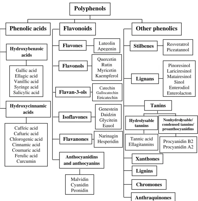
There are three main classes of polyphenols: phenolic acids, flavonoids, and other phenolics (figure 1).
Figure 1. Types of polyphenols (Martinez et al., 2017).
Nonhydrolysable/ condensed tannins/ proanthocyanidins Resveratrol Piceatannol Pinoresinol Lariciresinol Matairesinol Sinol Enterodiol Enterolacton
Polyphenols
Phenolic
acids
Flavonoids
Other phenolics
Hydroxybenzoic acids Gallic acid Ellagic acid Vanillic acid Syringe acid Salicylic acid Hydroxycinnamic acids Caffeic acid Caftaric acid Chlorogenic acid Cinnamic acid Coumaric acid Ferulic acid Curcumin Flavones Luteolin Apegenin Flavonols Quercetin Rutin Myricetin Kaempferol Isoflavones Flavan-3-ols Flavanones Catechin Gallocatechin Epicatechin Genestein Daidzin Glycitein Equol Narinagin Hesperidin Anthocyanidins and anthocyanins Malvidin Cyanidin Peonidin Stilbenes Lignans Tanins Xanthones Lignins Anthraquinones Chromones Hydrolysable tannins Tannic acid Ellagitannins Procyanidin B2 Procyanidin A2 17
2.1.1. Phenolic acids
Phenolic acids are a subclass of the larger phenolics category, occurring in food plants as esters or glycosides conjugated with other natural compounds such as flavonoids, alcohols, hydroxyfatty acids, sterols, and glucosides. Structurally, phenolic acids are phenols that possess one carboxylic acid moiety that can be directly attached either to the aromatic ring (benzoic acid derivatives) or attached to an alkyl residue (hydroxycinnamic acid derivatives). Although the basic skeleton remains the same, phenolic acids differ in the number and position of the hydroxyl groups on the aromatic ring (Losada-Barreiro and Bravo-Díaz, 2017). The majority of phenolic acids are linked through ester, ether, or acetal bonds either to structural components of the plant (cellulose, proteins, lignin) or to larger polyphenols (flavonoids), or smaller organic molecules (glucose, quinic, maleic, or tartaric acids) or other natural products (terpenes) (Goleniowski et al., 2013).
2.1.2. Flavonoids
Flavonoids comprise the most abundant class of plant polyphenols with more than 9000 of identified chemical structures (Wang et al., 2018). They share a carbon skeleton of diphenyl propanes, two benzene rings (A and B) joined by a linear three carbon chain. This central chain usually forms a closed pyran ring (C) with one of the benzene rings (figure 2). According to the degree of oxidation of the C ring, the hydroxylation pattern of the nucleus, and the substituent at carbon 3, the flavonoids can thus be classified into different subclasses: flavones, isoflavones, flavanols (catechins), flavonols, flavanones, anthocyanins, and proanthocyanidins (Losada-Barreiro and Bravo-Díaz, 2017).
Figure 2. Basic skeleton or structure of flavonoids (Wang et al., 2018).
Flavonoids are the most studied class of polyphenols. Although they are low molecular weight secondary metabolites, their chemical diversity, size, three-dimensional shape, and physical and biochemical properties allow them to interact with multiple targets to influence biological activity in plants, animals, humans and microbes. Consequently, many therapeutic properties have been assigned to flavonoids (Francisco et al., 2014).
2.1.3. Other phenolics
There are at least two major classes of tannins: i: hydrolyzable and nonhydrolyzable (also known as condensed) tannins and ii: proanthocyanidins and procyanidins. Structurally, hydrolyzable and nonhydrolyzable tannins are richly hydroxlyated oligomers or polymers of hydroxybenzoic acids such as gallic acid or flavan-3-ols such as catechin, respectively. High-molecular-weight condensed tannins may contain 50 or more flavan-3-ols subunits attached by carbon–carbon bonds (Selma et al., 2009). They are highly astringent and noticeable in unripe fruits.
Stilbenes are well-known class of naturally occurring phytochemicals. They bear classical C6-C2-C6 structures with two hydroxyl groups on the A ring and one on the B ring. Stilbenes are characterized by a double bond connecting the phenolic rings. These compounds are stress metabolites produced in response to fungal infection. Though known as plant defence compounds, stilbens have an enormous diversity of beneficial human health effects. One of the most relevant and extensively studied stilbene is resveratrol found largely in grapes (Martinez et al., 2014; Losada-Barreiro and Bravo-Díaz, 2017).
Lignans are found in all plants and show enormous structural diversity, with their molecular backbone consisting of two phenylpropane (C3-C6) units. There is a very good recent review of the health effects of lignans. The most lignans that are of special interest owing to their many powerful health benefits are tetrahydrofurofuran and sesamin (Martinez et al., 2014).
2.2. Biological activities of polyphenols
Natural polyphenols have shown numerous biological activities and health benefits for prevention and treatment of age-related diseases, cancers, heart diseases, etc.
2.2.1. Antioxidant activity
Among the notable bioactivities of phenolic compounds, the antioxidant activities have been widely studied especially their acclaimed capability to scavenge ROS and RNS. This activity is frequently cited to be the key property underlying the prevention and/or reduction of oxidative stress-related chronic diseases and age-related disorders (Quideau et al., 2011). Phenolics can also interrupt the propagation stage of the lipid autoxidation chain reactions as effective radical scavengers or act as metal chelators to convert hydro-peroxides or metal pro-oxidants into stable compounds. Plant polyphenols can also act as antipro-oxidants by chelating metal ions such as iron and cupper ions that are involved in the •OH formation through Haber-Weiss/Fenton-type reactions (Procházková et al., 2011). They may reduce the catalytic activity of enzymes involved in ROS and RNS generation. Finally, polyphenols can stimulate antioxidant activities of other enzymes such as CAT (Quideau et al., 2011).
While phenolic compounds are strong antioxidants, it should be pointed out that when a phenolic molecule loses an electron or when it acts as a reducing agent, the molecule itself becomes a radical; its oxidized intermediates can also become pro-oxidants. Interaction between polyphenols and transition metal ions can also result in pro-oxidant formation (Zhang and Tsao, 2016). Polyphenols therefore can be a double-edged sword; on the one hand, when used properly in the form of food or functional food, they are strong antioxidants against excess oxidative stress such as ROS, thus beneficial to health, on the other; they can display pro-oxidant activity when consumed in high doses by taking supplements (Bouayed and Bohn, 2010). The antioxidant potential of phenolic compound depends on the existence of a C2=C3 double bond in conjugation with a C4-carbonyl group, the number and position of
hydroxyl groups in the molecule, the presence of methoxyl groups, and less saccharides connection (Wang et al., 2018).
2.2.2. Anti-inflammation activity
Various investigations have shown that different polyphenols modulate the activity of arachidonic acid metabolizing enzymes such as cyclooxygenase, lipoxygenase, and NOS. Inhibition of these enzymes reduces the production of aminoacids, prostaglandins, leukotrienes, and NO which are among the key mediators of inflammation (Hussain et al., 2016). Moreover, a large number of polyphenols have been reported to inhibit the expression of pro-inflammatory cytokines, which is coupled in some cases to the enhancement of anti-inflammatory cytokines. Polyphenols also exert their beneficial action by modulating monocyte adhesion during the inflammatory process. Flavonoids can modulate the cascade of molecular events leading to the overexpression of inflammatory mediators. These include inhibition of transcription factors, nuclear molecules, protein kinases and mitogen-activated protein kinases (Kim et al., 2014).
2.2.3. Antimicrobial activity
The antimicrobial activity of polyphenols occurring in medicinal plants has been extensively investigated against a wide range of microorganisms. Polyphenols, especially flavan-3-ols, flavonols, and tannins, have wide spectrum and higher antimicrobial activity in comparison with other polyphenols. Cited polyphenols are also able to suppress a number of microbial virulence factors (such as inhibition of biofilm formation, reduction of host ligands adhesion, and neutralization of bacterial toxins) and show synergism with antibiotics (Daglia, 2012). For instance, therapeutic activities of polyphenols against influenza virus, canine distemper virus, hepatitis virus, and Escherichia coli, have been attributed to chemical structures in particular patterns of methoxylation, glycosylation and hydroxylation (Carvalho et al., 2013).
2.2.4. Anticancer activity
The anti-cancer activities of polyphenols against a wide range of cancers have been documented. Several mechanisms underlying the anticancer activity of polyphenols are proven, including modulation of ROS and RNS, modulation of inflammation- related factors, modulation of estrogen receptor, detoxification of xenobiotics, induction of apoptosis, modulation of cell cycle and their effects on the cellular signaling system. Among these are effects on nuclear factors, such as NF-κB or activator protein 1, which play central roles in cellular signaling cascades, regulating DNA transcription, gene expression in response to different stimuli, cell proliferation and survival (Abdal Dayem et al., 2016; Niedzwiecki et
al., 2016).
2.2.5. Cardiovascular protective activity
Numerous studies have demonstrated the health benefits of polyphenols, and special attention has been paid to their beneficial effects against cardiovascular disease. Polyphenols present vasodilator effects and are able to improve lipid profiles and attenuate the oxidation of low density lipoproteins. In addition, they present clear anti-inflammatory and antithrombotic effects and can modulate apoptotic processes in the vascular endothelium. It has been suggested that most of these effects are a consequence of the antioxidant properties of polyphenols (Quinones et al., 2012).
2.2.6. Neuroprotective activity
Recently, a rapidly growing number of polyphenolic compounds with neuroprotective effects have been described. Neuroprotective effects of polyphenols can be divided into the following categories: i: neuroprotective action through antioxidant pathways, ii: interaction with signaling pathways, iii: neuroprotection through modulation of neural mediators and enzymes like acetylcholine and acetylcholinesterase, vi: inhibition of N-methyl-d-aspartate neurotoxicity and v: anti-amyloidogenic effect (Ebrahimi and Schluesener, 2012).
2.2.7. Anti-aging activity
There is great interest in using polyphenol organic compounds to modify or retard the aging process. Several polyphenols augment the lifespan of multiple invertebrate and vertebrate species, in higher organisms in the presence of dietary modification. Polyphenols may influence aging by acting on genes in energy-regulatory intracellular pathways that are believed to play important roles in the aging process. In addition, or together with this process, polyphenols may alter concentrations of ROS, transcription factors, and act via epigenetic mechanisms. Polyphenols may also modulate organ systems through their effect on intercellular signaling molecules including nitrous oxide and pro-inflammatory cytokines (Cherniack, 2016).
2.2.8. Other biological activities
Growing evidence from animal studies supports some other activities of polyphenols such as anti-diabetic activity (Kim et al., 2016), prevention of obesity and obesity-related chronic diseases (Wang et al., 2014), amelioration liver injury (Janel and Noll, 2014), attenuation microarchitecture bone deterioration (Shen et al., 2014).
4. Toxicity of plant
Plants used in traditional medicine are assumed to be safer than pharmaceuticals. This safety is based on their natural origin and also their long usage in the treatment of diseases according to knowledge accumulated over centuries (Philomena, 2011). However, recent scientific reports have demonstrated that several medicinal plants used in phytomedicine are potentially toxic, and some are even mutagenic and/or carcinogenic (Nasri and Shirzad, 2013). It cannot therefore be taken for granted that medicinal plants are void of toxic effects. Some authors have thus recommended that pharmacological studies on medicinal plants should always be accompanied by toxicological screening (Cos et al., 2006).
The toxicity in medicinal natural products may originate from i: dearth of reports on the side effect of medicinal plants, ii: error in botanical identification, iii: accidental ingestion of cardiotonic plants, iv: inappropriate combinations in phytotherapy, v: interference of medicinal plants and vi: conventional pharmacological therapy and contamination with heavy metals (Goldman, 2001; Wojcikowski et al., 2004).
The toxicity for medicinal plant therefore depends on many different factors, such as: their purity, medicinal plants combinations, route of administration, absorption, bioavailability, period of exposure, number of exposures, physical form of the toxicant (solid, liquid, or gas), and reported adverse effects (Ogunlana, 2012 ; Püssa, 2014).
The toxicity of plant is based on their chemical constituents that are classified into: lectins or hemagglutinins, enzyme inhibitors, alkaloids, cyanogenic glycosides, phytoestrogens, glucosinolates, coumarin, toxic amino acids, toxic lipids, oxalates, fluoroacetates, saponins, etc. (Püssa, 2014). They act by altering specific mechanisms involving enzymes, receptors and even genetic material at particular cells and tissues (Chandra et al., 2012).
4.1. Definition of toxicity
Toxicity is defined as "the potential of a substance to exert a harmful effect on humans or animals, and a description of the effect and the conditions or concentration under which the effect takes place" (Yu et al., 2005).
4.2. Assessment of toxicity
Toxicology is an aspect of pharmacology that deals with the adverse effect of bioactive substance on living organisms. In order to establish the safety and efficiency of a new drug, toxicological studies are very essential experiments in animals like mice, rat, etc. No drug substance is used clinically without its laboratory safety assessment at preclinical phase. Toxicological studies help to make a decision whether a new drug should be adopted for clinical use or not (Alam et al., 2006).
The primary aim of toxicological assessment of any medicinal plant is to identify adverse effects that may be associated with its use and to determine limits of exposure level at which such effects occur, therefore avoiding potential harmful effects when used as medicine (Sims
et al., 2010; Ifeoma and Oluwakanyinsola, 2013).
Generally, toxicity assessment tests can be divided into two categories: the first category comprises tests that are designed to evaluate the overall effects of compounds on experimental animals. Individual tests in this category differ from each other basically in regard to the duration of the test and the extent to which the animals are evaluated for general toxicity. These tests are classified as acute, sub-acute, chronic and subchronic toxicity tests (Loomis and Hayes, 1996). The last category of tests consists of those that are designed to evaluate specific types of toxicity in detail. The subacute, chronic and subchronic toxicity tests do not detect all forms of toxicity, but they may reveal some of the specific toxicities and indicate the need for more detailed studies. Thus, this last category of tests has been developed for the determination of effects of compounds on the fetus in a pregnant animal (teratogenic tests), on