Pépite | Réponse osmo-inélastique du disque intervertébral : caractérisation expérimentale et modélisation constitutive
169
0
0
Texte intégral
(2) Thèse de Amil Derrouiche, Université de Lille, 2018. 2 © 2018 Tous droits réservés.. lilliad.univ-lille.fr.
(3) Thèse de Amil Derrouiche, Université de Lille, 2018. OSMO-INELASTIC RESPONSE OF INTERVERTEBRAL DISC: EXPERIMENTS AND CONSTITUTIVE MODELING. 3 © 2018 Tous droits réservés.. lilliad.univ-lille.fr.
(4) Thèse de Amil Derrouiche, Université de Lille, 2018. 4 © 2018 Tous droits réservés.. lilliad.univ-lille.fr.
(5) Thèse de Amil Derrouiche, Université de Lille, 2018. Contents Remerciements ................................................................................................................................................... 7 Introduction ........................................................................................................................................................ 8 Chapter 1: General considerations .................................................................................................................... 12 1.1. Public health issue ............................................................................................................................. 13 1.2. Anatomical aspects ............................................................................................................................ 13 1.3. Microstructural aspects ..................................................................................................................... 15 1.4. Nucleus pulposus ............................................................................................................................... 18 1.5. Annulus fibrosus ................................................................................................................................ 19 1.6. Experimental pre-requests................................................................................................................. 20 1.7. Thesis organization ............................................................................................................................ 21 1.8. References ......................................................................................................................................... 21 Chapter 2: Experimental characterization ......................................................................................................... 26 Part 1: Osmo-inelastic response of the intervertebral disc ............................................................................ 26 2.1.1. Partial introduction ......................................................................................................................... 27 2.1.2. Materials and methods ................................................................................................................... 29 2.1.3. Results ............................................................................................................................................ 35 2.3.4. Discussion ....................................................................................................................................... 38 2.1.5. Partial conclusion ............................................................................................................................ 41 2.1.6. References ...................................................................................................................................... 42 Part 2: Pre-strain effect on the chemo-torsional response of the intervertebral disc .................................... 46 2.2.1. Partial introduction ......................................................................................................................... 47 2.2.2. Materials and methods ................................................................................................................... 49 2.2.3. Results ............................................................................................................................................ 54 2.2.4. Discussion ....................................................................................................................................... 60 2.2.5. References ...................................................................................................................................... 65 Part 3: Osmo-inelastic response of the annulus fibrosus ............................................................................... 69 2.3.1. Partial introduction ......................................................................................................................... 70 2.3.2. Materials and methods ................................................................................................................... 72 2.3.3. Results ............................................................................................................................................ 79 2.3.4. Discussion ....................................................................................................................................... 87 2.3.5. Partial conclusions .......................................................................................................................... 89 2.3.6. References ...................................................................................................................................... 89 Part 4: Poisson’s ratio in annulus fibrosus and osmo-inelastic coupling ........................................................ 94 2.4.1. Partial introduction ......................................................................................................................... 95 2.4.2. Materials and methods ................................................................................................................... 97 2.4.3. Results .......................................................................................................................................... 102. 5 © 2018 Tous droits réservés.. lilliad.univ-lille.fr.
(6) Thèse de Amil Derrouiche, Université de Lille, 2018. 2.4.4. Discussion ..................................................................................................................................... 108 2.4.5. Partial conclusions ........................................................................................................................ 113 2.4.6. References .................................................................................................................................... 113 Chapter 3: Constitutive modeling and simulation ............................................................................................ 117 Part 1: A chemo-mechanical constitutive model for osmo-inelastic effects in the annulus fibrosus ............ 117 3.1.1. Partial introduction ....................................................................................................................... 118 3.1.2. Model formulation ........................................................................................................................ 120 3.1.3. Finite element computations and comparison with experiments .................................................. 131 3.1.4. Partial conclusions ........................................................................................................................ 142 3.1.5. References .................................................................................................................................... 143 Part 2: A finite element model for time-dependent biomechanics of the intervertebral disc ...................... 148 3.2.1. Partial introduction ....................................................................................................................... 149 3.2.2. Finite element computations ........................................................................................................ 150 3.2.3. Results and discussion .................................................................................................................. 157 3.2.4. Partial conclusions ........................................................................................................................ 161 3.2.5. References .................................................................................................................................... 162 Conclusion ...................................................................................................................................................... 166 Research perspectives..................................................................................................................................... 168. 6 © 2018 Tous droits réservés.. lilliad.univ-lille.fr.
(7) Thèse de Amil Derrouiche, Université de Lille, 2018. Remerciements Merci à Caroline Frederix, Jean-Marc Lefebvre, Jean-François Ganghoffer et Gregory Chagnon pour avoir accepté de jugé mon travail de thèse et pour les discussions passionantes pendant la soutenance. Cette thèse n’aurait pu s’achever sans mon directeur, Fahmi Zaïri. Tu m’as guidé lorsque je me perdais, tu m’as rassuré lorsque je doutais, tu as toute ma reconnaissance. Merci pour ton soutien et pour les hectolitres de café. Ces remerciements seraient incomplets sans une pensée pour Ameni, Anouar, Faten, Yan et Li qui m’ont aidé dans mes recherches, cette thèse est aussi la vôtre. Je souhaite également remercier mes collègues Christian, Hamza, Qiang et Karim pour ces moments passés en votre compagnie. Fahed, merci pour tes encouragements et tes conseils. Ma famille te remercie encore pour t’être occupé de mon grand-père. Papa, Maman, vous avez toujours souhaité le meilleur pour nous, j’espère vous rendre fiers et être un jour un bon parent comme vous l’avez été. Mon épouse, mon plus grand soutien, je remercie Dieu pour cet inestimable cadeau. Nous voilà tous les deux docteurs, une nouvelle étape s’ouvre à nous. Mes frères Jamir, Sliman et Nadir je souhaite que nous soyons toujours proches et que vous soyez heureux, merci pour tout. Anissa et Souad bienvenue et prenez soin de mes frères. Mes grands-parents, ai-je déjà vu quelqu’un de plus courageux que vous, vous êtes des modèles pour moi, que Dieu vous garde. Ma grande famille, tantes, oncles, cousins, cousines sachez que je serai toujours là pour vous. Ma famille de Marseille, vous m’avez accompagné pendant ces années, merci Saïd, Djemy, Yacine mon bof et Kaïna la meilleure des belles sœurs. Mourad, Ayoub, Naïm, Moustaf, Sofian, Hind, Moh, Sarah, Myriam et tous les autres qui m’ont accompagné le long de ces années merci à vous. Merci à ceux que j’ai croisé de loin ou de près, pour vos messages, vos sourires et votre aide. A mes proches, qui nous ont quitté pendant cette thèse, je ne saurai oublier les moments passés avec vous.. 7 © 2018 Tous droits réservés.. lilliad.univ-lille.fr.
(8) Thèse de Amil Derrouiche, Université de Lille, 2018. Introduction The life is a complex concept that allows us to move and grow at the macroscopic scale. In the history of mechanics, living body were described with the same mathematical tools than inert body before the consideration of multi-scale organization. A living body is an automaintained system requiring a description in relation with the biological, mechanical and chemical features. The first one can explains the microstructural development essential for the second one working, while the third one is strongly coupled with the second. A correct description of the life being is based on these three distinct disciplines which should interact. From global description of our body towards cellular description, the intermediate scale is essential, in which our organs and soft tissues are studied for the development of more accurate treatments. The word “biomechanics” finds its origin in the beginning of the twentieth century but this discipline is considered millenary with the oldest known prosthesis old by 3000 years. Biomechanics progresses by the consideration of the specificity of our tissues. That leads to the development of new adapted scientific tools as continuum biomechanics with the capability to describe life. As an application of mechanical concepts on the biology, the biomechanics heads towards mechanobiology with the aim to understand living beings in relation with molecular constitution. The mutual effects are now highlighted especially for the study of biological changes induced by the time or the environment. The study of living being is based on application of mechanics strongly coupled with biology in the aim to understand physiology and development of disease towards enhancement of medical techniques. Nowadays, artificial devices have to be designed in order to interact with living tissues with the required care.. 8 © 2018 Tous droits réservés.. lilliad.univ-lille.fr.
(9) Thèse de Amil Derrouiche, Université de Lille, 2018. Understand the inevitable alteration is difficult without a correct description of a healthy tissue. It is a way to improve the medical devices with biomimetic materials usable in sport performance, plastic surgery or tissue engineering. Biomechanics is an interdisciplinary domain where biologists, chemists and physicists work with medical doctors for the improvements of our quality of life. In the context of changes in the lifestyle and the population aging, the aim of the present thesis is to focus on spine, considered as the most complicated join of our body. This fundamental element presents hierarchical organization with hard and soft tissues: vertebrae, intervertebral discs (IVDs) and ligaments. It is subjected to troubles for more and more people while being not well understood in the healthy state. The back pain concerns a growing part of the population with unclear multiple sources. The chronic pain founds often its origin in the IVD disorder. This soft tissue connecting two adjacent vertebrae is a fibrocartilage able to resist at high stresses induced by spine movements. The complex organization of the IVD, with nucleus pulposus in center and annulus fibrosus around, and the strong local heterogeneity make the understanding complicated. IVD malfunction is due to several interacting factors, involving chemical, biological and mechanical effects. This thesis is dedicated to the experimental and theoretical/numerical description of the healthy IVD in relation with the mechanical and the biochemical environment. A two-scale description is employed with a study at the IVD scale and at the AF scale. The thesis dissertation is divided into three Chapters. Chapter 1 provides the general biomechanical and biochemical considerations on IVD. Chapter 2 is dedicated to the experimental characterization of the osmo-inelastic coupling in healthy IVD and is divided into two parts. The first part is focused on the experimental study performed on functional units extracted from cervical spine of mature sheep. Multiaxial mechanical experiments provide some insights on the source of inelastic effects in relation with osmolarity by varying mechanical loading path. The second part reports. 9 © 2018 Tous droits réservés.. lilliad.univ-lille.fr.
(10) Thèse de Amil Derrouiche, Université de Lille, 2018. the experimental observations on the intrinsic response of AF in relation with microstructure, biochemical environment and strain-rate. An interpretation of the osmo-inelastic mechanisms is proposed at the two scales. Chapter 3 is dedicated to the constitutive modeling and simulation of osmo-inelastic coupling in healthy IVD and is divided into two parts. The first part is devoted to the formulation of a chemo-mechanical constitutive model taking into account the osmo-inelastic couplings in relation with heterogeneous and anisotropic features. This model is a new approach in the description of soft tissues able to reproduce osmo-induced volumetric changes at the AF scale in relation with microstructure, osmolarity and water content. The present formulation is implemented into a finite element program and used to reproduce the AF intrinsic response using the experimental observations reported in Chapter 2. In the second part, the constitutive model is applied to a finite element model of C5-C6 functional unit constructed from computed tomography. Comparisons with experimental data for different neck movements allows us to analyze the local fields in healthy IVD. Some elements towards a better understanding of the mechano-biological coupling in healthy IVD close this chapter. General conclusions and Research perspectives are presented at the end of the document.. 10 © 2018 Tous droits réservés.. lilliad.univ-lille.fr.
(11) Thèse de Amil Derrouiche, Université de Lille, 2018. 11 © 2018 Tous droits réservés.. lilliad.univ-lille.fr.
(12) Thèse de Amil Derrouiche, Université de Lille, 2018. Chapter 1: General considerations. 12 © 2018 Tous droits réservés.. lilliad.univ-lille.fr.
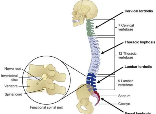
(13) Thèse de Amil Derrouiche, Université de Lille, 2018. Chapter 1: General considerations. 1.1. Public health issue The spine is composed by vertebrae and soft tissues, including intervertebral discs (IVDs) which provide flexibility and softening, with ligaments avoiding too large displacements. In 16 developed countries, 47% of pains are located to the back and deteriorated IVDs are the second causes of chronic pain (Maetzel and Li, 2002; Breivik et al., 2006; Vos et al., 2016). Direct costs of back pain are estimated around 3 billion euros in France with significant impact on quality of life (Depont et al., 2010). This statistic increases in industrialized western countries (Frymoyer and Cats-Baril, 1991; Schmidt et al., 2007; Raspe, 2008; Hoy et al., 2012) due to changes in lifestyle. Multiple biological and sociological factors are predominant in back pain apparition. There is correlation with physical inactivity and overweight which is increasingly common in our society with time spent on watching TV or on computer (Adams and Hutton, 1985; Skoffer and Foldspang, 2008). Compared to standing posture, sitting posture increases disc pressure which is associated with IVD degeneration and back pain (Makhsous et al., 2009; Aranjan Lione, 2013).This trouble is a public health issue and it justifies public health policies to prevent bad movements or posture which could cause back pain.. 1.2. Anatomical aspects 1.2.1. Spine The spine or vertebral column provides the main support for our body, while protecting spinal cord from injury. The spine is classically separated into three distinct regions, at both functional and anatomical levels: the cervical, thoracic and lumbar regions, see Figure 1.1. There are 23 intervertebral discs placed between each vertebra along the spine, except for the articulation between the first and second cervical vertebrae. Two adjacent vertebrae are interconnected by the IVD and ligaments and constitute the smallest physiological motion segment of the spine.. 13 © 2018 Tous droits réservés.. lilliad.univ-lille.fr.
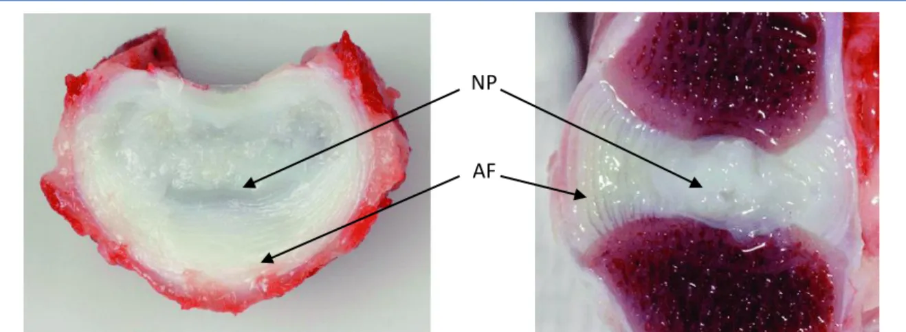
(14) Thèse de Amil Derrouiche, Université de Lille, 2018. Chapter 1: General considerations This segment, well-known as functional spine unit (FSU) is described the elementary element exhibiting the same biomechanical characteristics than the entire spine (White et al., 1990).. Figure 1.1. Functional spine unit and vertebral column (White et al., 1990).. 1.2.2. Intervertebral discs The IVD is a vascular fibrocartilaginous medium that provides flexibility and softening of the spine thanks to a fibrous ring (annulus fibrosus: AF) and a gelatinous nucleus (nucleus pulposus: NP), see Figure 1.2. At the structural level, IVD is a hydrophilic reinforced medium where the solid phase and the fluid phase interact by osmotic effect with the microstructure (Urban and Roberts, 1995; Bibby et al., 2001; Kramer, 2009). NP is a gelatinous subcomponent, which transfers axial loads from the spine towards AF. At the micro-structural scale, the IVD is composed by collagen, proteoglycans (PGs) macromolecules and cellular population, varying in content according to the location.. 14 © 2018 Tous droits réservés.. lilliad.univ-lille.fr.
(15) Thèse de Amil Derrouiche, Université de Lille, 2018. Chapter 1: General considerations. NP. AF. Figure 1.2. Transverse (left) and sagittal (right) section through a L5-L6 IVD of a 2-years old chondrodystrophic dog, showing a fibrocartilaginous NP, a widened transition zone, and a normally structured AF (Smolders et al., 2012).. 1.3. Microstructural aspects 1.3.1. Collagen Collagen is the most abundant protein of the human body. Nineteen types exist with predominance of type one in the AF and two in the NP. With length of 10 to 100 μm and diameters of between 100 and 200 nm (Inoue, 1981; Rannou et al., 2004), they define certain mechanical properties. Collagens that account for 44 to 51% of the dry mass of the IVD (Kraemer, 2009) are distributed differently according to the zones. Indeed, first type of collagen fibers is highly organized with strong tensile modulus that contributes to the AF resistance and help supporting a multiaxial mechanical loading. Second type is tangled with PGs macromolecules mainly in the NP as illustrated in Figure 1.3. The collagen fibers exhibit an increase in content in the AF, generating graded mechanical properties from inner part to outer part. Moreover, they present a variable orientation with approximately 23° in the IVD dorsal part and approximately 43° in the IVD ventral part (Holzapfel et al., 2005).. 15 © 2018 Tous droits réservés.. lilliad.univ-lille.fr.
(16) Thèse de Amil Derrouiche, Université de Lille, 2018. Chapter 1: General considerations. (a) (b) Figure 1.3. Collagen fiber organization: (a) in AF, (b) in NP (Inoue, 1981).. 1.3.2. Proteoglycans The PGs represent the second major solid element of IVD with 5 to 15% of the weight (Bibby et al., 2001). They are organized as macro-aggregate with the remarkable particularity of being negatively charged. They consist of a core protein filament and glycosaminoglycan (GAG) chains, see Figure 1.4.. Glycosaminoglycans. Proteoglycan. Figure 1.4. Dark field electron micrograph of PGs macromolecules from bovine articular cartilage (×120000) and schematic representation of a PG macromolecule with GAGs chain. The bar at the lower right equals 0.5 µm (Rosenberg et al., 1975).. 16 © 2018 Tous droits réservés.. lilliad.univ-lille.fr.
(17) Thèse de Amil Derrouiche, Université de Lille, 2018. Chapter 1: General considerations The high polarity of the GAG gives the hydrophilic property to the PGs by osmotic effect, and then they act as lubricant and shock absorber in the IVD. The IVD hydration remains relatively constant thanks to osmotic effect and water is sucked especially when the disc is compressed. The induced internal osmotic pressure governs the mechanical behavior of the IVD while creating the essential internal fluid flow for the nutrient supply.. 1.3.3. Cellular population and nutrients supply The IVD consists thus in PGs molecules, collagen fibers and internal fluid, which represent 90 to 95% of the disc (Bibby et al., 2001). The rest is minority molecules and cellular population designed for the production of PG and collagen. The IVD contains relatively few cells, compared to other articular cartilages, with approximately 3000 cells/mm3 in the NP, 9000 cells/mm3 in the AF and 15000 cells/mm3 in the vertebral endplates (Maroudas et al., 1975; Rannou et al., 2000; Bibby et al., 2001). Two main types of cells are found in the IVD: fibroblasts, which are responsible for the production of first type of collagen mainly in the outer part of the AF, and chondrocytes, producing PGs and second type of collagen, are found in AF and NP (Bibby et al., 2001). The IVD is vascularized by arterioles during childhood, and then it becomes quickly avascular. The homeostasis depends thus on the nutrients supply and wastes evacuation. These phenomena depend on the internal fluid exchange with the closest vessels in the vertebral bodies and in the periphery of AF.. 1.3.4. Degeneration IVD is a wet tissue mainly composed by water, with 80% at birth and 60% in great age (Gu et al., 1999). Thanks to the fluid phase, IVD thickness decreases during the day and is restored during the night (de Puky, 1935). The age-dependency of fluid content induces changes in mechanical properties leading to degeneration; IVDs lose flexibility and become rigid 17 © 2018 Tous droits réservés.. lilliad.univ-lille.fr.
(18) Thèse de Amil Derrouiche, Université de Lille, 2018. Chapter 1: General considerations (Campana et al., 2011), then IVD diseases can occur as herniation, see Figure 1.5. Fixed charge density of PGs contributes to hydration, nutrition and compressive strength of the tissue (Urban et al., 1979). Alteration in the fixed charge density due to loss of PGs content (Lyons et al., 1981; Urban and Holm, 1986; Antoniou et al., 1996) may explain the degeneration process due to increase in solid matrix stresses, decrease in fluid content, and bad nutrients supply inside tissue (Wills et al., 2016).. Figure 1.5. Human lumbar IVD: (a) Healthy disc from a young adult, (b) Healthy disc from a middle-aged adult, (c) Herniated disc with signs of aging. A small quantity of NP (arrow) has herniated through the posterior annulus in response to severe mechanical loading.. 1.4. Nucleus pulposus At centrum, with 80% water content, and non-organized collagen fibers network, the NP is an isotropic incompressible gelatinous medium (Rannou et al., 2000). The NP, representing 30 to 60% of the total surface area, absorbs and distributes the loads from the spine. Its cellular population, being mainly composed by chondrocytes, has an active metabolism ensuring a renewal of PGs and second type of collagen. The type of fibers changes gradually in nature by approaching the AF. The extracellular matrix of the nucleus is equivalent to that of cartilage, 18 © 2018 Tous droits réservés.. lilliad.univ-lille.fr.
(19) Thèse de Amil Derrouiche, Université de Lille, 2018. Chapter 1: General considerations with second type of embedded collagen fibers with large PGs macromolecules. Thanks to the water contribution, the NP generates hydrostatic pressure under axial loads (Nachemson and Morris, 1964; Wilke et al., 1999; Goins et al., 2005). The proportion of water is estimated at 90% during childhood and decreases around 80% at 20 years old, to less than 70% after 60 years old (Iatridis et al., 1997). The PGs content can decrease by 30% with aging depending on lifestyle with induced decrease in osmotic pressure. The NP (and inner part of the AF) is connected to the vertebral body via the vertebral endplates. Main contributor to the nutrients supply, the fluid flow is promoted by their porosity and their thickness about 0.6 mm.. 1.5. Annulus fibrosus The second part of IVD, around nucleus is AF. It is composed by PGs macromolecules embedded. However, in annulus, collagen fibers are regularly oriented to form a fiber reinforced deformable solid material (Ghosh et al., 1977). Collagen network gives to annulus mechanical resistance permitting to support high stresses. Indeed, AF has a very specific organization, it is composed by concentric lamellae around the nucleus (Marchand and Ahmed, 1990). Each lamella is reinforced by collagen fibers with angle and content depending on location. There is a regular circumferential evolution between ventro-lateral and dorsal part and an increase in content from inner part to outer part (Holzapfel et al., 2005). Moreover, there is 90° variation of the fiber angle between each lamella (Ghosh et al., 1977). Under compression, NP presses against AF, leading to circumferential tension. The graded property of AF in terms of collagen fibers content, and thus tensile strength, is accompanying with decrease in water content compared with the NP. Non-reinforced interlamellar zones between each lamellae have been recently studied (Tavakoli et al., 2016) especially due to their contribution to shear resistance of AF considered as main mode of damage.. 19 © 2018 Tous droits réservés.. lilliad.univ-lille.fr.
(20) Thèse de Amil Derrouiche, Université de Lille, 2018. Chapter 1: General considerations. 1.6. Experimental pre-requests Experimental characterization of the biological tissue is a challenge. The variability and representativeness of the specimen mechanical response depends on many factors. The main problem in the human characterization comes from the samples provenance and the low available samples. The age of donators exceed 70 years old for the in-vitro studies (Panjabi et al., 1991; McBride et al., 1995; Weis et al., 1996). Therefore, the results are not representative of population and cannot provide correct behavior of the healthy IVD. In-vivo studies allow to include more young population but mechanical parameters are uncertain. The passage to the animal model was the solution of some authors against the lack of human samples. The ovine spine is close to human IVDs (see Figure 1.6). So, this is a good way to describe biomechanical behavior of IVDs (Wilke et al., 1997; Alini et al., 2008). The bovine spine is a good alternative, permitting extraction of biggest samples. Animal models are useful to have better understanding of healthy IVD behavior, however experiments need to be validated with human tissues and results have to be discussed.. Figure 1.6. Motion range of different species (Alini et al., 2008).. 20 © 2018 Tous droits réservés.. lilliad.univ-lille.fr.
(21) Thèse de Amil Derrouiche, Université de Lille, 2018. Chapter 1: General considerations Another requirement comes from the samples storage. Properties of biological tissues are altered ex-vivo within hours (Fung, 1981; Brown et al., 2003). Freezing implies microstructure damages (Bass et al., 1997) and decrease in PGs content resulting on chemical disorders. The osmotic pressure is thus altered as the mechanical behavior. Yet, some studies show freezing influence is negligible in compression, flexion or torsion (Smeathers and Joanes, 1988; Gleizes et al., 1998). The fresh tissues with short storage appear as good compromise.. 1.7. Thesis organization The complex behavior of healthy spine is studied since several years but a lot of features are not completely understood yet. This PhD thesis is focused on the structural and intrinsic response of the healthy IVD. Experimental characterization in relation with chemical environment and mechanical loading conditions are performed in Chapter 2. In Chapter 3, the observed chemo-mechanical couplings are then used to create a constitutive model able to reproduce the intrinsic response and to simulate the healthy IVD biomechanics.. 1.8. References Adams, M.A., Hutton, W.C., 1985. The effect of posture on the lumbar spine. The Journal of Bone and Joint Surgery 67, 625-629. Alini, M., Eisenstein, S.M., Ito, K., Little, C., Kettler, A.A., Masuda, K., Melrose, J., Ralphs, J., Stokes, I., Wilke, H.J., 2008. Are animal models useful for studying human disc disorders/degeneration? European Spine Journal 17, 2-19. Antoniou, J., Steffen, T., Nelson, F., Winterbottom, N., Hollander, A.P., Poole, R.A., Aebi, M., Alini, M., 1996. The human lumbar intervertebral disc: evidence for changes in the biosynthesis and denaturation of the extracellular matrix with growth, maturation, ageing, and degeneration. Journal of Clinical Investigation 98, 996-1003. Aranjan Lione, K., 2013. Risk factors forchronic low back pain. Journal of Community Medicine and Health Education 4, 1000271. Bass, E.C., Duncan, N.A., Hariharan, J.S., Dusick, J., Bueff, H.U., Lotz, J.C., 1997. Frozen storage affects the compressive creep behavior of the porcine intervertebral disc. Spine 22, 2867-2876. Bibby, S.R., Jones, D.A., Lee, R.B., Yu, J., Urban, J.P., 2001. The pathophysiology of the intervertebral disc. Joint Bone Spine 68, 537-542. Breivik, H., Collett, B., Ventafridda, V., Cohen, R., Gallacher, D., 2006. Survey of chronic pain in Europe: Prevalence, impact on daily life, and treatment. European Journal of Pain 10, 287333. 21 © 2018 Tous droits réservés.. lilliad.univ-lille.fr.
(22) Thèse de Amil Derrouiche, Université de Lille, 2018. Chapter 1: General considerations Brown, J.D., Rosen, J., Kim, Y.S., Chang, L., Sinanan, M.N., Hannaford, B., 2003. In-vivo and in-situ compressive properties of porcine abdominal soft tissues. Studies in Health Technology and Informatics94, 26-32. Campana, S., Charpail, E., de Guise, J.A., Rillardon, L., Skalli, W., Mitton, D., 2011. Relationships between viscoelastic properties of lumbar intervertebral disc and degeneration grade assessed by MRI. Journal of the Mechanical Behavior of Biomedical Materials 4, 593599. Depont, F., Hunsche, E., Abouelfath, A., Diatta, T., Addra, I., Grelaud, A., Lagnaoui, R., Molimard, M., Moore, N., 2010. Medical and non-medical direct costs of chronic low back pain in patients consulting primary care physicians in France. Fundamental and Clinical Pharmacology 24, 101-108. de Puky, P., 1935. The physiological oscillation of the length of the body. Acta Orthopaedica Scandinavica 6, 338-347. Frymoyer, J.W., Cats-Baril, W.L., 1991. An overview of the incidences and costs of low back pain. The Orthopedic Clinics of North America 22, 263-271. Fung, Y.C., 1981. Biomechanics: mechanical properties of living tissues. New York: SpringerVerlag. Ghosh, P., Bushell, G.R., Taylor, T.F., Akeson, W.H., 1977. Collagens, elastin and noncollagenous protein of the intervertebral disk. Clinical Orthopaedics and Related Research 129, 124-132. Gleizes, V., Viguier, E., Feron, J.M., Canivet, S., Lavaste, F., 1998. Effects of freezing on the biomechanics of the intervertebral disc. Surgical and Radiologic Anatomy 20, 403-407. Goins, M.L., Wimberley, D.W., Yuan, P.S., Fitzhenry, L.N., Vaccaro, A.R., 2005. Nucleus pulposus replacement: an emerging technology. The Spine Journal 5, 317S-324S. Gu, W.Y., Mao, X.G., Foster, R.J., Weidenbaum, M., Mow, V.C., Rawlins, B.A., 1999. The anisotropic hydraulic permeability of human lumbar anulus fibrosus. Influence of age, degeneration, direction, and water content. Spine 24, 2449-2455. Holzapfel, G.A., Schulze-Bauer, C.A.J., Feigl, G., Regitnig, P., 2005. Single lamellar mechanics of the human lumbar anulus fibrosus. Biomechanics and Modeling in Mechanobiology 3, 125-140. Hoy, D., Bain, C., Williams, G., March, L., Brooks, P., Blyth, F., Woolf, A., Vos, T., Buchbinder, R., 2012. A systematic review of the global prevalence of low back pain. Arthritis and Rheumatism 64, 2028-2037. Iatridis, J.C., Setton, L.A., Weidenbaum, M., Mow, V.C., 1997. Alterations in the mechanical behavior of the human lumbar nucleus pulposus with degeneration and aging. Journal of Orthopaedic Research 15, 318-322. Inoue, H., 1981. Three-dimensional architecture of lumbar intervertebral discs. Spine 6, 139146. Kramer, J., 2009. Intervertebral disk diseases: causes, diagnosis, treatment, and prophylaxis. New York: Thieme. Lyons, G., Eisenstein, S.M., Sweet, M.B., 1981. Biochemical changes in intervertebral disc degeneration. Biochimica et Biophysica Acta 673, 443-453. Maetzel, A., Li, L., 2002. The economic burden of low back pain: a review of studies published between 1996 and 2001. Best Practice and Research Clinical Rheumatology16, 23-30. Makhsous, M., Lin, F., Bankard, J., Hendrix, R.W., Hepler, M., Press, J., 2009. Biomechanical effects of sitting with adjustable ischial and lumbar support on occupational low back pain: evaluation of sitting load and back muscle activity. BMC Musculoskeletal Disorders 10, 111. Marchand, F., Ahmed, A.M., 1990. Investigation of the laminate structure of lumbar disc anulus fibrosus. Spine 15, 402-410. 22 © 2018 Tous droits réservés.. lilliad.univ-lille.fr.
(23) Thèse de Amil Derrouiche, Université de Lille, 2018. Chapter 1: General considerations Maroudas, A., Stockwell, R.A., Nachemson, A., Urban, J., 1975. Factors involved in the nutrition of the human lumbar intervertebral disc: Cellularity and diffusion of glucose in vitro. Journal of Anatomy 120, 113-130. McBride, A.D., Mukherjee, D.P., Kruse, R.N., Albright, J.A.,1995. Anterior screw fixation of type II odontoid fractures. A biomechanical study. Spine 20, 1855-1860. Nachemson, A., Morris, J.M., 1964. In vivo measurements of intradiscal pressure. Discometry, a method for the determination of pressure in the lower lumbar discs. The Journal of Bone and Joint Surgery46, 1077-1092. Panjabi, M., Dvorak, J., Crisco, J., Oda, T., Hilibrand, A., Grob, D.,1991. Flexion, extension, and lateral bending of the upper cervical spine in response to alar ligament transections. Journal of Spinal Disorders 4, 157-167. Rannou, F., Poiraudeau, S., Corvol, M., Revel, M., 2000. Biochimie et biologie du disque intervertébral. Revue du Rhumatisme67, 214-218. Rannou, F., Mayoux-Benhamou, M.A., Poiraudeau, S., Revel, M., 2004. Disque intervertébral et structures voisines de la colonne lombaire : anatomie, biologie, physiologie et biomécanique. EMC - Rhumatologie-Orthopédie 1, 487-507. Raspe, H., 2008. Management of chronic low back pain in 2007-2008. Current Opinion in Rheumatology 20, 276-281. Rosenberg, L., Hellmann, W., Kleinschmidt, A.K., 1975. Electron microscopic studies of proteoglycan aggregates from bovine articular cartilage. The Journal of Biological Chemistry 250, 1877-1883. Schmidt, C.O., Raspe, H., Pfingsten, M., Hasenbring, M., Basler, H.D., Eich, W., Kohlmann, T., 2007. Back pain in the German adult population: Prevalence, severity, and sociodemographic correlates in a multiregional survey. Spine 32, 2005-2011. Skoffer, B., Foldspang, A., 2008. Physical activity and low-back pain in schoolchildren. European Spine Journal 17, 373-379. Smeathers, J. E., Joanes, D. N., 1988. Dynamic Compressive Properties of Human Lumbar Intervertebral Joints: A Comparison between Fresh and Thawed Specimens. Journal of Biomechanics 21, 425-33. Smolders, L.A., Bergknut, N., Grinwis, G.C., Hagman, R., Lagerstedt, A.S.., Hazewinkel, H.A., Tryfonidou, M.A., Meij, B.P., 2012. Intervertebral disc degeneration in the dog. Part 2: Chondrodystrophic and non-chondrodystrophic breeds. The Veterinary Journal 195, 292-299. Tavakoli, J., Elliott, D.M., Costi, J.J., 2016. Structure and mechanical function of the interlamellar matrix of the annulus fibrosus in the disc: inter-lamellar matrix of the inter-vertebral disc. Journal of Orthopaedic Research 34, 1307-1315. Urban, J.P., Maroudas, A., Bayliss, M.T., Dillon, J., 1979. Swelling pressures of proteoglycans at the concentrations found in cartilaginous tissues. Biorheology 16, 447-464. Urban, J.P.G., Holm, S.H., 1986. Intervertebral disc nutrition as related to spinal movements and fusion. Tissue Nutrition and Viability, 101-119. New York: Springer. Urban, J.P.G., Roberts, S., 1995. Development and degeneration of the intervertebral discs. Molecular Medicine Today 1, 329-335. Vos, T., Allen, C., Arora, M., et al. 2016. Global, regional, and national incidence, prevalence, and years lived with disability for 310 diseases and injuries, 1990-2015: a systematic analysis for the Global Burden of Disease Study 2015. Lancet 388, 1545-1602. Weis, J.C., Cunningham, B.W., Kanayama, M., Parker, L., McAfee, P.C., 1996. In vitro biomechanical comparison of multistrand cables with conventional cervical stabilization. Spine 21, 2108-2114. White, A.A., Panjabi, M.M., 1990. Clinical Biomechanics of the Spine. JB Lippincott Company.. 23 © 2018 Tous droits réservés.. lilliad.univ-lille.fr.
(24) Thèse de Amil Derrouiche, Université de Lille, 2018. Chapter 1: General considerations Wilke, H.J., Kettler, A., Claes, L.E., 1997. Are sheep spines a valid biomechanical model for human spines? Spine 22, 2365-2374. Wilke, H.J., Neef, P., Caimi, M., Hoogland, T., Claes, L.E., 1999. New in vivo measurements of pressures in the intervertebral disc in daily life. Spine24, 755-762. Wills, C.R., Malandrino, A., van Rijsbergen, M.M., Lacroix, D., Ito, K., Noailly, J., 2016. Simulating the sensitivity of cell nutritive environment to composition changes within the intervertebral disc. Journal of the Mechanics and Physics of Solids 90, 108-123.. 24 © 2018 Tous droits réservés.. lilliad.univ-lille.fr.
(25) Thèse de Amil Derrouiche, Université de Lille, 2018. 25 © 2018 Tous droits réservés.. lilliad.univ-lille.fr.
(26) Thèse de Amil Derrouiche, Université de Lille, 2018. Chapter 2: Experimental characterization Part 1: Osmo-inelastic response of the intervertebral disc1 Abstract The intervertebral disc (IVD) exhibits a complex inelastic response characterized by relaxation, hysteresis during cyclic loading and rate-dependency. All these inelastic phenomena are dependent on the surrounding chemical environment in which the physiological fluid reacts with the soft tissue by osmotic effect. The coupling between osmotic and inelastic effects which usually appears together is far from being fully established. The research design adopted in the present part of the Chapter 2 is to study the behavior changes of the IVD in different mechanical and chemical conditions in the aim to highlight the existence of the osmo-inelastic couplings. Eighteen non-frozen specimens, extracted from cervical spines of mature sheep, were tested under different loading paths (tension, compression and torsion) at various loading rates and saline concentrations. Analysis of variance shows that the osmotic and inelastic effects are statistically significant (p<0.05) in tension and in compression. The osmo-inelastic features are found higher in compression than in tension, and almost imperceptible in torsion. An inverse chemical effect is observed between tension and compression due to modulation of the intradiscal pressure and the interactions between annulus fibrosus and nucleus pulposus. An interpretation of the underlying osmo-inelastic mechanisms is proposed in which two sources of inelastic effects are identified, i.e. extracellular matrix rearrangements and fluid exchange created by osmotic effect. Keywords: Intervertebral disc; Chemo-mechanical coupling; Inelastic effects; Mechanical loading state; Osmoinelastic mechanisms.. 1. This Part of this chapter is based on the following paper: Amil Derrouiche, Fahmi Zaïri, Fahed Zaïri, Osmoinelastic response of the intervertebral disc, Journal of Engineering in Medicine, in press.. 26 © 2018 Tous droits réservés.. lilliad.univ-lille.fr.
(27) Thèse de Amil Derrouiche, Université de Lille, 2018. Chapter 2: Experimental characterization Part 1: Osmo-inelastic response of the intervertebral disc. 2.1.1. Partial introduction Chronic low back pain or neck pain is increasingly affecting people worldwide; it becomes the main reason for long-term disabilities in developed countries with a major socio-economic impact (Vos et al., 2016). Although the exact origin of the chronic back pain remains unclear, its correlation with the intervertebral disc (IVD) degeneration is commonly accepted (Cheung et al., 2009). The IVD degeneration is due to several interacting factors including biological and biomechanical malfunctions (Vergroesen et al., 2015). The natural effect of aging has, for consequence, to disturb the delicate homeostasis of the IVD, entering with the time in a vicious loop in which mechanical and biological phenomena are indeed interconnected. The IVD mechanical response at the macroscopic scale is entirely governed by the rearrangement of the collagen fibers and proteoglycans (PGs) that interact with a liquid phase composed of water, mobile charges and small proteins. In the healthy IVD, the negatively charged PGs generate an osmotic pressure through the liquid phase attraction. The decrease in PGs content with degeneration is accompanied by a water loss with age (Vergroesen et al., 2015): 70% water in healthy IVD and 61% water in Thompson grade III (Thompson et al.,1990; Gu et al.,1999). The IVD mechanical response is directly related to chemical disorders (Iatridis et al., 2005). Indeed, every change in osmolarity has a direct effect on the IVD mechanical behavior with variations in fluid content inside extracellular matrix and modifications in chemical equilibrium. The osmolarity variations are naturally present in the IVD via the internal fluid flow (Ferguson et al., 2004) due to the disc movement. The connection of the IVD degeneration with sustainable chemical changes and the related changes in mechanical properties may be also understood as a chemo-mechanical coupling. As a mechanical dashpot organ in the spine, the IVD exhibits a complex history-dependent response characterized by relaxation, hysteresis during cyclic loading and rate-dependency (Asano et al., 1992 ; Yingling et al., 1997; Smeathers and Joanes, 1998; Race et al., 2000; 27 © 2018 Tous droits réservés.. lilliad.univ-lille.fr.
(28) Thèse de Amil Derrouiche, Université de Lille, 2018. Chapter 2: Experimental characterization Part 1: Osmo-inelastic response of the intervertebral disc Holzapfel et al., 2005; Kemper et al., 2007; Costi et al., 2008; Newell et al., 2016, 2017; Tavakoli and Costi, 2018). The inelastic behavior, that assures a more even distribution of local stresses, is a fundamental characteristic of the healthy IVD provided by its two main subcomponents, i.e. inner nucleus pulposus (NP) and outer annulus fibrosus (AF). The respective role of NP and AF during motion of the functional spine unit (FSU) has to be understood through their interactions and their specificities, e.g. NP migration (Seroussi et al., 1989; Osti and Fraser, 1992; Balkovec and McGill , 2012; Kim et al., 2017) and AF micro-structure (Cassidy et al., 1989; Marchand and Ahmed , 1990; Newell et al., 2017). Cyclically loaded FSUs exhibit a hysteretic response manifested by a difference between loading and unloading paths. This phenomenon highlights the dissipative capability of the IVD and its dashpot role. The deviation from a purely elastic response is due to the presence of a viscous stress in the extracellular matrix which is strongly dependent on the level of microstructural interactions between the constituents, i.e. PGs and collagen fibers. The hysteresis also depends on the fluid content in the IVD which is altered by osmolarity (Adams et al., 1996; McMillan et al., 1996; Iatridis et al., 1997), driving fluid outward during loading and inward during unloading (Drost et al., 1995). The loadingunloading process contributes to provide nutrients under normal circumstance. However, at high rate magnitudes, it generates high local stresses which may affect cell metabolism (Chan et al., 2011). Under usual loads and without degeneration, the IVD response is totally recovered without any effect of previous imposed chemo-mechanical loadings. This total recovery phenomenon excludes micro-alterations; It depends on PG macromolecules rearrangement, collagen fibers-PGs interactions, fluid-PGs interactions and chemo-mechanical coupling. The loading rate affects the osmotic gradient between the NP and the surrounding fluid, and then the chemo-mechanical coupling. The latter is responsible for the time-dependent behavior of the IVD. Although the coupling between inelastic phenomena and chemical effects have been. 28 © 2018 Tous droits réservés.. lilliad.univ-lille.fr.
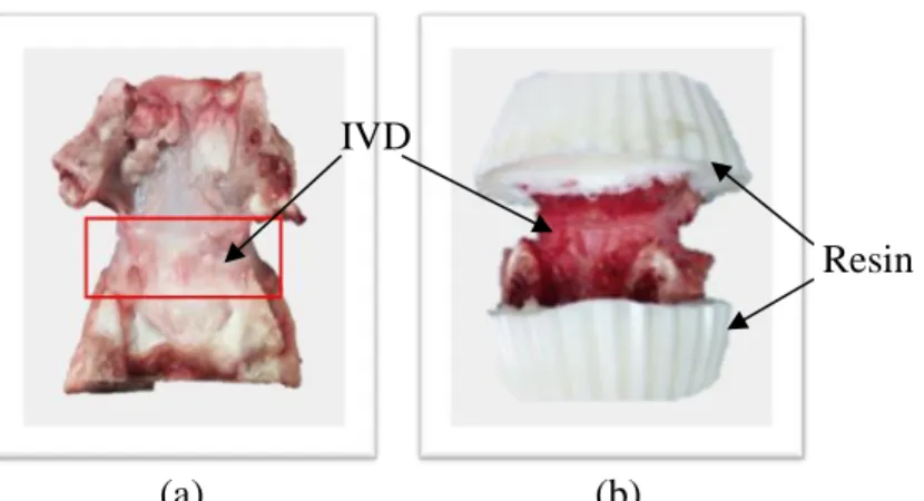
(29) Thèse de Amil Derrouiche, Université de Lille, 2018. Chapter 2: Experimental characterization Part 1: Osmo-inelastic response of the intervertebral disc very recently investigated (Emanuel et al., 2018; Vergroesen et al., 2018), the inherent chemomechanical mechanisms are far from being fully established, especially regarding the mechanical loading path. Establishing the relationship between the osmo-inelastic response and the mechanical loading path would allow highlighting the origin of the coupling. The present part of the Chapter 2 is focused on the chemical sensitivity of the FSU inelastic response in relation with the mechanical loading path. In-vitro experiments were performed for different osmolarity and loading rate conditions in order to provide insights on the timedependent chemo-mechanical response of the FSU. The response was examined in tension, compression and torsion. A plausible explanation of the inherent chemo-mechanical coupling is discussed under the guidance of our experimental observations.. 2.1.2. Materials and methods 2.1.2.1. Specimen preparation Nineteen FSUs from cervical spines were harvested from mature sheep cadavers (8-10 months) obtained from a local abattoir. Sheepwas selected in reason of the vertebrae size that facilitates cutting operations and easily fits our testing machine. The selected specimens were stored at 4°C at maximum two days before testing. Note that although the responses of fresh and frozen FSUs are expected to be different (Callaghan and McGill, 1995; Bass and al., 1997), the storage effects in terms of temperature and duration are still unknown (Newell et al., 2017). That is why fresh specimens were used in this study. The entire cervical spines extracted within two days after animal death were first separated from muscles and ligaments by hand using a surgical tool, and then separated into nineteen FSUs with a band saw, consisting in « bone-disc-bone » segment without neural arches. The cutting was carefully performed in order to assure a sufficient bone segment size. External cracks in bone segment due to cutting operation were carefully examined, and after a visual inspection, only eighteen intact FSUs were chosen for 29 © 2018 Tous droits réservés.. lilliad.univ-lille.fr.
(30) Thèse de Amil Derrouiche, Université de Lille, 2018. Chapter 2: Experimental characterization Part 1: Osmo-inelastic response of the intervertebral disc mechanical testing. Immediately after extraction, the FSU was cleaned and then immerged in 9 g/L saline solution at room temperature during 100 minutes. Since the storage condition after animal death in dry cold room decreases the water content, immerging specimens in that solution increases its hydration content to reach the chemical equilibrium (Costi et al., 2002) when IVD height remains constant. Note that ex-vivo conditions increase the equilibrated water content compared to in-vivo conditions. Due to the irregular shape of the vertebra, the bone segment was embedded in polyurethane resin Axson F180 to obtain two parallel plans. The specimen preparation procedure is illustrated in Figure 2.1.1. The superior and inferior parts of the FSU were glued to metal plates in order to be gripped between clamps of the testing machine. The applied loadings were lower than the maximal physiological movements in order to keep normal working conditions and to avoid any damage in IVDs or in both bone-resin interface and resin-metal plate gluing.. IVD Resin. (a) (b) Figure 2.1.1. Preparation of FSU specimen (dorsal view): (a) extraction of IVD and two adjacent bone segments, (b) bone segments embedded in resin.. 2.1.2.2. Methods In-vitro experiments were performed under controlled-environment using a displacementcontrolled universal-axial testing machine Instron-5500 equipped with a 5 kN load cell and an angle-controlled universal-torsion testing machine Instron-8874 equipped with a 100 Nm load. 30 © 2018 Tous droits réservés.. lilliad.univ-lille.fr.
(31) Thèse de Amil Derrouiche, Université de Lille, 2018. Chapter 2: Experimental characterization Part 1: Osmo-inelastic response of the intervertebral disc cell. Throughout the mechanical loading, the FSUs were immerged in a saline solution. The osmolarity was controlled by varying the NaCl content in water from 0 g/L hypo-osmotic solution to 36 g/L hyper-osmotic solution. The temperature influence is not considered in the present work, and all measurements were performed at room temperature. The results reported in this investigation follow the experimental procedure illustrated in Figure 2.1.2.. Figure 2.1.2. Experimental design for two successives loading-unloading tests (1: mechanical rest, 2: successive low-strain compressions, 3: loading-unloading test).. A two-step preconditioning was applied in the aim to adapt the IVD tissue to its new environment after changes in NaCl content and to ensure reproducibility of the mechanical response. The two-step preconditioning consists in a period of 30 minutes of mechanical rest without load and successive low-strain compressions which generate inter-vertebral pressure and promote a fluid exchange with the environment (Urban and McMullin, 1985; Adams et al., 1987; Ferguson et al., 2004). In particular, the fluid exchange during this preconditioning avoids 31 © 2018 Tous droits réservés.. lilliad.univ-lille.fr.
(32) Thèse de Amil Derrouiche, Université de Lille, 2018. Chapter 2: Experimental characterization Part 1: Osmo-inelastic response of the intervertebral disc internal imbalances due to changes in solution osmolarity and allows penetration of NaCl inside IVD. The reliability of the preconditioning can be verified by performing two successive relaxation tests. In the test, a compressive displacement was progressively increased until reaching a prescribed value and then maintained constant during 1 hour.. [NaCl] = 9 g/L Displacement = 2 mm. [NaCl] = 9 g/L Displacement = 2 mm. (a) [NaCl] = 9 g/L. [NaCl] = 9 g/L. (b) Displacement = 2 mm. Displacement = 2 mm. (c) Figure 2.1.3. Relaxation response: (a) two successive relaxation tests performed on the same FSU, (b) displacement level effect (1: 3 mm, 2: 2 mm, 3: 1 mm) and (c) osmolarity effect (1: 0 g/L, 2: 9 g/L, 3: 18 g/L).. 32 © 2018 Tous droits réservés.. lilliad.univ-lille.fr.
(33) Thèse de Amil Derrouiche, Université de Lille, 2018. Chapter 2: Experimental characterization Part 1: Osmo-inelastic response of the intervertebral disc The results of two successive relaxation tests are reported in Figure 2.1.3a for a reference saline concentration of 9 g/L and a compressive displacement of 2 mm. The load begins to substantially decrease in the first relaxation period and then evolves towards an end corresponding to an obvious stabilized state. Since the two relaxation curves are superimposed, a total recovery is obtained between the repeated tests. This result assesses our test method in which the mechanical and structural features of the FSU specimen remain intact. As an inelastic feature that depends on the chemical state of the IVD, the stabilized response is function of the compressive displacement level (Figure 2.1.3b) and the NaCl concentration (Figure 2.1.3c). Although the applied displacements seem substantial, no failure was reported. The preconditioning allows the chemo-mechanical equilibrium of the FSU before experiments. Experiments on FSU specimens are, however, repeatable only if the loading is within the physiological range in order to avoid all damage phenomena in the unit. Even after 10,000 cycles, a total recovery of the FSU response was observed by Johannessen et al. (2004). Although in some studies thousands of preconditioning cycles are applied, the reproducibility of the mechanical response is generally reached upon only first three cycles (Newell et al., 2017). In the present study, the FSUs were preconditioned by 10 cycles with low amplitude of 0.1 mm (at a rate of 5×10⁻² mm s-1) in order to adapt the IVD tissue to its new environment. This step is particularly important due to the storage conditions in dry cold room, in terms of relative humidity and temperature, which decrease the water content in IVD and its thickness. Experiments were sufficiently slow to ensure fluid circulation, chemical equilibrium and to avoid dynamic effects. A recovery period of 30 minutes was imposed between two successive experiments in order to restore both mechanical and chemical equilibrium. The measurements on the same FSU were repeated after each preconditioning and the total recovery was observed. The FSU chemo-mechanical response was studied in tension, compression and torsion. These loading types provide fundamental information about physiological motions of the FSU such. 33 © 2018 Tous droits réservés.. lilliad.univ-lille.fr.
(34) Thèse de Amil Derrouiche, Université de Lille, 2018. Chapter 2: Experimental characterization Part 1: Osmo-inelastic response of the intervertebral disc as lateral bending, flexion-extension and axial rotation. Experiments were performed by successively increasing the loading rates: 5×10⁻⁴ mm s-1, 5×10⁻³ mm s-1, 5×10⁻² mm s-1 for axial experiments and 0.01 deg s-1, 0.05 deg s-1, 0.1 deg s-1 for twisting experiments. The axial displacement and the twist were limited to a maximum value of 0.25 mm and 2 deg, respectively. The maximum twist angle value of 2 deg corresponds to a neck movement of 50 deg (Anderst et al., 2015). The osmolarity effect was studied by varying the NaCl content in solution: 0 g/L, 9 g/L, 18 g/L and 36 g/L in order to change the environment from hypo-osmotic condition to hyper-osmotic condition and the respective mechanical response. During the physiological movements, the ionic repartition in IVD is inhomogeneous due to water exchange. These experiments permit a better understanding of changes in mechanical response as a result of chemical changes.. 2.1.2.3. Statistical analysis Statistical analysis was carried out to determine standard deviation and error bars on the load and torque values at a 95% confidence. The data presented in the next section represent the mechanical response on one IVD. A p-value < 0.05 was considered to be statisticallysignificant. For both the compression (n=5) and the torsion (n=7), at the same loading rate and the same NaCl concentration, one-way analysis of variance (ANOVA) indicates a p-value < 0.05. A twoway ANOVA without replication is then performed to other FSUs (n=6) in order to indicate the statistical significance of the loading rate and osmolarity effects. Although the present study is based on a relatively small number of specimens (n=18), the goal is to provide qualitative trends allowing to highlight the existence of the osmo-inelastic coupling with our validated protocol.. 34 © 2018 Tous droits réservés.. lilliad.univ-lille.fr.
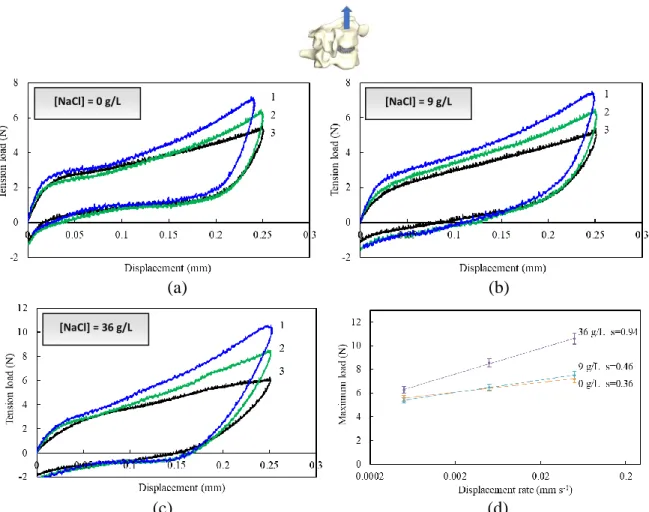
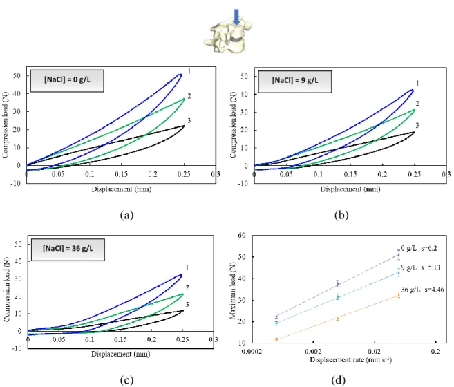
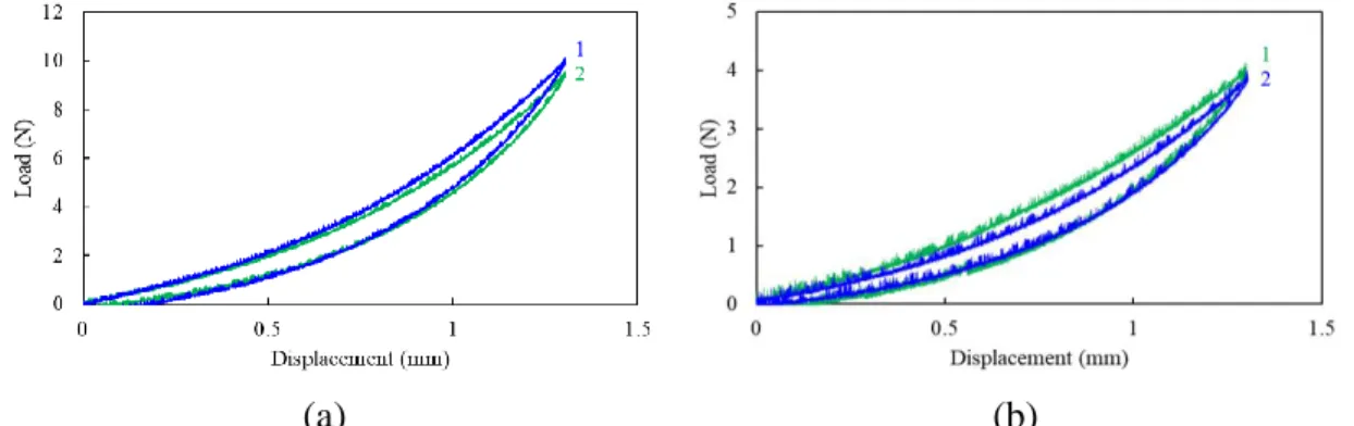
(35) Thèse de Amil Derrouiche, Université de Lille, 2018. Chapter 2: Experimental characterization Part 1: Osmo-inelastic response of the intervertebral disc. 2.1.3. Results 2.1.3.1. Tension The load-displacement curves under tension are plotted in Figure 2.1.4 for different NaCl concentrations and loading rates. The two latter effects are both statistically significant, with a p-value of 0.015 and 0.002, respectively.. [NaCl] = 0 g/L. [NaCl] = 9 g/L. (a). (b). [NaCl] = 36 g/L. (c) (d) Figure 2.1.4. Tension load-displacement responses at three different rates (1: 5×10 -2 mm s-1, 2: 5×10⁻³ mm s-1, 3: 5×10⁻⁴ mm s-1) for (a) 0 g/L, (b) 9 g/L, (c) 36 g/L NaCl concentration, and (d) maximum load-rate response.. The FSU tension response exhibits different stages including an initial stiff response followed by a gradual rollover to hardening. The response depends markedly on the applied loading rate. Higher the loading rate, stiffer the response. The unloading path is different from the loading one, and a residual set appears upon complete unloading, i.e. zero load. The osmolarity affects. 35 © 2018 Tous droits réservés.. lilliad.univ-lille.fr.
(36) Thèse de Amil Derrouiche, Université de Lille, 2018. Chapter 2: Experimental characterization Part 1: Osmo-inelastic response of the intervertebral disc the residual set. Due to its effect on the water content and the microstructure, the increase in NaCl concentration induces a decrease in IVD height. Higher axial strains are then applied on the tissue when the NaCl concentration is increased. The maximum tension load is reported in Figure 2.1.4d as a function of the loading rate, such that a straight-line fit adequately describes the results. The slope, characterizing the rate sensitivity, increases with the NaCl concentration. The osmolarity effect is more important at high loading rate. The rate sensitivity is two time higher between hypo-osmotic and hyper-osmotic conditions.. 2.1.3.2. Compression The compression is a more common mechanical loading applied to IVDs, resulting from the head weight, muscles tension or loads carried by the individual. The FSU load-displacement curves under compression are plotted in Figure 2.1.5. The loading rate effect and the osmolarity effect on the FSU response in compression are obviously statistically significant (p<0.001 for both effects). Chemo-mechanical couplings are then highlighted especially in compression. The FSU compressive response exhibits important energy dissipation, indicating again the dashpot role of the IVD. The osmolarity modifies the compressive response in shape and in magnitude. In particular, the observed inflexion point is delayed with the increasing osmolarity. Also, the residual set increases when the osmolarity increases. More interestingly, the osmolarity effects are inversed in compression and in tension. Indeed, higher the NaCl concentration, softer the response. As reported in Figure 2.1.5d, the rate sensitivity decreases with the NaCl concentration. Moreover, the impact of the loading rate on the FSU response is more important in compression than in tension. At the end of the unloading path, the load, for which the displacement is zero, is the same whatever the conditions.. 36 © 2018 Tous droits réservés.. lilliad.univ-lille.fr.
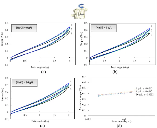
(37) Thèse de Amil Derrouiche, Université de Lille, 2018. Chapter 2: Experimental characterization Part 1: Osmo-inelastic response of the intervertebral disc. [NaCl] = 0 g/L. [NaCl] = 9 g/L. (a). (b). (c). (d). [NaCl] = 36 g/L. Figure 2.1.5. Compression load-displacement responses at three different rates (1: 5×10⁻² mm s-1, 2: 5×10⁻³ mm s-1, 3: 5×10⁻⁴ mm s-1) for (a) 0 g/L, (b) 9 g/L, (c) 36 g/L NaCl concentration, and (d) maximum load-rate response.. 2.1.3.3. Torsion The spine twisting is also a very common physiological loading mode. The loading rate effect on the FSU torsion response is reported in Figure 2.1.6 for different NaCl concentrations. A pvalue > 0.05 is found for both effects in torsion. At low twist angle no appreciable effect of the loading rate can be observed on the torsional stiffness. The FSU response became rate sensitive only athigh twist angle. The chemical sensitivity is not dependent on the loading rate, as illustrated by Figure 2.1.6d in which the rate sensitivity is almost unchanged with the concentration.. 37 © 2018 Tous droits réservés.. lilliad.univ-lille.fr.
(38) Thèse de Amil Derrouiche, Université de Lille, 2018. Chapter 2: Experimental characterization Part 1: Osmo-inelastic response of the intervertebral disc. [NaCl] = 9 g/L. [NaCl] = 0 g/L. (a). (b). [NaCl] = 36 g/L. (c) (d) Figure 2.1.6. Torsion load-displacement responses at three different rates (1: 0.1 deg s-1, 2: 0.05 deg s-1, 3: 0.01 deg s-1) for (a) 0 g/L, (b) 9 g/L, (c) 36 g/L NaCl concentration, and (d) maximum torque-rate response.. 2.3.4. Discussion Our results show a significant effect of the osmolarity on the axial inelastic response and an almost imperceptible effect on the torsional inelastic response. Figure 2.1.7 provides our interpretation of the inherent osmo-inelastic mechanisms by means of a schematic representation in which inelastic and osmolarity effects are separated. The IVD consists of randomly distributed PG macromolecules and organized collagen fibers. The chemical reaction between negatively charged PG macromolecules and mobile ions of the saline solution governs the fluid exchange with the environment. The fluid flow, induced by external ionic imbalance, is dependent on the mechanical loading path, i.e. tension, compression or torsion, and implies osmotic force and IVD swelling (Lanir, 1987). 38 © 2018 Tous droits réservés.. lilliad.univ-lille.fr.
(39) Thèse de Amil Derrouiche, Université de Lille, 2018. Chapter 2: Experimental characterization Part 1: Osmo-inelastic response of the intervertebral disc. (a) (b) (c) Figure 2.1.7. Osmo-inelastic mechanisms in (a) tension, (b) compression and (c) torsion.. The FSU time-dependent response is intrinsically linked with the variation of fluid content in the IVD and with the PG macromolecules rearrangement. The inherent chemo-mechanical coupling is strongly dependent on the water and NaCl content, and is shown in our experiments for the FSU under various mechanical loading types. The water content in the IVD is reduced with increasing NaCl concentration leading to reduced chemical exchanges. The rate sensitivity is found higher in compression than in tension, and almost imperceptible in torsion. The NaCl content has a strong impact on the FSU compression rate-dependency (Figure 2.1.5d). By contrast, the rate-dependency in tension (Figure 2.1.4d) is slightly affected by the saline concentration and nearly not affected in torsion (Figure 2.1.6d). In torsion, the overall response is mainly due to collagen fibers reorientation. In hyper-osmotic environment, although the fluid flow is limited (Lai et al., 1991; Ateshian et al., 2004; Vergroesen et al., 2018), our results show a time-dependence of the chemo-mechanical response. The higher chemical sensitivity in compression than in tension is directly linked to the solely increase in fixed charge, i.e. chemical imbalances. The response in Figure 2.1.3 of an IVD compressed and stimulated by a chemical loading gives some insights on the fluid flow contribution. Increasing the compression level. 39 © 2018 Tous droits réservés.. lilliad.univ-lille.fr.
(40) Thèse de Amil Derrouiche, Université de Lille, 2018. Chapter 2: Experimental characterization Part 1: Osmo-inelastic response of the intervertebral disc (Figure 2.1.3b) or the NaCl concentration (Figure 2.1.3c) decreases the fluid content and leads to an increasing omostic pressure attempting to neutralize the mechanical loading. With the applied compressive displacement, the fluid content in IVD decreases and the chemical unbalance increases. In reaction, the IVD swells in proportion to the applied compression to reach the chemo-mechanical equilibrium. The swelling implies higher chemical stresses in IVD which opposite to the applied compressive displacement. That results in the increase of the relaxed-load with the displacement as observed in Figure 3b. As shown in Figure 3c, the same effect is reported with the increase in NaCl concentration since the osmolarity increases the internal chemical stresses. By increasing the NaCl concentration, the IVD contracts to eject superfluous ionic component and the equilibrium response is quickly reached since there is more ionic component in solution. Under hypo-osmotic condition, for which the solution is poor in ionic component, the relaxation time increases. In hyper-osmotic environment, the extracellular matrix is saturated and the time necessary to achieve ionic equilibrium is shorter. The apparent viscosity seems to be thus related to the PG macromolecules rearrangement. This statement indicates that the macroscopic inelastic features are due to both microstructural rearrangements and fluid flow. The viscoelastic properties of the soft tissue are predominant at high rates whereas the fluid exchange requires longer times (Vogel and Pioletti, 2012; Vergroesen et al., 2018). As observed in Figure 2.1.3c, the equilibrium response is delayed by decreasing the osmolarity, and then by increasing the chemical exchanges. Our results bring some insights on the FSU chemo-mechanical response, and a better understanding of interactions between NP and AF is highlighted. The macro-dissipative response is generally attributed to the gelatinous NP but also the AF plays a major role in it (Liu et al., 2014; Emanuel et al., 2018). The NP is an essential element in the FSU behavior, its main role being to generate a hydrostatic pressure on the AF (Nachemson and Morris, 1964; Iatridis et al., 1997; Wilke et al., 1999; Goins et al., 2005). Tensile and compressive FSUs. 40 © 2018 Tous droits réservés.. lilliad.univ-lille.fr.
(41) Thèse de Amil Derrouiche, Université de Lille, 2018. Chapter 2: Experimental characterization Part 1: Osmo-inelastic response of the intervertebral disc exhibit opposite responses concerning the environment dependency since the internal pressure generated by the NP is reduced in tension. This observation is not common and must be associated to the fluid flow in the IVD (Ferguson et al., 2004; Schmidt et al., 2016). Indeed, the chemical-dependence is due to the difference in fluid mobility between tensile and compressive states. Under a compressive load the fluid is pushed out of the IVD leading to a chemical unbalance and the negative charge of the PGs generates an osmotic pressure through the attraction of water. By contrast, the tensile state decreases the PGs density and leads to a lesser chemical sensitivity. With the increase in osmolarity the IVD constrains to push out excess mobile ions contained in the fluid which results in a force opposing the tensile load and matching the compressive load. Upon the complete unloading, the amount of fluid loss in the IVD governs the extent of residual set. The latter is found lesser in compression than in tension due to higher fluid exchange in compression than in tension. With the increase in osmolarity, the PGs density increases, the IVD constrains and the residual set increases. Due to their concentric organization with ventral and dorsal parts that strain in opposing directions, the AF collagen fibers have a preponderant role under twisting. As a consequence, the fluid flow effect on the torsional inelastic response is reduced.. 2.1.5. Partial conclusion This study has been conducted to determine the chemical sensitivity of the FSU inelastic response under different mechanical loading paths. The osmotic and time-dependent properties have been considered at the same time to better understand the osmo-inelastic mechanisms and the biomechanical behavior. The torsional stiffness and rate-dependency were found chemically insensitive whereas an inverse chemical sensitivity was found between tension and compression. Although it is difficult to know to what extent the time-dependent behavior can be attributed to the PGs 41 © 2018 Tous droits réservés.. lilliad.univ-lille.fr.
(42) Thèse de Amil Derrouiche, Université de Lille, 2018. Chapter 2: Experimental characterization Part 1: Osmo-inelastic response of the intervertebral disc rearrangement and the fluid flow, an interpretation of the inherent chemo-mechanical coupling was proposed. The chemo-mechanical coupling is strong. Indeed, the PGs density depends on the chemical environment affecting the fluid content, and in the meantime, the mechanical loading modulates the PGs density which changes the chemical sensitivity and the level of microstructural interactions. Due to the body weight and muscles tension, the FSU is constantly submitted to a compressive loading stimulating the fluid exchanges with the increase in PGs density which increases the chemical exchanges and the nutrient supply in vivo.. 2.1.6. References Adams, M.A., Dolan, P., Hutton, W.C., 1987. Diurnal variations in the stresses on the lumbar spine. Spine 12, 130-137. Adams, M.A., McNally, D.S., Dolan, P., 1996. “Stress” distributions inside intervertebral discs. The effects of age and degeneration. Journal of Bone and Joint Surgery 78, 965-972. Anderst, W.J., Donaldson, W.F., Lee, J.Y., Kang, J.D., 2015. Cervical motion segment contributions to head motion during flexion\extension, lateral bending, and axial rotation. The Spine Journal 15, 2538-2543. Asano, S., Kaneda, K., Umehara, S., Tadano, S., 1992. The mechanical properties of the human L4-5 functional spinal unit during cyclic loading: the structural effects of the posterior elements. Spine 17, 1343-1352. Ateshian, G.A., Chahine, N.O., Basalo, I.M., Hung, C.T., 2004. The correspondence between equilibrium biphasic and triphasic material properties in mixture models of articular cartilage. Journal of Biomechanics 37, 391-400. Balkovec, C., McGill, S., 2012. Extent of nucleus pulposus migration in the annulus of porcine intervertebral discs exposed to cyclic flexion only versus cyclic flexion and extension. Clinical Biomechanics 27, 766-770. Bass, E., Duncan, N., Hariharan, J., 1997. Frozen storage affects the compression creep behavior of the porcine intervertebral disc. Spine 22, 2867-2876. Callaghan, J.P., McGill, S.M., 1995. Frozen storage increases the ultimate compression load of porcine vertebrae. Journal of Orthopaedic Research 13, 809-812. Cassidy, J.J., Hiltner, A., Baer, E., 1989. Hierarchical structure of the intervertebral disc. Journal Connective Tissue Research 23, 75-88. Chan, S.C.W., Ferguson, S.J., Gantenbein-Ritter, B., 2011. The effects of dynamic loading on the intervertebral disc. European Spine Journal 20, 1796‑1812. Costi, J.J., Hearn, T.C., Fazzalari, N.L., 2002. The effect of hydration on the stiffness of intervertebral discs in an ovine model. Clinical Biomechanics 17, 446-455. Costi, J.J., Stokes, I.A., Gardner-Morse, M.G., Iatridis, J.C., 2008. Frequency-dependent behavior of the intervertebral disc in response to each of six degree of freedom dynamic loading: solid phase and fluid phase contributions. Spine 33, 1731-1738. 42 © 2018 Tous droits réservés.. lilliad.univ-lille.fr.
Figure
+7
Documents relatifs
