HAL Id: dumas-02345525
https://dumas.ccsd.cnrs.fr/dumas-02345525
Submitted on 4 Nov 2019HAL is a multi-disciplinary open access archive for the deposit and dissemination of sci-entific research documents, whether they are pub-lished or not. The documents may come from teaching and research institutions in France or abroad, or from public or private research centers.
L’archive ouverte pluridisciplinaire HAL, est destinée au dépôt et à la diffusion de documents scientifiques de niveau recherche, publiés ou non, émanant des établissements d’enseignement et de recherche français ou étrangers, des laboratoires publics ou privés.
Facteurs associés à la survie chez les patients atteints
d’un cancer solide sortant vivant de réanimation entre
2005 et 2013
Hubert Gheerbrant
To cite this version:
Hubert Gheerbrant. Facteurs associés à la survie chez les patients atteints d’un cancer solide sortant vivant de réanimation entre 2005 et 2013. Médecine humaine et pathologie. 2019. �dumas-02345525�
AVERTISSEMENT
Ce document est le fruit d'un long travail approuvé par le
jury de soutenance et mis à disposition de l'ensemble de la
communauté universitaire élargie.
Il n’a pas été réévalué depuis la date de soutenance.
Il est soumis à la propriété intellectuelle de l'auteur. Ceci
implique une obligation de citation et de référencement
lors de l’utilisation de ce document.
D’autre part, toute contrefaçon, plagiat, reproduction illicite
encourt une poursuite pénale.
Contact au SID de Grenoble :
bump-theses@univ-grenoble-alpes.fr
LIENS
LIENS
Code de la Propriété Intellectuelle. articles L 122. 4
Code de la Propriété Intellectuelle. articles L 335.2- L 335.10
http://www.cfcopies.com/juridique/droit-auteur1
UNIVERSITÉ GRENOBLE ALPES UFR DE MÉDECINE DE GRENOBLE
Année : 2019
FACTORS ASSOCIATED WITH SURVIVAL OF PATIENTS WITH
SOLID CANCER ALIVE AFTER INTENSIVE CARE UNIT
DISCHARGE BETWEEN 2005 AND 2013
THÈSE
PRÉSENTÉE POUR L’OBTENTION DU TITRE DE DOCTEUR EN MÉDECINE
DIPLÔME D’ÉTAT
Hubert GHEERBRANT
THÈSE SOUTENUE PUBLIQUEMENT À LA FACULTÉ DE MÉDECINE DE GRENOBLE
Le : 18/10/2019
DEVANT LE JURY COMPOSÉ DE
Président du jury :
Monsieur le Professeur MORO-SIBILOT Denis
Membres :
Mme le Docteur TOFFART Anne-Claire (directrice de thèse)
Monsieur le Professeur TERZI Nicolas
Monsieur le Docteur LARAMAS Mathieu
L’UFR de Médecine de Grenoble n’entend donner aucune approbation ni improbation aux opinions émises dans les thèses ; ces opinions sont considérées comme propres à leurs auteurs.
6
Remerciements
Je commencerais par remercier les membres du jury et en particulier
Le Professeur Moro-Sibilot, mon premier patron. Il est le témoin de mes premiers pas maladroits de médecin. Sa rigueur bienveillante a incontestablement laissé une trace dans ma formation et mon exercice de docteur. Je vous en remercie.
Le Docteur Toffart, pour ton aide et ta patience durant ces 4 années de préparation de thèse. Je pense anticiper ta remarque sur mes éternelles phote d’aurtografes dont je m’en excuse d’avance. Le problème est franchement complexe. Ta passion et tes connaissances m’ont arrêté de voir que les contraintes dans les études. Je te dois mon choix du master 2.
Le Professeur Terzi et le Docteur Laramas dont je n’ai pas pu profiter de votre formation. Car il faut tout de même noter qu’a posteriori je ne suis pas l’interne le plus approprié pour une thèse d’oncologie/réanimation, n’ayant pas le DESC d’oncologie ni de stage en réanimation. Mais finalement, avec votre aide, je peux aujourd’hui présenter une thèse dont je suis fière. Merci
A l’équipe de pneumologie,
Au Professeur Pison, pour vos visites professorales dont j’en ai oublié les heures mais pas leurs enseignements (sauf peut être le samedi matin).
A Anne Laure, Marie, Johanna, Cécile, Sébastien, Christel, Boubou, Rita et Linda qui ont été au premier rang pour mesurer mes capacités de concentration aussi constante que le beau temps dans le nord. Je vous remercie de votre patience (seuil de tolérance un peu plus bas chez Anne Laure mais plus haut pour bien d’autre chose) et vos enseignements. Je suis heureux de prolonger mon travail avec vous.
Aux internes (et ex interne) de pneumologie, la mousse de lait du café au lait, la substantifique moelle de la médecine d’un hôpital trop petit pour contenir le génie. Mention particulière au grand, athlétique et génial Loic dont tes origines nordiques n’y sont pas un hasard. Je n’oublierai pas nos soirées interminables en onco tho sous tes douces paroles de ♪ je m'en vas guerroyer jolie mamieeee ♫, notre bref concours pas très catholique en onco tho ou à ta tenue de mafieux russe du pétrole à val tho. Je n’oublierai pas aussi la voix de la sagesse (ou pas…) qui tant bien que mal essayait de nous contenir avec Loic et qui m’a tant appris lors de mon 1er stage. Julie, je serai toujours content de te voir en passant par Chambéry.
Au Dr Amourette, qui m’a transmis sa passion pour la pneumologie.
A l’équipe de pneumologie de la Croix Rousse. Au Professeur Devouassoux, pour m’avoir donné une chance unique durant mon internat de bénéficier de votre expérience. Aux ARC qui après un long et fastidieux apprentissage ont réussi à me donner un ordre d’un coup de pied bien pesé sur le plafond de mon bureau.
A toute l’équipe de médecine interne du 3ème A : Fanny, la licorne et Aude que je retrouverais en maladie infectieuse quelques mois plus tard.
Parlons-en de la maladie infectieuse, comment mieux vous décrire le tableau : 5 filles et pas que des moitiés de fille : Aude, Margot, Olga, Alice et Sabrina. A cela vous ajoutez un Hubert qui
7
essayait de faire retenir tant bien que mal à son hippocampe atrophié des cours de stat et d’immuno. Je sais que l’ennui est mon meilleur somnifère et les gens lisses me fatiguent. C’est indéniablement, le semestre où je me suis le moins ennuyé. Je vous en remercie et serais toujours disponible à prendre un verre avec vous ou vous donner un cours de ski de fond . Aux chefs, le Dr Pavese, le Dr Pierre, le Dr Froidure, encore Fanny, Julie, le Dr Brion et le Pr Epaulard d’une patience envers un interne qui était persuadé que la rage est un virus non … une bactérie… Bref, merci pour votre patience.
A toute l’équipe médicale et para-médicale de pneumologie, d’oncologie et d’EFR pour ces jours de collaboration avec vous dont je ne souhaite pas de sitôt arrêter.
A mes parents, je vous dédie plus particulièrement cette thèse qui est la conclusion de mes années d’études. Vous m’avez guidé dans mes choix avec toujours la volonté, pas toujours facile à me faire comprendre, de ne pas avoir de regret plus tard. Petit bémol sur mes échecs en français et en anglais, qui malgré votre aide, c’est avéré être un vain combat. Mais globalement disons que votre mission est bien réussie. A ma mère attentionnée, patiente et clairvoyante envers moi, impatient, distrait et myope. Tu as toujours les bons conseils face à mes nombreuses questions. A mon père, j’espère pouvoir transmettre dignement les valeurs que tu m’as transmise et une pratique de la médecine qui reste un idéal pour moi.
A Clémentine, dont je sais que tu seras toujours là pour passer ½ heures au téléphone à parler de tout et de rien. A Edouard, mon capitalisme, libéraliste et ex-tortionnaire préféré. A Louis, car il faut toujours être en bon terme avec un petit frère qui fait de la boxe thaï, du rugby, de l’athlé, de l’apnée et qui a eu 18 au bac.
A mes cousins, cousines, oncles et tantes, repère solide, dont je mesure toute l’importance en grandissant. C’est un héritage fort qui n’existerait pas sans l’implication de mes grands-parents et les fameuses réunions de famille : Bon Papa et Bonne Maman Gheerbrant (dont sa mousse au chocolat est incontestablement ma madeleine de Proust), je ne vous oublierai jamais. A Mamina dont je pense avoir hérité un côté un peu électron libre. A Bon Papa Rouer et Pome toujours à l’écoute de mes doutes et interrogations durant nos longs mais trop rares (je m’en excuse) repas.
A Baptiste, amis depuis nos 1ère classes à la Présentation, Matthieu et nos parties de ping pong interminable et Benoit, la force tranquille, avec qui j’ai la chance de partager une amitié dont le temps et la distance ne pourra éroder.
A Grenoble, ses montagnes, ses fromages et ses amis
Adrien, l’éternel positif, partenaire (et quelques fois adversaire) de vélo, tu es devenu au fil des années un ami solide sur qui je peux compter. Dysmas, le dernier cow-boy de Grenoble, tes valeurs ont fait de toi un indéfectible optimiste. Qualité rare et précieuse qui fait de chacune de nos rencontres des bons moments. A la team ile de ré : la baronne, paulo, audrey, marine et momo dont le principal point commun est d’être tolérant envers nos différences. C’est ce qui fait toute la richesse de notre amitié. A Anne-Sophie, ma parisienne préférée, Victor et ta très sérieuse théorie des zones, Héloise et non conformisme réfléchi, Josepha, la souriante, Marie dont je sais toute l’amour que tes oreilles ont pour moi, Quentin, le meilleur des meilleurs et Thomas, le papa.
@ Grenoble hiver 2017-8 : Gauthier, Fanny, Joris, Camille encore Camille, Giovanni, Davy, Alison, Ludo, Jean Charles, Clément (rare nordique, qui me fait dire que l’on est vraiment les
8
meilleurs) et nos soirées à l’origine de la très select confrérie des amis de l’internat du 1er
semestre.
A mes nouveaux amis de master, Maxime, Nolwenn, Audrey et Sophie. A l’équipe 12 : Xavier et Matthieu pour s’être relayé pour accompagner le fou faire ses tours de cours, les doctorants, Solène, Stéphane, Ian, Alicia victimes anonymes de mes pauses de 16h. A Emie dont je sais à l’avance que le pot de thèse sera excellent. A Valérie qui n’avait probablement pas mesuré la difficulté d’être ma tutrice, mais qui a réussi à me transmettre son exigence scientifique qui était proche de 0 au début du stage. A tous dont l’intelligence m’a ouvert l’esprit sur une vie que j’avais trop centré sur la médecine. J’espère avoir encore nos longs débats après mon master 2.
Merci à tous.
Pardon,
A notre Terre qui supporte le poids de plus en plus pesant de la bêtise humaine et dont tu n’as en retour que notre mépris.
A nos abeilles perdues, nous mesurerons votre bienveillant et indispensable travail que lorsque les pommiers n’auront plus de pomme.
Aux arbres, rares témoins d’une noblesse que l’homme veut systématiquement couper mais dont vous en garde la trace au plus profond de votre matière.
9
Sommaire
ABSTRACT ... 9
ABREVIATIONS ... 14
INTRODUCTION ... 16
MATERIALS AND METHODS ... 17
Design and Setting ... 17
Study Population... 17 Data Collection ... 18 Statistical Analysis ... 19 RESULTS ... 19 Patient Characteristics ... 19 Outcome Analyses ... 20
Patient Presentations Following ICU Discharge ... 20
DISCUSSION ... 21 CONCLUSIONS ... 23 FIGURES ... 24 TABLES ... 26 SUPPLEMENTARY ... 32 REFERENCES ... 34
10
Hubert GHEERBRANT
Facteurs associés à la survie chez les patients atteints d'un cancer solide
sortant vivant de réanimation entre 2005 et 2013.
RÉSUMÉ :
Introduction : Lors de l’admission en unité de soins intensifs (USI), le pronostic à la sortie
d’USI des patients avec un cancer est une question importante mais difficile. Notre objectif était
d'évaluer les facteurs associés à la survie à 3 et 6 mois de ces patients à la sortie d’USI.
Méthodes : A partir de la base de données OutcomeRea ™, nous avons inclus les patients
d’oncologie sorties vivants de l’USI de l’hôpital universitaire de Grenoble entre décembre 2005
et novembre 2013.
Résultats : Sur les 361 patients atteints d’un cancer avec une admission non programmée en
USI, 253 (70%) sont sortis vivant. Le cancer primitif était principalement digestif (31%) et
thoracique (26%). La mortalité à 3 et 6 mois était respectivement de 33% et 41%. Les facteurs
pronostics indépendants à 6 mois comprenaient un indice de performance du groupe d'oncologie
coopérative orientale (ECOG-PS) de 3 ou 4 (OR: 3,71; IC 95%: 1,67–8,23), une maladie
métastatique (OR, 2,24; IC 95%: 1,22. –4,09), une admission liée à la progression du cancer
(OR: 2,64; IC 95%: 1,32–5,30), un score physiologique aigu simplifié II (SAPS II) de 45 à 58
(OR: 4,41; IC 95%: 1,85–10,53), et une décision de limitation des traitements à l’admission en
USI (OR: 3,89; IC 95%: 1,60–9,43). Parmi les patients avec un ECOG-PS de 0 ou 1 à
l'admission (n= 114), 61% (n= 57) ont récupéré un ECOG-PS de 0–2 à 6 mois. À 3 mois, 74
patients (55%) bénéficiaient un traitement anticancéreux, tandis que 13 (8%) étaient en soins
11
Conclusion : Les facteurs pronostics à 6 mois sont presque les mêmes que ceux associés à la
survie en USI. Nous observons que la plupart des patients ont récupéré un bon ECOG-PS à 6
mois et pouvaient bénéficier d’un traitement anticancéreux.
MOTS CLÉS : Cancer solide, réanimation, traitement oncologique, performans status, pronostic
12
Hubert GHEERBRANT
Factors associated with survival of patients with solid cancer alive after
intensive care unit discharge between 2005 and 2013.
RÉSUMÉ :
Introduction : At intensive care unit (ICU) admission, the prognosis after ICU discharge of
patients with cancer is an important but difficult issue. Our objective was to assess the factors
associated with 3 and 6 month survival of ICU cancer survivors.
Méthodes : Based on the French OutcomeRea™ database, we included solid cancer patients
discharged alive, between December 2005 and November 2013, from the medical ICU of the
University Hospital in Grenoble, France.
Résultats : Of the 361 cancer patients with unscheduled admissions, 253 (70%) were discharged
alive from ICU. The main primary cancer sites were digestive (31%) and thoracic (26%). The 3
and 6 month mortality rates were 33% and 41%, respectively. Factors independently associated
with 6 month mortality included Eastern Cooperative Oncology Group performance status
(ECOG-PS) of 3–4 (OR: 3.71; 95%CI: 1.67–8.23), metastatic disease (OR: 2.24; 95%CI: 1.22–
4.09), admission for cancer progression (OR: 2.64; 95%CI: 1.32–5.30), high simplified acute
physiologic score II (SAPS II) of 45 to 58 (OR: 4.41; 95%CI: 1.85–10.53), and treatment
limitation decision at ICU admission (OR: 3.89; 95%CI: 1.60–9.43). About the patients with an
ECOG-PS 0–1 at admission, 61% (n=57) displayed an ECOG-PS 0–2 at 6 months. At 3 months,
13
Conclusion : The factors associated with 6 month mortality were almost the same as those
known to be associated with ICU mortality. We have highlighted that most patients recovered an
good ECOG-PS at 6 months and could benefit from an anticancer treatment following ICU
discharge.
KEY WORDS :Solid cancer; intensive care unit; prognosis; anticancer treatments; performance
14
Abréviations
CI: confidence interval
ECOG: Eastern Copperative Oncology Group
ICU: intensive care unit
IQR: Interquartile Range
MD: missing data
OR: odds ratio
PS: performance status
TLD: treatment limitation decision
15
Factors Associated With Survival of Patients With Solid Cancer
Alive After Intensive Care Unit Discharge Between 2005 and 2013
Hubert Gheerbrant, MD1; Jean-François Timsit, MD, PhD2,3; Nicolas Terzi, MD, PhD4;
Stéphane Ruckly3; Mathieu Laramas, MD5; Matteo Giaj Levra, MD, PhD1; Emmanuelle Jacquet,
MD5; Loic Falque, MD1; Denis Moro-Sibilot, MD1; and Anne-Claire Toffart, MD, PhD1,6
1Department of Pneumology and Physiology, Grenoble-Alpes University Hospital, Grenoble,
France.
2Medical and Infectious Diseases ICU, APHP, Paris, France.
3Department of Biostatistics, OUTCOMEREA™, Bobigny, France.
4Department of Intensive Care and Reanimation, Grenoble-Alpes University Hospital, Grenoble,
France; INSERM, U1042, Grenoble-Alpes University Hospital, HP2, Grenoble, France.
5Cancer and Blood Diseases, Grenoble-Alpes University Hospital, Grenoble, France.
6Institute for Advanced Biosciences – INSERM U823 – Grenoble-Alpes University, Grenoble,
France.
Corresponding Author: Hubert Gheerbrant, Department of Pneumology and Physiology,
Grenoble-Alpes University Hospital, BP217, FR-38043 Grenoble Cedex 9.
Tel.: +33 (0)645 752 994; Email: hgheerbrant@chu-grenoble.fr
16
INTRODUCTION
In 2018, the World Health Organization (WHO) estimated the number of new cancer
occurrences at 18.1 million worldwide, and the specific cancer-related mortality at about
10 million. In addition, the WHO estimates that the global cancer incidence would increase by
more than 63% in 2040 as compared to 2018 (1). Furthermore, improvements in anticancer
treatments have improved the overall survival, which is associated with a significant increase of
cancer prevalence worldwide (2, 3). Nevertheless, improving patients’ life expectancy does not
exclude their fragility, given that approximately 5–10% of them will develop a life-threatening
disease requiring intensive care unit (ICU) admission (4).
This is an issue facing intensive care physicians, both in terms of the ICU admission of
these patients, as well as their management; These patients represent 15–20% of all ICU
admissions (5–8). Despite therapeutic improvements for cancer patients, an ICU admission is
still associated with a very poor medium-term prognosis (9, 10). While intra-hospital mortality is
estimated around 25–35% (5,6,11), with no significant difference as compared to patients
without cancer (11–13), the 1-year mortality often exceeds 70% (14–16). A better understanding
of the prognostic factors associated with ICU survival has been associated with an improved
patient selection upon ICU admission (4). However, while the factors associated with patients’
being still alive at ICU discharge are much less known, they are likewise less taken into account
at admission (17). Thus, it appears necessary to better understand these factors in order to better
identify the cancer patients that most likely could benefit from the ICU stay (18). The mortality
has proven to be largely associated with the general patient condition (Eastern Cooperative
Oncology Group performance status [ECOG-PS]) at ICU discharge (10, 19). This association
can be partly explained by the fact that the cancer management strategy is dependent on a
patient's ECOG-PS conditions (20, 21). To our knowledge, the evaluation of the oncologic
management pertaining to these patients discharged alive from intensive care has not been fully
17
This research work sought to further determine the factors associated with the survival
of cancer patients still alive at ICU discharge. We also sought to describe their general condition
and anticancer treatments following their ICU stay.
MATERIALS AND METHODS
Design and Setting
We conducted a retrospective analysis involving a French multicenter prospective observational
cohort entered into the OutcomeRea™ database (22). The study was approved by the appropriate ethics committee (Comité d’éthique des Centres d’Investigation Clinique de l’inter-région
Rhône-Alpes-Auvergne). An information letter was sent to each living patient providing him the
opportunity to refuse study participation.
The primary objective was to identify the factors associated with 3- and 6-month
mortality after ICU discharge. The secondary objectives were to assess the ECOG-PS and
anticancer treatments at 3- and 6-months .
Study Population
We included solid tumor patients admitted, between December 2005 and November 2013, to the
medical ICU of the Grenoble Alpes University Hospital in France. Patients were retrieved from
the OutcomeRea™ database, and we selected only those with an International Statistical
Classification of Diseases by the World Health Organization (ICD-10) related to solid tumor
(C00 to C97). Exclusion criteria were patients under 18 years of age at admission, cancer in
remission for over 5 years, hematological malignancy, lack of histological or cytological cancer
diagnosis upon ICU admission, programmed hospitalization for post-surgery or central venous
18
patient were independently considered, provided that they were separated by more than 3
months. If they were closer, only the first was taken into account.
Data Collection
Data related to both the ICU admission and stay were extracted from the OutcomeRea™
database. Complementary data relating to cancer history before and at 3- and 6-months
following ICU admission were retrieved from the patients’ computerized medical charts.
Primary tumor sites were defined as digestive (gastrointestinal, esophageal, liver, and pancreas),
thoracic (lung and mesothelioma), head and neck, genitourinary (including testicles),
gynecological (including breast), and other (endocrine, skin, brain, sarcoma, and rare cancers).
Other cancer-related data retrieved were: metastatic status at ICU admission, time from
diagnosis, anticancer treatments, and cancer status at the last oncological evaluation (newly
diagnosed or in recurrence, controlled or in remission for less than 5 years, progression). At
admission, we recorded the ECOG-PS (23), comorbidities using the Charlson comorbidity index
(24), reason for admission (thrombotic event, bleeding, complications of oncology therapy, or
not cancer-related), sepsis-related organ failure assessment (SOFA) score, as well as simplified
acute physiologic score II (SAPS II). Treatments applied within the ICU were also collected
(vasoactive drugs, invasive mechanical ventilation, or renal replacement therapy), along with
potential limitations regarding care decisions. Data regarding ECOG-PS and new anticancer
treatments administered were collected at 3- and 6-months following ICU discharge. The
follow-up was completed for all patients by either consulting the hospital medical chart or
19
Statistical Analysis
Continuous variables have been expressed as median (25–75% interquartile range [IQR]), and
categorical variables as numbers (percentage). The SAPS II and SOFA scores have been
expressed in points. Survival curves were obtained using the Kaplan-Meier method and
compared by means of the log-rank testing. Patients lost to follow-up on July 2017 were
considered censored. Multivariate analyses of variables associated (P ≤0.20) in a univariate
analysis with 3- and 6-month mortality following ICU discharge were performed using stepwise
logistic regressions. Results were expressed as odds ratios (ORs), with 95% confidence intervals
(CIs) and P values.
All tests were two-sided, and P values <0.05 were considered statistically significant.
All statistical analyses were performed using SAS 9.4 (SAS Institute, Cary, NC, USA).
RESULTS
Patient Characteristics
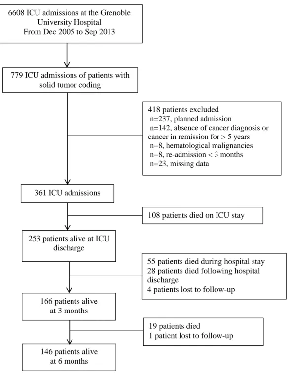
Of the 6,608 patients admitted between December 2005 and November 2013 to the Grenoble
ICU and recorded into the OutcomeRea™ database, 779 concerned cancer patients (Fig. 1).
After considering inclusion and exclusion criteria, 361 ICU admissions were selected. ICU
mortality was 30% (n=108), resulting in 253 studied patients. The median follow-up following
ICU discharge was 250 days (IQR 25–75%: 41–748).
Patient characteristics have been reported in Table 1. The main primary tumor sites
were digestive (n=79, 31%) and thoracic (n= 65, 26%). Almost half of the patients (n= 108,
45%) had a metastatic disease at ICU admission and 167 (68%) patients displayed a newly
diagnosed cancer or cancer in progression. Most patients (n=149, 59%) were treated by
20
192 (76%) patients. The main reasons for ICU admission were tumor progression in 60 (24%)
patients and not cancer-related in 117 (46%).
The median ICU length of stay was 4 days [IQR 25–75%, 2–9]. Upon their ICU stay,
the decision to withhold or withdraw life-sustaining treatments was made for 40 (16%) patients.
Outcome Analyses
After ICU discharge, the median hospital stay length was 12 days [IQR 25–75%, 5–23], and the
median survival was 173 days [IQR 25–75%, 18–622]. The 3- and 6-month mortality rates were
33% (n=83/249) and 41% (n=102/248), respectively (Fig. 1).
The univariate analysis results aimed to identify factors associated with 3- and 6-month
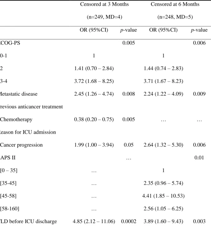
mortality are listed in Table 1. The multivariate analysis results are displayed in Table 2. The
main determinants of 3- and 6-month mortality were an ECOG-PS of 3 or 4, metastatic disease at
ICU admission, ICU admission for cancer progression, and treatment limitation decision taken
within the 2 days preceding ICU discharge. Having been treated with chemotherapy prior to ICU
admission was associated with an improved 3-month survival. A high SAPS II was only
associated with 6-month mortality.
The median survival was 332 days [IQR 25–75%, 35–1476] in patients previously treated
with chemotherapy, and 286 days [IQR 25–75%, 54–690] in those never treated with
chemotherapy prior to ICU admission (Supplementary Table 1). With respect to survival
curves (Supplementary Fig. 1), we observed that the curves crossed between 3- and 6-months .
Patient Presentations Following ICU Discharge
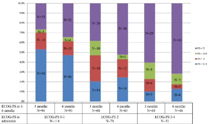
Of the patients with an ECOG-PS of 0–1 at admission, 70% (n=66) and 61% (n=57) displayed
21
patients with an ECOG-PS 3–4 at admission exhibited an ECOG-PS of 0–2 at 3 or 6 months,
respectively.
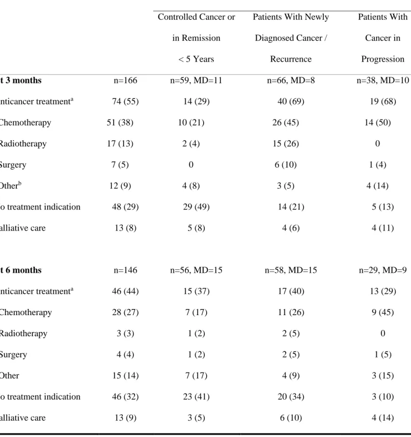
At 3 months post-ICU discharge, 74 (55%) patients received anticancer treatments, while
13 (8%) were in exclusive palliative care (Table 3). The other patients had no anticancer
treatment indication. Most patients with a newly diagnosed cancer, cancer recurrence (n=40,
69%), or cancer in progression (n=19, 68%) and still alive at 3 months were treated, mostly with
chemotherapy. At 6 months, 46 (44%) patients received anticancer treatment, while 13 (9%)
were in exclusive palliative care.
Concerning the 40 patients with a treatment limitation decision prior to ICU discharge, 12
(30%) were alive at 3 months, and 9 (23%) at 6 months. At 3 months, four (33%) benefited from
anticancer treatment, five (42%) had no treatment indication, whereas one was in exclusive
palliative care.
DISCUSSION
In this large mono-center study, we have reported the 3- and 6-month survival data, along with
the characteristics of cancer patients discharged alive from ICU. The usual prognostic factors
were proven to be associated with 3- and 6-month survival. Most patients with an ECOG-PS of
0–1 at ICU admission displayed a good ECOG-PS (0–2) at 3 (70%) and 6 (61%) months. Of
note is that 2/3 of the patients admitted in ICU with newly cancer/cancer recurrence/cancer
progression and alive at ICU discharge did benefit from anticancer treatment at 3 months.
One of the study strengths include the large variety of data recorded, such as data relating
to ICU admission and care, cancer history before ICU to 6 months after ICU discharge with only
few missing data (less than 2% for vital status at 6 months). To our knowledge, this is only the
second study that specifically investigated the prognostic factors of cancer patients following
ICU discharge (18), with only very few studies having reported patient characteristics following
single-22
center patient recruitment limits to a certain extent the extrapolation of our results to other
centers. In spite of only few missing data concerning patient characteristics at ICU admission
and survival, it proved difficult to retrospectively collect the ECOG-PS at 3- and 6-months .
Moreover, as the inclusion period was until 2013, we were unable to investigate patients treated
with targeted therapy or immune checkpoint inhibitors.
The ICU mortality of our patient cohort was 30%, thus in line with other studies (5, 11,
12, 17). The 3- and 6-month mortality rates (33% and 41%) of patients alive at ICU discharge
were likewise similar to the 90-day mortality rate reported in the Auclin et al. (28%) study and
120-day mortality rate from the Vincent et al. study (41.6%) (12, 18). In our study, the factors
independently associated with 3- and 6-month mortality were in accordance with data previously
published for ICU-admitted cancer patients (ECOG-PS, metastatic disease, cancer progression,
SAPS II and treatment limitation decision) (12, 27, 28). No cancer primary site was identified as
a prognostic factor, conversely to other studies that reported lung cancer to be an independent
predictor of in-hospital mortality (18). Interestingly, we have revealed that prior chemotherapy
was independently associated with a superior 3-month survival. This had not been described
previously, but may be partly explained by our interest in an ICU survivor cohort. Furthermore,
we have observed that the survival curves actually crossed, with a superior survival of patients
pretreated with chemotherapy before 6 months and a poorer survival for those pretreated with
chemotherapy thereafter.
In ICU survivors, most patients with an ECOG-PS of 0–1 at ICU admission displayed an
ECOG-PS of 0–2 at 3- and 6-months . Conversely, numerous patients with an ECOG-PS of 3–4
at ICU admission died prior to 3 months post-discharge. No other study has so far reported the
ECOG-PS data following ICU discharge in relation with prior ECOG-PS. It is of note that
Soares et al. revealed the ECOG-PS at 6 months to be 3–4 in 9.5% of hospital survivors (17).
With respect to anticancer treatment at 3- and 6-months post-ICU discharge, we reported
23
typically with chemotherapy. Interestingly, only few patients alive at 3- and 6-months were in
exclusive palliative care. However, we were unable to assess whether palliative care
implementation was modified or delayed by ICU admission. Only very few studies have reported
anticancer treatments following ICU discharge. Considering patients still alive at hospital
discharge, Soares et al. reported that 37% of these patients benefited from anticancer treatment,
such as surgical resection (7%), radiation therapy (34%), and chemotherapy (80%) (17). In 35
(34%) patients, the initially scheduled anticancer treatment plan required either reduction or
modification. These authors also reported that poor ECOG-PS was the only factor associated
with a lower probability of receiving the initially scheduled treatment plan (OR, 0.20; 95%CI
0.05–0.87; P = 0.032). In a smaller cancer patient cohort, 30 patients (68%) of the 44 ICU
survivors with available clinical information were able to undergo a specific anticancer treatment
following hospital discharge. In brief, one patient underwent surgical treatment, two received a
combination of chemotherapy and radiation therapy, and 27 remaining ones were treated with
chemotherapy alone (29).
CONCLUSIONS
Considering the cancer patients alive at ICU discharge, 52% had an ECOG-PS of 0–2 at 3
months, while 55% benefited from an anticancer treatment. Of note, most patients with a good
ECOG-PS before ICU admission displayed a good ECOG-PS following ICU discharge. These
results should be taken into account when deciding upon ICU admission. At that particular time,
it is paramount to have a sound concept concerning the patient’s general condition and his/her
anticancer treatment opportunities following ICU discharge. Moreover, it would be of great
interest to evaluate how ICU admission actually does modify oncological projects. Only a
24
Figure 1.
Patient Flow chart. ICU = intensive care unit.779 ICU admissions of patients with solid tumor coding
418 patients excluded
n=237, planned admission
n=142, absence of cancer diagnosis or cancer in remission for > 5 years n=8, hematological malignancies n=8, re-admission < 3 months n=23, missing data
6608 ICU admissions at the Grenoble University Hospital
From Dec 2005 to Sep 2013
253 patients alive at ICU discharge
108 patients died on ICU stay
166 patients alive at 3 months
55 patients died during hospital stay 28 patients died following hospital discharge
4 patients lost to follow-up
19 patients died
1 patient lost to follow-up 146 patients alive
at 6 months 361 ICU admissions
25
Figure 2.
ECOG performance status at 3- and 6-months according to ECOG performance status at intensive care unit admission (40 missing data at 3 months and 46 missing data at26
TABLE 1.
Main Patient Characteristics Associated With 3- and 6-Month Mortality From Intensive Care Unit Admission in Patients Who Were Discharged Alive From ICU (Univariate Analysis)Censored at 3 Months (n=249, MD=4) Censored at 6 Months (n=248, MD=5) At Admission (n=253)
OR (95%CI) p-value OR (95%CI) p-value
Patient Characteristics Female gender 77 (30) 1.12 (0.63 – 1.99) 0.69 0.91 (0.52 – 1.59) 0.75 Age (/ year) 64 [55 – 71] 1.02 (1 – 1.04) 0.13 1.01 (0.99 – 1.04) 0.17 ECOG-PS (MD=10) <0.01 <0.01 0-1 114 (47) 1 1 2 78 (32) 1.83 (0.96 – 3.5) 2.12 (1.15 – 3.9) 3-4 51 (21) 4.86 (2.38 – 9.95) 5.07 (2.48 – 10.39)
Charlson comorbidity index 1 [0 – 3] 1.02 (1 – 1.03) 0.80 0.94 (0.82 – 1.09) 0.44
Cancer Characteristics
27
Digestive 79 (31) 1.22 (0.69 – 2.14) 0.50 1.05 (0.61 – 1.82) 0.86
Thoracic 65 (26) 1.49 (0.83 – 2.67) 0.19 1.9 (1.07 – 3.36) 0.03
Head and Neck 32 (13) 0.42 (0.17 – 1.06) 0.07 0.38 (0.16 – 0.92) 0.03
Gynecological 29 (12) 1.13 (0.49 – 2.56) 0.78 1.1 (0.5 – 2.45) 0.81
Genito-urinary 28 (11) 1.0 (0.43 – 2.33) 1 0.7 (0.3 – 1.63) 0.41
Other 23 (9) 0.68 (0.26 – 1.80) 0.44 0.93 (0.39 – 2.24) 0.87
Metastatic disease (MD=12) 108 (45) 2.29 (1.33 – 4) <0.01 2.46 (1.45 – 4.16) <0.01
Time since cancer diagnosis (days) (MD=6) 222 [72 – 841] 1.0 (1 – 1) 0.67 1.0 (1 – 1) 0.25
Previous anticancer treatment
Surgery 119 (47) 0.60 (0.35 – 1.02) 0.06 0.60 (0.36 – 1) 0.05
Radiotherapy 81 (32) 0.71 (0.4 – 1.27) 0.25 0.86 (0.5 – 1.48) 0.58
Chemotherapy 149 (59) 0.71 (0.42 – 1.21) 0.20 1 (0.6 – 1.67) 0.99
Cancer status (MD=6) 0.19 0.03
Controlled or in remission for <5 years 80 (32) 1 1
Newly diagnosed / recurrence 102 (41) 1.41 (0.73 – 2.69) 1.65 (0.88 – 3.08)
28
ICU Admission
From home / emergency vs other hospital ward 120 (47) / 133 (53) 1.0 (0.59 – 1.69) 1 1.35 (0.82 – 2.25) 0.24
Reason of ICU admissiona
Tumor progression 60 (24) 2.67 (1.47 – 4.88) <0.01 2.72 (1.49 – 4-95) <0.01
Thrombotic event 18 (7) 1.66 (0.63 – 4.39) 0.30 2.44 (0.91 – 6.54) 0.08
Bleeding 28 (11) 1.13 (0.49 – 2.56) 0.78 1.1 (0.5 – 2.45) 0.81
Complications of anticancer treatment 61 (24) 0.84 (0.45 – 1.59) 0.60 1.14 (0.63 – 2.06) 0.67
Not related to cancer 117 (46) 0.53 (0.31 – 0.91) 0.02 0.4 (0.24 – 0.68) <.01
Glasgow score 15 [12 – 15] 1.00 (0.93 – 1.06) 0.87 1.00 (0.94 – 1.06) 0.90 SAPS II 46 [36 – 58] 1.02 (1 – 1.03) 0.03 1.02 (1.0 – 1.03) 0.02 SOFA score 4 [1 – 7] 1.07 (1 – 1.14) 0.06 1.06 (0.99 – 1.13) 0.12 ICU Stay Vasopressors 88 (34.8) 1.17 (0.68 – 2.03) 0.57 1.04 (0.61 – 1.77) 0.88 Number of days 4 [2.5 – 5] 1.03 (0.95 – 1.13) 0.45 1.02 (0.90 – 1.15) 0.82 Invasive ventilation 103 (40.7) 0.8 (0.46 – 1.37) 0.41 0.78 (0.47 – 1.31) 0.35 Number of days 6 [3 – 12] 0.99 (0.96 – 1.02) 0.55 0.99 (0.96 – 1.03) 0.65
29
Renal replacement therapy 22 (9) 0.93 (0.36 – 2.37) 0.87 0.82 (0.33 – 2.03) 0.66
Chemotherapy 4 (2) … … … …
TLD before ICU discharge 40 (16) 6.53 (3.11 – 13.74) <0.01 3.22 (1.25 – 8.29) <0.02
Length of stay in ICU (days) 4 [2 – 9] 0.99 (0.96 – 1.02) 0.42 0.99 (0.96 – 1.01) 0.30
CI = confidence interval, ICU = intensive care unit, OR = odds ratio, MD = missing data, PS = performance status, TLD = treatment limitation
decision.
30
TABLE 2.
Multivariate Analysis of Characteristics Associated With 3- and 6-Month Mortality Censored at 3 Months(n=249, MD=4)
Censored at 6 Months
(n=248, MD=5)
OR (95%CI) p-value OR (95%CI) p-value
ECOG-PS 0.005 0.006
0-1 1 1
2 1.41 (0.70 – 2.84) 1.44 (0.74 – 2.83)
3-4 3.72 (1.68 – 8.25) 3.71 (1.67 – 8.23)
Metastatic disease 2.45 (1.26 – 4.74) 0.008 2.24 (1.22 – 4.09) 0.009
Previous anticancer treatment
Chemotherapy 0.38 (0.20 – 0.75) 0.005 … …
Reason for ICU admission
Cancer progression 1.99 (1.00 – 3.94) 0.05 2.64 (1.32 – 5.30) 0.006 SAPS II … 0.01 [0 – 35] … 1 [35-45] … 2.35 (0.96 – 5.74) [45-58] … 4.41 (1.85 – 10.53) [58-160] … 2.56 (1.05 – 6.25)
TLD before ICU discharge 4.85 (2.12 – 11.06) 0.0002 3.89 (1.60 – 9.43) 0.003
CI = confidence interval, ICU = intensive care unit, OR = odds ratio, TLD = treatment limitation
31
Table 3.
Anticancer Treatment at 3- and 6-months Following Intensive Care Unit Discharge Cancer status at admission in ICU (MD=3)Controlled Cancer or
in Remission
< 5 Years
Patients With Newly
Diagnosed Cancer / Recurrence Patients With Cancer in Progression At 3 months n=166 n=59, MD=11 n=66, MD=8 n=38, MD=10 Anticancer treatmenta 74 (55) 14 (29) 40 (69) 19 (68) Chemotherapy 51 (38) 10 (21) 26 (45) 14 (50) Radiotherapy 17 (13) 2 (4) 15 (26) 0 Surgery 7 (5) 0 6 (10) 1 (4) Otherb 12 (9) 4 (8) 3 (5) 4 (14) No treatment indication 48 (29) 29 (49) 14 (21) 5 (13) Palliative care 13 (8) 5 (8) 4 (6) 4 (11) At 6 months n=146 n=56, MD=15 n=58, MD=15 n=29, MD=9 Anticancer treatmenta 46 (44) 15 (37) 17 (40) 13 (29) Chemotherapy 28 (27) 7 (17) 11 (26) 9 (45) Radiotherapy 3 (3) 1 (2) 2 (5) 0 Surgery 4 (4) 1 (2) 2 (5) 1 (5) Other 15 (14) 7 (17) 4 (9) 3 (15) No treatment indication 46 (32) 23 (41) 20 (34) 3 (10) Palliative care 13 (9) 3 (5) 6 (10) 4 (14)
ICU = intensive care unit, MD = missing data.
a3 missing data regarding cancer status at 3- and 6-months ; bTargeted therapy, immunotherapy,
32
Supplementary figure 1.
Estimation of survival according to previous chemotherapy (Kaplan Meier).33
SUPPLEMENTARY TABLE 1.
Patient Characteristics According To Previous Chemotherapy No Chemotherapy Before ICU Admission n=104 Chemotherapy Before ICU Admission n=149 p-valueSurvival (Kaplan Meier) 332 (35 – 1476) 286 (54 – 690) 0.17
Type of cancer
Digestive 33 (32) 46 (31) 0.88
Thoracic 17 (16) 48 (32) <.01
Head and Neck 14 (13) 18 (12) 0.75
Gynecological 7 (7) 22 (15) 0.05 Genito-urinary 24 (23) 4 (3) <.01 Metastatic disease (MD=12) 26 (27) 82 (56) <.01 Cancer status (MD=6) <.01 Controlled or in
remission for <5 years
39 (39) 41 (28)
Newly diagnosed /
recurrence
48 (48) 54 (37)
In progression 13 (13) 52 (35)
34
REFERENCES
1. Bray F, Ferlay J, Soerjomataram I, et al: Global cancer statistics 2018: GLOBOCAN
estimates of incidence and mortality worldwide for 36 cancers in 185 countries. CA Cancer J
Clin 2018; 68:394–424
2. Siegel RL, Miller KD, Jemal A: Cancer statistics, 2018. CA Cancer J Clin 2018; 68:7–30 3. Colonna M, Boussari O, Cowppli-Bony A, et al: Time trends and short term projections of
cancer prevalence in France. Cancer Epidemiol 2018; 56:97–105
4. Shimabukuro-Vornhagen A, Böll B, Kochanek M, et al: Critical care of patients with cancer.
CA Cancer J Clin 2016; 66:496–517
5. Soares M, Silva UVA, Teles JMM, et al: Validation of four prognostic scores in patients with cancer admitted to Brazilian intensive care units: results from a prospective multicenter study. Intensive Care Med 2010; 36:1188–1195
6. Cooke CR, Feemster LC, Wiener RS, et al: Aggressiveness of Intensive Care Use Among Patients With Lung Cancer in the Surveillance, Epidemiology, and End Results-Medicare Registry. Chest 2014; 146:916–923
7. Koutsoukou A: Admission of critically ill patients with cancer to the ICU: many uncertainties remain. ESMO Open 2017; 2:e000105
8. Escher M, Ricou B, Nendaz M, et al: ICU physicians’ and internists’ survival predictions for patients evaluated for admission to the intensive care unit. Ann Intensive Care 2018; 8:108 9. Mokart D, Pastores SM, Darmon M: Has survival increased in cancer patients admitted to
the ICU? Yes. Intensive Care Med 2014; 40:1570–1572
10. Puxty K, McLoone P, Quasim T, et al: Survival in solid cancer patients following intensive care unit admission. Intensive Care Med 2014; 40:1409–1428
11. Ostermann M, Ferrando-Vivas P, Gore C, et al: Characteristics and Outcome of Cancer Patients Admitted to the ICU in England, Wales, and Northern Ireland and National Trends Between 1997 and 2013. Crit Care Med 2017; 45:1668–1676
12. Auclin E, Charles-Nelson A, Abbar B, et al: Outcomes in elderly patients admitted to the intensive care unit with solid tumors. Ann Intensive Care 2017; 7:26
13. Taccone FS, Artigas AA, Sprung CL, et al: Characteristics and outcomes of cancer patients in European ICUs. Crit Care 2009; 13:R15
14. Caruso P, Ferreira AC, Laurienzo CE, et al: Short- and long-term survival of patients with metastatic solid cancer admitted to the intensive care unit: prognostic factors. Eur J Cancer
Care (Engl) 2010; 19:260–266
15. Parakh S, Piggin A, Neeman T, et al: Outcomes of haematology/oncology patients admitted to intensive care unit at The Canberra Hospital. Intern Med J 2014; 44:1087–1094
16. Murphy K, Cooksley T, Haji-Michael P: Short- and long-term outcomes of patients with solid tumours following non-surgical intensive care admission. QJM 2018; 111:379–383
35
17. Soares M, Toffart AC, Timsit JF, et al: Intensive care in patients with lung cancer: a multinational study. Ann Oncol 2014; 25:1829–1835
18. Vincent F, Soares M, Mokart D, et al: In-hospital and day-120 survival of critically ill solid cancer patients after discharge of the intensive care units: results of a retrospective
multicenter study-A Groupe de recherche respiratoire en réanimation en Onco-Hématologie (Grrr-OH) study. Ann Intensive Care 2018; 8:40
19. Normilio-Silva K, de Figueiredo AC, Pedroso-de-Lima AC, et al: Long-Term Survival, Quality of Life, and Quality-Adjusted Survival in Critically Ill Patients With Cancer. Crit
Care Med 2016; 44:1327–1337
20. Hanna N, Johnson D, Temin S, et al: Systemic Therapy for Stage IV Non-Small-Cell Lung Cancer: American Society of Clinical Oncology Clinical Practice Guideline Update. J Clin
Oncol 2017; 35:3484–3515
21. Sohal DPS, Mangu PB, Khorana AA, et al: Metastatic Pancreatic Cancer: American Society of Clinical Oncology Clinical Practice Guideline. J Clin Oncol 2016; 34:2784–2796
22. Morvan AC, Hengy B, Garrouste-Orgeas M, et al: Impact of species and antibiotic therapy of enterococcal peritonitis on 30-day mortality in critical care-an analysis of the
OUTCOMEREA database. Crit Care 2019; 23:307
23. Oken MM, Creech RH, Tormey DC, et al: Toxicity and response criteria of the Eastern Cooperative Oncology Group. Am J Clin Oncol 1982; 5:649–655
24. Quan H, Li B, Couris CM, et al: Updating and validating the Charlson comorbidity index and score for risk adjustment in hospital discharge abstracts using data from 6 countries. Am
J Epidemiol 2011; 173:676–682
25. Ha FJ, Weickhardt AJ, Parakh S, et al: Survival and functional outcomes of patients with metastatic solid organ cancer admitted to the intensive care unit of a tertiary centre. Crit
Care Resusc 2017; 19:159–166
26. Roshdy A: One more lesson from a great man! The intensive care ethical dilemma exposed.
Trends Anaesth Crit Care 2018; 22:4–7
27. Soares M, Salluh JI, Ferreira CG, et al: Impact of two different comorbidity measures on the 6-month mortality of critically ill cancer patients. Intensive Care Med 2005; 31:408–415 28. Fisher R, Dangoisse C, Crichton S, et al: Short-term and medium-term survival of critically
ill patients with solid tumours admitted to the intensive care unit: a retrospective analysis.
BMJ Open 2016; 6:e011363
29. Roques S, Parrot A, Lavole A, et al: Six-month prognosis of patients with lung cancer admitted to the intensive care unit. Intensive Care Med 2009; 35:2044–2050