HAL Id: tel-02469015
https://tel.archives-ouvertes.fr/tel-02469015
Submitted on 6 Feb 2020
HAL is a multi-disciplinary open access archive for the deposit and dissemination of sci-entific research documents, whether they are pub-lished or not. The documents may come from teaching and research institutions in France or abroad, or from public or private research centers.
L’archive ouverte pluridisciplinaire HAL, est destinée au dépôt et à la diffusion de documents scientifiques de niveau recherche, publiés ou non, émanant des établissements d’enseignement et de recherche français ou étrangers, des laboratoires publics ou privés.
release dynamics and docking site occupancy at single
central synapses
Kris Blanchard Tapia
To cite this version:
Kris Blanchard Tapia. Subthreshold Ca2+-dependent modulation of vesicle release dynamics and
docking site occupancy at single central synapses. Neurons and Cognition [q-bio.NC]. Université Sorbonne Paris Cité; Universidad de Chile, 2018. English. �NNT : 2018USPCB206�. �tel-02469015�
Université Paris Descartes - Universidad de Chile
École doctorale Cerveau, Cognition, Comportement (Ed3C)Sujet de la thèse
Subthreshold Ca
2+-dependent modulation of vesicle release
dynamics and docking site occupancy at single central
synapses
présentée par
Kris Blanchard Tapia
pour obtenir le grade de
Docteur en Neurosciences de l’Université Paris Descartes, Doctor en Ciencias con Mención en Biología Molecular,
Celular y Neurociencias de la Universidad de Chile
Soutenue le 26 novembre 2018, devant le jury composé de: Brigitte Van Zundert Rapporteur
Ursula wyneken Rapporteur Cecilia Vergara Examinateur Stéphane Dieudonné Examinateur Pierre Vincent Examinateur Federico Trigo Examinateur Isabel Llano Directeur de thèse Juan Bacigalupo Directeur de thèse
Université Paris Descartes - Universidad de Chile
École doctorale Cerveau, Cognition, Comportement (Ed3C)Título de la Tesis
Subthreshold Ca
2+-dependent modulation of vesicle release
dynamics and docking site occupancy at single central
synapses
presentada por
Kris Blanchard Tapia
para obtener el grado de
Docteur en Neurosciences de l’Université Paris Descartes, Doctor en Ciencias con Mención en Biología Molecular,
Celular y Neurociencias de la Universidad de Chile
Fecha de la defensa: 26 de noviembre de 2018. Jurado compuesto por: Brigitte Van Zundert Revisor
Ursula wyneken Revisor Cecilia Vergara Jurado Stéphane Dieudonné Jurado Pierre Vincent Jurado Federico Trigo Jurado
Isabel Llano Director de tesis Juan Bacigalupo Director de tesis
FACULTAD DE CIENCIAS UNIVERSIDAD DE CHILE INFORME DE APROBACION
TESIS DE DOCTORADO
Se informa a la Escuela de Postgrado de la Facultad de Ciencias que la Tesis de Doctorado presentada por el candidato
Kris Blanchard Tapia
Ha sido aprobada por la comisión de evaluación de la tesis como requisito para optar al grado de Doctor en Ciencias con mención en Biología Molecular Celular y
Neurociencias, en el examen de Defensa de Tesis rendido el día 26 de noviembre de 2018.
Comisión de Evaluación de la Tesis:
Dra. Brigitte Van Zundert (Chile)……….. Dra. Ursula Wyneken (Chile)………. Dra. Cecilia Vergara (Chile)………... Dr. Stéphane Dieudonné (Francia)………. Dr. Pierre Vincent (Francia)……….... Dr. Federico Trigo (Francia)……….. Directores de Tesis:
Dra. Isabel Llano (Francia)………....……….…….……….. Dr. Juan Bacigalupo (Chile)…..………..……...
vi
Table of contents
1 Introduction ... 1
1.1 Subthreshold somatic depolarization enhances neurotransmitter release in cerebellar molecular layer interneurons and other central synapses ... 1
1.2 Possible role of protein kinase C in the subthreshold somatic depolarization-mediated facilitation of neurotransmitter release ... 7
1.3 Synapse-specific differences in the number of docking sites and their occupancy probability as key parameters in the subthreshold somatic depolarization-mediated synaptic facilitation ...11
1.4 The importance of studying the mechanisms of modulation of synaptic transmission at the level of single synaptic contacts ...14
1.5 Approaches for the study of single synaptic contacts in brain slices ...20
2 Thesis project proposal ...23
3 Hypothesis ...25
4 General objective ...25
5 Specific objectives ...26
6 Methods ...27
6.1 General methods used in this work ...27
6.2 Preparation of cerebellar slices ...29
6.3 Electrophysiology ...30
6.3.1 Electrophysiology for uncaging experiments in pairs of molecular layer interneurons ...30
6.3.2 Pure electrophysiological recordings in pairs of molecular layer interneurons ...32
6.4 Fluorescence imaging and synaptic contact identification ...33
6.5 Photolysis of DM-Nitrophen ...36
6.6 Calcium imaging ...39 6.7 Determination of the readily releasable pool at a single synaptic contacts 40
vii
6.8 Counting single fusion events and estimating docking site occupancy
probability ...43
6.9 Data Analysis ...46
7 Results ...47
7.1 Calcium photolysis and release of the readily releasable pool at single synaptic contacts ...47
7.2 Subthreshold calcium rises modulates the readily releasable pool size and its kinetics at single synaptic contacts ...55
7.3 Time dependency of the effects of subthreshold calcium rises on synaptic transmission ...68
7.4 Calcium imaging at single synaptic contacts ...74
7.5 Docking site occupancy probability accounts for the subthreshold calcium-dependent modulation of GABA release in single synaptic contacts ...78
7.6 Depolarization-induced changes in presynaptic calcium modulates vesicular release dynamics ...86
8 Discussion ...91
8.1 Approaches for release and measurement of the readily releasable pool in single synaptic contacts ...92
8.2 Modulation of the RRP and docking site occupancy probability by subthreshold calcium levels and its impact on the features of the postsynaptic response ...97
8.3 Differences in synaptic output (facilitation/depression) depend on both the number and occupancy of docking sites and the timing at which the effect is evaluated ...102
8.4 Possible mechanisms for the subthreshold calcium dependent modulation of the RRP and its relation to the analog-to-digital facilitation phenomenon in MLIs ...105
9 Appendix ...110
9.1 Tables ...110
viii
I. Figures index
Figure 1-1. Vesicular release model for a single synaptic contact between cerebellar MLIs. ...13 Figure 1-2. Traditional synapses used as synaptic transmission models and a typical central synapse. ...19 Figure 6-1. Main components of the electrophysiological setup for simultaneous dual whole-cell patch-clamp recordings and photolysis experiments. ...28 Figure 6-2. Dorsal and caudal view of the rat brain. ...30 Figure 6-3. Fluorescence image and synaptic site localization of a synaptically connected pair of MLIs. ...35 Figure 6-4. RRP release by Ca2+ uncaging from the cage compound DM-nitrophen (DMNP)...38 Figure 6-5. Comparison between the optimal situation where a high level of desynchronization of single fusion events is obtained vs. the typical postsynaptic response evoked by presynaptic Ca2+ uncaging at single synaptic contacts. ...42 Figure 6-6. Quantal analysis and estimation of the RRP for each sweep. ...45 Figure 7-1. Direct control of GABA release at single synaptic contacts by photo-release of caged Ca2+. ...52 Figure 7-2. Latency histograms of the laser-evoked PSCs. ...54 Figure 7-3. Experimental approach to evaluate the effect of subthreshold Ca2+ transients on asynchronous and evoked synaptic transmission at single synaptic contacts. ...58 Figure 7-4. Peak amplitude, synaptic charge and rate of failures for control and test conditions. ...61 Figure 7-5. All latencies, first latencies and time to peak for control and test conditions. ...62 Figure 7-6. Peak amplitude, synaptic charge and rate of failure for control and test conditions for 2 second delay. ...71
ix
Figure 7-7. Comparison of miniature GABAergic event frequency between control and test conditions for the 2 second delay. ...72 Figure 7-8. All latencies, first latencies and time to peak for control and test conditions for 2 second delay. ...73 Figure 7-9. Modulation of Ca2+ levels in the sub- and suprathreshold range. ...77 Figure 7-10. Representative experiment showing the approach for quantal analysis of the RRP in single synaptic contacts. ...80 Figure 7-11. Quantal analysis of RRP release in control and test conditions for 200 ms and 2 s delay. ...84 Figure 7-12. Differences between the “extreme points” along the distribution of the (test-ctrl) values for RRPavg and docking site occupancy probability. ...85 Figure 7-13. Presynaptic subthreshold somatic depolarization enhances AP-evoked release in MLIs. ...90
x
II. Résumé
Dans plusieurs neurones du SNC l'activité somato-dendritique infraliminaire peut se propager passivement dans l'axone et augmenter transitoirement la transmission synaptique spontanée et évoquée par le potentiel d'action de manière dépendante du Ca2+. Les mécanismes sousjacents à ce type de plasticité synaptique, appelée
facilitation "analogique" ou "analogo-digitale", restent largement inconnus pour la plupart des synapses centrales, principalement en raison de la difficulté à réaliser des enregistrements directs des petits boutons présynaptiques. Ici, nous utilisons de la photolyse de Ca2+ et de l'imagerie aux niveau des terminaisons présynaptiques
individuelles des interneurones de la couche moléculaire du cervelet (ICM), combinées à des enregistrements électrophysiologiques avec la technique du patch. Nous décrivons un nouveau mécanisme de facilitation analogo-digitale qui est dépendant du Ca2+ infraliminaire et dans lequel la fraction et la cinétique du pool
de vésicules dites “prêtes à être libérées” (readily releasable pool ou RRP en anglais), est modulée par des modifications de la probabilité d'occupation des sites d'ancrage dans des contacts synaptiques individuels. Nos résultats ajoutent une nouvelle dimension à la compréhension de la manière dont l'activité infraliminaire module le flux d'information dans les circuits neuronaux
xi
III. Abstract
In several neurons of the CNS, subthreshold somatodendritic activity can spread passively into the axon and transiently enhance spontaneous and spike-evoked synaptic transmission in a Ca2+-dependent and graded manner. Available evidence
about the underlying mechanism of this type of synaptic plasticity, called “analog” or “analog to digital” facilitation (ADF), remains largely incomplete for the majority of central synapses, mainly due to the experimental inaccessibility to the small presynaptic boutons. Here we use both Ca2+ photolysis and imaging at
individual presynaptic terminals of the rat cerebellar molecular layer interneurons (MLIs), combined with whole-cell paired recordings from synaptically connected MLIs, to report a novel subthreshold Ca2+-dependent mechanism for ADF whereby
the fraction and the kinetics of the pool of vesicles available for immediate release, the readily releasable pool (RRP), are modulated by changing the docking site occupancy probability in single synaptic contacts. Our results add a new dimension in the understanding of how subthreshold activity modulates information flow in neuronal circuits.
IV. Mots clés; Keywords:
Analog signaling, Analog-to-digital, Docking Sites, GABAergic interneurons, Readily Releasable Pool, Single synaptic contacts, Synaptic plasticity.
xii
V. List of abbreviations
1. NT: neurotransmitter
2. AP: Action potential
3. SSD: subthreshold somatic depolarization
4. ADF: Analog-to-digital facilitation
5. MLI: Molecular layer interneuron
6. VDCC: Voltage-dependent calcium channels
7. PKC: Protein kinase C
8. NMDA: N-methyl-D-aspartate
9. DAG: Diacylglycerol
10. RRP: Readily releasable pool
11. pNR: Probability of neurotransmitter release
12. PSC: Postsynaptic current
13. p: Probability of a single quantum to be released
xiii
15. OGB-1: Oregon Green BAPTA-1
xiv
A mi amada esposa, quien con su amor y coraje me recuerda día a día el real sentido del éxito.
A mí amada hija, por darme la oportunidad de experimentar una fuerza tan poderosa, y por alegrar mi mundo con su inocencia y belleza.
xv
VI. Acknowledgment
First of all, I would like to thank Federico, Alain and Isabel for accepting me into the lab. It’s been amazing to work in Paris doing beautiful experiments. Thank you, Fede, for giving me the chance to explore and to think freely, and also for putting up with my stubbornness; I have learned a lot from you. Thank you, Isabel for your support and guidance. Thank you, Alain, for your support and advice; it’s a pleasure to discuss results with you. A special thanks to Professor Juan Bacigalupo for your support, advice and trust over the years, none of these would have been possible without your help and mentorship; I have no words to express my gratitude.
I would like to thank to the members of the thesis committee: Dra. Brigitte Van Zundert, Dra. Ursula Wyneken, Dr. Stéphane Dieudonné, Dr. Pierre Vincent, Dra. Magdalena Sanhueza, and Dra. Cecilia Vergara. I really appreciate your help, advice and support.
Thank you to all the past and present members of the Cell Physiology Lab, it is been a real pleasure to work in the lab.
Thank you to David and Celine for being so nice to me in spite of my poor English and French. Thank you to Philippe and Boris, it was a pleasure to share the working space with you. Thank you to Brandon for always smiling and your
xvi
willingness to help. Thank you to Michael for being such a nice guy, it was great to work next to you the last year. Thank you, Thibault, for helping me every time I had a question related to science or the French language. A super special thank you to Jorge, Gerardo, Taka, Camila, Lupe, Javier, Merouann, Bastian, Laura Castro, Laura Gomez, Enrico and Maria, you are totally awesome!!! I am very happy to have met you. Thanks to Patrick, Linda and Chantal for being so kind and helpful.
Thank you to all my Parisian-Chilean friends; it’s been a real pleasure to share part of my life with you. A special thank you to “los terribles voisins” Vale and Enrique, for your friendship and support.
Thank you to my parents for your love and the enormous effort you have put into my education. Thanks to my big brother Israel for walking with me through life and for your unconditional love. Thanks to my little sisters Nathalie and Ambar for your love and support, you are amazing. Thanks to all my friends for the advices and amazing conversations.
Thanks to my mother-in-law Lita for your love and support. A super mega thank to my beloved wife for all your love, patience, kindness and bravery, you are wonderful. It is been amazing to share my life with you. Finally, I would like to thank to my baby Violeta, you are everything to me now, thank you for making me a better person.
1
1 Introduction
1.1 Subthreshold somatic depolarization enhances neurotransmitter release in cerebellar molecular layer interneurons and other central synapses
It is well known that the probability of neurotransmitter (NT) release depends on the recent history of synaptic activity, which can influence residual calcium (Ca2+)
and modulate the amount of NT released when an action potential (AP) arrives to the presynaptic terminal (short-term synaptic plasticity). Traditionally, AP-dependent transmission has been considered as the only fast way of communication between the somatodendritic compartment and the presynaptic terminals (Shu et al., 2006). However, a growing body of evidence suggests that the neuronal activity on which short-term synaptic plasticity depends, not only involves intracellular Ca2+ changes as a result of the "all-or-nothing" (or “digital”)
nature of the AP, but also involves the subthreshold, AP-independent or “analog” synaptic activity that produces changes in membrane potential without reaching the AP threshold. On invertebrate preparations it was demonstrated long ago that the application of subthreshold presynaptic depolarization can increase the amount of NT released to the synaptic cleft (Shimahara and Tauc, 1975; Nicholls and Wallace, 1978). In mammals, this feature was demonstrated more recently by
2
recording synaptic activity in pairs of connected neurons (Shu et al., 2006; Alle and Geiger, 2006). Direct recordings from hippocampal mossy fiber boutons in slices have revealed that subthreshold somatic depolarization (SSD) can passively spread hundreds of microns into the axon and modulate the process of NT release at the mossy fiber bouton–CA3 pyramidal neuron synapse (Alle and Geiger, 2006). In the Calyx of Held (where direct pre- and postsynaptic recordings is achievable), it has been shown that subthreshold depolarization of the presynaptic terminal is capable of potentiating NT release by a mechanism that involves both, an increase in the basal Ca2+ levels (Awatramani et al., 2005) and an increase in the
AP-dependent Ca2+ influx as a consequence of a change in the AP shape (Hori and
Takahashi, 2009). In cortical neurons, it has been proposed that changes in the AP shape may be mediating the potentiation induced by the application of SSD (Shu et al., 2006). However, no changes have been found in the axonal Ca2+ levels(neither
basal nor AP-evoked) during this so-called “analogue signaling” in hippocampal mossy fibers (Scott et al., 2008). Given the discrepancies, it has been proposed that different forms of analog (or analog-to-digital1) facilitation (ADF), that are Ca2+
-dependent or Ca2+-independent, can co-exist in different synapses and with
different contributions (Scott et al., 2008).
3
Little is known about the molecular mechanisms underlying this form of synaptic plasticity; however, important advances were recently made in this direction using cerebellar molecular layer interneurons (MLIs) as a model (Bouhours, et al., 2011). In these cells, SSD pulses are electrotonically spread to the presynaptic terminals, where they produce an enhancement of NT release through a signaling pathway involving the activation of the P/Q type, voltage-dependent Ca2+ channels (VDCC),
followed by an increase in the basal Ca2+ levels and a local activation of protein
kinase C (PKC) (Bouhours, et al., 2011); the target of PKC that mediates this effect is unknown. Additionally, for the same preparation it has been also proposed that the SSD-evoked Ca2+ influx facilitates the AP-evoked Ca2+ entry and enhances NT
release (Christie et al., 2011). Interestingly, in the same study was reported that 4 out of 10 MLI-MLI recorded pairs showed a depression instead of a facilitation when the SSD pulse was preceded by the application of a high frequency train of action potentials. More recently, some controversy has arisen because the group of Christie did not see any SSD-mediated facilitation in MLIs in PKC knockout animals. Instead, they attributed the presence of analog signaling to a broadening of the AP due to the rapid inactivation of the Kv3 type of potassium channels in the
presynaptic terminals, which modifies the currents accounting for the AP waveform, directly enhancing the spike-evoked Ca2+ influx independently of PKC
4
(Rowan and Christie, 2017). Previous studies showed that both, the application of NMDA and the application of SSD in cerebellar MLIs can significantly increase the frequency of miniature GABAergic events (Glitsch and Marty, 1999), thus suggesting that physiological subthreshold activity can efficiently modulate the axonal function. Additionally, the application of SSD also produces an increase in the frequency of the so-called "preminis" (Trigo et al., 2010), which correspond to synaptic events that emerge from the autocrine activation of the presynaptic GABAergic receptors in the axon of MLIs. It was recently shown that these preminis can have an impact on the interneuron excitability, because the voltage changes that they produce are transmitted to the soma antidromically (de San Martin JZ et al., 2015). Furthermore, since preminis are prominent during the formation of the molecular layer (P11-15), it is thought that they could guide the formation of the neuronal circuits in growing GABAergic neurons through an increase in the release probability of newly formed synaptic contacts by a positive feedback mechanism.
During the last decade, a growing body of evidence suggests that many of the neurons in the nervous system can express ionotropic receptors in the axonal terminals that can modify the membrane potential locally. For example, in several preparations in which the SSD-mediated facilitation effect has been studied, the
5
presynaptic terminal expresses GABAA or glycine receptors [calyx of Held
(Turecek and Trussell, 2001); mossy fibers (Ruiz et al., 2003; Alle and Geiger, 2007); MLIs (Pouzat and Marty, 1999; Trigo et al., 2007)]. In addition, unlike the somatodendritic compartment, where the activation of these receptors is inhibitory (or mixed inhibitory/excitatory), their activation in the axonal compartment is excitatory, leading to membrane depolarization, increase in the axonal Ca2+ levels
and increase in NT release (Trigo et al., 2008). Consequently, it has been proposed that the activation of these presynaptic receptors and the SSD-mediated facilitation could share a common signaling pathway.
In summary, the analog-to-digital signaling, by which subthreshold synaptic activity reaches the presynaptic terminals and modules the AP-evoked process of NT release, has deep implications in the way the NS encodes and processes information. Indeed, in a “digital” synapse NT release follows the all-or-nothing nature of the AP, hence being a binary process that either does (“1”) or does not (“0”) happen, while in an analog synapse the dynamic range in which information can be transferred is very large (Rama, Zbili, Debanne, 2015). Thus, a hybrid type of synaptic transmission where subthreshold activity modulates the following AP-dependent release will largely increase the amount of information that can be transmitted in neural circuits. Furthermore, if we consider that many neurons in the
6
mammalian brain possess axons with lengths of a few hundred microns, it became clear that the extent of SSD penetration can result in that a high proportion of presynaptic terminals are modulated; for example, the space time constant (distance at which the change in the membrane potential has experienced a decrease of 63%) in hippocampal neurons where ADF has been described is approximately 430 μm (Alle and Geiger, 2006).
It would be very helpful, to determine the synaptic processes controlling NT release in central synapses, to describe the specific synaptic parameters that are being modulated during ADF, their differences between individual synaptic contacts of the same neuron, and from different neuronal types at different developmental stages.
7
1.2 Possible role of protein kinase C in the subthreshold somatic depolarization-mediated facilitation of neurotransmitter release
Protein kinase C is a family of kinases that are implicated in controlling the function of others proteins by the phosphorylation of their hydroxyl group of Ser/Thr amino acid residues. The conventional isoforms of PKC (PKCα, PKCβI/βII, and PKCγ) are expressed in several tissues (including the brain) and require the binding of the lipid second messenger diacylglycerol (DAG) and Ca2+
for their full activation (Hirokazu Hirai, 2017). The effects of the activation of the conventional isoforms of PKC by DAG (through its C1 binding domain) or directly
by Ca2+ (through its C
2 domain) have been implicated in many studies with the
induction of synaptic enhancement/augmentation (Francis et al., 2002, Majewski and Iannazzo, 1998, Stevens and Sullivan, 1998). There is also evidence that PKC plays a critical role in the phenomenon of post-tetanic potentiation2, which results
from a high-frequency or “tetanic” stimulation of 30-50 or 100 Hz (commonly, 50 Hz for 5 seconds). (Alle et al., 2001, Beierlein et al., 2007, Brager et al., 2003; Fioravante et al., 2011; Korogod et al., 2007; Lee et al., 2008; Wierda et al., 2007). Because post-tetanic potentiation as well other types of short-term synaptic plasticity (including depression) are elicited by different temporal patterns of presynaptic activity, it has been hypothesized that they may contribute to the
2. The short-term synaptic plasticity is often separated into three categories according to their temporal scales: facilitation (which lasts for tens of milliseconds), augmentation (lasts for seconds) and potentiation (also called post-tetanic potentiation), that lasts for minutes. It is thought that all depend in one way or another on the residual presynaptic Ca2 + levels acting in different targets.
8
phenomenon of temporal filtering by facilitating or impeding the transmission of different patterns of activity (Fortune and Gary, 2000). Additionally, post-tetanic potentiationis thought to correspond to one of the mechanisms that significantly contribute to the formation of short-term memory, synaptic filtering and information processing in the nervous system (Abbott and Regehr, 2004; Klug et al., 2012; Silva et al., 1996).
Many forms of regulation of neurotransmitter release are mediated through an increase in the size of the readily releasable pool (RRP) (fraction of vesicles in the active zone available for immediate release upon strong stimulation) and/or by an increase in the probability of NT release (pNR) (Pan and Zucker, 2009, Regehr et al., 2009, Zucker and Regehr, 2002).
In a recent study in the calyx of Held (Chu et al., 2014) it was demonstrated that, depending on the developmental stage of the animal, the activation of different conventional PKC isoforms is capable of mediating post-tetanic potentiation either by an increase in pNR or through an increase in the RRP; however, the PKC substrate that could be mediating these processes is unknown.
From a functional perspective, knowing which of the two mechanisms (changes in pNR or the RRP size) is governing the properties of NT release in different
9
synapses is very important, since they have very different outcomes in response to the application of high frequency stimulation trains (Pan and Zucker, 2009; Thanawala and Regehr, 2013): an increase in RRP proportionally increases the amplitude of the synaptic responses (an effect similar to increasing the number of postsynaptic receptors), while an increase in pNR has also the effect of increasing the use-dependent depression of the response. In this way under tetanic stimulation, the total amount of NT released is doubled if the RRP is doubled, while it essentially remains unchanged if pNR doubles (Chu et al., 2014).
Proteins of the release machinery (SNAP25, MUNC18-1, GAP-43, and synaptotagmin-1) and some voltage-dependent ion channels (K+ and Ca2+) have
been identified as putative presynaptic substrates of PKC, whose phosphorylation could be involved in the phenomenon of synaptic plasticity. However, with the notable exception of MUNC-18, evidence for a role of the rest of the proteins in the PKC-mediated potentiation is either incomplete or has been ruled out (Parfitt and Madison, 1993; Finley et al., 2003; Hulo et al., 2002; Nagy et al., 2006). MUNC18-1 is an essential component of the vesicle fusion complex and is indispensable in the process of NT release (Verhage et al., 2000; Zilly et al., 2006). Interestingly, MUNC18-1 presents multiple sites of phosphorylation by PKC (Barclay et al., 2003; Fujita et al., 1996); phosphorylation of these sites has been
10
previously implicated in vesicle replenishment after intense stimulation (Nili et al., 2006) and in the control of RRP size in GABAergic and glutamatergic synapses (Toonen et al., 2006). Additionally, it is known that MUNC18-1 is rapidly phosphorylated by PKC under depolarizing conditions (Craig et al., 2003; de Vries et al., 2000). All these observations make the MUNC18-1 protein the most likely candidate to mediate the effects of PKC activation during subthreshold somatic depolarizations.
11
1.3 Synapse-specific differences in the number of docking sites and their occupancy probability as key parameters in the subthreshold somatic depolarization-mediated synaptic facilitation
Central mammalian synapses differ substantially among each other in several key parameters like quantal size (postsynaptic response to the release of NT from a single vesicle), pNR and short-term synaptic plasticity. One of the most basic parameters within the set of synaptic heterogeneities is the size. It is known that differences in synaptic size can reach up to an order of magnitude, and that the dimensions of the presynaptic active zones and the postsynaptic densities, as well as the number of synaptic vesicles and the number of postsynaptic receptors, vary in direct proportion with the synaptic size (Harris and Stevens, 1988, Schikorski and Stevens, 1997, Nusser et al., 1997). Recently, it was demonstrated in the Calyx of Held (by local electrophysiological recordings in the presynaptic terminal, Sheng et al., 2012) and in hippocampal glutamatergic synapses (by optical quantal analysis, Holderith et al., 2012; see later section 1.5: Approaches for the study of single synaptic contacts in brain slices) that the number of Ca2+ channels and the
corresponding pNR increases linearly with the size of the active zone.
In a recent publication using MLIs of the cerebellum as a model, the differences between presynaptic parameters (such as the number of docking sites and release
12
probability) and postsynaptic parameters (such as the amplitude and kinetics of the synaptic currents) were explored at the level of single synaptic contacts. This was carried out using an approach that allows studying individual contacts by simultaneously recording the pre- and postsynaptic activity in pairs of connected neurons in slices of rat cerebellum (Pulido et al., 2015), based on the fact that a significant percentage of the MLI-MLI connections consist of single synaptic contact with a single active zone and a single postsynaptic density (Nusser et al., 1997; Kondo and Marty, 1998).
Pulido et al. (2015) used a simple binomial model (briefly explained in Figure 1-1) to propose that the differences observed between individual synaptic contacts could emerge as a consequence of differences in the number of docking sites (NDS), which would increase in direct proportion with the synaptic size. They also suggested that the application of depolarizing subthreshold pulses could significantly modify the docking site occupancy, which would have a direct impact on the synaptic performance during high-frequency stimulation (40 Hz). In this way, variations in the synaptic size might account for the differences that were reported in both presynaptic (release probability) and postsynaptic parameters such as the peak amplitude and the kinetics of the postsynaptic current (PSC). This neuronal property could be particularly important in synapses with a marked
13
saturation of postsynaptic receptors such as in GABAergic synapses between MLIs (~70%), since in these synapses an increase in the amount of released NT would not generate a substantial change in the amplitude of the PSCs, unless both an increase in the amount of NT released as well as an increase in the number of postsynaptic receptors occurs in a coordinated way. Thus, in the long-term, a fixed synaptic size would markedly decrease the efficiency of the transmission process.
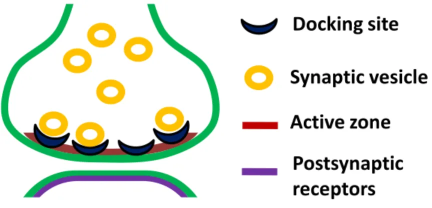
Figure 1-1. Vesicular release model for a single synaptic contact between cerebellar MLIs.
The figure illustrates the active zone, the docking sites, the docked vesicles (RRP), the recycling vesicle pool (which fill the empty docking sites after release) and the postsynaptic receptors of a single synaptic contact. This central synapse, unlike the typical models of synaptic transmission (the calyx of Held, the amphibian neuromuscular junction, or the squid giant synapse), has a single active zone associated with a single postsynaptic density. In this model of Pulido et al., (2015), NT release is described according to the following assumptions: a synapse contains "N" independent docking sites, each of which has a probability () of being occupied by a vesicle. At the same time, a docked vesicle has a probability (p) of fusing with the plasma membrane and releasing its content to the synaptic cleft. After the exocytosis, each empty docking site will be refilled according to a replacement constant rate "R".
Docking site Synaptic vesicle Active zone
Postsynaptic receptors
14
1.4 The importance of studying the mechanisms of modulation of synaptic transmission at the level of single synaptic contacts
One of the ultimate goals of neuroscience is the detailed understanding of the processes by which humans and other animals with a nervous system are capable of sensing and processing the environmental information in order to perform an action that allows an adequate adaptation to a particular situation. Virtually all the evidence accumulated to this day support the idea that all this amazing capacity of the nervous system depends on the ability to generate and propagate electrical signals between its essential neuronal components during the process of synaptic transmission.
The importance of the electrical activity for the physiology of the nervous system was highlighted more than two centuries ago by the Italian physicist and anatomist Luigi Galvani (Piccolino, 1998), and since then the research has mainly focused on understanding the cellular and molecular bases by which this electrical activity allows the transmission of information in a fast and efficient way between excitable cells3, and how this activity relates to animal behavior. Consistent with
this idea, most of the information that we have today about the physiological aspects of synaptic transmission has been obtained by electrophysiological approaches. Although this approximation has a series of remarkable advantages
3. Those cells that present some type of electrical signaling as a result of a differential gene expression pattern that allows them to express ion channels through which they are able to generate and propagate an electrical signal: neurons, muscle cells, glia, endocrine pancreatic cells, endocrine pituitary cells, adrenal medulla cells, gametes and some endothelial cells.
15
(e.g., high temporal resolution of the synaptic current), electrophysiological recordings have proved insufficient to study the functional properties of synaptic transmission at the level of individual synaptic contacts. This is mainly because most electrophysiological recordings usually reflect the averaged electrical activity of many different synapses that are activated in a determined time interval. This is very relevant if we consider that mammalian central synapses differ widely in morphological and functional aspects, even within the same family of synapses, where the pre- and postsynaptic cells belong to the same neuronal type (Branco and Staras, 2009).
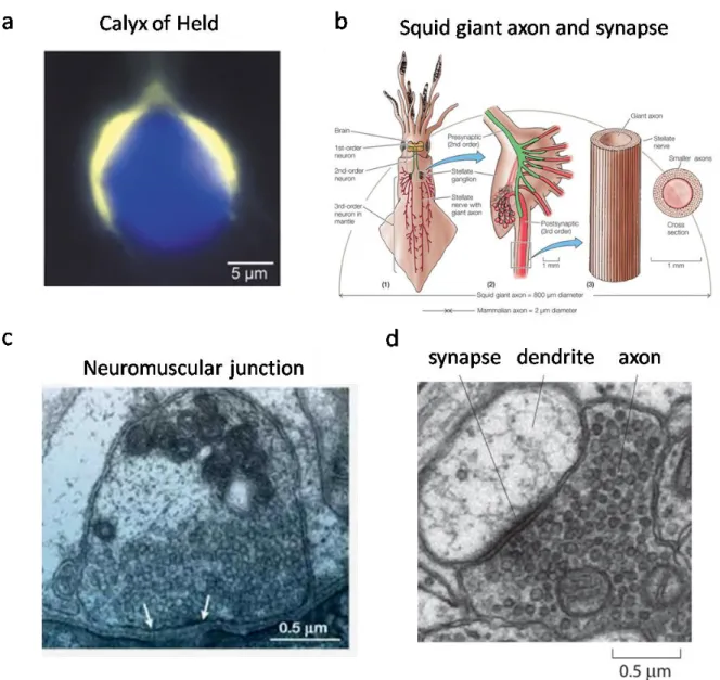
On the other hand, the experimental preparations that have been commonly used for the study of synaptic physiology correspond to "special" types of synapses that do not represent the vast majority of the central synapses. These are: the amphibian neuromuscular junction, the squid giant synapse, and the mammalian Calyx of Held. These synapses have been very useful as models; however, they markedly differ in several important aspects with the great majority of the central synapses; for example, central synapses are usually smaller (up to orders of magnitude) and possess well-defined synaptic contacts that are morphologically and functionally differentiable from each other (Schikorski and Stevens, 1997, Nusser et al., 1997). As mentioned before, one important parameter of the synapse is the size, especially
16
if we consider that a decisive and often limiting aspect in the study of central synapses is the relationship between the amplitude of the response evoked by an AP and the quantal response. Unlike the amphibian neuromuscular junction, the squid giant synapse or the Calyx of Held, where the quantal responses produce only modest fluctuations in the AP-evoked response, in the central synapses the quantal and AP-evoked responses can be similar in magnitude, hence the influence of a single quantum in the AP-evoked response becomes much more significant.
Although the probability of a single quantum to be released (p) does not differ markedly between central synapses and the traditional models of synaptic transmission, these last synapses have a quite distinctive feature that makes them very special: they present a very low probability of failures in the transmission process, thus ensuring that NT is released every time that an AP arrives to the presynaptic terminal. In the amphibian neuromuscular junction and the squid giant synapse, a low failure rate confers to the animal the ability to reliably move away from potential life-threatening dangers, while the Calyx of Held confers the ability to efficiently locate the source of a sound. These exceptional features are a direct consequence of the high number of actives zones (hundreds) in the presynaptic terminal, which allow them to have both a high probability of an AP generating NT release (synaptic reliability), and also a high average postsynaptic response
17
(synaptic efficacy). Contrary to this, a typical central synapse in the brain has a single active zone in the presynaptic terminal and presents a high rate of failures, which translates directly into a low synaptic reliability and efficacy in the process of synaptic transmission. Although this may be seen as a disadvantage, in evolutionary terms this feature plays a fundamental role: it allows the neurotransmitter release process to be modulated within a wide dynamic range, which is extremely important for the phenomenon of synaptic plasticity and in consequence for the processes of memory and learning (Kandel, Schwartz, and Jessell, 2000). A comparison between the sizes of the traditional synaptic models and a typical central synapse is presented in Fig.1-2.
Considering the arguments presented so far, it should be evident that studying synaptic transmission and its mechanisms of modulation, as for example the SSD-mediated facilitation in more representative models of synaptic transmission and at the level of individual synapses is crucial. Nevertheless, it is also clear that the experimental interrogation of a single and small synapse that is immersed in the complexity of brain tissue is an extremely difficult task. It is absolutely necessary to overcome this barrier in order to understand the details of the mechanisms that could be governing the different aspects of synaptic physiology and that may
18
account for the differences that have been observed among different synapses, for example, the differences in Ca2+ dependency during the SSD-mediated facilitation.
19
Figure 1-2. Traditional synapses used as synaptic transmission models and a typical central synapse.
(a) Image of the calyx of Held and its large presynaptic terminal in the mammalian brain stem. The calyx was filled with Lucifer Yellow. The presynaptic terminal and the postsynaptic cell body are presented in pseudocolors, yellow and blue, respectively (adapted from Meinrenken, Borst, and Sakmann 2003). (b) Giant synapses in the squid nervous system. The squid brain possesses “first-order” giant neurons (left) whose activation excites second-order giant neurons (middle) that make connections with the stellate ganglia at the squid mantle. The giant axons of several third-order giant neurons (~1 mm in diameter; right) radiate from the ganglia to the mantle muscles and cause contraction (Animal Physiology, Fourth edition, box extension 12.3 Giant Axons). (c) Electron micrograph of a cross-section through the nerve terminal of the frog neuromuscular junction where the actives zones are indicated by the white arrows (adapted from S. O. Rizzoli and W. J. Betz, 2005). (d) Electron micrograph of a central synapse in the brain (image by Linnaea Ostroff and adapted from Cell Biology by the Numbers by Ron Milo, Rob Phillips).
20
1.5 Approaches for the study of single synaptic contacts in brain slices
Given the extreme difficulty of studying NT release at the level of single synaptic contacts, the problem has usually been solved by the use of cellular cultures. In these preparations the accessibility to the small and well-defined cellular structures makes relatively easy to induce the exocytosis of NT by direct electrical stimulation of the presynaptic terminals, to visualize the release events by performing imaging experiments and to perform recordings of synaptic current from single synaptic contacts (Liu and Tsien, 1995, Ryan et al., 1997, Forti et al., 1997). However, the relevance of these results is uncertain, since it is highly probable that the synaptic function is altered in this type of preparations. A more physiological approach, although more difficult, is to search for pairs of synaptically connected neurons in brain slices, since in some cases a single synaptic contact is established between them (Gulyás et al., 1993, Kondo and Marty, 1998).
One approach that has been extensively used is the technique called “minimal synaptic stimulation of single synapses”, where extracellular electrical stimulation attempts to trigger release from a single presynaptic fiber, for example, in CA3 to CA1 connections in the hippocampus (Stevens and Wang, 1995). However, with this methodology it becomes very difficult to control of the extracellular
21
stimulation well enough to ensure that a single presynaptic fiber is being stimulated.
The exocytosis-inducing effect of the α-latrotoxin has previously been used to study synaptic transmission between cerebellar MLIs at single synaptic contacts (Auger and Marty, 1997). This toxin, when applied in low doses, is able to stochastically activate one synaptic site in periods of about one minute, resulting in a burst of quantal signals arising from a single contact. This method has been used to study the number, the unitary conductance and the opening probability of the postsynaptic channels in GABAergic synapses (Auger et al., 1997) and to study the replenishment of vesicle and the degree of desensitization of AMPA receptors in glutamatergic synapses (Crowley et al., 2007). However, the main disadvantage of this approach is that it does not allow identifying the sites that are activated and is difficult to control the duration and intensity of the presynaptic stimulus.
On the other hand, at the level of individual glutamatergic synapses the so-called "quantal optical analysis" method has been used to study synaptic vesicular release; the method relies in visualizing the Ca2+ rises produced by the opening of
NMDA receptors on the postsynaptic dendritic spines (Yuste et al., 1999; Oertner et al., 2002). This approach has provided convincing information in favor of
22
multivesicular release in synapses where previous results obtained by using the minimal synaptic stimulation approach had been interpreted in favor of the one-site-one-vesicle hypothesis4. These Ca2+ imaging experiments, while providing
relatively high spatial resolution of synaptic events, do not reliably reflect the current through the postsynaptic receptors, and lack the temporal resolution required to analyze in detail the effects of receptor activation (as can be obtained by measuring the synaptic currents).
The ideal approach to study synapses at the level of individual synaptic contacts with high spatial and temporal resolution should be capable of detecting individual synaptic release events.
A method that allows studying single synaptic contacts between cerebellar MLIs without the disadvantages of the previous methodologies was recently developed. It combines the photorelease of Ca2+ from a photosensitive "cage" in the
presynaptic terminal and the simultaneous measurement of postsynaptic currents by dual pre-and postsynaptic whole-cell recordings. This methodology has already been used successfully to estimate the size of the RRP at individual synaptic contacts in the same preparation used in the present study (Trigo et al., 2012).
4. Hypothesis that propose that although a synaptic contact can contain several docked vesicles in the active zones, only one of them fuses with the plasma membrane and releases its content as consequence of the arrival of an action potential to the terminal.
23
2 Thesis project proposal
The discovery of the dual nature of the rapid communication between the soma and the presynaptic terminals (through the generation of APs and by analog-to-digital signaling) has profound implications in our understanding of the mechanisms by which the brain processes information: the analog signaling phenomenon can significantly modify the synaptic output that determines the functioning of cellular circuits, either through an increase in the amount of information transmitted by a particular neuron or by modulating the synaptic activity in groups of neurons subjected to fluctuations of membrane potential, such as those produced by oscillatory activity (Alle and Geiger, 2008).
Although today we know that this type of signaling is capable of modulating NT release in both excitatory and inhibitory synapses, we still have much to discover about the mechanisms by which this modulatory effect is set; for example, it is still unknown whether the Ca2+-dependency of this phenomenon is a particular feature
of some experimental preparations or whether it is a general mechanism in central synapses. On the other hand, we do not know what are the parameters associated with the vesicular cycle that could be modified during SSD-mediated facilitation and if they exhibit or not variations among individual synaptic contacts within the same neuron.
24
In cerebellar MLIs, a SSD-dependent increase in both asynchronous release (during SSD) and AP-evoked release (after SSD) has been reported (Glitsch and Marty, 2009; Trigo et al., 2012; Bouhours, et al., 2011; Christie et al., 2011). It has also been suggested that the strength of the effect on AP-evoked NT release could depend on the availability of fusion-competent vesicles to release their content upon the arrival of an AP (Christie et al., 2011).
The evidence presented so far suggests that the analog signaling phenomenon in MLIs of the cerebellum could act through a mechanism that involves a subthreshold Ca2+-dependent modification of the dynamics of the occupation of the
docking sites; this mechanism may depend on the activation of PKC and the downstream phosphorylation of some of the proteins of the release machinery, like MUNC18-1. This would generate changes in the amount of NT that is released both asynchronously as well as evoked by a suprathreshold increase in Ca2+ levels,
which in turn would generate changes in the reliability and/or efficacy of the synaptic transmission.
In this thesis work, we studied the relationship between the ADF phenomenon and the synaptic parameters accounting for NT release (like peak amplitude, charge and synaptic latency of postsynaptic currents). Additionally, we studied how
25
possible changes on these parameters are associated with the occupancy probability of docking sites, and the size of the RRP at single synaptic contacts of cerebellar MLIs. In order to do this, we used a methodology that combines optical and electrophysiological techniques (see methods) that provides the temporal and spatial resolution required to perform direct measurements of the synaptic activity at the single synaptic level.
3 Hypothesis
The subthreshold Ca2+ rises reached during analog-to-digital facilitation in
cerebellar MLIs can effectively modulate GABA release by a mechanism that involves an increase in the occupancy probability of docking sites, which translates directly into an increase in the RRP size and an enhancement of the synaptic reliability/efficacy in single synaptic contacts.
4 General objective
To explore the effects of the direct manipulation of presynaptic subthreshold Ca2+
levels on the synaptic parameters controling the features of the postsynaptic response, and their possible differences between individual GABAergic MLI-MLI synapses of the rat cerebellum.
26
5 Specific objectives
1. To study the nature of the RRP at the single synaptic level and its possible modulation by subthreshold Ca2+ rises.
2. To directly determine the number of docking sites (NDS), their occupancy probability () and its putative modulation by subthreshold Ca2+ changes.
3. To explore the effects of modulating subthreshold Ca2+ influx on the features
of PSC by the application of presynaptic SSD in pairs of synaptically connected MLIs.
27
6 Methods
6.1 General methods used in this work
In this thesis work we studied quantal release in single synaptic contacts between MLIs of the rat cerebellar cortex. Individual synaptic connections were interrogated by measuring quantal events evoked postsynaptically by presynaptic Ca2+ uncaging with a localized laser spot and after establishing a recording from a
synaptically connected pair of MLIs. Direct control of Ca2+ levels in the sub- and
suprathreshold range was made by varying the laser intensity. The presynaptic cell was recorded with an intracellular solution (IS) containing a high millimolar concentration of the Ca2+ cage DM-nitrophen and the fluorescent dye Alexa 594 to
allow identification. In some experiments, the Ca2+ indicator Oregon Green
BAPTA-1 (OGB-1) was included in the IS to monitor the laser-evoked Ca2+
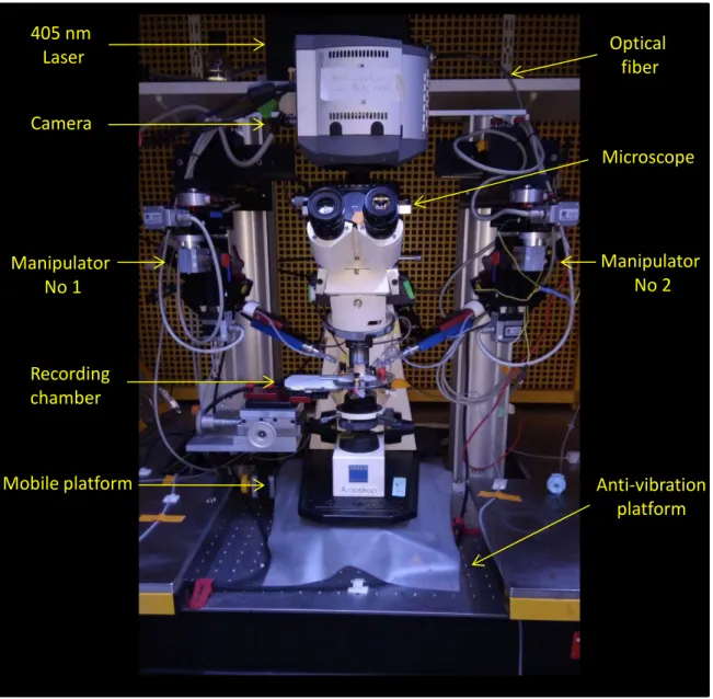
changes. The postsynaptic cell was recorded using a KCl-based IS to obtain large quantal sizes. Alexa 488 was included to distinguish the postsynaptic MLI from the presynaptic MLI. An additional set of experiments were performed using purely electrophysiological interrogation of synaptic contacts in pairs of connected MLIs, where subthreshold somatic depolarizing steps were delivered to modulate synaptic transmission. Figure 6-1 shows the principal components of the setup for simultaneous dual patch-clamp recording and photolysis experiments.
28
Figure 6-1. Main components of the electrophysiological setup for simultaneous dual whole-cell patch-clamp recordings and photolysis experiments.
Camera Manipulator No 1 Manipulator No 2 Microscope
Mobile platform Anti-vibration
platform Recording
chamber 405 nm
29
6.2 Preparation of cerebellar slices
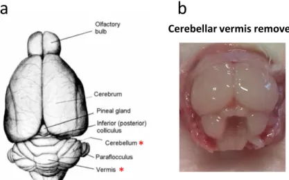
Sagittal cerebellar slices from young (11–17 days old) Sprague–Dawley rats were obtained according to the established procedures used in the Brain Physiology Lab (Llano et al., 1991). Rats were killed by direct decapitation in accordance with institutional guidelines for ethical procedures. The cerebellar vermis was carefully dissected (see Figure 6-2) and placed in ice-cold bicarbonate-buffered solution (BBS; see composition below) that was constantly gassed with a mixture of 5% (vol/vol) CO2 and 95% O2.
The meninges that were still attached to the surface of the vermis were carefully removed by using tweezers under a stereomicroscope in order to facilitate the slicing procedure. 200 μm thick slices were cut with a vibroslicer (VT1200S; Leica) in ice-cold BBS and then placed in an incubating chamber at 34°C for 45 min, to recover from the slicing damage. Thereafter, they were kept at room temperature until use. The slices were transferred to the recording chamber and used during a period of up to 8 h after the decapitation procedure. The composition of the BBS for cutting, storing slices and recording was (in mM): 115 NaCl, 2.5 KCl, 1.3 NaH2PO4, 26 NaHCO3, 25 glucose, 5 Na-Pyruvate, 2 CaCl2, and 1 MgCl2,
with an osmolality of 300 mOsm/kg, and pH 7.4 when gassed with 5% (vol/vol) CO2 and 95 O2.
30
Figure 6-2. Dorsal and caudal view of the rat brain.
(a) Illustration of the dorsal aspect of the brain. Red asterisks
denote the structures relevant for this work: the cerebellum and the cerebellar vermis (https://instruct.uwo.ca/anatomy/530/ratpix.pdf).
(b) Picture showing the caudal view of the head of a rat where the
vermis has been removed.
6.3 Electrophysiology
6.3.1 Electrophysiology for uncaging experiments in pairs of molecular layer interneurons
Molecular layer interneurons were selected based on their location in the molecular layer and the soma size, ranging from 6 to 8 m. MLIs were voltage-clamped with the whole-cell patch-clamp technique using a double EPC-10 USB amplifier (HEKA Elektronik). In these experiments we used a high [Cl-] in the postsynaptic
IS in order to optimize the amplitude of PSCs and readily detect individual fusion events. The composition of the postsynaptic IS was as follows (in mM): 150 KCl, 1 EGTA, 10 Hepes, 0.1 CaCl2, 4.6 MgCl2, 4 Na2ATP, 0.4 NaGTP, and 0.08 Alexa
Cerebellar vermis removed
* *
31
488, with an osmolality of 300 mOsm/kg, and pH 7.3. The composition of the presynaptic IS was as follows (in mM): 95 K-Gluconate, 5.6 KCl, 50 Hepes, 0.5 MgCl2, 4.2 CaCl2, 5 Na2ATP, 0.2 NaGTP, 5.2 KOH, 5
1-(2-nitro-4,5-dimethoxyphenyl)-N,N,N′,N′-tetrakis[(oxycarbonyl)methyl]-1,2-ethanediamine (DM-nitrophen), 0.08 Alexa 594, and 10 GABA, with an osmolality of 300 mOsm/kg, and pH 7.3. GABA was included in the IS to prevent rundown of the PSCs by the emptying of synaptic vesicles, as reported previously by Bouhours et al., 2011. The experiments were performed at room temperature (20–22 °C) and the recording chamber was continuously perfused at a rate of 1–2 ml/min with gassed BBS. Holding potentials were −60 mV for both presynaptic and postsynaptic neurons and the reported values for Vm were corrected for the calculated liquid junction potential of 13.8 mV and 1.4 mV, giving final Vm values of -73.8 mV and -61.4 mV for the presynaptic and postsynaptic cells, respectively. Patch electrodes (micropipettes) were pulled from borosilicate glass to a tip resistance of ~ 4.5 MΩ when filled with the postsynaptic IS. Series resistance was in the range of 10–35 MΩ and was not compensated. Cells were discarded when the series resistance value was larger than 35 MΩ or if it varied more than 20%. Synaptic connectivity was tested by delivering short depolarizing voltage steps of 1 ms to 0 mV to the presynaptic cell. Immediately after the connectivity test was
32
successful (as ascertained by the presence of postsynaptic currents), TTX (0.2 µM final concentration) was included in the bath solution throughout the entire experiment in order to minimize contamination from AP dependent neurotransmitter release. Recordings from both pre- and postsynaptic cells were acquired at a sampling rate of 50 kHz and low-pass filtered at 2.9 KHz. Most data were obtained from cells located in the proximal 1/3 part of the molecular layer (basket cells). However, interneurons located in the outer 2/3 of the molecular layer (stellate cells) were also included.
6.3.2 Pure electrophysiological recordings in pairs of molecular layer interneurons
Molecular layer interneurons were selected following morphological criteria and voltage-clamped with the whole-cell patch-clamp technique. The composition of the IS for the pre and postsynaptic cells was as follows (in mM): 145 KCl, 10 GABA, 0.02 EGTA, 10 HEPES, 0.1 CaCl2, 4.6 MgCl2, 4 Na2ATP, 0.4 NaGTP,
with an osmolality of 300 mOsm/kg, and pH of 7.3. Synaptic connectivity between the two cells was tested by delivering depolarizing voltage steps of 1 ms to 0 mV to each of them (they were dialyzed with the same IS, thus connectivity could be detected in both directions). Holding potential was set at −75 mV for both presynaptic and postsynaptic MLIs. The subthreshold somatic depolarization
33
protocol used in these experiments consisted of a 2 s pulse from -75 mV of holding potential (Vm= -76.4 mV after correcting by the junction potential) to a value just below the spike threshold (usually -50 mV, corresponding to -51.4 of Vm). The spike-evoked PSC accounting for the properties of presynaptic GABA release was evoked by applying a short depolarizing step of 1 ms to 0 or 10 mV to the presynaptic cell. Series resistance was in the range 10–35 MΩ and was 50 % compensated. Cells were discarded when the series resistance value was larger than 35 MΩ or if it varied more than 20%. Recordings in both pre and postsynaptic cells were acquired at a sampling rate of 50 kHz and low-pass filtered at 2.9 KHz.
6.4 Fluorescence imaging and synaptic contact identification
The localization of putative individual synaptic contacts was made by alternating the excitation of the fluorescent dyes Alexa 594 and Alexa 488 that were included in the presynaptic and postsynaptic IS, respectively (see composition above). In the juvenile MLIs used in this work, the presynaptic axon and the postsynaptic somatodendritic compartment are readily distinguished by their characteristic ramification pattern and neurite thickness. The functional identification of a putative individual synaptic contact was made by photoreleasing Ca2+ from the
photosensitive Ca2+chelator DM-nitrophen (Kaplan and Ellis-Davies, 1988; see
34
obtained from a light emitting diode (LED) system (Optoled; Cairn Research) equipped with 2 LEDs resulting in outputs at 470/40 nm and 572/35 nm wavelength by using excitation filters (Chroma Technology). The emitted fluorescence was collected after emission filters of 520/40 nm and 630/60 nm (Chroma Technology) and directed to an EM CCD camera (Ixon, 512 × 512 pixels; Andor Technology). Reconstruction of the morphology was made by taking single full frames pictures with excitation of Alexa 488 or Alexa 594 and varying the focal depth. ImageJ (National Institutes of Health; https://imagej.nih.gov/ij/) was used to make the stacks (at 1μm increments) and the composite images. Figure 6-3 shows and example for the reconstruction of a pair of connected MLIs and the localization of the interrogated synaptic contact.
35
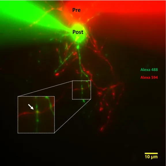
Figure 6-3. Fluorescence image and synaptic site localization of a synaptically connected pair of MLIs.
The image corresponds to the composite of the individual z-projections of the stacks taken at 594 nm and 488 nm of excitation. Cells are presented with pseudocolors: presynaptic in red, postsynaptic in green. The arrow indicates the site chosen for uncaging.
Pre
Post
Alexa 488
36
6.5 Photolysis of DM-Nitrophen
Photolysis of DM-nitrophen (Kaplan and Ellis-Davies, 1988) was implemented with a 405 nm wavelength diode laser (DeepStar 405; Omicron). The light was focused through a 63× Zeiss objective as a ~5 µm diameter spot in the focal plane of the objective and viewed with an electron multiplying charge-coupled device (EM CCD) camera at 0.25 μm per pixel (Ixon, 512 × 512 pixels; Andor Technology). The position and the approximate size of the laser spot was checked every experimental day by looking at the fluorescence of HPTS (8-Hydroxypyrene-1,3,6-Trisulfonic Acid; 100 µM), which is excited by the 405 nm laser light. Upon photolysis, the affinity of DM-nitrophen for Ca2+ decreases
around 6,000,000-fold and its Kd increases from 5 nM to 3 mM, resulting in a pulse
of free Ca2+. Lyophilized DM-nitrophen (Synaptic Systems) was dissolved in KOH
solution in a 1:4 DM-nitrophen/K+ proportion (50 mM of final concentration) and
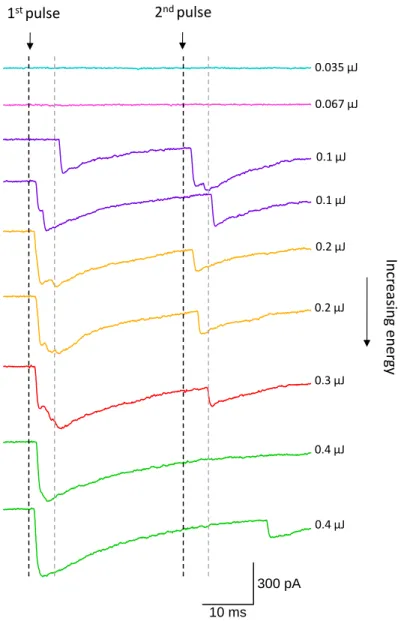
stored at -20°C. The potassium salt was then added to the IS the same day of the experiment. For the control protocols of uncaging used in this work, a single laser pulse that releases the RRP was applied in single varicosities. For the test protocol, the same RRP-releasing pulse was preceded by a train of five low intensity pulses at 10 Hz and separated by 200 ms or 2 s from the RRP-releasing pulse. The ranges of energies for the subthreshold train and the suprathreshold pulse was
0.0085-37
0.098 µJ and 0.1–0.4 µJ, respectively. These energies were set during individual experiments and varied for each synapse. The energies for the train of subthreshold laser pulses was set to reach the maximal level that does not produce a postsynaptic response within the first 5 ms after triggering the laser. The RRP-releasing nature of the suprathreshold pulse was confirmed by the lack of response to a second pulse of the same energy that was delivered 30 ms later (Figure 6-4). During photolysis of DM-nitrophen, two net OH- ions are formed for each Ca2+ ion
released. In order to minimize the alkalinization on photolysis, 50 mM Hepes was included in the IS (see composition above). At this concentration of Hepes a change of ~ 0.37 pH units is still expected. Nevertheless, in similar experimental conditions, this pH change had no effect on synaptic transmission (Trigo et al., 2012).
The number of repetitions that can be performed under these experimental conditions is limited. The minimum and maximal number of repetitions obtained in this work was 10 and 46, respectively. This restriction is not a consequence of photodamage produced by the 405 nm laser because it does not happen when other cage compounds like glutamate or GABA are photolised in the extracellular milieu (Trigo et al., 2009b) using the exact same technique. It may be caused by oxidizing
38
byproducts of photolysis (nitrosoacetophenone; Kaplan and Ellis-Davies, 1988), but the exact mechanism of this phenomenon remains to be explored.
Figure 6-4. RRP release by Ca2+ uncaging from the cage compound DM-nitrophen (DMNP).
The traces correspond to a group of PSCs evoked by the photorelease of Ca2+ at different laser
energies. Laser timing is indicated by the black dashed lines. First and second pulses were separated by 30 ms; the grey dashed lines correspond to the end of the 5 ms interval after triggering the laser. Any postsynaptic event that has a first latency within this range was considered as part of the RRP. From the figure it is possible to observe that there was no second response for laser energies of 0.4 µJ (last 2 traces in green), thus we concluded that at this energy the RRP was fully released by the first laser pulse.
10 ms 300 pA In cr easin g en er gy 1st pulse 2nd pulse 0.035 µJ 0.067 µJ 0.1 µJ 0.1 µJ 0.2 µJ 0.2 µJ 0.3 µJ 0.4 µJ 0.4 µJ
39
6.6 Calcium imaging
For Ca2+ imaging experiments, 200 µM of the high-affinity indicator (K
d=170 nM)
Oregon Green BAPTA-1 (OGB-1) (Invitrogen) was added to the presynaptic IS (see composition above) to monitor changes of Ca2+ upon photolysis of DMNP.
The axonal compartment of the recorded cell was identified by using Alexa 594 and the selected varicosity for uncaging was imaged after letting the dye diffuse for at least 10 min after break-in. A subregion of the CCD comprising sixteen 4 × 4 binned pixels, giving a resolution of 1 μm, was imaged in 2.11 ms exposure at 259.7 Hz. Fluorescence was corrected for background, determined in pixels at the periphery of the subregion. The traces shown in this work are averaged responses from individual varicosities in which averaging was done from at least 5 repetitions. The experiments were performed in the same experimental set-up as detailed for the uncaging work (see above). Imaging data was analyzed with IGOR Pro (Wavemetrics) using a routine written in the laboratory by Brandon M. Stell and Jorge Ramirez. The fluorescence signals are reported as F/F0 corresponding
to changes in background subtracted fluorescence with respect to the values before stimulation (F: F- F0), normalized to the pre-stimulus values (F0). For the analysis
of the kinetics of the axonal Ca2+ signal, exponential functions were fitted to the
40
6.7 Determination of the readily releasable pool at a single synaptic contacts
The onset kinetics of all the detected events was analyzed for a time window of 100 ms after the laser pulse and histograms of synaptic latency were constructed. Single exponential fit of the histograms were used to determine the event distribution and to set a criteria to estimate whether a fusion event belonged to the RRP. A time corresponding to 4 time constant (4 =10 ms) in the histogram of all events (in the 100 ms time window), at which 98% of the events have occurred, was defined to associate an event with the laser pulse. Considering this limit, normal and cumulative histograms of the first latencies were constructed to determine the superior limit to associate a first event with the laser pulse. A time limit corresponding to t = 5 ms (at which almost 100 % of the first events have occurred) was defined as the superior limit to associate a first event with the laser pulse. Hence, in this work any event (or series of events) was considered as part of the RRP if it had latency values up to 10 ms and first latency values were below than or equal to 5 ms. Peak amplitude, synaptic charge (from 0-150 ms), rate of failures, first and all latencies and time to peak values were calculated for all the PSCs responses associated with the release of the RRP. These synaptic parameters were calculated for each of the individual and interleaved PSCs in control and test