HAL Id: tel-02180569
https://tel.archives-ouvertes.fr/tel-02180569
Submitted on 11 Jul 2019HAL is a multi-disciplinary open access
archive for the deposit and dissemination of sci-entific research documents, whether they are pub-lished or not. The documents may come from teaching and research institutions in France or abroad, or from public or private research centers.
L’archive ouverte pluridisciplinaire HAL, est destinée au dépôt et à la diffusion de documents scientifiques de niveau recherche, publiés ou non, émanant des établissements d’enseignement et de recherche français ou étrangers, des laboratoires publics ou privés.
Characterization of monogenic enteropathies
Fabienne Charbit-Henrion
To cite this version:
Fabienne Charbit-Henrion. Characterization of monogenic enteropathies. Immunology. Université Sorbonne Paris Cité, 2016. English. �NNT : 2016USPCB059�. �tel-02180569�
ACKNOWLEDGMENTS
I had the opportunity of doing a wonderful and exciting 3-year PhD internship. Since the beginning of this project with my Master internship in January 2012, I had the honor of working with brilliant scientists and clinicians. My thesis would never have been possible without the support, the help and the emulation that I received from so many people, within the lab, within the Institut and through the GENIUS European network. To all of you, please accept my sincere thanks.
To Frank,
Since the beginning, you trusted me with the new “Early-onset IBD” axis that you have been creating within the lab. You let me be at the front line, while fixing my mistakes each time it was needed! Thank you for the trust you put in me.
To Nadine,
Your honesty and integrity, both on a scientific and human scale, as well as your immeasurable knowledge, are humbling. You are a role model that will be hard to follow. Thank you for your availability, your guidance and your encouragements.
To the members of the Jury,
I would like to thank Pr Janneke Samsom and Pr Harry Sokol for accepting to review my thesis manuscript. I also thank Pr Aleixo Muise for accepting to judge my work, and Pr Stanislas Lyonnet for accepting to preside the jury. It is an honor, and a great source of anxiety (!), to defend my work in front of all of you.
To all the lab,
You welcomed me with open arms almost five years ago. I admire your passion and dedication to your research, which do not keep you from being fun and caring! It has been an honor to join your ranks.
To Bernadette, you taught me everything at the bench. I think that, together, we have helped building this project, slowly but steadily, and with strong bases. It has been a real pleasure working close to you. I will miss our chocolate breaks!
To Marianna, you joined our “VEO-IBD team” two years ago. Since then, we have made great progress, thanks to your scientific rigor and your skills. I learned a lot by your side.
To past and present members of the “VEO-IBD team”, Anaïs, Jan, Elie, Sabine, Rémi, thank you for your enthousiasm!
To Bénédicte, I am excited to work side by side with you in the clinics! I admire your commitment. But most of all, I love your energy and sense of humor! Thank you for your daily coaching! My special thanks to all members of Nadine’lab: to Valérie and Bertrand for your kind support, to Julien, Nicolas and Marion for so many jokes and laughs, to Natalia for your advice, to Aurélie and Pamela for sharing teapots, to Benoit for your skills in Western Blot, to Rute for our exciting conversations, to Corinne for showing microscopical wonders! I wish the best of luck to new PhD students: Rémi, Iris, and Roman.
To our closest collaborators,
To Frédéric, Eva, Marie-Claude, Fabienne, and Aude from the “Auto-immunity team”, and to Sylvain and Christelle from the “XIAP team”, thank you for so many interesting discussions. To Nicolas Garcelon,
Without you, the GENIUS and IMMUNOBIOTA databases would only exist in my mind! You spent so much time trying to “translate” my ideas in computer-language! Thank you for your friendship and patience.
To Vincent Benoit,
Thanks to you, we have now a wonderful website! To Imagine’s “ethical team”,
To Pauline, thank you for your amazing organization and energy! To Elisabeth, Pauline, and Nicholas, thank you for all the rigorous work you did, which was extremely helpful.
To Sylvain H,
Our targeted panel would never have existed without you. I learned a lot in genetics and NGS analysis while designing the panel with you. You are a great teacher! Thank you for your help, your commitment and your availability!
Last, but not least, to my family,
I already said and wrote how much you all mean to me. But I feel that even if I were to write it every day, it would never be enough.
To my parents,
You’ve filled my childhood with so much love and happiness. If I am happy today, both professionally and personally, it is thanks to you. I hope to make you proud. I love you, “comme des milliers de soleils éclatés…”
To my brother,
You are one of the person that I admire the most. Your passion, your integrity, and your creativity amaze me. I wish you all the happiness in the world.
To my parents in-law,
Thank you for welcoming me with open arms, for making me improve my English, but most of all, thank you for raising your son to be a Mensch.
To my wonderful Princess and my little Prince Charming,
Comes the sun, the sparks of happiness and cheekiness in your eyes are my daily achievements. Comes the night, your smiles and kisses are my greatest reward. My biggest purpose is to love you and cherish you both.
To my husband,
You are my husband, my lover, my best friend, my everything. Thank you for your faith in me, and for supporting my endless years of study! In your eyes, I feel strong and beautiful. There is no word to describe how much you mean to me, but I will try to show it to you every day. I love you.
SUMMARY
Background: Mendelian mutations causing monogenic enteropathies are identified in an increasing
number of genes and are responsible for either chronic inflammatory diseases (frequently called VEO-IBD for very early-onset inflammatory bowel diseases) or for congenital diarrheal disorders (CDD). Management of many patients with monogenic enteropathies requires difficult therapeutic decisions and heavy treatments, such as hematopoietic stem cell transplantation for VEO-IBD patients, or total parenteral nutrition and intestinal transplantation for CDD patients. Early molecular diagnosis is crucial to define the most pertinent treatment and increase life expectancy. During my thesis, I introduced in the laboratory big data management tool (e. g. online dedicated database) and applied next-generation sequencing tools (whole exome sequencing (WES) and targeted gene panel sequencing (TGPS)) to a cohort of patients suffering from monogenic enteropathies in order to characterize them phenotypically and genetically.
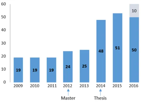
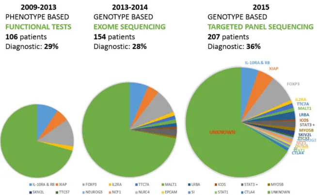
Methods: My thesis was divided in 4 steps. In Step 1, patients (n=216 in January 2016, n=260 in
August 2016) recruited through a French research protocol (Immunobiota, 12 centers) and European network (GENIUS, 33 centers) were phenotypically characterized through an online dedicated database. Following precise phenotyping, molecular diagnoses were obtained by Sanger sequencing of candidate genes suggested by functional tests in Step 2. Step 3 was the adaptation of WES for our cohort of patients (59 patients were sequenced in trio and 11 sequenced by themselves or in duo) and lastly, in Step 4, TGSP was designed and applied to our cohort (173 patients without a molecular diagnosis).
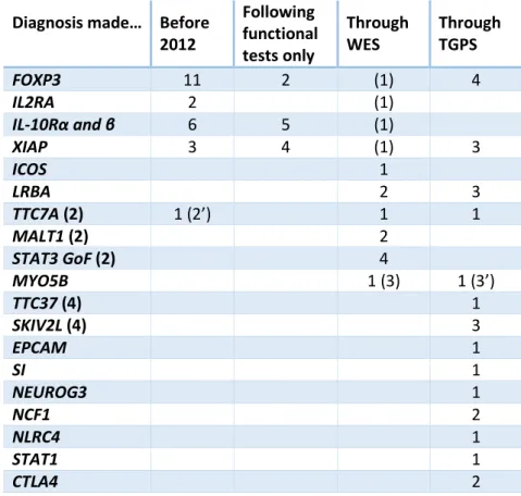
Findings: The cohort gathered 57 patients including 22 with a molecular diagnosis in January 2012, and 216 patients including 70 with a diagnosis in January 2016, corresponding to a global diagnosis rate of 1/3. Approximately 50 new patients are recruited each year, with blood samples taken from each patient, both parents and siblings. During this period, 11 diagnoses were obtained by a phenotype-based approach, with identification of mutations notably in IL-10R (4 patients) and XIAP (4 patients). Eleven patients obtained a genetic diagnosis by WES including two siblings with a MALT1 deficiency responsible for an IPEX-like syndrome. Because of the increasing number of genes involved in monogenic enteropathies, we developed, in collaboration with Genomics, Bioinformatics and Translational Genetics platforms from the Institut IMAGINE, a custom-made TGPS gathering 68 genes responsible for either VEO-IBD or CDD. The sequencing of all negative patients (n=173) on this panel allowed to identify 28 new diagnoses (among which 8 were made in patients included before 2012).
Interpretation: This work lead to the identification of the genetic diagnosis in 1/3 patients. The
close investigations of phenotype-genotype correlations highlighted frequent overlaps among monogenic enteropathies. Following completion of this work, we suggest to use TGPS as a first-line genetic test in addition to a precise phenotyping of the patient. Depending on the results, TGPS will either reach an early molecular diagnosis crucial to optimize treatments in a cost-effective manner, or allow to perform further genetic analysis notably by WES.
Table of Contents
ACKNOWLEDGMENTS ... 3 SUMMARY ... 7 PUBLICATIONS LIST ... 11 ABBREVIATIONS ... 13 ... 17 PREAMBULE ... 19I. DEFECTS OF THE EPITHELIAL BARRIER ... 21
I.A-The role of epithelium in the intestinal barrier ... 21
I.A.1. Barrier role of mucus ... 23
I.A.2- Role of endoplasmic reticulum stress in epithelial barrier function ... 25
I.A.3- ATG16L1 and the role of autophagy in epithelial cells ... 27
I.A.4- Monogenic enteropathies with predominant epithelial defect. 29 I.A.4.1- Microvillus inclusion disease (MVID) ... 29
I.A.4.2- Epithelial dysplasia (ED) ... 31
I.B- Defective innate immunity at the intestinal barrier ... 34
I.B.1- Defects in pattern recognition receptors (PRR) ... 34
I.B.1.1- MyD88 ... 34
I.B.1.2- NEMO/IκK ... 37
I.B.1.3- NOD2... 41
I.B.2- XIAP ... 42
I.B.3- Defects in production of Reactive Oxygen Species ... 45
I.C- Defects in the effector functions of the adaptive immune system ... 51
II. DEFECTS IN REGULATION OF THE INTESTINAL BARRIER ... 52
II.A- Defects in intrinsic regulation of signaling pathways ... 52
II.A.1- Hyperinflammation due to over production of IL-1β ... 52
II.A.2- Intestinal inflammation induced by STAT3 gain of function mutations ... 54
II.A.3- Inflammation due to loss of NF-κB regulation in A20 deficiency ... 55
II.B- Defects in extrinsic immunoregulatory mechanisms ... 59
II.B.1- Defects in IL-10 signaling pathway ... 59 II.B.2- Defects in generation and/or functions of regulatory T-cells . 63 II.B.2.1- Lessons from FOXP3 deficiency in mice and humans 63
II.B.2.2- IL2RA or CD25 deficiency ... 71
II.B.2.3- LRBA and CTLA4 ... 72
III. OVERLAPPING SYNDROMES: LESSONS FROM MUTATIONS IN TTC7A AND TRICHO-HEPATO-ENTERIC SYNDROME ... 75
III.A- TTC7A ... 75
III.B- THES and POLA1: implications of RNA/DNA metabolism ... 76
... 83
I. PHENOTYPIC CHARACTERIZATION OF PATIENTS WITHIN THE COHORT ... 86
I.A- Description of the IMMUNOBIOTA Cohort ... 87
I.B- Molecular characterization of the cohort ... 89
II. FIRST IDENTIFICATION OF A MUTATION WITH A FOUNDER EFFECT IN INTERLEUKIN-10 RECEPTOR 2 GENE ... 91
III. DEFICIENCY IN MUCOSA ASSOCIATED LYMPHOID TISSUE LYMPHOMA TRANSLOCATION 1 (MALT1): A NOVEL CAUSE OF IPEX-LIKE SYNDROME ... 105
IV. TARGETED NEXT-GENERATION SEQUENCING PANEL IN MONOGENIC ENTEROPATHIES: AN EFFECTIVE FIRST-LINE GENETIC TEST ... 117
... 147
... 159
... 163
Identification of two novel mutations gain-of-function of STAT3 responsible for severe enteropathies ... 165
Refractory monogenic Crohn’s disease due to X-linked inhibitor of apoptosis deficiency ... 171
Atypical Manifestation of LPS-Responsive beige- like anchor (LRBA) Deficiency Syndrome as an Autoimmune Endocrine Disorder without Enteropathy and Immunodeficiency. ... 173
A New Case of Enteric Anendocrinosis: An Extremely Rare Cause of Congenital Malabsorptive Diarrhea and Diabetes Secondary to Mutations in Neurogenin-3 ... 181
Table 6. Summary of IL-10 and IL-10R defective patients. ... 199
Table 7. Summary of FOXP3-mutated patients. ... 205
PUBLICATIONS LIST
Main publications:
Charbit-Henrion, F., Jeverica, A.K., Begue, B., Markelj, G., Parlato, M., Avcin,
S.L., Callebaut, I., Bras, M., Parisot, M., Jazbec, J., et al. (2016). Deficiency in
Mucosa Associated Lymphoid Tissue Lymphoma Translocation 1 (MALT1): A
Novel Cause of Ipex-Like Syndrome. J Pediatr Gastroenterol Nutr. Epub ahead of
Fabienne Charbit-Henrion, Bernadette Bègue, Anaïs Sierra, Nicolas Garcelon,
Frédéric Rieux-Laucat, Marie-Claude Stolzenberg, Bénédicte Neven, Isabelle Loge,
Capucine Picard, Sandra Pellegrini, Zhi Li, GENIUS Group, Jorge Amil Dias,
Nadine Cerf-Bensussan*, Frank M. Ruemmele*, First identification of a mutation
with a founder effect in interleukin-10 receptor 2 gene, in preparation
Fabienne Charbit-Henrion, Mariana Parlato, Sylvain Hanein, Bernadette Begue,
Rémi Duclaux-Loras, Sabine Rakotobe, Jan Nowak, Julie Bruneau, Cécile
Fourrages, Olivier Alibeu, Frédéric Rieux-Laucat, Eva Lévy, Marie-Claude
Stolzenberg, Fabienne Mazerolles, Sylvain Latour, Christelle Lenoir, Alain Fischer,
Capucine Picard, GENIUS Group*, Marina Aloi*, Jorge Amil Dias*, Mongi Ben
Hariz*, Anne Bourrier*, Christian Breuer*, Anne Breton*, Jiri Bronski*, Stephan
Buderus*, Mara Cananzi*, Stéphanie Coopman*, Clara Crémilleux*, Alain
Dabadie*, Clémentine Dumant-Forest*, Odul Egritas Gurkan*, Alexandre Fabre*,
Aude Fischer*, Marta German Diaz*, Yago Gonzalez-Lama*, Olivier Goulet*,
Graziella Guariso*, Neslihan Gurcan*, Matjaz Homan*, Jean-Pierre Hugot*, Eric
Jeziorski*, Evi Karanika*, Alain Lachaux*, Peter Lewindon*, Rosa Lima*,
Fernando Magro*, Janos Major*, Georgia Malamut*, Emmanuel Mas*, Istvan
Mattyus*, Luisa Mearin*, Jan Melek*, Victor Manuel Navas-Lopez*, Anders
Paerregaard*, Cecile Pelatan*, Bénédicte Pigneur*, Isabel Pinto Pais*, Julie
Rebeuh*, Claudio Romano*, Nadia Siala*, Caterina Strisciuglio*, Michela
Tempia*, Patrick Tounian*, Dan Turner*, Vaidotas Urbonas*, Stéphanie Willot*,
Frank Ruemmele, Nadine Cerf-Bensussan ; Targeted next-generation sequencing
panel in monogenic enteropathies: an effective first-line genetic test, submitted
Publications in collaboration:
Coelho, R., Peixoto, A., Amil-Dias, J., Trindade, E., Campos, M., Magina, S.,
Charbit-Henrion F., Lenoir, C., Latour, S., Magro, F., et al. (2016). Refractory
monogenic Crohn's disease due to X-linked inhibitor of apoptosis deficiency. Int J
Colorectal Dis 31, 1235-1236.
Shahrzad Bakhtiar, Eva Levy, Frank Ruemmele, Fabienne Charbit-Henrion,
Frédéric Rieux-Laucat, Nadine Cerf-Bensussan, Peter Bader, Ulrich Paetow,
Atypical Manifestation of LPS-Responsive beige- like anchor (LRBA) Deficiency
Syndrome as an Autoimmune Endocrine Disorder without Enteropathy and
Immunodeficiency, Front Immunol. Epub ahead of print
Germán-Díaz, Marta; Cruz-Rojo, Jaime; Rodriguez-Gil, Yolanda;
Charbit-Henrion, Fabienne; Cerf-Bensussan, Nadine; Manzanares-López Manzanares,
Javier; Moreno-Villares, José; A New Case of Enteric Anendocrinosis: An
Extremely Rare Cause of Congenital Malabsorptive Diarrhea and Diabetes
Secondary to Mutations in Neurogenin-3, in revision
ABBREVIATIONS
AIE Autoimmune enteropathy
AMP Anti-microbial peptide
ATG16L1 Autophagy related 16-like 1
ATG5 Autophagy related 5
BCL B-cell lymphomas
BIM BCL-2-interacting mediator
BM Bone marrow
CARD11 Caspase recruitment domain-containing protein 1 CBF-β Core-binding factor subunit beta
CCR4 C-C chemokine receptor type 4
CD Crohn's disease
CDD Congenital diarrheal disorders CGD Chronic granulomatous diseases
CID Combined Immunodeficiency
CNS2 Conserved noncoding sequence 2 CTE Congenital tufting enteropathy CTLA4 Cytotoxic T lymphocyte antigen-4 CVID Common variable immunodeficiency
DHR Dihydrorhodamine
DSS Dextran sulfate sodium
ED Epithelial dysplasia
EDA-ID Anhidrotic ectodermal dysplasia with immunodeficiency EDA-R Ectodysplasin receptor
EPCAM Epithelial cell adhesion molecule
ER Endoplasmic Reticulum
ESPGHAN European Society of Pediatric Gastroenterology, Hepatology, And Nutrition FFAR2 Free fatty acid receptor 2
FOXP3 Forkhead box P3
GALT Gut-associated lymphoid tissue
GENIUS GENetically and/or ImmUne mediated enteropathieS GFP Green fluorescent protein
GGPP Geranylgeranyl pyrophosphate
GI Gastro-intestinal
GoF Gain of function
HIDS Hyperimmunoglobulinemia D and periodic fever syndrome
HIES Hyper IgE syndrome
HLA Human leukocyte antigen
HLH Hemophagocytic lymphohistiocytosis HMG-CoA Hydroxy-méthyl-glutaryl-coenzyme A HSCT Hematopoietic stem cell transplantation IBD Intestinal bowel disease
IEC Intestinal epithelial cells
IFN Interferon gamma
IgA Immunoglobulin A
IL-10 Interleukin 10
IMBT Immunobiota
iNKT Invariant natural killer T cells
IPEX Immune dysregulation, polyendocrinopathy, enteropathy, X-linked syndrome
IQR Interquartile range
IRAK-4 IL-1R-associated kinase 4 IRF4 IFN regulatory factor 4
IRGM GTPase family M
iTreg Induced Treg
IκBα Inhibitor of kappa B, alfa
IκK I-kappa B kinase
JAK1 Janus kinase 1
KLRG1 Killer cell lectin-like receptor subfamily G member 1-positive
KO Knock-out
LAMP2 Lysosomal-associated membrane protein LC3 Microtubule-associated protein 1 light chain 3
LoF Loss of function
LRBA Lipopolysaccharide responsive beige-like anchor protein
LRR Leucine-rich repeats
LUBAC Linear ubiquitin chain assembly complex MAIT Mucosal-associated invariant T-cells
MALT1 Mucosa-associated lymphoid tissue lymphoma translocation protein 1 MAP Mitogen activated proteins
MCL1 Myeloid leukemia cell differentiation 1 MCP-1 Monocyte chemoattractant protein 1
MDP Muramyl-dipeptide
meso-DAP Meso-L-Ala-y-D-Glu, meso-diaminopimelic acid MHC Major histocompatibility complex
MIA Multiple intestinal atresia
miRNA Micro RNA
MKD Mevalonate kinase deficiency
MLN Mesenteric lymph nodes
MUC2 Mucin 2
MVID Microvillus inclusion disease
MyD88 Myeloid differentiation primary response gene 88
MYO5B Myosin 5B
NADPH Nicotinamide adenine dinucleotide phosphate NFAT Nuclear factor of activated T-cells
NF-κB Nuclear factor-kappa B
NLR NOD-like receptor
NLRC4 NLR family-Card domain containing 4
NOD2 Nucleotide-binding oligomerization domain 2
NO Nitric oxide
NPC1 Niemann-Pick disease type C1
OL-EDA-ID Osteopetrosis and lymphoedema associated to EDA-ID
OR Odd ratios
OTU Ovarian tumor domain
PAMP Pathogen-associated molecular pattern
PAS Periodic acid Schiff
PI3K Phosphatidylinositol 3 kinase PI4KIIIα Phosphatidylinositol 4-kinase III alfa PID Primary immunodeficiencies
POLA1 DNA polymerase alfa 1
Poly(IC) Polyinosinic-polycytidylic acid PRR Pattern recognition receptors
Rac1 Ras-related C3 botulinum toxin substrate 1 RAG1 Recombination activating gene 1
RhoA Ras homolog gene family, member A RIG-1 Retinoid acid-inducible gene-1
RIP1 Receptor-interacting serine/threonine-protein kinase 1 RIPK2 Receptor-interacting serine/threonine-protein kinase 2 ROR RAR-related orphan receptor gamma
ROS Reactive oxygen species
RUNX1 Runt-related transcription factor 1 SCFA Short-chain fatty acids
SCID Severe combined immunodeficiencies
SH3 SRC Homology 3
siRNA Small interfering RNA SLE Systemic lupus erythematosus SOCS1 Suppressor of cytokine signaling 1
SPF Specific-pathogen free
SPINT2 Serine Peptidase Inhibitor, Kunitz Type, 2 STAT1 Signal transducer and activator of transcription 1 STING Stimulator of type 1 IFN gene
STX3 Syntaxin 3
STXBP2 Syntaxin 3 binding protein
TAK1 Transforming growth factor beta-activated kinase 1
TBK1 TANK Binding Kinase 1
TCR T-cell receptor
TGF-β Transforming growth factor beta TGPS Targeted gene panel sequencing
Th1 T helper type 1
THES Trichohepatoenteric syndrome
TLR Toll-like receptor
TNBS 2,4,6-trinitro benzene sulfonic acid TNFα Tumor necrosis factor alfa
TRAF2 TNF receptor-associated factor 2
Treg Regulatory T cells
TTC7A Tetratricopeptide repeat domain 7A
UC Ulcerative colitis
UPR Unfolded protein response
VEO-IBD Very early onset inflammatory bowel disease
WES Whole exome sequencing
WGS Whole genome sequencing
WT Wild type
XBP1 X-box-binding protein 1
XIAP X-linked apoptosis inducing protein XLP-2 X-lymphoproliferative disease
XLPDR X-linked reticulate pigmentary disorder
PREAMBULE
Intestinal barrier: of mice and men
With a surface of approximately 300-400m², the intestinal mucosa is the largest interface of the human body. It is made of a single layer of epithelial cells supported by a layer of connective tissue called lamina propria, and is strongly interconnected with a considerable number of immune cells of hematopoietic origin assembled in the gut associated lymphoid system. Altogether they form a highly regulated barrier able to cope with several challenges. On one hand, the primary function of the intestinal epithelium is the digestion and absorption of nutrients, water and electrolytes. On the other hand, the intestinal barrier must restrict body access to undigested food antigens and to the considerable and complex community of microbes, which settle after birth in the intestinal lumen, where they find the metabolic resources to thrive. Microbial density increases along the gastrointestinal tract and reaches impressive concentrations of 1011 to 10 12 bacteria and archaea per
gram of luminal content in the distal gut, which can be seen as the ecological site on Earth the most densely populated by microbes (Cerf-Bensussan and Gaboriau-Routhiau, 2010). As a whole, the intestinal microbiota may contain over 100 fold more genes than the human genome and encode a broad spectrum of enzymatic activities, which can considerably enlarge host metabolic capacities (Gill et al., 2006). Hosts and their microbiota are thus thought to have evolved mutualistic relationships that are largely based on metabolic and energy exchanges. Yet this huge mass of microbes at the intestinal surface must be kept in check. Hosts have evolved multiple mechanisms, which cooperate within a highly dynamic network of interactions implicating both intestinal epithelial cells and immune cells to preserve the symbiotic relationship with the intestinal microbiota, while retaining the ability to recognize and react swiftly against pathogens.
The intricate relationship between hosts and intestinal microbes has been extensively analyzed over the past 20 years in animal models and through physiopathogenic studies of human inflammatory bowel diseases (IBD). It is now admitted that, in polygenic IBD such as Crohn’s disease (CD) or ulcerative colitis (UC), inflammation results from abnormal host immune responses to the microbiota. Genome wide association studies have pinpointed over 200 genetic polymorphisms, which may affect the function of host pathways that are important to build and regulate the gut immune barrier (Liu et al., 2015). Yet, altogether these polymorphisms account for less than 14% of the risk to develop IBD, and it is increasingly clear that environmental factors play a central role in jeopardizing host-microbiota interactions (McGovern et al., 2015). Therefore, the exact contribution of genetic variants to inflammation remains difficult to delineate. However, a growing
number of rare Mendelian diseases have and are being identified in children as a cause of severe intestinal inflammation of very early onset (VEO-IBD). Analysis of the expanding number of genes, which can be affected by mutations causing intestinal inflammation thus provides precious information on host pathways indispensable to build and regulate the gut barrier in humans (Uhlig, 2013).
In the INTRODUCTION, I have therefore chosen to illustrate how the different components of the intestinal barrier cooperate to protect the host and to maintain intestinal homeostasis, through the analysis of monogenic enteropathies. This description has been completed, when considered useful, by data concerning selected variants predisposing for human polygenic IBD and by data in mouse models. The first part of this chapter is focused on the role of the epithelium in the gut barrier. The importance of mucus and of two key cellular mechanisms, endoplasmic reticulum stress and autophagy, are discussed. Two monogenic diseases affecting epithelial differentiation and functions, microvillus inclusion disease and congenital tufting enteropathy/epithelial dysplasia, are described. The second part discusses the contribution of innate immune pathways to the gut barrier by highlighting the roles of MyD88, NEMO, NOD2/XIAP and reactive oxygen species production. The third part illustrates how defective regulation of innate immune signaling pathways (over-production of IL-1β, STAT3 gain of function, loss of regulation of the NF-κB pathway in A20 deficiency), lack of regulation by IL-10 or by regulatory T cells, can lead to severe intestinal inflammation. The last part describes syndromes combining defects in epithelial and hematopoietic compartments due to mutations in TTC7A, in TTC37 or SKIV2L (tricho-hepato-enteric syndrome) or in POLA1. A novel as yet unsuspected role of the nucleic acid homeostasis pathway in intestinal inflammation is highlighted.
I. DEFECTS OF THE EPITHELIAL BARRIER
I.A-The role of epithelium in the intestinal barrier
he intestinal epithelium is made of a single monolayer of epithelial cells (IEC), which arise from stem cells located at the bottom of the crypts. The latter differentiate into several cell types, distributed along crypts and villi in the small intestine and within colonic glands in the large intestine. The majority of IEC are absorptive enterocytes, which predominate in the small intestine where they are essential to the digestion and absorption of nutrients. Goblet cells, specialized in the production of mucins increase in numbers along the gut and are very dense in the colon. Paneth cells, which produce large amounts of antimicrobial proteins (AMPs), are located at the bottom of the crypts of the small intestine, notably in the ileum. Enteroendocrine cells, responsible for the secretion of numerous hormones regulating digestive functions (Peterson and Artis, 2014) and Tuft cells, recently identified as the main source of interleukin 25 (IL-25) in response to parasite infection (Gerbe and Jay, 2016), are interspersed between other cell types. The intestinal epithelium is entirely renewed every 3-5 days and this rapid epithelial cell renewal, one of the highest in the human body, allows rapid restoration of an intact epithelial barrier in case of injury.
The role of the epithelium in maintaining intestinal homeostasis is discussed below through the description of selected models of mice harboring mutations in genes associated with polygenic IBD which impair relationships between hosts and their microbiota and of two monogenic human diseases, microvillus inclusion disease, and congenital tufting enteropathy (also called epithelial dysplasia). The latter are severe epithelial defects that primarily result in loss of intestinal absorption as well as impaired epithelial barrier function.
Figure 1, from (Cerf-Bensussan and Gaboriau-Routhiau, 2010)- Schematic representation of host– microbiota interactions in the healthy and inflamed gut*
*All throughout the manuscript, legends of Figures written in italic are copied from the quoted article/review.
To facilitate the discussion of genetically modified mouse models, I have briefly introduced two models of chemically-induced colitis which are widely used to analyze the consequences of gene inactivation in the mouse intestinal barrier.
Dextran sulfate sodium (DSS) colitis
Feeding mice for several days with dextran sulfate sodium (DSS) polymers in the drinking water induces a very reproducible acute colitis. DSS is believed to act via a direct toxic effect on IEC by damaging the mucus layer that is rendered penetrable to bacteria. Owing to the diurnal drinking cycle of mice, the animals can partly recover from the effects of DSS-induced damage during the first days until prolonged bacterial contact with the epithelium causes colitis. It shows the importance of the inner mucus layer in protecting the colon (Johansson and Hansson, 2016). Since DSS induces a severe colitis in mice lacking T and B cells, adaptive immunity is thought to play a minor or no role, and DSS colitis is more particularly useful to study the contribution of innate immune mechanisms to intestinal inflammation (Wirtz and Neurath, 2007).
Colites induced by 2,4,6-trinitro benzene sulfonic acid (TNBS) or by oxazolone
In these two models, colitis is induced by intrarectal instillation of TNBS (2,4,6-trinitro benzene sulfonic acid) or of oxazolone dissolved in ethanol, after skin pre-sensitization or not. In both models, ethanol is required to break the mucosal barrier, while TNBS or oxazolone induce an adaptive immune response against hapten-modified antigens, either autologous or microbiota derived proteins (Wirtz and Neurath, 2007). The nature of the T cell response is however different, TNBS inducing a T helper type 1 immune response (Th1, dominated by the production of interferon (IFN ) and tumor necrosis factor α (TNFα)) while oxazolone rather induces Th2 cells (producing notably IL-13).
I.A.1. Barrier role of mucus
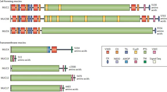
Mucus is produced by goblet cells that are found along the entire gastrointestinal tract, from mouth to rectum. Goblet cells are however more particularly numerous in the stomach and in the colon. They produce mucins, which are very large and abundantly glycosylated proteins. In addition to their important glycosylation, that enables a “water-holding” effect, mucins can form massive aggregates (McGuckin et al., 2011). Twenty mucin-encoding genes have been reported in humans, the major airway mucins being MUC5A and MUC5B, whereas MUC2 is the main intestinal mucin (Thornton et al., 2008). Two major group of intestinal mucins can be distinguished, transmembrane mucins that form the glycocalix and gel-forming mucins such as MUC2 that are produced by goblet cells and are released by protease cleavage into the lumen. Work by the group of Johansson and Hansson has shown that the thickness of the mucus layer varied along the digestive tract. In the small intestine, the mucus layer is thin, fluid and even discontinuous, likely to facilitate nutrient absorption. In contrast, in the colon, where the density of bacteria increases considerably, mucus is organized into two layers, one central fluid layer, in which bacteria can thrive and forage for mucin-derived sugars, and one external very thick layer almost impermeable to bacteria at steady state (Johansson and Hansson, 2016).
Figure 2, from (Johansson and Hansson, 2016): The domain structures of gel-forming and transmembrane intestinal mucins expressed in the small intestine. The proline, threonine and serine (PTS) domains become heavily O‑glycosylated to form the mucin domains. The non-PTS parts of the gel-forming mucins are rich in cysteine amino acids and form compact structures. AMOP; adhesion-associated domain; C8, conserved 8 cysteines domain; CK, cysteine knot domain; NIDO, nidogen domain; Signal Seq, signal sequence domain; SEA, sea urchin sperm protein, enterokinase, and agrin domain; TIL, trypsin inhibitor-like cysteine rich domain; TM, transmembrane domain; VWC, Von Willebrand factor type C domain; VWD, Von Willebrand factor type D domain.
The key role of mucins in the maintenance of gut homeostasis was demonstrated in 2006 with the first description of Muc2-/- mice. These mice developed spontaneous colitis at 5 weeks of age which
aggravated with time. Histological analysis showed mucosal thickening, increased proliferation, and superficial erosions. Strikingly, analysis with microbial probes revealed many bacteria adhering to the intestinal mucosa, demonstrating the key role of the mucus in allowing bacterial segregation from the intestinal surface. Muc2-/- mice were more sensitive to DSS colitis than heterozygous Muc2+/- or wild type (WT) mice, with observation of many crypt abscesses instead of mucosal ulcerations. Notably, even though heterozygous Muc2+/- mice were less sensitive to DSS than Muc2
-
mice, they were also more sensitive to DSS than WT mice, pointing out a dose-effect (Van der Sluis et al., 2006).
Mucus deficiency in human intestinal diseases?
Colonic expression of MUC2 is markedly diminished in UC patients (Van Klinken et al., 1999), along with the loss of glandular cells observed during chronic inflammation. As in mice, the loss of MUC2 is associated with enhanced bacterial adhesion to the colonic mucosa, thereby resulting in excessive microbial signaling to epithelial cells and increased risk of microbial translocation across
epithelium. The loss of MUC2 thus participates to a vicious circle, which maintains colonic inflammation. However, there has never been yet any example in humans of monogenic enteropathy due to mutations in mucins genes. This may be explained by some functional redundancy between mucins in humans. Thus, mucin genes contain long regions of repeated sequences that are rich in GC nucleotides and share important homology (Thornton et al., 2008). Of note, any whole exome sequencing (WES) reveals at least 5-10 rare variants in mucin genes. Ascribing a causative effect to one of these variants is extremely difficult since nobody carries the “normal” sequence of all genes. Yet, as discussed below, other genetic factors can impair mucus production in humans, and may thereby predispose to intestinal inflammation.
Figure 3, from (Thornton et al., 2008) – Repetitive domains in mucins
I.A.2- Role of endoplasmic reticulum stress in epithelial barrier
function
The endoplasmic reticulum (ER) is the cellular organelle in which proteins are synthetized, modified (for instance by glycosylation), and folded (notably by establishing disulfide bonds). Accumulation of improperly folded proteins causes ER stress that triggers the unfolded protein response (UPR). This response aims at restoring protein-folding homeostasis by three main mechanisms: i) transient reduction in protein translation; ii) increase in folding capacity; iii) initiation of apoptosis when ER stress cannot be resolved (Janssens et al., 2014).
Figure 4, from (Janssens et al., 2014) – The three UPR pathways in humans
One of the three UPR pathways activates the splicing of XBP1 (X-box-binding protein 1), a transcription factor that induces expression of chaperone molecules required for correct protein folding. All cells that produce large amounts of proteins such as goblet cells, Paneth cells and plasma cells are therefore prone to ER stress. Kaser and colleagues engineered mice deleted of Xbp1 specifically in IEC. These mice exhibited ER stress and spontaneous enteritis with complete loss of Paneth cells and reduction of goblet cells, associated with a considerable increase in activation of the transcription factor (nuclear factor-kappa B) NF-κB. When challenged by DSS, Xbp1
mice displayed more severe colitis with increased areas of mucosal erosions, edema, and cellular infiltration along with increased crypt loss compared to WT littermates, whereas heterozygous mice displayed an intermediate phenotype (Kaser et al., 2008). Interestingly, overexpression of HLA-B27, a protein prone to misfolding in humanized rats (human leukocyte antigen HLA-B27, expressed with human β2-microglobulin in transgenic rats) leads to spontaneous colitis and arthritis, thereby mimicking inflammatory disorders observed in a subset of HLA-B27 expressing individuals (Milia et al., 2009). Currently, no human monogenic disease has been described in genes related to ER stress or implicated in UPR. However, XBP1 variants predisposing to both CD and UC have been found, suggesting that unresolved ER stress might predispose to intestinal inflammation in humans (Adolph et al., 2012).
I.A.3- ATG16L1 and the role of autophagy in epithelial cells
Autophagy is one of the most evolutionary conserved cellular processes triggered by fasting. It is a complex process involving multiple proteins which activates the formation of vesicles containing cytoplasm and cytoplasmic organelles, and their fusion with lysosomes where these components can be degraded. Through this “self-digestion”, cells can salvage nutrients and maintain vital cellular functions during fasting. Cells can eliminate damaged organelles, misfolded proteins, but also invading microorganisms in a process using similar cellular mechanisms called xenophagy. Numerous diseases have been linked to autophagy defects, including neurodegenerative, liver, cardiac, tumoral and dysimmune diseases (Levine and Kroemer, 2008).
Following the identification of a non-synonymous coding variant of ATG16L1 (Thr300Ala) predisposing to adult IBD, Cadwell and colleagues were the first to generate mouse models to study the role of Atg16L1 but also of Atg5, which form a complex involved in the formation of autophagosomal vesicles. Since complete inactivation of either gene was lethal, they engineered mice with hypomorphic defective protein expression of Atg16L1 or Atg5 restricted deletion to IEC. They observed that Atg16L1- and Atg5-deficient Paneth cells displayed marked abnormalities in the granule exocytosis pathway, attested by disordered, diminished or diffuse lysozyme staining. Their examination of biopsies from CD patients homozygous for the ATG16L1 risk allele showed similar abnormalities in Paneth cell granules (Cadwell et al., 2008).
Two years later, Cadwell and colleagues used the same Atg16L1 hypomorphic mice to suggest that intestinal inflammation happened as the consequence of a “multi-hit” process. Unexpectedly they observed that Atg16L1 hypomorphic mice, rederived in specific-pathogen free (SPF) animal facility, were indistinguishable from WT littermates and failed to display aberrant packaging of the granule protein lysozyme in Paneth cells. In this SPF setting, they demonstrated that Paneth cell abnormalities were triggered by persistent norovirus infection. Triggering was strain specific as non-persistent strains did not induce Paneth cells abnormalities. Persistent-strain infected mice had also to be challenged by DSS to aggravate inflammation. Conversely treatment with either TNFα or IFN blocking antibodies, or broad spectrum antibiotics, drastically reduced inflammatory lesions. Overall, the authors suggested that IBD occur along a “multi-hit” model involving genetic predisposition and several environmental triggers which cooperate to ultimately induce inflammation mediated by Th1 cytokines (Cadwell et al., 2010).
The role of the ATG16L1 (Thr300Ala) was further investigated by the group of R. Xavier, who developed knock-in mice homozygous for this variant. These authors showed that the variant promoted cleavage by caspase 3 and 7 and thereby resulted in lesser amount of functional ATG16L1 protein. They observed that mice raised in SPF conditions displayed not only Paneth cells abnormalities comparable to those described by Cadwell et al, but also enlarged goblet cells
reminiscent of those described in mice with epithelial inactivation of ATG5. In this model in which the variant was expressed in all cells, the authors also observed defective bacterial clearance and increased IL-1β secretion by macrophages infected with Salmonella typhimurium. Yet curiously, they did not report any evidence of intestinal inflammation (Lassen et al., 2014). The exact role of ATG16L1 deficiency in intestinal inflammation remains however incompletely understood and is likely to involve other cells than IEC. Cadwell and coworkers have recently shown that mice with hypomorphic ATG16L1 displayed an intestinal hyperimmune signature notably characterized by enhanced transcription of type 1 IFN genes in response to infection by Citrobacter rodentium. Unexpectedly, this conferred protection against this enteropathogen. Protection was abolished by eliminating macrophages (Marchiando et al., 2013). Along the same line, Mazmanian and coworkers have recently shown that ATG16L1-/- macrophages produced more inflammatory cytokines upon activation by bacterial products and were less efficient in triggering IL-10 production by regulatory T cells than WT cells. This effect was ascribed to a role of ATG16L1 in the non-canonical autophagy pathway and was comparable to the one observed in macrophages lacking nucleotide-binding oligomerization domain 2 (NOD2). This intracellular sensor of bacteria-derived muramyl-dipeptide (MDP), which was the first genetic predisposing factor implicated in human IBD (see below) was shown to physically recruit ATG16L1 (Chu et al., 2016). Altogether these data indicate that ATG16L1 dysfunction may predispose to intestinal inflammation via multiple mechanisms involving epithelial cells but also immune cells of hematopoietic origin. Recent work in humans further indicated that AGT16L1 is involved in the negative control of the mitochondrial antiviral signaling pathway and that production of type 1 IFN was abnormally increased in colon cancer cells harboring the ATG16L1 (Thr300Ala) mutation, an effect correlated with increased survival to colon cancer (Grimm et al., 2016). It is not excluded that this mechanism may conversely predispose to protracted intestinal inflammation.
Autophagy and human intestinal diseases?
In humans, susceptibility to CD conferred by the risk allele ATG16L1 (Thr300Ala), is relatively weak with an odd ratio of 1.38 (McGovern et al., 2015). In addition, the variant is found in up to 50% in European-derived populations. Yet, it is interesting to stress that a common exonic (c.313C>T) variant in a second gene involved in autopaghy, Immunity-related GTPase family M (IRGM) was also shown to predispose to CD. The exonic variant is thought to jeopardize the regulation of IRGM transcription by microRNA196 (miRNA196) up-regulated in CD tissues. Recent work indicated that IRGM is a partner of both NOD2 and ATG16L1 and plays a central role in the activation of the core autophagy machinery (Chauhan et al., 2015). Presently, no human monogenic disease has however been ascribed to an autophagy defect.
I.A.4- Monogenic enteropathies with predominant epithelial defect
Congenital diarrheal disorders (CDD) are a vast group of monogenic enteropathies that usually start within the first days or weeks of life, or even in utero. They affect primarily or exclusively IEC functions. They are characterized by an intractable diarrhea which may be isolated or part of a syndrome. This severe diarrhea leads rapidly to life-threatening dehydration and serum electrolyte imbalance. CDD are often associated with intestinal failure requiring long-term parenteral nutrition or even bowel transplantation. Their phenotype and genotype have been extensively characterized (Canani et al., 2015). Chronic diarrhea is ascribed to either secretory or osmotic mechanism. Secretory diarrhea is caused by an excessive electrolytes and water flux from the intestinal mucosa towards the luminal content. Micovillus inclusion disease (MVID) or congenital tufting enteropathy/epithelial dysplasia (ED) are typical secretory diarrheas. Osmotic diarrhea is due to the presence of undigested and/or non-absorbed nutrients, which attract water flux. Osmotic diarrhea may be improved by dietary changes, notably by excluding nutrients that cannot be digested. CDD can be divided into three subsets: defects in enterocyte structure, defects in digestion/absorption, or defects in enteroendocrine cells differentiation. I will only describe CDD due to defects in enterocyte structure, which highlight the barrier role of the epithelial monolayer.
I.A.4.1- Microvillus inclusion disease (MVID)
MVID is an autosomal recessive monogenic enteropathy characterized by a profuse, watery intractable diarrhea (up to 150ml/kg/day water loss at birth) that can even start in utero with the formation of hydramnios. Mutations were first described in MYO5B, a member of the myosin family (Muller et al., 2008; van der Velde et al., 2013). The defect in enterocyte structure in MVID is the loss of the apical brush border associated with intracellular microvillus inclusions (Canani et al., 2015). MVID diagnosis is made by examination of small bowel biopsies that show accumulation of periodic acid Schiff (PAS) reactivity (thick and blur PAS staining at the apical side), and/or abnormal expression of the brush border enzyme CD10 in the cytoplasm of enterocytes. Visualization of microvillus inclusions can be further demonstrated by electron microscopy. Even though MYO5B is ubiquitously expressed, extra intestinal symptoms are rare, perhaps due to partial redundancy of MYO5A and MYO5C in other organs (van der Velde et al., 2013). Nevertheless severe cholestasis can be observed in some patients, due to abnormal polarization of hepatocytes (Girard et al., 2014).
WES of MVID patients with normal MYO5B sequence and milder phenotype led to identify mutations in Syntaxin-3 (STX3) (Wiegerinck et al., 2014), an apical receptor that regulates protein trafficking and vesicle fusions in IEC. In addition, a mild MVID phenotype has been reported in patients with familial hemophagocytic lymphohistiocytosis (HLH) type 5 due to mutations in STX3 protein binding 2 (STXBP2, also known as MUNC18-2) (Stepensky et al., 2013). Accumulation of PAS-positive granules and thicker CD10 staining at the apical side of enterocytes as well as abnormal kidney epithelial cells were observed, accounting for the severe watery diarrhea and nephropathy described in these patients. Intestinal and kidney defects persisted after correction of the lymphocyte defect by hematopoietic stem cell transplantation (HSCT) indicating that loss of function of STXBP2 is responsible for abnormal cellular trafficking not only in cytotoxic T cells but also in polarized epithelia.
Mouse models of MVID
Several mouse models recapitulate intestinal epithelial lesions in MIVD. In 2007 Sato and colleagues generated Rab8A-/- mice, which displayed diarrhea and progressive wasting after 2.5 weeks. They died at 5 weeks, likely from intestinal failure as their small intestines were swollen and contained undigested milk. As Rab8 was thought to regulate basolateral transport in polarized kidney epithelial cells, the authors examined basolateral and apical markers in IEC. Unexpectedly, basolateral markers (like Low-Density Lipoprotein receptor and Na+, K+ATPase) showed proper
localization whereas apical markers (dipeptidyl peptidase IV, alkaline phosphatase, sucrase-isomaltase, and oligopeptide transporter 1) were markedly decreased in the apical membrane and accumulated intracellularly. Kidney epithelial cells and hepatocytes were normal. Mice exclusively lacking Rab8A in IEC displayed the same phenotype. Examination of IEC from conditional Rab8A -/-mice with mosaic expression of Rab8 found large sub-apical vacuoles identical to those in fully
deficient Rab8A-/- mice in Rab8-negative but not in Rab8-positive IEC. Electron microscopy
examination of IEC from Rab8A-/- mice showed marked shortening of microvilli at three weeks, enlarged organelles with electron-dense materials strongly positive for the apical marker dipeptidyl peptidase IV but also for the endosome/lysosome marker LAMP2 (lysosomal-associated membrane protein) while Rab8 did not colocalize with this marker in WT IEC. The authors concluded that Rab8 deficiency prevented normal polarization and resulted in mislocalization of brush border peptidases and transporters to lysosomes, where they were degraded. Digestion and absorption were massively impaired, leading to rapid malnutrition and death. Because of the similarities between phenotypes in Rab8A-/- mice in MVID patients, the authors sequenced RAB8 but failed to identify any mutation in 3 MVID patients (Sato et al., 2007).
In 2012, specific inactivation of Cdc42 in IEC led to the delayed development of MVID in 10% of the mutant mice, which died at 6 months of age with an average body weight reduction of 60-70%, compared to WT littermates. Apart from large intracellular vacuolar structures, epithelial cells displayed additional features of MIVD, notably cell division defects, reduced capacity for clonal expansion and differentiation into Paneth cells, and increased apoptosis in stem cells. It was shown that Cdc42 deficiency impaired Rab8 activation and its association with multiple effectors. The authors suggested that defects of the stem cell niche might cause MVID (Sakamori et al., 2012). In 2014, mice with selective intestinal deletion of Rab11a were engineered, as ubiquitous Rab11a deletion was lethal during embryogenesis. Rab11a-/- mice started to die one week after birth and displayed histological features of MVID with cytoplasmic accumulation of apical shorter microvilli and microvillus inclusion bodies. In addition, Rab8a was mislocalized. As Rab11a was also mislocalized in Rab8a -/- mice and in one MYO5B-mutated patient, the authors pointed out
functional relationships between Rab11a, Rab8a and myosin Vb in vivo (Sobajima et al., 2014). No patients with mutations in CDC42 or RAB11A have been reported yet.
The first intestine-specific Myo5b-deficient (Myo5bfl/fl;Vil-CreERT2) mouse model was only generated very recently. Mice developed severe diarrhea within 4 days after tamoxifen induction. They recapitulated histological hallmarks of MYO5B deficient patients with subapical accumulation of PAS and alkaline phosphatase staining, almost complete absence of apical microvilli, appearance of microvillus inclusions, and enlarged intercellular spaces by electron microscopy (Schneeberger et al., 2015). Using a tamoxifen inducible model, Weis and colleagues further showed that Myo5B loss at 8 weeks of age reduced duodenal brush border enzymes but induced much less prominent microvillus inclusions than at 2 weeks of age, suggesting that Myo5b deficiency may differ with age (Weis et al., 2016).
Altogether, human MVID and related mouse models have allowed unravelling the complementary roles of Myo5B, Rab8 and Rab11 in the establishment of polarization in IEC, which is essential for their proper absorptive function.
I.A.4.2- Epithelial dysplasia (ED)
Epithelial dysplasia (ED), also known as congenital tufting enteropathy (CTE), is another CDD due to a structural defect of epithelium. As MVID, ED patients display severe, watery, intractable
diarrhea since birth with severe failure to thrive. ED can be isolated or part of a syndrome with superficial punctate keratitis and choanal atresia.
ED was first ascribed to recessive autosomal EPCAM mutations in 2008 in patients with chronic diarrhea only. EpCAM (for epithelial cell adhesion molecule) is a transmembrane protein, located at IEC basolateral membrane, which regulates cellular adhesion and proliferation. Examination of small bowel biopsies shows villus atrophy and crypt hyperplasia. Observation of epithelial tufts made of round-shape IEC in teardrop-like formation is considered to be pathognomonic (Sivagnanam et al., 2008). Additional features include increased expression of desmoglein, and increased length and number of desmosomes. As ED patients get older, they can display lamina
propria T-cell infiltration, even sometimes in the absence of visible tufts (Goulet et al., 2007). Mouse models provide some clues to why EpCAM deficiency might mimic dysimmune enteropathy (see below).
The c.498insC EpCAM mutation, which abolishes protein expression, results in the most severe phenotype. This mutation is shared by families originating from Kuwait and Qatar. Delimitation of the minimal common haplotype suggests a founder effect 5,000-6,000 years ago (Salomon et al., 2011). Patients with expression of a truncated protein display a milder phenotype, which permits weaning from parenteral nutrition in late childhood (Salomon et al., 2011; Sivagnanam et al., 2008; Sivagnanam et al., 2010). The only extra-intestinal symptom reported is polyarthritis in a few patients.
Mutations in a second gene called Serine Peptidase Inhibitor, Kunitz Type, 2 (SPINT2) have been described as a cause of ED. Such mutations were initially described in patients with syndromic congenital sodium diarrhea, e. g. associated with anal or choanal atresia, hypertelorism and corneal erosions. Mutations in SPINT2, and notably the common c.488A>G mutation, were subsequently described in patients displaying the same symptoms but also epithelial tufts reminiscent of those observed in patients with EPCAM mutations (Canani et al., 2015; Heinz-Erian et al., 2009).
Mouse models of ED
Analysis of mice harboring hypomorphic Spint2 mutations indicated that SPINT2, a transmembrane serine protein inhibitor also called hepatocyte growth factor activator inhibitor type 2, is involved in organogenesis and in epithelial homeostasis by regulating multiple cellular processes, including bioavailability of growth factors, ion fluxes and paracellular permeability (Szabo et al., 2009). In 2009, Nagao et al reported that EpCAM-/- mice died in utero by E12.5 with growth retardation,
other groups engineered EpCAM-/- mice, which were alive at birth. Mice generated by Guerra and
colleagues however failed to thrive, and died soon after birth because of hemorrhagic diarrhea. Histological features of ED were found, including intestinal tufts, villous atrophy and colon crypt hyperplasia, whereas other organs were normal. The authors reported loss of membrane localization and increased intracellular accumulation of cadherin and β-catenin (Guerra et al., 2012). E-cadherin is the main intestinal E-cadherin. It is localized along the basolateral surfaces and apical junctions of IEC and is essential to intercellular adhesion between epithelial cells and epithelial etancheity. Of note, mice expressing a dominant negative N-cadherin mutant that prevents surface localization of endogenous E-cadherin, displayed severe inflammation with polymorphous infiltration of lamina propria (Hermiston and Gordon, 1995), reminiscent of the inflammatory features described in some ED patients.
EpCAM-/- mice generated by Lei et al also died shortly after birth. These authors described reduced
expression of tight junction proteins Claudins 2, 3, 7, and 15 and loss of claudin-7. Tight junctions in intestinal epithelium were morphologically abnormal with a disrupted network. Sulfo-NHS-biotin injected into the intestinal lumen of E18.5 EpCAM-/- mice abnormally diffused at the lateral
membrane, a result ascribed by the authors to down-regulation of claudins. Claudins being responsible for paracellular permeability, the authors further showed that Na+-selective paracellular
permeability was reduced while Cl--selective permeability remained normal, reminiscent of the
findings in the Claudin-15 mutant mouse (Lei et al., 2012). Lastly, in 2014, Sivagagnam’s laboratory and colleagues used Cre-LoxP recombination to engineer mice lacking exon 4 of
EpCAM. EpCAM-/- mice died shortly after birth with growth retardation and histological features of
ED including epithelial tufts, enterocyte crowding, altered desmosomes, and intercellular gaps. EpCAM and Claudin-7 expression were low and the two proteins failed to colocalize, a result confirmed in ED patients. Permeability and intestinal cell migration were increased. The authors suggested a pathogenic scheme in which reduced expression of EpCAM and claudin-7 resulted in the development of histological abnormalities including tufting, intercellular gaps, increased desmosomes, and villous atrophy, which secondary lead to increased proliferation and migration of IEC, as well as to loss of barrier function. The exact role of claudin 7 remains however unclear and the defect in EPCAM may impair desmosomes and intercellular adhesion independently of claudin 7 (Mueller et al., 2014).
I.B- Defective innate immunity at the intestinal barrier
I.B.1- Defects in pattern recognition receptors (PRR)
Pattern recognition receptors (PRR) are innate receptors that recognize pathogen-associated molecular patterns (PAMPs) of microorganisms. They include notably Toll-like receptors (TLR), NOD-like receptors (NLRs), and RNA helicases like RIG-1 (retinoid acid-inducible gene-1). Many mouse models of PRR defects have been engineered. Unexpectedly, individual PRR defects did not lead to profound immunodeficiency nor to spontaneous intestinal inflammation. Similarly, monogenic diseases due to mutations in genes coding for PRR display mild or absent phenotypes. These observations point out the important redundancy, which exists between PRR to prevent infections (Alain Fischer and Antonio Rausell, submitted) and control intestinal homeostasis. Therefore only three molecules will be discussed in this chapter. The first one is MyD88 (for myeloid differentiation primary response gene 88), a scaffold protein bridging most TLR with intracellular signaling. The second one is NEMO/IκK indispensable to switch on the NF-κB pathway downstream most PRR. The last molecule is NOD2, as rare variants in this PRR represent the main genetic risk factor for CD in Caucasians, and as it is an important partner of X-linked apoptosis inducing protein (XIAP) in which loss of functions mutations are one monogenic cause of severe colitis in humans (see below).
I.B.1.1- MyD88
MyD88 is a key downstream adaptor coupling IL-1 and IL-18 receptors and all TLR except TLR3 to NF-κB and mitogen-associated protein (MAP) kinase signaling pathways.
In 2006, Rakoff-Nahum and colleagues engineered Il-10-/-/MyD88-/- mice. They showed that
Il-10-/-/MyD88-/- mice were completely free of all signs of intestinal disease throughout more than
1.5 years, contrary to Il-10-/-mice that developed enterocolitis, mild to moderate weight loss, and increased mortality. These results led the authors to suggest that chronic colitis in Il-10-/-mice is
related to the loss of regulation of immune responses triggered by recognition of commensals by TLR via the MyD88-dependent signaling pathway (Jobin, 2010; Rakoff-Nahoum et al., 2006).
Figure 5, from (O'Neill and Bowie, 2007) – MyD88 at the crossroads of TLR pathways
In 2010, Asquith et al further studied MyD88 contribution to intestinal barrier homeostasis. Following H. hepaticus infection, intestinal and systemic inflammation was dramatically reduced in Rag-/-/MyD88-/- mice compared to Rag-/- mice (lacking recombinant activating gene, thus with complete absence of T and B cells). The luminal burden was reduced in the prior mouse model. Then, they generated bone marrow (BM) chimeras from irradiated Rag-/- mice that received bone
marrow from either Rag-/- or Rag-/-/MyD88-/- mice and infected them with H. hepaticus. They found a complete absence of intestinal and systemic inflammation in mice reconstituted with MyD88-deficient BM, demonstrating that hematopoietic, and not epithelial cells, were responsible for transmitting the MyD88-dependent inflammatory response following bacterial infection (Asquith et al., 2010). In keeping with Rakoff-Nahum et al findings, WT mice infected with H. hepaticus developed severe typhlocolitis after treatment with anti-IL-10R blocking antibody whereas
MyD88-/- mice did not.
Even under strict SPF conditions, Rag-/-/MyD88-/- mice died progressively (30% survival at day 25
after weaning) without signs of wasting or dysimmunity, whereas Rag-/-/MyD88+/- mice had 100% survival. In chimeric mice with MyD88-deficiency in the hematopoietic system only, survival was normalized, highlighting an important and distinctive role of MyD88 in IEC homeostasis. Rag -/-reconstituted with MyD88-/--BM maintained normal anti-microbial peptides (AMP) expression after
H. hepaticus infection, demonstrating that epithelial MyD88 signaling efficiently drives AMP expression in the colon (Asquith et al., 2010). Taken together, these data indicate that MyD88
activates a cytoprotective signaling pathway in IEC, but triggers inflammatory responses in response to H. hepaticus infection in innate immune cells of hematopoietic origin (Jobin, 2010).
Figure 6, from (Jobin, 2010)- Dual function of MyD88 signaling in the intestine
Autosomal recessive mutations in MyD88 were first described in 2008. Heterozygous carriers were free of symptoms. As discussed above, loss-of function MyD88 mutations had relatively mild consequences with only narrow susceptibility to some pyogenic bacterial infections (Streptococcus
pneumoniae, Staphylococcus aureus, and Pseudomonas aeruginosa responsible for invasive meningitis, sepsis, arthritis, osteomyelitis, and abscesses) while resistance to common fungi, parasites, viruses was normal. Of note, deficiency in IRAK-4 (IL-1R-associated kinase 4), situated immediately downstream MyD88 in the signaling pathway, see figure 5) phenocopied MyD88 deficiency. In keeping with a preponderant role of innate immunity in early childhood, MyD88 and
IRAK-4 deficient patients displayed their first invasive bacterial infection before the age of two (90% cases). In contrast, susceptibility to invasive bacterial infections disappeared after 10 years of age, probably thanks to the development of adaptive immune responses. Noninvasive infections mostly of the skin and upper respiratory tract were however observed in adult patients, suggesting that the protective role of MyD88 described in murine IEC may also exist in human epithelial cells (Picard et al., 2011).
Recently, analysis of transcriptome from MyD88 mutated patients revealed drastically reduced responses to TLR2 agonists (90% less than those of healthy subjects). TLR2 being known to be
crucial for the recognition of Gram-positive bacteria (S. pneumoniae and S. aureus), this result explains at least partially the narrow range of infections displayed by MyD88-mutated patients (Alsina et al., 2014).
I.B.1.2- NEMO/IκK
Figure 7, from (Picard et al., 2011)- NEMO signaling
Gender-conditioned phenotypes in human NEMO deficiency
The IκB kinase enzyme complex (IκK) is the core element of the NF-κB pathway. Specifically, IκK phosphorylates the inhibitor of kappa B α (IκBα) which, at steady state, is bound to NF-κB transcription factors and masks their nuclear localization signals, keeping them sequestered in an inactive state in the cytoplasm. Phosphorylation results in the dissociation of IκBα from NF-κB, which can then migrate into the nucleus and activate transcription of multiple genes, participating in numerous cell processes and notably in innate and adaptive T cell responses.
Mutations in IKBKG gene, which encodes for NEMO/IκK subunit were first described in 2000 in familial incontinentia pigmenti. This monogenic skin disease is an X-linked dominant disorder prenatally lethal in males. Affected females displayed highly variable abnormalities of skin, hair, nails, teeth, eyes and central nervous system. NF-κB activation was defective in skin fibroblasts from affected fetuses. Affected females survived because of X-chromosome dizygosity and
negative selection of cells carrying the mutant X-chromosome. The most common mutation reported in this article was a large deletion of NEMO gene encompassing exons 4 to 10, which resulted from recombination between two repeated regions of strong homology located in intron 3 and the 3’ part of exon 10 respectively (Smahi et al., 2000).
Figure 8, from (Smahi et al., 2000) – Recombination event leading to NEMO partial deletion
In 2001, Döffinger and colleagues linked NEMO mutations to another syndrome called X-linked recessive anhidrotic ectodermal dysplasia with immunodeficiency (EDA-ID) that arises in males. They reported hypomorphic mutations in the gene IKBKG in 12 males with EDA-ID and in two patients with osteopetrosis and lymphoedema associated to EDA-ID (OL-EDA-ID). As germline loss-of-function mutations in IKBKG are lethal in male fetuses, the authors showed that EDA-ID and OL-EDA-ID mutations impaired but did not fully abolish NF-κB signaling. Notably, they linked patients’ abnormal immune features to impaired cell responses to lipopolysaccharide (LPS), IL-1β, IL-18, TNFα and CD40L. Therefore, loss of regulation of NF-κB signaling is responsible for combined developmental and immunological defects (Doffinger et al., 2001).
Incontinentia pigmenti and EDA-ID form a syndromic continuum as attested by some case reports. The first one described a female patient with incontinentia pigmenti who suffered from immunodeficiency because of a persistent expression of the mutated X-chromosome found in peripheral blood cells until the age of 3.5 years, after which the X-chromosome inactivation pattern was completely skewed (Martinez-Pomar et al., 2005). The second one described an insertion responsible for a premature stop codon (at position 49). Surprisingly, the affected boy displayed only a severe immunodeficiency. Puel and colleagues showed that reinitiation of translation happened from a methionine located immediately downstream the stop codon, resulting in a truncated NEMO protein that was sufficient for normal fetal development of epithelia but insufficient for normal immune function (Puel et al., 2006).