HAL Id: tel-02180591
https://tel.archives-ouvertes.fr/tel-02180591
Submitted on 11 Jul 2019HAL is a multi-disciplinary open access
archive for the deposit and dissemination of sci-entific research documents, whether they are pub-lished or not. The documents may come from teaching and research institutions in France or abroad, or from public or private research centers.
L’archive ouverte pluridisciplinaire HAL, est destinée au dépôt et à la diffusion de documents scientifiques de niveau recherche, publiés ou non, émanant des établissements d’enseignement et de recherche français ou étrangers, des laboratoires publics ou privés.
Dissecting the signaling pathways controlling
inflammation during Gram-negative bacterial infections :
the role of ALPK1, TIFA and TRAF6 during Shigella
flexneri infection
Milica Milivojevic
To cite this version:
Milica Milivojevic. Dissecting the signaling pathways controlling inflammation during Gram-negative bacterial infections : the role of ALPK1, TIFA and TRAF6 during Shigella flexneri infection. Bacte-riology. Université Sorbonne Paris Cité, 2017. English. �NNT : 2017USPCB061�. �tel-02180591�
Université Paris Descartes
Ecole doctorale BioSPC
Equipe « Signalisation Cellulaire et Infections Bactériennes »
Institut Cochin - INSERM U1016, CNRS UMR8104
, Université Paris Descartes
Dissecting the signaling pathways
controlling inflammation during
Gram-negative bacterial infections:
The role of ALPK1, TIFA and TRAF6 during Shigella
flexneri infection
Par Milica Milivojevic
Thèse de doctorat d’Infectiologie
Dirigée par Dr. Cécile Arrieumerlou
Présentée et soutenue publiquement le 16 Novembre 2017
Devant un jury composé de :
Dr. Agathe SUBTIL
– Rapporteur
Dr. Thomas HENRY
– Rapporteur
Dr. Laurence ARBIBE
– Examinatrice
Pr. Olivier DUSSURGET
– Examinateur
Dr. Emmanuel LAPLANTINE
– Examinateur
[1]
Contents
Resume ... 3
Abbreviations ... 4
INTRODUCTION ... 7
1 Epithelial cells in immunity ... 7
1.1 Immune sensing ... 9 1.1.1 PAMPs ... 9 1.1.2 DAMPs ... 11 1.1.3 PRRs ... 11 TLRs ... 12 1.1.3.1.1 TLRs in epithelial cells ... 14 NLRs ... 15
1.1.3.2.1 NOD1 and NOD2 ... 16
1.1.3.2.2 NLRs and the inflammasome ... 17
1.1.3.2.3 Non-canonical inflammasome ... 18
1.1.4 Downstream signaling and NF-B activation ... 19
1.1.5 Cytokines ... 22
2 Intracellular Pathogens and Immunity ... 24
2.1 Shigella flexneri: A model pathogen ... 24
2.1.1 Epidemiology ... 24
2.1.2 Virulence plasmid and T3SS ... 25
2.1.3 Infection Cycle... 28
Macrophage escape ... 29
Epithelial cell entry ... 30
Vacuolar escape and cellular dissemination ... 30
Intracellular detection of bacteria ... 32
2.1.4 Effectors interfering with immunity ... 34
Osp genes ... 34
IpaH ... 36
2.2 Mechanism of bystander cell activation ... 37
3 RNAi screen and Identification of candidates... 40
3.1 TRAF6... 41
3.2 TIFA ... 42
[2]
ALPK1 ... 47
RESULTS... 49
DISCUSSION ... 95
1 HBP delivery and detection ... 96
1.1 Endocytosis/ macropinocytosis?... 96
1.2 Intracellular lysis? ... 97
1.3 Cytosolic bacterial replication? ... 97
1.4 T3SS-dependent delivery ... 99
1.5 HBP sensing? ... 100
2 Bystander cell activation? ... 102
3 The role of ALPK1 ... 103
3.1 HBP receptor? ... 103
3.2 ALPK1 substrates and interaction partners? ... 104
3.3 Role of ALPK1 in immune cells? ... 105
3.4 ALPK1, a wider implication? ... 105
3.5 Therapeutic potential? ... 106
4 Concluding remarks ... 107
[3]
Resume
Epithelial cells represent the first line of defense against pathogens and play an active role in innate immunity. Via local secretion of cytokines, they are able to orchestrate the immune response against invading pathogens. The activation of both intracellular and extracellular pathogen recognition receptors leads to a complex signaling cascade, resulting in the activation of the transcription factor nuclear factor B (NF-B) and the subsequent production of pro-inflammatory cytokines. However, the molecular mechanisms governing this process have not been fully elucidated. The Gram-negative bacterium Shigella flexneri is an important human pathogen and the causative agent of bacillary dysentery. This disease is characterized by acute inflammation of the colon resulting in the destruction of the intestinal tissue and, in severe cases, death. S. flexneri can invade and replicate within colonic epithelial cells. Following detection of the bacteria, both infected and uninfected bystander cells initiate inflammatory signaling pathways, which result in massive interleukin-8 (IL-8) production by the latter.
Using S. flexneri as a model of infection, we have identified a novel signaling pathway, which is central to the activation of NF-B and the subsequent production of IL-8 during Gram-negative bacterial infections. Following the cytosolic detection of bacteria, the protein TRAF-interacting factor with forkhead-associated domain (TIFA) forms oligomers, a process dependent on its threonine at position 9 and the forkhead-associated domain. These oligomers interact with TNF receptor associated factor (TRAF) 6, leading to its oligomerization and the subsequent activation of NF-B . In addition, we show that oligomerization of TIFA is dependent on the kinase alpha-kinase (ALPK)1 and that this pathway is activated in response to the detection of the bacterial metabolite heptose-1, 7-bisphosphate (HBP). These observations could be extended to the enteroinvasive pathogen Salmonella typhimurium as well as the extracellular bacteria Neisseria meningitidis. Our results therefore demonstrate the central role of the ALPK1-TIFA-TRAF6 signaling pathway in response to HBP of both intracellular and extracellular Gram-negative bacterial pathogens, and offer a better understanding of the molecular mechanisms governing the epithelial cell immune response to pathogenic bacteria.
[4]
Abbreviations
AHNAK: Neuroblast differentiation-associated protein ALPK1: Alpha kinase 1
ANXA2: Annexin 2
AP-1: Activator protein 1
ASC: Apoptosis-associated speck-like protein containing a CARD
ATP: Adenosine triphosphate
BAG2: BCL2 associated athanogene 2
BCV: Bacteria-containing vacuole
CARD: Caspase activation and recruitment domain
CBM: CARD-MALT-BCL
cGAMP : cyclic guanosine monophosphate–adenosine monophosphate
cGAS: cyclic GMP–AMP synthase
CLR: C-type lectin receptor
DAG: Diacylglycerol
DAMP: Danger associated molecular pattern EPEC: Enteropathogenic Escherichia Coli ERK: Extracellular signal-regulated kinase FAE: Follicle associated epithelium
FHA: Forkhead-associated
GBP: Guanylate binding protein
GEF: Guanine nucleotide exchange factor
GTP: Guanosine-5'-triphosphate
HBP: Heptose-1, 7-bisphosphate
HGMB: High-mobility group protein B
HP1: Hetrochromatin protein 1
HSP: Heat shock protein
IBD: Inflammatory bowel disease
IEC: Intestinal epithelial cell
ie-DAP: -d-glutamyl-meso-diaminopimelic acid
IFN: Interferon
[5]
IL- Interleukin
IRAK: IL-1 receptor associated kinase
IB: Inhibitor of B
JNK: Jun N-terminal kinase
KD: Knock-down
Kdo: Ketodeoxyoctonic acid
KO: Knock-out
LAMP: Lysosome-associated protein
LPS: Lipopolysaccharide
LTA: Lipoteichoic acid
M cell: Microfold cell
MAL: MYD88 adaptor-like protein
MAPK: Mitogen activated protein kinases MD-2: Myeloid differentiation factor 2
MDP: Muramyl dipeptide
MOI: Multiplicity of infection
mRNA: messenger RNA
MSK: Mitogen and stress activated protein kinase
MSU: Monosodium urate
Myd88: Myeloid differentiation primary response gene 88
NAG: N-acetylglucosamine
NBD: N-terminal protein binding domain NEMO: NF-B Essential Modulator NF-B : Nuclear factor κB
NK: Natural Killer
NLR: NOD-like receptor
NOD: Nucleotide-binding oligomerization domain PAMP: Pathogen associated molecular pattern
PGN: Peptidoglycan
PI(4,5)P2: Phosphatidylinositol 4,5-bisphosphate
PKC: Protein kinase C
PMA: phorbol 12-myristate 13-acetate
[6] PRR: Pathogen recognition receptor RING: Really interesting new gene
RIP2: Receptor-interacting serine/threonine-protein kinase 2 RISC: RNA-induced silencing complex
RLR: RIG-like receptor RNAi: RNA interference
ROCK: Rho-associated protein kinase SARM: Sterile α- and armadillo-motif SCV: Salmonella-containing vacuole STING: Stimulator of IFN genes T3SS: Type 3 secretion system T4SS: Type 4 secretion system TAB: TAK1-binding protein
TAK1: Transforming growth factor- activated kinase-1
Th: T helper
TIFA: TRAF interacting forkhead associated TIR: Toll-interleukin 1 receptor
TLR: Toll-like receptor
TNFα: Tumor necrosis factor α
TRAF: TNF receptor associated factor TRAM: TRIF-related adaptor molecule
TRIF: TIR domain containing adaptor protein inducing IFN
[7]
INTRODUCTION
An organism’s ability to detect and respond quickly and efficiently to invading pathogens is paramount to its survival. The innate immune system is charged with the task of recognising microbes and initiating the primary immune response. This is crucial in orchestrating the adaptive immune response which follows and shaping the outcome of infection. The cells of the innate immune system are equipped with an array of extracellular and intracellular receptors capable of recognising conserved microbial components. This results in a complex downstream signalling cascade, which ultimately leads to the production of pro-inflammatory cytokines and chemokines. Epithelial cells represent the first line of defence and play a central role in the establishment of this primary response.
Bacteria of the Shigella genus are important human pathogens, which cause the disease bacillary dysentery. This disease is characterized by acute inflammation and destruction of the intestinal epithelium. Shigella have adapted to an intracellular lifestyle and employ a number of mechanisms to interfere with host cell signalling processes and to ensure their survival, replication and spread within epithelial cells. Understanding the molecular mechanisms involved in bacterial sensing and the establishment of the innate immune response has been the aim of my thesis.
The first part of this introduction is aimed at reminding the reader of the different mechanisms employed by epithelial cells to sense bacterial pathogens, with a focus on Gram-negative bacteria. I will then go on to describe the different signalling pathways implicated in this process. The second part will be focused on the Gram-negative bacteria S. flexneri, the model pathogen used in this work. The third and final part is aimed at introducing the reader to the three proteins which we have identified as playing an important role during Gram-negative bacterial infections.
1 Epithelial cells in immunity
Epithelial cells line the cavities of organisms. Due to their exposure to the external environment, they are continuously faced with the challenge of protecting the organism from incoming pathogens. For this reason, they are often referred to as the sentinels, representing the first line of defense. They provide both a physical barrier, due to the presence of intercellular tight junctions, as well as a chemical one, via the secretion of antimicrobial peptides. Whilst they themselves are not considered immune cells, they are equipped with a number of innate mechanisms for sensing and responding to infection.
[8]
Intestinal epithelial cells (IECs) are of particular interest since they are continually exposed to residing microorganisms, termed the microbiota (Sansonetti, 2004). Whilst they must manage this constant contact with a huge number of commensals, they must also respond effectively and appropriately to potential breaches of this homeostasis from invasive pathogenic bacteria. Their ability to do so is paramount in orchestrating the adaptive immune response which follows.
Figure 1. Anatomy of the small intestine and colon (Abreu, 2010). Intestinal epithelial cells (IECs), linked
via tight-junctions, form a single cell barrier separating the lumen from the lamina propria. The vast majority of this layer is made up of enterocytes. Goblet cells secrete mucus, which forms a gel-like protective layer, separating IECs from the commensals. Microfold (M) cells, overlying the Peyer’s patches, transport luminal antigens and microorganisms to the immune cells in the lamina propria. Resident macrophages, dendritic cells, and both B and T lymphocytes are all found in the lamina propria.
IECs are polarized cells with an apical pole on the luminal side of the intestine and a basolateral pole. They are composed of five cell types. The enterocytes are the most abundant in both the small and large intestine (Figure 1) and are the ones that I will be referring to when using the term IECs. They form tight junctions between them, creating an impermeable barrier. They also actively secrete antimicrobial peptides capable of directly killing bacteria, and secrete cytokines, which coordinate the action of both innate and adaptive immune cells. There are three secretory cell types: goblet cells, Paneth cells and enteroendocrine cells. The main function of goblet cells is to secrete mucus, which forms a protective layer and separates the microbiota from the rest of the
[9]
IECs (Kim and Ho, 2010). Paneth cells are mainly found in the small intestine and are capable of secreting granules filled with microbicidal proteins (Ayabe et al., 2000). The enteroendocrine cells are important for sensing nutrients and releasing peptide hormones, which mediate digestion but also participate in the inflammatory response (Worthington et al., 2017). Finally, there are the microfold (M) cells. They are generally in specialized regions called the follicle associated epithelium and overlie the Peyer’s patches. They serve to “sample” the environment by transepithelially transporting antigens and microorganisms to the underlying immune cells, which include resident macrophages, dendritic cells as well as B and T lymphocytes (Kraehenbuhl and Neutra, 2000).
1.1 Immune sensing
Innate immune recognition, unlike adaptive, relies on a limited number of germline-encoded receptors termed Pathogen Recognition Receptors (PRRs). They are mainly expressed in cells of the innate immune system such as dendritic cells and macrophages but IECs possess them as well. Whilst the repertoire and spatial distribution of these PRRs may differ between these cell types, PRRs allow IECs to sense and respond to infection. PRRs sense infection either by directly binding to exogenous Pathogen Associated Molecular Patterns (PAMPs), or indirectly through endogenous danger signals termed Danger Associated Molecular Patterns (DAMPs).
1.1.1 PAMPs
The concept of PAMPs was first introduced by Charles Janeway Jr in 1989 (Janeway, 1989). PAMPs are characteristic molecular motifs conserved between groups of pathogens including fungi, viruses and bacteria. These motifs are highly evolutionarily conserved since they are usually central to the microbes physiology (Medzhitov and Janeway, 1997). PAMPs can be both intracellular as well as present on the surface of the microorganisms. They vary in their composition and can be composed of lipids, proteins, lipoproteins, monosaccharides or nucleic acids. Since they are absent in the host, they provide an exogenous signal to the cell of a pathogenic presence and promote an immune response. Bacteria have a number of PAMPs, which can be recognized by the host. Some PAMPs, such as DNA, are present in all bacteria. Indeed, bacterial DNA, like that of viruses, is recognized as a PAMP since they have unmethylated CpG-DNA whereas, in mammals, it tends to be methylated (Häcker et al., 2002). Other PAMPs are only present in certain subsets of bacteria. Bacteria such as Salmonella typhimurium, Escherichia coli and Listeria monocytogenes possess a flagellum, an organelle which provides the bacterium with motility. The main protein component of flagella, flagellin, is a PAMP which elicits an immune response following recognition (Ciacci-Woolwine et al., 1998). Bacteria, such
[10]
as the pathogens Shigella flexneri, S. typhimurium, pseudomonas aeruginosa, and enteropathogenic Escherichia coli (EPEC) have a type 3 secretion system (T3SS), which is structurally related to flagella (Blocker et al., 2003) and can also be recognized as PAMP (Miao et al., 2010).
Figure 2. Schematic representation of the cell walls of Gram-positive and Gram-negative bacteria.
(Adapted from Akira et al., 2006). The cytoplasmic lipid membrane of Gram-positive bacteria is covered by a thick wall made up of peptidoglycan and containing lipoproteins and lipoteichoic acids. In Gram-negative bacteria this peptidoglycan wall is much thinner and lacks lipoteichoic acid. In addition, it is covered by an outer membrane made up of lipopolysaccharide (LPS). The LPS is made up of the Lipid A anchor, a core oligosaccharide unit and the variable outer O antigen.
Bacteria can be divided into two large groups based on their surface composition: Gram-positive and Gram-negative bacteria (Figure 2). They both possess a cytoplasmic lipid membrane surrounded by the cell wall, which is made up of peptidoglycan (PGN). This is a polymer of N-acetylglucosamine and N-acetylmuramic acid, crosslinked by short peptides (Dziarski, 2003). In Gram-positive bacteria, this cell wall is thick and contains teichoic acids, both lipoteichoic acids (LTA) and wall teichoic acids. Peptidoglycan and LTA both constitute bacterial PAMPs, which can be recognized by the host. Gram-negative bacteria, on the other hand, have a much thinner PGN wall lacking teichoic acids. This wall is covered by an outer lipid membrane made up of lipopolysaccharide (LPS). The LPS is composed of 3 domains; a hydrophobic lipid moiety embedded in the outer membrane, termed lipid A; a relatively conserved oligosaccharide core attached to the lipid A via ketodeoxyoctonic acid (Kdo), which influences permeation properties of the outer membrane; and the variable O-antigen, containing a variable number of repeating saccharide units which contributes to bacterial antigenicity and serospecificity (Wang and Quinn, 2010). Lipid A, also known as the endotoxin, is the immunogenic component of LPS and is
Lipid A
Core oligosaccharide O antigen
[11]
responsible for LPS-induced sepsis (Raetz and Whitfield, 2002). Recently, it has also been shown that heptose-1,7-bisphosphate (HBP), a cytosolic intermediate of the LPS biosynthetic pathway, represents a potent new PAMP (Gaudet et al., 2017). This will be discussed in further detail later on.
1.1.2 DAMPs
It has become increasingly clear that cells do not only respond following the recognition of pathogen associated molecules, but can also respond to endogenous danger signals termed DAMPs. This concept was first proposed by Polly Matzinger who suggested that cells were less concerned with non-self and more so with the presence of danger (Matzinger, 1994, 2002). It now seems that inflammation and the immune response is triggered by a combination of the two. Unlike PAMPs, DAMPs are endogenous molecules, which are released following tissue stress or damage. Their production is not limited to the presence of infection and can be induced through sterile injury and disease. To date, many DAMPs have been identified. DAMPs can be either intracellular or extracellular in origin and vary enormously in their size and composition (Schaefer, 2014). These include small molecules like uric acid (Kono et al., 2010) or ATP (McDonald et al., 2010), to whole proteins such as high-mobility group protein B1 (HMGB1) and heat shock proteins (HSP) (Wheeler et al., 2009). They are sensed largely by the same PRRs responsible for the sensing of PAMPs, which will be discussed in the next section. The result of DAMP recognition can lead to a number of different outcomes such as autophagy and inflammation, and is associated with a number of diseases including sepsis (Kung et al., 2012), Crohn’s disease (Pastorelli et al., 2011), autoimmune diseases such as systemic lupus erythematosus (Urbonaviciute et al., 2008) and cancer (Huang et al., 2015).
1.1.3 PRRs
A number of different PRRs have been described to date. They can be divided into four groups: RIG-like-receptors (RLRs), Nucleotide Oligomerization Domain (NOD)-like receptors (NLRs), C-type lectin receptors (CLRs) and Toll-like receptors (TLRs). The former two are cytoplasmic while the latter two are membrane-associated receptors present either at the cellular surface or in intracellular compartments such as endosomes. RLRs are mainly associated with the recognition of double- and single-stranded RNA from viruses whilst CLRs recognize carbohydrates and are mainly involved in fungal recognition. I will focus on TLRs and NLRs which, amongst other triggers, can be activated by bacteria.
[12] TLRs
The discovery of TLRs revolutionized the field of innate immunity. The protein Toll was first identified in the anti-fungal response in Drosophila (Lemaitre et al., 1996). A year later, a human homologue of Toll, TLR4, was shown to lead to the production of inflammatory cytokines when constitutively active (Medzhitov et al., 1997). Since then, much work has gone into the characterization of this family of proteins. Currently, there are 10 functional TLRs in humans and 12 in mice, with TLRs 1-9 conserved between the two species (Figure 3). They are type I transmembrane proteins with an ectodomain comprised of leucine rich repeats (LRRs) important in the recognition of PAMPs, a transmembrane domain and an intracellular tail required for downstream signaling called the Toll-Interleukin 1 receptor (TIR) domain (Kawai and Akira, 2010). They can be located either at the plasma membrane or in intracellular compartments and are expressed in cells both of immune origin and non-immune origin such as epithelial cells. They recognize diverse molecules including lipids, proteins, lipoproteins and nucleic acids from bacteria, viruses, parasites and fungi, with each TLR having a distinct PAMP recognition specificity (Akira et al., 2006). Ligand binding to the ectodomain is accompanied by receptor dimerization. Adaptor proteins, which themselves possess a TIR domain, are recruited via TIR-TIR interactions. This serves as a platform for the formation of higher order complexes, which will set off a signaling cascade resulting in the production of cytokines (Gay et al., 2014).
Figure 3. Toll-like receptors (TLRs) and their ligands. (Adapted from Kaufmann, 2007). TLRs 1, 2 and
4-6 are located at the plasma membrane whilst TLRs 3 and 7-9 are associated with membranes of intracellular compartments.
[13]
The TLRs most associated with the recognition of bacterial PAMPs are TLR2, TLR4, TLR5 and TLR9. TLR9 is located in the endosomes and recognizes unmethylated CpG DNA, which is present in bacteria but rarely in mammalian cells (Häcker et al., 2002). TLR5 is expressed at the cell surface and is responsible for the recognition of flagellin of both positive and Gram-negative bacteria such as L. monocytogenes and S. typhimurium, respectively (Hayashi et al., 2001). The founding member and, by far, the most-well studied is TLR4. It is particularly important in Gram-negative bacterial infections due to its central role in LPS recognition (Poltorak et al., 1998). Indeed mice, which do not possess a functional TLR4 receptor, are much more susceptible to infections with Gram-negative bacteria such as Haemophilus influenza and Klebsiella pneumoniae (Branger et al., 2004; Wang et al., 2002). Interestingly, TLR4 does not function alone since the myeloid differentiation factor 2 (MD-2) was found to be indispensable in response to LPS challenge both in vitro and in vivo (Nagai et al., 2002; Shimazu et al., 1999) MD-2 associates with the extracellular domain of TLR4 and structural studies have shown that five of the six lipid chains of lipid A, the immunogenic unit of LPS, bind to the MD-2 hydrophobic pocket with the sixth binding to TLR4 (Park et al., 2009). In addition, the glycosylphosphatidylinositol-linked protein CD14 acts as a co-receptor (Poltorak et al., 1998). LPS released from certain Gram-negative bacteria associates with LPS binding protein (LBP), an acute-phase protein present in the bloodstream. This LPS-LBP complex can then bind to CD14, expressed on the cell surface of phagocytes, which transfers the LPS to MD-2 and TLR4 (Poltorak et al., 1998; Shimazu et al., 1999; Ulevitch and Tobias, 1995). Whilst TLR4 confers protection to Gram-negative bacterial infections by its recognition of LPS, it also exacerbates endotoxic shock. Indeed, mice with mutations in TLR4 or MD-2 are hyporesponsive to LPS (Hoshino et al., 1999; Nagai et al., 2002; Qureshi et al., 1999) and TLR4 Knock Out (KO) mice are protected from E.coli-induced lethal septic shock (Roger et al., 2009).
TLR2, like TLR4, is located at the plasma membrane. It recognizes a variety of PAMPs from a wide range of microorganisms including bacterial lipoproteins and PGN and LTA from Gram-positive bacteria. TLR2 generally forms heterodimers with TLR1 or 6, which confers the specificity of recognition (Takeuchi et al., 2001, 2002). The TLR2-TLR1 heterodimer recognizes triacetylated lipopeptides, which bind hydrophobic pockets of both TLR1 and TLR2 (Jin et al., 2007). TLR6 lacks this hydrophobic pocket, therefore the TLR2-6 heterodimer is associated with the recognition of diacetylated lipopeptides (Kang et al., 2009). In addition, TLR2 can interact with the co-receptors CD36 and CD14 (Hoebe et al., 2005; Janot et al., 2008; Jimenez-Dalmaroni et al., 2009). TLR2 has been implicated in a number of bacterial infections, and most Gram-positive bacteria activate it to some extent (Oliveira-Nascimento et al., 2012). TLR2-deficient mice are much more susceptible to infections with bacteria such as Staphylococcus aureus (Takeuchi et
[14]
al., 2000) or L. monocytogenes (Torres et al., 2004). Its role in Gram-negative bacterial infections is less pronounced although some have been described. For example, TLR2 KO mice have higher bacterial burdens during S. typhimurium infection; however bacterial resolution and mortality rates are not altered compared to wild-type (WT) mice (Seibert et al., 2010). In the context of Gram negative bacterial infections, TLR4 has the defining role, with TLR2 playing more of a synergistic role. Indeed, double KO mice have increased susceptibility to Leptospira and Klebsiella infection compared to single TLR4 KO mice (Chassin et al., 2009; Spiller et al., 2007). This is also true during septic shock since a double KO of TLR4 and TLR2 results in 100% survival rates in mice in a model of sepsis using E. Coli or S. typhimurium (Spiller et al., 2008).
As previously mentioned, PRRs are not only involved in the sensing of PAMPs but DAMPs as well. TLR9 can sense mitochondrial DNA released into the circulation following injury (Zhang et al., 2010). TLR2 and TLR4 have been particularly implicated in the sensing of a wide range of DAMPs. Due to their presence at the extracellular surface, they are involved in sensing soluble proteoglycans of the extracellular matrix (Frey et al., 2013) as well as the normally intracellular molecules HGMB1, HSP and histones, which have been released from dying cells (Schaefer, 2014). Exactly how these structurally varied DAMPs are capable of activating the same PRRs is not completely clear.
1.1.3.1.1 TLRs in epithelial cells
Whilst TLRs are largely associated with sensing of pathogens by immune cells such as macrophages and dendritic cells, they are also present in epithelial cells where they play an important role. Expression of human TLRs 1-9 have all been detected in IECs, at least at RNA level, particularly in the colon (Abreu, 2010). However, unlike immune cells, IECs are constantly exposed to predominantly unharmful bacteria. They therefore regulate the expression of their TLRs both quantitatively as well as spatially to avoid constant activation, which would be deleterious for the host. TLR3 is abundantly expressed throughout the intestine whilst TLR2 and TLR4 are thought to only be expressed at low levels (Cario and Podolsky, 2000). Looking closer at TLR4, IECs, for example, do not express the CD14 co-receptor. In addition, TLR4 and MD-2 are expressed at low levels under steady state conditions (Abreu et al., 2001) but can be upregulated under inflammatory conditions such as following Interferon (IFN) and Tumor Necrosis Factor (TNF)α stimulation (Abreu et al., 2002; Suzuki et al., 2003). Indeed, an increase of TLR4 and MD-2 expression is observed in inflammatory bowel disease (IBD), a disease characterized by chronic inflammation (Vamadevan et al., 2010).
[15]
In addition to regulation of expression levels, TLR localization is also controlled. TLR5 localizes exclusively to the basolateral pole of polarized IECs in order to only detect bacteria, which have breached the intestinal barrier (Gewirtz et al., 2001). Similarly, the expression of TLR4 and MD-2, whilst low, is localized to the basolateral pole (Fusunyan et al., 2001; Vamadevan et al., 2010) although they were found to be highly expressed on the apical side during Crohn’s disease (Cario and Podolsky, 2000). Another study found TLR4 to be localized intracellularly at the Golgi apparatus, requiring LPS internalization for its activation, thus avoiding activation by extracellular LPS (Hornef et al., 2003). TLR9 may also be differently distributed in IECs than immune cells. Studies on mouse colonic epithelial cells showed that TLR9 was expressed both apically and basolaterally but not in endosomes (Lee et al., 2006). Stimulation of apical TLR9 leads to inhibition of Nuclear Factor -B (NF-B) activation whilst basolateral stimulation leads to its activation.
NLRs
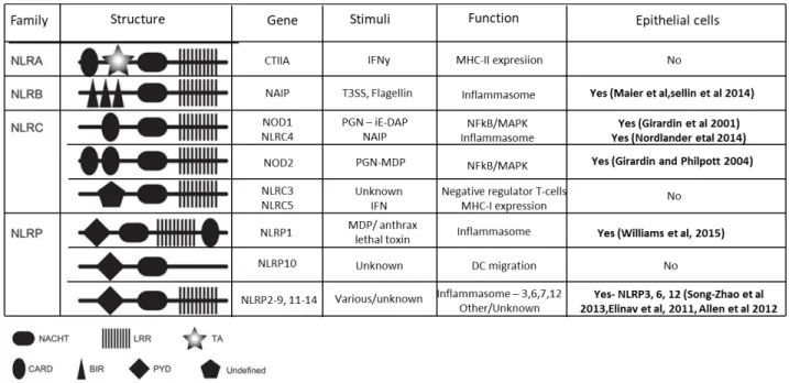
Unlike TLRs, which are membrane bound receptors, NLRs are cytosolic PRRs and recognize both PAMPs and DAMPs. The downstream signaling cascades triggered following NLR activation promote a number of cellular processes such as inflammasome assembly, immune signaling and autophagy (Motta et al., 2015). NLRs share an N-terminal protein binding domain (NBD), C-terminal leucine rich repeats and a central Nucleotide NOD domain. The NOD domain is also known as the NACHT domain and consists of seven conserved motifs. The family can be subdivided into four families NLRA, NLRB, NLRC and NLRP (Ting et al., 2008). In humans, only one member of each the NLRA and NLRB families is expressed; CIITA and NAIP respectively. The NLRC subfamily consists of six members: NLRC1 (NOD1), NLRC2 (NOD2), NLRC3 (NOD3), NLRC4 (IPAF), NLRC5, and NLRX1 and is characterized by the presence of a Caspase Recruitment Domain (CARD) domain, at least in NOD1, NOD2 and NLRC4. The NLRP family is characterized by the presence of an N-terminal pyrin domain. Whilst the expression of NLRs is found largely in immune cells, not all of them have been found to be expressed in epithelial cells (Pott and Hornef, 2012). Those found in IECs are NAIP (Maier et al., 2007), the NLRCs NOD1 and NOD2 (Philpott and Girardin, 2004) as well as NLRC4 (Nordlander et al., 2014; Sellin et al., 2014) and the NLRPs 1, 3, 6 and 12 (Allen et al., 2012; Elinav et al., 2011; Song-Zhao et al., 2014; Williams et al., 2015) (Figure 4).
[16]
Figure 4. NOD-Like Receptors (NLRs) structure and function. (Adapted from (Motta et al., 2015). LLR,
leucine-rich repeats; TA, transactivation; CARD, caspase recruitment domain; BIR, baculoviral inhibition of apoptosis protein repeat; PYD, pyrin domain.
1.1.3.2.1 NOD1 and NOD2
NOD1 was the first NLR family member to be identified (Bertin et al., 1999). NOD2, which is closely related to NOD1 but has an extra CARD domain, was described shortly thereafter (Ogura et al., 2001). The two proteins were shown to be able to activate NF-B and mitogen activated protein kinases (MAPKs) in response to infection with intracellular Gram-negative bacteria. This observation was first attributed to the recognition of LPS (Girardin et al., 2001; Inohara et al., 2001). However, further studies showed that it was, in fact, PGN that was recognized by these two proteins. NOD2 recognizes muramyl dipeptide (MDP), a component common to both Gram-negative and Gram-positive bacteria (Girardin et al., 2003a) whereas NOD1 recognizes -d-glutamyl-meso-diaminopimelic acid (iE-DAP) mainly present in Gram-negative bacteria (Chamaillard et al., 2003; Girardin et al., 2003b). It has been suggested that both NOD1 and NOD2 interact directly with their cognate ligands via the LRR region (Grimes et al., 2012; Laroui et al., 2011). Without stimulation, NOD1 and NOD2 exist in a monomeric auto-inhibited states in the cytosol (Caruso et al., 2014). Upon ligand binding, a conformational change occurs leading to their homo-oligomerization and the recruitment of the receptor-interacting serine/threonine-protein kinase 2 (RIP2) through homotypic CARD-CARD interactions. This process is necessary for the downstream activation of NF-B and the MAPK (Hasegawa et al., 2008).
[17]
How PGN enters the cytosol depends on the bacterial model. Certain bacteria invade the cell making peptidoglycan fragments available in the cytosol. This is the case for the sensing of the Gram-negative bacteria S. flexneri (Girardin et al., 2001), enteroinvasive E. coli (Kim et al 2004) as well as by the Gram-positive bacteria L. monocytogenes (Opitz et al., 2006) by NOD1. Other modes of delivery include direct injection into the cytosol by the Gram-negative bacteria Helicobacter pylori (Viala et al., 2004) as well as outer membrane vesicles from H. pylori, Neisseria gonorrhoeae and P. aeruginosa (Kaparakis et al., 2010). Others have found that PGN can enter cells through endocytosis (Lee et al., 2009; Marina-García et al., 2009). Whilst NOD1 is important for sensing of PGN, in vivo studies have only shown moderate effects of NOD1 KO in mice (Philpott et al., 2014). This is likely due to the presence of other receptors such as TLRs, which activate the same downstream signaling pathways thus conferring a certain level of redundancy (Park et al., 2009). Consistently, the importance of NOD1 signaling is enhanced in TLR unresponsive cells (Kim et al., 2004).
1.1.3.2.2 NLRs and the inflammasome
The other NLRs expressed by epithelial cells; NAIP, NLRC4, and NLRPs 1, 3, 6 and 12 are all involved with inflammasome assembly. The inflammasome is a multimeric protein complex, which activates caspase-1 (Martinon et al., 2002). This is a necessary step in the maturation of the pro-inflammatory cytokines IL-1 and IL-18, which are expressed as inactive precursors in the cytoplasm. It also leads to a caspase-1-dependent inflammatory cell death termed pyroptosis characterized by IL-1 release (Bergsbaken et al., 2009). Activation is triggered by the recognition of PAMPs or DAMPs by specific NLRs. Upon activation, NLRs oligomerize via their NBD. They then recruit pro-caspase-1 either directly via the CARD domain, as is the case for NLRC4, or through the CARD-pyrin containing adaptor associated speck-like protein (ASC). This results in caspase-1 activation and the subsequent cleavage of pro-IL1 and pro-IL-18. Since the inflammasome is mainly associated with cells of hematopoietic origin, most studies into its activation and function have been performed in these cells. However, a growing number of evidence has brought to light the importance of the inflammasome in epithelial cells (Sellin et al., 2015).
In IECs NLRP6 and NLRP12 are thought to play a regulatory role and to be involved in the maintenance of intestinal homeostasis (Chen, 2014). NLRP6, for example, seems to be particularly important in goblet cells for the production of mucus (Wlodarska et al., 2014). NLRP3 is one of the most well studied NLRs and is activated by a large number of stimuli including bacteria, viruses, fungi as well as DAMPs such as ATP and hyaluronan (Menu and Vince, 2011). It is therefore thought that it does not directly recognize a specific ligand per se but rather that it
[18]
senses changes within the cell such as potassium efflux (Muñoz-Planillo et al., 2013) or mitochondrial factors such as the production of ROS (Zhou et al., 2011). A unique characteristic of NLRP3 is that it requires a two-step activation. The first priming step following TLR stimulation leads to NF-B activation and results in the transcription of the caspase-1 and IL-1 genes as well as NLRP3 itself (Bauernfeind et al., 2009) although more recently, studies have shown that there is also transcription independent priming which is rather dependent of post-translational modifications such as deubiquitination (Juliana et al., 2012) and phosphorylation (Song et al., 2017). The second step is induced by the NLRP3 activating agent and leads to its oligomerization and inflammasome activation. A number of bacterial pathogens have been shown to activate NLRP3 in macrophages. These include both Gram-positive bacteria including S. pneumoniae, L. monocytogenes, and S. aureus, as well as the Gram-negative bacteria N. gonorrhea, P. aeruginosa and S. typhimurium (Menu and Vince, 2011). In epithelial cells, its role is less clear although one study has suggested that NLRP3 may protect against Citobacter rodentium colonization and spread (Song-Zhao et al., 2014).
In mice, the NAIP/NLRC4 inflammasome is activated in macrophages in response to a number of Gram-negative bacterial pathogens including S. typhimurium, P. aeruginosa and S. flexneri (Miao et al., 2006; Suzuki et al., 2007). The mouse NAIP5 and NAIP 1/2 recognize flagellin and the T3SS respectively (Zhao et al., 2011). The only NAIP in humans was thought to recognize proteins present in the T3SS of certain bacteria but not flagella (Yang et al., 2013). However, another recent study has shown that an isoform present in primary macrophages, but not in certain cell lines, was able of detecting flagellin (Kortmann et al., 2015). Recognition of the ligand by NAIP leads to its association with the downstream NLR, NLRC4. NLRC4 is responsible for the downstream recruitment of caspase via direct CARD-CARD domain interactions. In epithelial cells, NAIP and NLRC4 have been shown to have a protective role during S. typhimurium and C. rodentium infection with Nlrc4 KO mice showing increased bacterial loads compared to wild-type mice, especially early on in infection (Nordlander et al., 2014; Sellin et al., 2014). The activation of the NAIP/NLRC4 inflammasome is thought to control S. typhimurium infection by leading to the cell death of infected cells, thus favoring their extrusion from the epithelial lining and limiting infection (Sellin et al., 2014).
1.1.3.2.3 Non-canonical inflammasome
In addition to the NLR-mediated inflammasome assembly, the inflammasome can also be activated independently of NLRs. This non-canonical inflammasome is based on the direct activation of caspase-11 in mice or the human counterparts, caspases 4/5. These caspases have been shown to directly bind LPS and are thus thought to be the intracellular receptors of LPS
[19]
(Hagar et al., 2013; Kayagaki et al., 2013; Meunier et al., 2014; Shi et al., 2014). The non-canonical inflammasome has been shown to play an important role in the restriction of a number of bacterial infections. In murine macrophages, caspase-11 activation can induce pyroptosis, which increases clearance of S. typhimurium (Aachoui et al., 2013). In epithelial cells, the non-canonical inflammasome is required for IL-18 but not IL-1 secretion in IECs of both mice and humans (Knodler et al., 2014a). In human epithelial cells, the bacteria S. flexneri, S. typhimurium and E. Coli all cause caspase-4-dependent cell death favoring expulsion of infected cells from the intestinal epithelium (Knodler et al., 2014a; Kobayashi et al., 2013).
1.1.4 Downstream signaling and NF-B activation
Whilst different PRRs exist, many of the downstream signaling pathways converge. The signaling pathways, apart from those activated following inflammasome assembly which results in IL-1 and IL-18 production, lead to the activation of the NF-B and the MAPKs extracellular signal-regulated kinase (ERK), p38 and Jun N-terminal kinase (JNK). This results in the production of pro-inflammatory cytokines, which are responsible for the pro-inflammatory response associated with infection.
NF-B was first identified in B cells as a nuclear factor that binds the enhancer element of the immunoglobulin (Ig) light-chain gene (Sen and Baltimore, 1986). It is now known that NF-B proteins are ubiquitously expressed and are a family of transcription factors that control the transcription of many different genes in the cell ranging from its own regulation, to cell survival and apoptosis and, of course, immune signaling and inflammation (Vallabhapurapu and Karin, 2009). The family consists of five proteins, which can form homo- or heterodimers and all possess a Rel homology domain responsible for dimerization and DNA binding (Ghosh et al., 1998). The most abundant and the most ubiquitously expressed of these dimers is the p65/p50 heterodimer. Due to its central role in gene transcription, NF-B activation is tightly regulated. In steady state, it is present in an inactive form in the cytoplasm. The inhibitor of B (IB ) protein ensures this by occluding its nuclear localization site (Verma et al., 1995). In order for NF-B to be released, IB must be phosphorylated and ubiquitinated, tagging it for degradation by the proteasome (Chen et al., 1995; Henkel et al., 1993). Two pathways of NF-B activation exist, the canonical and the non-canonical pathways, of which I will only discuss the former. In the canonical pathway, the complex responsible for IB phosphorylation is IB Kinase (IKK). This multimeric complex is composed of 2 catalytic subunits IKKα and IKK as well as a non-catalytic regulatory subunit NF-B Essential Modulator NEMO (DiDonato et al., 1997; Rothwarf et al., 1998; Yamaoka et al., 1998). Activation of the IKK complex requires the phosphorylation of the IKKα and IKK subunits
[20]
(Mercurio et al., 1997). Transforming growth factor- activated kinase-1 (TAK1) is central to this process (Wang et al., 2001).
Following TLR recognition of a ligand and dimerization, TIR-containing adaptor proteins are recruited via TIR-TIR domain interactions. Five have been described to date in the literature; Myd88, Myd88 adaptor-like protein (MAL), TIR domain containing adaptor protein inducing IFN (TRIF), TRIF-related adaptor molecule (TRAM) and sterile α- and armadillo-motif-containing protein (SARM) (O’Neill and Bowie, β007). Myd88 is the most common and is utilized by all of the TLRs apart from TLR3. Its recruitment is followed by the recruitment of the IL-1 receptor associated kinase (IRAK) 4, which subsequently interacts with IRAK1 and IRAK 2 (Lin et al., 2010; Motshwene et al., 2009). IRAK1 and 2 possess motifs, which interact with the E3 ubiquitin ligase Tumor necrosis factor Receptor Associated Factor (TRAF) 6. Ubiquitination, although well characterized as a process targeting proteins for proteasomal degradation, is emerging as highly important in immune signaling events (Hu and Sun, 2016). It is a sequential three-step process involving ubiquitin-activating (E1), ubiquitin-conjugating (E2), and ubiquitin-ligating (E3) enzymes, which can conjugate either single ubiquitin molecules or ubiquitin chains to a lysine residue on target protein. These modifications can be “read” by ubiquitin binding proteins and lead to downstream signaling events (Husnjak and Dikic, 2012).
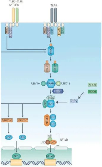
Following its recruitment, TRAF6 undergoes autoubiquitination. TAK1 forms a complex with the regulatory proteins TAK1-binding protein (TAB)1, TAB2 and/or TAB3. Whilst TAB1 enhances TAK1 kinase activity (Shibuya et al., 1996), TAB2 and TAB3 are capable of binding to the ubiquitin chains of TRAF6 (Kanayama et al., 2004). This is an essential step in the activation of TAK1 since mutants of TAB, which do not bind these ubiquitin chains, are unable to activate the kinase (Kanayama et al., 2004; Xia et al., 2009). TAK1 is a serine/threonine kinase and plays a central role in the activation of NF-B since it is the kinase responsible for the phosphorylation and activation of the IKK complex (Wang et al., 2001). It is not completely clear how this is achieved but NEMOs capacity to bind K63 polyubiquitinated chains may play a part in this by bringing the two complexes into proximity (Wu et al., 2006). Signaling via the NLRs NOD1 and NOD2 also leads to the activation of TAK1. However, this does not happen through the recruitment of the IRAK kinases but rather through RIP2. RIP2 is recruited to NOD via CARD-CARD interactions (Inohara et al., 1999). It undergoes K63-linked ubiquitination and interacts with TAB2/TAB3 leading to the activation of TAK1 and subsequent activation of IKK and NF-B (Hasegawa et al., 2008) (figure 5).
[21]
Figure 5. Signalling cascade activated following TLR and NOD 1/2 activation. (Adapted from Liew et
al., 2005). TLR dimerization following ligand recognition leads to the recruitment of adaptor proteins such as MAL and Myd88. This is followed by the activation of IRAK4, IRAK1 and IRAK2. IRAKs 1 and 2 interact with the ubiquitin ligase TRAF6, which activates TAK1. TAK1 can also be activated following NOD1 and NOD2 stimulation, resulting in the activation of the kinase RIP2 and subsequently TAK1. TAK1 in turn activates the IKK complex, which tags IB for proteasomal activation thus releasing NF-B, which then translocates to the nucleus and exerts its activity as a transcription factor. TAK1 also phosphorylates MAPKKs (MKK), which activate the MAPKs such as p38 and ERK. This step is necessary for the activation of the transcription factor AP-1 which, like NF-B, induces transcription of proinflammatory cytokines.
In addition to its role in phosphorylating IKK, TAK1 is also central in the activation of the MAPKs JNK, ERK and p38. These proteins are involved with the regulation of transcriptional responses mediated by external signals (Whitmarsh, 2007). The MAPK signaling cascade is a three-step process whereby a MAPK kinase kinase (MAPKKK) phosphorylates and activates a MAPK Kinase (MAPKK), which subsequently activates the aforementioned MAPKs by dual phosphorylation of the Thr–X–Tyr activation motif. TAK1 is itself a MAPKKK, and is responsible for the activation of p38 and JNK whilst also participating indirectly to the activation of ERK via IKK-induced proteolysis
[22]
of the NF-B subunit precursor protein p105 (Beinke et al., 2004). Activated MAPKs have many targets and functions and can phosphorylate their substrates either in the cytoplasm or translocate to the nucleus to exert this function (Yang et al., 2003). JNK and p38, for example, are important for the activation of another transcription factor called activator protein 1 (AP-1). This transcription factor is a dimeric complex most commonly formed by JUN and Fos proteins in mammals (Eferl and Wagner, 2003). It plays a key role in regulation of cell proliferation, differentiation and inflammatory processes and controls the expression of a number of cytokines. p38 and ERK activate Mitogen and stress activated protein kinases 1 and 2 (MSK1/2), which, among many other roles, phosphorylate histone H3, making DNA more accessible to transcription factors (Arthur, 2008).
1.1.5 Cytokines
Upon infection, the result of the activation of the transcription factors is ultimately the production of pro-inflammatory cytokines, which are necessary for the immune response that follows. Whilst IECs can produce a number of different cytokines, by far the most abundantly produced during bacterial infections is interleukin-8 (IL-8). The IL-8 promoter possesses both AP-1 and NF-B DNA binding sites (Roebuck, 1999). It is, in fact, a chemokine and a strong chemoattractant for polymorphonuclear cells (PMNs), which express the chemokine receptors CXCR1 and CXCR2. This leads to their recruitment to the infected tissue. These cells are the first to be recruited and play a key role in bacterial clearance. Indeed, strong IL-8 production and neutrophil influx is observed following infection with invasive bacteria such as S. typhimurim, S. flexneri and L. monocytogenes (Eckmann et al., 1993; Sansonetti et al., 1999). Other cytokines, which are also produced by epithelial cells include interleukin-6 (IL-6) and TNFα. IL-6 participates in neutrophil recruitment (Fielding et al., 2008) as well as other inflammatory processes such as the acute phase response. It also plays a role in both T and B lymphocyte activation (Ataie-Kachoie et al., 2014). TNFα has many roles both in inflammation as well as normal physiological processes. It is an activator of NF-B via the TNF receptor promoting the further production of cytokines including IL-6 (Shalaby et al., 1989), IL-8 (Kolios et al., 1996) and TNFα itself (Philip and Epstein, 1986). TNFα, along with IL-1 , which is also capable of activating NF-B , is one of the key mediators of endotoxic shock (Dinarello, 1991).
As previously mentioned, the two cytokines IL-1 and IL-18 are produced following inflammasome activation. Both of these cytokines belong to the IL-1 family of cytokines. The family is particularly associated with the effects of acute inflammation such as fever, vasodilation and hypertension (Dinarello, 2009). Whilst the main producers of IL-1 are macrophages, epithelial cells do produce
[23]
it as well (Franchi et al., 2012; Knodler et al., 2014a). IL-1 has many roles both in inflammation and beyond. It is capable of stimulating NF-B activation through signaling via the IL-1 receptor and thus all the associated downstream effects. IL-1 also leads to the increased expression of adhesion molecules favoring infiltration of inflammatory cells (Beck-Schimmer et al., 1997; Smith et al., 1988). It functions as a co-stimulator for T cells along with an antigen or mitogen and is important for polarizing T helper 17 (Th17) cells (Acosta-Rodriguez et al 2007). Unlike pro-IL-1 , pro-IL-18 is constitutively expressed throughout the gastrointestinal tract under steady state conditions (Puren et al 1999). IL-18 is particularly important for inducing the production of IFN by T lymphocytes in conjunction with IL-12 (Tominaga et al., 2000). IFN is important in restricting pathogen intracellular replication as is seen during Francisella tularensis and S. flexneri infections (Le-Barillec et al., 2005; Lindgren et al., 2007; Way et al., 1998). Similarly, IL-18 is also important for Natural Killer (NK)-derived IFN as well as attracting and activating NK cell granule secretion during S. typhimurium infection (Müller et al., 2016).
[24]
2 Intracellular Pathogens and Immunity
In spite of all the defense mechanisms developed by the host to protect the organism from pathogens, certain bacteria manage to establish infection. Some cause disease by colonizing the extracellular surface of epithelial cells such as H. pylori and EPEC, whilst others have opted for an intracellular lifestyle. Enteroinvasive pathogens that target IECs include both Gram-negative and Gram-positive bacteria. These bacteria use different mechanisms in order to gain access to the IEC cytoplasm where they can survive and replicate. The result of such infections is acute inflammation of the gut although specific symptoms and severity vary depending on the pathogen. An important Gram-positive intestinal pathogen is L. monocytogenes, which causes the disease listeriosis. This disease has a variety of symptoms depending on the infected individual, from gastroenteritis in healthy individuals to meningitis in immunocompromised patients and abortions in pregnant women (Cossart, 2011). Salmonella are Gram-negative enteroinvasive bacteria causing a range of diseases, including gastroenteritis, bacteremia, enteric fever and focal infections. There are over 2500 serovars in the Salmonella enterica species, defined on the basis of their flagella and LPS (LaRock et al., 2015). The most commonly studied is Salmonella typhimurim, a non typhoidal strain, which generally causes acute gastroenteritis in humans. In our work, we have used the Gram-negative bacterial pathogen S. flexneri as a model.
2.1 Shigella flexneri: A model pathogen
2.1.1 Epidemiology
Bacteria of the genus Shigella are Gram-negative bacteria belonging to the family Enterobacteriacae and are the cause of the disease shigellosis, otherwise known as bacillary dysentery. This disease can vary in severity from watery diarrhea to severe inflammatory dysentery characterized by blood and mucus in the stool, and accompanied by abdominal cramps and fever (Schroeder and Hilbi, 2008). It is the most prevalent cause of bloody diarrhea in developing countries, accounting for anywhere between 80 - 165 million cases of infection per year (Bowen, 2017). It is thought to be associated with around 600 000 deaths per year, mainly in children under the age of 5, although these estimates vary (Mani et al., 2016). It is spread via the faeco-oral route, either by direct contact or through contaminated food and water. As few as 10-100 microorganisms are thought to be enough to cause disease (DuPont et al., 1989). The current treatment for shigellosis is antibiotic treatment; however multi-drug resistance has become a growing concern (Phalipon and Sansonetti, 2007). A vaccine is highly desirable but current efforts thus far have been unsuccessful (Mani et al., 2016).
[25]
The genus Shigella is divided into 4 subgroups S. boddyi, S. sonnei, S. dysenteriae and S. flexneri which, apart from S. sonnei, can be further divided into several serotypes. The serotypes are defined by the O-antigen, the outer most part of the LPS (Lindberg et al., 1991). The first 2 species are generally associated with the milder form of the disease whilst S. dysenteria is the cause of the most devastating epidemic outbreaks and represents the most severe form of dysentery largely due to the presence of the Shiga toxin (Phalipon and Sansonetti, 2007). S. flexneri is the principal cause of endemic shigellosis in developing countries and is the most studied and well characterized. It is the strain used in this work.
2.1.2 Virulence plasmid and T3SS
Genetic studies have shown that there is only 1.5% divergence between Shigella and non-invasive E. coli (Lan and reeves, 2002). Shigella have acquired a 213 kb virulence plasmid allowing them to adapt to a facultative intracellular lifestyle (Sansonetti 1982, Sasakawa 1986). This plasmid, pWR100, encodes around 100 genes (Buchrieser et al., 2000). A 31 kb region is of particular importance for bacterial entry into host cells (Maurelli et al., 1985; Sasakawa et al., 1989). It encodes the Mxi/spa proteins, which form the T3SS, a syringe like complex extending a needle into the external milieu. It spans both the outer and inner bacterial membranes and is necessary for bacterial entry into host cells (Blocker et al., 2001; Tamano et al., 2000). Around 25 Gram-negative bacterial species possess such an apparatus including Chlamydia, pseudomonas, Yersinia and Salmonella (Cornelis 2006). Whilst architectural differences are observed between species, they all possess a conserved core related to the flagellar T3SS (Diepold and Armitage, 2015).
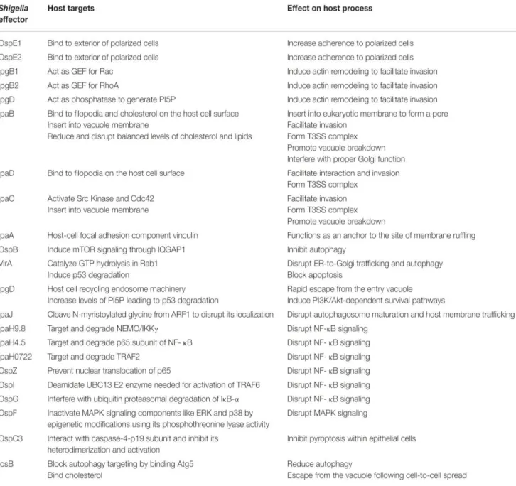
The T3SS is capable of penetrating the host cell and translocating proteins from the bacterial cytoplasm into the host cytoplasm. These proteins, called effectors, are able to interfere with a number of cellular processes to facilitate bacterial entry, survival and spread, and regulate inflammation (Figure 6). In S. flexneri, the expression of these genes is under tight regulation with the key trigger being the temperature switch to 37°C (Tobe et al., 1991). This leads to the increased expression of the transcription activator VirF, which in turn leads to the production of VirB (Tobe et al., 1993). VirB controls the expression of the entry region (Le Gall et al., 2005). Whilst the needle complex is assembled at 37°C, it is only weakly active (Allaoui et al., 1993) with the effectors stored in the cytoplasm associated to chaperone proteins (Ménard et al., 1994). Contact of bacteria with host cells, or the dye Congo red, constitutes a secretion signal leading to a rapid burst of protein secretion (Ménard et al., 1994; Parsot et al., 1995). This activation signal leads to the production of a second set of genes necessary for the intracellular phase of the life cycle and regulated by the transcriptional activator MixE (Mavris et al., 2002).
[26]
Figure 6. List of Shigella effectors and their function (Killackey et al., 2016).
Over 25 proteins are involved in the needle complex assembly, which consists of 2 pairs of rings joined together and spanning the inner and outer bacterial membrane, and the needle, which protrudes out (Cornelis, 2006) (Figure 7). Electron microscopy studies on the needle complex of both S. typhimurium and S. flexneri, which share structural similarities, have revealed much about its architecture (Blocker et al., 1999, 2001; Kubori et al., 1998; Sani et al., 2007; Tamano et al., 2000). The basal body has a length of around 32 nm and a width of 20-40 nm whilst the needle is around 45 nm in length and 7 nm in width (Tamano et al., 2000). An internal 2-3 nm channel spans
[27]
the complex (Blocker et al., 2001). The first step in assembly is the formation of the basal body, which consists of the periplasmic and inner membrane rings made up of the proteins MxiJ and MxiG. These proteins interact with MixD and MixM, which form the outer membrane ring (Blocker et al., 1999; Schuch and Maurelli, 2001; Tamano et al., 2000). Once assembled, the rest of the needle structure can be formed. It is composed of a major subunit MxiH and a minor subunit MxiI, which form an extracellular helical polymer (Blocker et al., 2001; Cordes et al., 2003; Tamano et al., 2000).
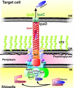
Figure 7. The S. flexneri Mxi-Spa T3SS (Schroeder and Hilbi, 2008). The basal body spans the bacterial
inner-membrane and outer-membrane with the hollow needle protruding into the extracellular medium. Upon contact with the host cell, IpaB and IpaC insert into the membrane forming a pore, stabilized by IpaD. Effectors, which are stored in the cytoplasm linked to chaperone proteins, can then be unfolded and translocated into the host cell.
The whole process is tightly controlled by a number of proteins, which are associated with the basal body. Spa33 is central to the recruitment and export of T3SS-associated proteins (Morita-Ishihara et al., 2006). It interacts with the proteins MxiK, MxiN, Spa32 and the ATPase spa47. MxiK and MxiN are necessary for MxiH transport to the needle complex (Jouihri et al., 2003). Spa32 controls the length of the needle (Magdalena et al., 2002). Spa47 is thought to provide the energy required for the unfolding of T3SS substrates, chaperone release, and transmembrane transport as is the case with the Salmonella ATPase InvC (Akeda and Galán, 2005) since the translocation of proteins is an energetically unfavorable process and fully folded proteins cannot pass through the channel.
[28]
2.1.3 Infection Cycle
The T3SS allows Shigella to invade and survive within the cellular cytoplasm, but to understand the symptoms of the disease, one must look at the whole infection cycle. Much of Shigella’s life cycle was established in the 80s and 90s using S. flexneri and has been very well studied since, although many of the molecular mechanisms still remain to be determined. One challenge with studying S. flexneri infection has been the lack of appropriate animal models since it is a human and primate only pathogen. A ligated ileal loop model in rabbits has been used for much of the phenotypical characterization as it leads to invasion and an acute inflammation with the associated symptoms. However, limited availability of the necessary immunological tools and practical issues make this model more difficult to use (Phalipon and Sansonetti, 2007). In mice, S. flexneri does not cause disease that resembles that of the human disease in the intestine although a pulmonary model following intranasal inoculation has proved useful (van de Verg et al., 1995) as well as infection as in newborn mice (Fernandez et al., 2003). Other organisms, which have been used as models for S. flexneri infection are guinea pigs (Barman et al., 2011; Shim et al., 2007), piglets (Jeong et al., 2010) and, recently, zebrafish larvae (Mostowy et al., 2013). The latter may hold promise due to the ever-growing availability of molecular tools and the possibility of in vivo imaging of the transparent larvae (Lieschke and Currie, 2007). More work will be needed to establish the relevance of this model in terms of human disease.
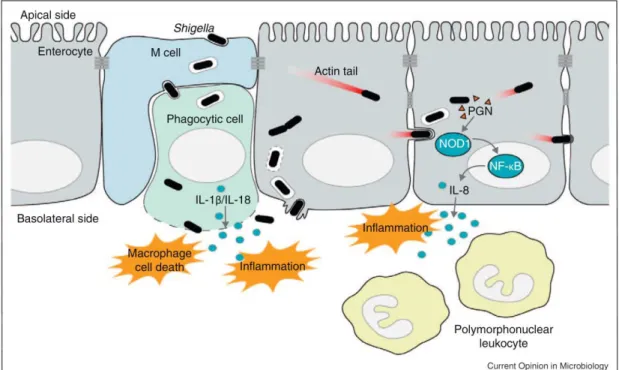
The general infection cycle is as follows (Figure 8). Following ingestion, S. flexneri travels through the intestine to the colon where it infects colonic epithelial cells. Whilst invasion is possible at the apical pole of polarized IECs (Carayol and Tran Van Nhieu, 2013), the majority occurs via the basolateral pole (Mounier et al., 1992). In order to get access to the underside, S. flexneri uses transcytosis via M cells (Wassef et al., 1989), and is quickly engulfed by the resident macrophages. S. flexneri is able to escape from the macrophage and induces its own uptake into epithelial cells. Once inside the cell, bacteria can replicate and perform intracellular and intercellular movement, infecting cells along the epithelium. Epithelial cells are capable of sensing infection and respond by producing the chemokine IL-8. PMNs migrate to the site of infection in response, a key step in bacterial clearance. However, in the process, PMNs also destabilize the integrity of the epithelium, resulting in the symptoms associated with bacillary dysentery. Each step of this process involves a complex interlay of bacterial and host factors, which will now be discussed in further detail.
[29]
Figure 8. S. flexneri infection cycle (Ashida et al., 2011).
Macrophage escape
Shigella’s first challenge in avoiding an untimely end is escaping destruction by macrophages. S. flexneri is capable of escaping the phagocytic vacuole and inducing what was thought to be apoptosis in the infected macrophage (Zychlinsky et al., 1992). It has since been shown that it is in fact pyroptotic cell death, dependent on the activation of caspase-1 (Hilbi et al., 1998) and accompanied by the release of the pro-inflammatory cytokines IL-1 and IL-18. The bacterial effector IpaB is central to this process. It co-localizes with activated caspase-1 on the bacterial surface, in the cytoplasm and on vesicular membranes of infected macrophages (Schroeder et al., 2007). The spontaneous formation of IpaB oligomers, which can insert into the vacuolar membrane causes an ion flux and subsequent disintegration of the vacuolar membrane (Senerovic et al., 2012). This event leads to the activation of caspase-1 via a NLRC4-dependent mechanism (Senerovic et al., 2012; Suzuki et al., 2007). Although this inflammatory cell death serves as a danger signal, it, in fact, seems to be beneficial for the bacteria by facilitating entry. Indeed, blocking IL-1 activity in a rabbit ligated ileal loop model of infection led to a decrease in bacterial invasion (Sansonetti et al., 2000). Furthermore, in a IL-1 KO model of lung infection, the inflammatory response was reduced but with similar bacterial clearance (Arondel et al., 1999).
[30] Epithelial cell entry
Once escaped from the dying macrophage, the bacterium enters into epithelial cells. Unlike macrophages, which are phagocytic by nature, epithelial cells are not. Whilst S. flexneri has no identified cognate receptor, it adheres to epithelial cells by binding the hyaluronan receptor CD44 and α5 1 integrin (Skoudy et al., 2000; Watarai et al., 1996). Recent evidence suggest that prior
to this, the bacteria interact with cellular filopodia, which facilitate their contact with the cellular body (Romero et al., 2011). Once in contact with the host cell, S. flexneri is able to induce its own uptake in a T3SS-dependent manner. The effectors IpaB, IpaC and IpaD, whose expression is under control of VirB, are necessary for this process (Ménard et al., 1993). IpaB and IpaC associate separately with the chaperone protein IpgC in the bacterial cytoplasm but can form a complex together once secreted by the bacterium (Ménard et al., 1994). IpaD has self-chaperoning activity and locates at the tip of the needle where it acts as a plug to avoid premature secretion (Espina et al., 2006; Johnson et al., 2007). Exposure to bile salts is thought to lead to the recruitment of IpaB to the tip where it associates with IpaD (Olive et al., 2007; Sani et al., 2007; Veenendaal et al., 2007). From this position, it can sense the host cell membrane components and recruit IpaC (Epler et al., 2009). IpaB then inserts into the host cell membrane and, along with IpaC, forms the 2.5 nm wide pore (Blocker et al., 1999). The efficiency of insertion is increased by IpaD (Picking et al., 2005). The formation of this translocon is necessary for the translocation of other bacterial effectors into the host cytoplasm and entry via macropinocytosis. A number of bacterial effectors including IpgB1, IpgB2 and IpaC as well as host components such as Rho GTPases and kinases lead to the induction of complex cytoskeletal rearrangements, which results in the formation of lamelipodia or “ruffles”, enveloping and engulfing the bacterium (Carayol and Tran Van Nhieu, 2013). Another effector, IpgD, is also important for the formation of the ruffles with deletion mutants causing much smaller entry foci but not showing a defect in invasion. This is attributed to its hydrolytic activity on Phosphatidylinositol 4,5-bisphosphate (PI(4,5)P2), ,which
destabilizes cortical actin, leaving more monomers free to polymerize at the invasion site (Niebuhr et al., 2002).
Vacuolar escape and cellular dissemination
Once inside the cell, the bacteria find themselves within a vacuole from which they must escape in order to gain access to the cytoplasm, their replicative niche. This vacuolar rupture is induced as soon as 10-15 min post infection and can be visualized in real time using galectin-3 as a marker of disassembled membranes (High et al., 1992; Paz et al., 2010; Ray et al., 2010; Sansonetti et al., 1986). Unlike the IpaB-dependent rupture, which has been suggested in macrophages, efficient vacuolar rupture was dependent on the recruitment of host Rab-GTPase Rab11 to the
[31]
bacterial entry site (Mellouk et al., 2014). This is mediated by IpgD since the deletion mutant did not recruit Rab11 and showed delayed and incomplete vacuolar rupture (Mellouk et al., 2014). It has recently been shown that macropinosomes other than the bacteria containing vacuole (BCV) are formed during this internalization process (Weiner et al., 2016) (Figure 9). Their formation is dependent on the presence of IpgD. The availability of the macropinosomes is correlated to the efficiency of vacuolar rupture; the fewer the macropinosomes, the slower the rupture. It is, in fact, to these macropinosomes that Rab11 is recruited and they interact directly with the BCV during vacuolar lysis (Weiner et al., 2016).
Figure 9. Role of IpgD during bacterial entry and vacuolar rupture. (Weiner et al., 2016). During WT S.
flexneri infection, macropinosomes are formed along with the BCV. They recruit Rab GTPases and participate in vacuolar rupture. The ΔipgD S. flexneri mutant causes less membrane ruffling and less macropinosome formation. This results in delayed vacuolar rupture.
Once in the cytoplasm, S. flexneri can move both intracellularly and intercellularly (Bernardini et al., 1989; Monack and Theriot, 2001). This is achieved through the action of the bacterial protein IcsA/ VirG. It localizes to one pole of the bacterium and can interact with host cell factors, including the neuronal Wiskott-Aldrich syndrome protein (N-WASP) and the Arp2/Arp3 complex. This serves as an actin nucleator and catalyzes the directed elongation of an actin tail, propelling the bacteria through the cytoplasm (Egile et al., 1999). This intracellular movement is also necessary for the intercellular spread. Bacteria target tricellular junctions where bacteria-containing pseudopodia are engulfed by neighboring cells via clathrin-mediated endocytosis (Fukumatsu et al., 2012). The virG deletion mutant is not compromised in its ability to invade epithelial cells. However, it is unable of cell-cell spread and thus forms a microcolony in the cell, which it invades.