HAL Id: tel-02426233
https://tel.archives-ouvertes.fr/tel-02426233
Submitted on 2 Jan 2020HAL is a multi-disciplinary open access archive for the deposit and dissemination of sci-entific research documents, whether they are pub-lished or not. The documents may come from teaching and research institutions in France or abroad, or from public or private research centers.
L’archive ouverte pluridisciplinaire HAL, est destinée au dépôt et à la diffusion de documents scientifiques de niveau recherche, publiés ou non, émanant des établissements d’enseignement et de recherche français ou étrangers, des laboratoires publics ou privés.
Camille Chalvin
To cite this version:
Camille Chalvin. Sclareol biosynthesis in clary sage and its regulation. Plant breeding. Université Paris Saclay (COmUE), 2019. English. �NNT : 2019SACLS194�. �tel-02426233�
and its regulation
Thèse de doctorat de l'Université Paris-Saclay préparée à l’Université Paris-Sud
École doctorale n°567 Sciences du Végétal : du gène à l’écosystème (SDV) Spécialité de doctorat: Biologie
Thèse présentée et soutenue à Gif-sur-Yvette, le 12/07/2019, par
Camille Chalvin
Composition du Jury : Marianne Delarue
Professeure, Université Paris-Sud (IPS2) Présidente Sylvie Baudino-Caissard
Professeure, Université Jean Monnet, Saint-Etienne (LBVpam) Rapporteur Andréa Hemmerlin
Chargée de recherche, CNRS (IBMP, Strasbourg) Rapporteur Johannes Panten
Responsable R&D, Symrise AG Examinateur
Françoise Granet
Manager d’expertise, Michelin Examinateur
Michel Dron
Professeur émérite, Université Paris-Sud (IPS2) Directeur de thèse Adnane Boualem
Directeur de recherche, INRA (IPS2) Invité Stéphanie Drevensek
Chargée de projet, Adeprina (IPS2) Invité
N N T : 2 0 1 9 S A C L S
1
Table of contents
Table of contents... 1 Résumé en français... 7 1. Introduction... 7 2. Résultats ... 72.1. Localisation de la biosynthèse du sclaréol ... 7
2.2. Identification de l’origine métabolique du sclaréol par marquage isotopique ... 8
2.3. La biosynthèse du sclaréol est-elle régulée par les jasmonates ? ... 9
2.4. Etude de la diversité naturelle de la sauge sclarée ... 9
3. Revue de la littérature ... 10
4. Perspectives ... 11
Introduction ... 13
1. Ambergris and sclareol ... 13
1.1. The search for substitutes to ambergris ... 13
1.2. Interest in increasing sclareol global production ... 14
2. Clary sage ... 15
2.1. Botanical description of clary sage ... 15
2.2. Sclareol biological function in clary sage ... 16
2.3. Clary sage commercial use ... 17
2.4. First advances in clary sage genetics and biotechnology ... 19
2.5. Clary sage breeding in France ... 20
3. Terpene biosynthesis and function in plants ... 21
3.1. Specialized metabolism and terpenoids ... 21
3.2. The diverse biological functions of terpenes ... 22
3.3. Industrial exploitation of terpenes ... 24
3.4. Terpene biosynthesis ... 25
3.5. Terpene biosynthesis regulation ... 32
4. Glandular trichomes ... 35
4.1. Capitate and peltate glandular trichomes of the Lamiaceae ... 35
4.2. Clary sage glandular trichomes ... 37
2
5. Terpene production by clary sage ... 39
5.1. Essential oil production in aerial parts ... 39
5.2. Sclareol biosynthesis and secretion ... 40
5.3. Terpene production in roots ... 42
6. Aim of the project... 43
Chapter 1 ... 45
Mass spectrometry imaging to localize sclareol biosynthesis ... 45
1. Introduction... 45
2. Materials and methods ... 47
2.1. Plant material ... 47
2.2. Scanning electron microscopy ... 47
2.3. Metabolite localization by LDI-FT-ICR ... 47
2.4. Sclareol and linalyl acetate quantification by GC-MS ... 48
3. Results ... 49
3.1. Different accumulation patterns for sclareol and linalyl acetate ... 49
3.2. Distinct roles of different trichome types in sclareol production ... 52
3.3. A clary sage glabrous mutant produces less sclareol and linalyl acetate ... 56
4. Discussion ... 58
4.1. Sclareol is mainly produced by glandular trichomes ... 58
4.2. Some sclareol could also be produced by other epidermal cells ... 60
4.3. Engineering higher glandular trichome density to increase sclareol yield ... 61
Chapter 2 ... 63
A retro-biosynthetic approach to decipher sclareol metabolic origin ... 63
1. Introduction... 63
1.1. Metabolic crosstalk between MVA and MEP pathways ... 63
1.2. Towards clary sage metabolic engineering ... 64
2. Materials and methods ... 66
2.1. Plant material ... 66
2.2. Treatment with MVA or MEP pathway-specific inhibitors ... 66
2.3. Sclareol and linalyl acetate quantification by GC-MS ... 67
2.4. Isotope labeling and analysis by 13C-NMR or GC-MS ... 67
3. Results ... 71
3.1. Sclareol and linalyl acetate are highly abundant terpenes of clary sage calyx surface ... 71
3
3.3. Comparison of results obtained for sclareol and linalyl acetate ... 79
3.4. The case of the acetate group of linalyl acetate ... 80
3.5. The mixed origin of the sesquiterpene β-caryophyllene ... 82
4. Discussion ... 84
4.1. Biosynthetic origin of mono-, sesqui- and diterpenes in clary sage and other plants... 84
4.2. Metabolic engineering of terpene production in plants ... 85
4.3. Perspectives for clary sage metabolic engineering ... 88
Chapter 3 ... 93
Is sclareol biosynthesis regulated by jasmonates? ... 93
1. Introduction... 93
1.1. Regulation of plant specialized metabolism by jasmonates ... 93
1.2. How to use jasmonate signaling to enhance the production of compounds of interest .. 94
1.3. Regulation of clary sage specialized metabolism by jasmonates: state of the art ... 95
2. Materials and methods ... 97
2.1. Calyx development stages ... 97
2.2. MeJA treatment by feeding ... 97
2.3. MeJA treatment by spraying... 97
2.4. Sclareol and linalyl acetate quantification by GC-MS ... 98
3. Results ... 99
3.1. Regulation of sclareol biosynthesis during calyx development ... 99
3.2. MeJA treatment on flowers by feeding ... 101
3.3. MeJA treatment on flowers by spraying ... 105
4. Discussion ... 105
4.1. MeJA apparently does not impact sclareol production, but further protocol optimization is needed ... 105
4.2. Other potential ways to manipulate sclareol biosynthesis regulation ... 107
Chapter 4 ... 109
Clary sage natural diversity ... 109
1. Introduction... 109
1.1. The use of natural genetic diversity for breeding ... 109
1.2. Impact of environmental conditions on terpene production and genetic diversity ... 110
2. Materials and methods ... 111
2.1. Plant material ... 111
4
2.3. Field trial ... 113
2.4. Sclareol and linalyl acetate quantification by GC-MS ... 114
3. Results ... 115
3.1. Croatian populations show genetic polymorphism in MEP pathway genes ... 115
3.2. Choice of the populations to be analyzed in the field... 117
3.3. Comparison of flowering date and metabolite content of the different populations .... 119
4. Discussion ... 122
4.1. Complex relationships between genetic diversity, chemical diversity and environmental conditions ... 122
4.2. Perspectives for the study of clary sage natural diversity... 124
4.3. Vatican White plants flower earlier and produce less specialized metabolites ... 125
Conclusion ... 127
1. Context and objectives of the project ... 127
2. Perspectives for research on sclareol production in clary sage ... 127
3. Perspectives for clary sage genetic improvement ... 129
Annex ... 131
Review: Genetic control of glandular trichome development ... 131
1. Introduction... 131
1.1. Why study the genetic control of glandular trichome development? ... 131
1.2. Which species could serve as model(s)? ... 132
2. Description of glandular trichome morphology and development ... 133
3. Genes controlling glandular trichome initiation ... 135
3.1. Transcription factors... 135
3.2. Cyclins ... 137
3.3. Regulatory complexes ... 139
3.4. Genes involved in hormonal signaling ... 140
4. Genes controlling later steps of glandular trichome development ... 140
4.1. Cytoskeleton regulators... 140
4.2. Cuticle deposition regulators ... 141
5. Evolution of glandular trichome development regulators ... 142
5.1. Evidence supporting the conservation of glandular trichome development regulators in the Solanaceae ... 142
5.2. Some regulators appear to be conserved in distant plant families, while the others are likely to have evolved independently ... 142
5
6. Perspectives ... 144 References ... 147
7
Résumé en français
1. Introduction
Le sclaréol est un terpène produit par les organes floraux de la sauge sclarée (Salvia sclarea), en particulier par les calices. Il est utilisé pour l’hémisynthèse de l’ambroxide, un composé très apprécié en parfumerie pour son odeur ambrée et ses propriétés fixatrices augmentant la ténacité des parfums. Le sclaréol utilisé pour produire de l’ambroxide est extrait à partir de sauge sclarée cultivée. En réponse au développement de l’utilisation de l’ambroxide en parfumerie fonctionnelle (détergents, shampooings…), la demande mondiale de sclaréol est actuellement en hausse, stimulant la recherche de moyens permettant d’en accroître la production.
Le projet PISTILL, porté par l’équipe « Développement des fleurs et des carpelles » de l’Institut de Sciences des Plantes de Paris-Saclay, a pour objectif d’augmenter le rendement de la production de sclaréol via l’amélioration génétique ciblée de la sauge sclarée. Le travail présenté dans ce manuscrit visait à compléter les connaissances actuelles sur la biosynthèse du sclaréol et sa régulation chez la sauge sclarée, avec un double objectif: d’une part, orienter le projet PISTILL vers des stratégies prometteuses pour l’augmentation du rendement en sclaréol, et d’autre part, améliorer notre compréhension générale de la production des terpènes chez les plantes. Nous avons entrepris de préciser l’origine cellulaire et métabolique du sclaréol dans les calices de sauge sclarée, et d’identifier les mécanismes de régulation contrôlant son accumulation. Nous avons également commencé à étudier comment la diversité naturelle de la sauge sclarée pourrait être exploitée. Cette étude ouvre des perspectives intéressantes pour la recherche fondamentale sur la production des terpènes chez les plantes, tout en mettant en évidence des pistes prometteuses pour l’amélioration génétique ciblée des performances de la sauge sclarée.
2. Résultats
2.1. Localisation de la biosynthèse du sclaréol
L’épiderme des calices de sauge sclarée est caractérisé par une forte densité de trichomes glandulaires. Chez de nombreuses espèces de plantes, ces structures multicellulaires sont
8
spécialisées dans la production de terpènes (Lange and Turner, 2013). La présence de sclaréol a été détectée dans les différents types de trichomes glandulaires de la sauge sclarée (peltés et capités) (Schmiderer et al., 2008). Cependant, d’autres cellules de l’épiderme du calice pourraient également sécréter du sclaréol (Caissard et al., 2012). Dans le premier chapitre de ce manuscrit, nous présentons plusieurs éléments appuyant l’hypothèse selon laquelle le sclaréol est principalement sécrété par les trichomes glandulaires. La répartition spatiale du sclaréol à la surface des calices a été analysée par imagerie par spectrométrie de masse, et les résultats indiquent que le sclaréol est particulièrement abondant au niveau des trichomes glandulaires capités. De plus, des mutants de sauge sclarée dépourvus de ces trichomes (obtenus via une mutagénèse à l’EMS) produisent moins de sclaréol. Dans le futur, l’identification de la mutation (ou des mutations) à l’origine de ce phénotype améliorerait notre compréhension du développement des trichomes glandulaires et de la production du sclaréol chez la sauge sclarée.
2.2. Identification de l’origine métabolique du sclaréol par marquage isotopique
La biosynthèse des terpènes débute par la condensation d’un nombre variable d’unités à cinq carbones : l’isopentényl diphosphate (IPP) et son isomère le diméthylallyl diphosphate (DMAPP). Chez les plantes, l’IPP et le DMAPP sont produits par deux voies métaboliques différentes : la voie du mévalonate dans le cytoplasme (voie MVA) et la voie du méthylérythritol-phosphate dans les chloroplastes (voie MEP). L’activité de ces deux voies alimente deux pools d’IPP et de DMAPP physiquement séparés au sein de la cellule. Selon la localisation subcellulaire des terpènes-synthases impliquées, la production d’un terpène donné ne dépend généralement que de l’un de ces deux pools (Lipko and Swiezewska, 2016). Lorsqu’elles sont exprimées de manière transitoire dans des feuilles de tabac (Nicotiana benthamiana), les terpènes-synthases impliquées dans la biosynthèse du sclaréol sont adressées aux chloroplastes (Caniard et al., 2012), le compartiment où la voie MEP est localisée. Cependant, l’origine métabolique du sclaréol n’avait pas encore été étudiée. Les approches développées dans le deuxième chapitre de ce manuscrit mettent en évidence les contributions respectives des voies MVA et MEP à la biosynthèse de trois terpènes dans les calices de sauge sclarée : le sclaréol, l’acétate de linalyle et le β-caryophyllène. Nous avons
9
réalisé un marquage isotopique en fournissant du glucose-1-13C à des tiges d’inflorescence coupées. L’analyse des résultats par RMN du 13C indique que le sclaréol et l’acétate de linalyle proviennent tous deux de la voie MEP, comme la plupart des mono- et diterpènes (Reed and Osbourn, 2018). En revanche, le β-caryophyllène semble être d’origine mixte, alors que la plupart des sesquiterpènes proviennent de la voie MVA (Reed and Osbourn, 2018) ; mais ce résultat nécessite d’être confirmé.
2.3. La biosynthèse du sclaréol est-elle régulée par les jasmonates ? La biosynthèse des terpènes est stimulée en réponse à des signaux développementaux ou environnementaux via l’activation de voies de signalisation spécifiques. Le renforcement de l’activité de ces voies de signalisation représente une autre possibilité d’accroître la production d’un terpène d’intérêt (Lu et al., 2016). Cette stratégie nécessite l’identification préalable des voies de signalisation stimulant la biosynthèse du terpène d’intérêt. Étant donné que la production de nombreux terpènes est activée par les jasmonates (Colinas and Goossens, 2018; Wasternack and Strnad, 2019), le troisième chapitre de ce manuscrit examine l’impact de ces phytohormones sur la production de sclaréol dans les calices de sauge sclarée. Du méthyljasmonate en solution a été fourni à des tiges d’inflorescence coupées ou appliqué sur plante entière par spray. Cependant, les expériences réalisées n’ont pas permis de démontrer une implication des jasmonates dans la régulation de la production de sclaréol. Deux possibilités restent plausibles: soit les jasmonates ne sont pas impliqués dans la régulation de la biosynthèse de sclaréol, soit le protocole expérimental doit encore être optimisé.
2.4. Etude de la diversité naturelle de la sauge sclarée
Les conditions environnementales et les polymorphismes génétiques sont responsables de variations de la teneur en terpènes entre différentes populations de la même espèce végétale. La pression de sélection exercée par les conditions de croissance peut conduire à la sélection de modifications génétiques modulant les niveaux de terpènes et rendant la plante plus adaptée à son environnement (Figueiredo et al., 2008; Moore et al., 2014). La sauge sclarée est présente à l’état sauvage dans divers types d’environnements du bassin méditerranéen et d’Asie occidentale (Wagner et al., 2012; Nasermoadeli and Rowshan,
10
2013). Les populations de sauge sclarée cultivées ne représentent probablement qu’un échantillon de la diversité naturelle de la sauge sclarée en termes de polymorphismes génétiques et de teneur en sclaréol. Le quatrième chapitre de ce manuscrit explore la diversité génétique et phénotypique de populations sauvages croates originaires de trois types d’environnements différents: côte, montagne et plateau. Bien que des polymorphismes génétiques soient détectables dans les gènes de biosynthèse du sclaréol, les populations croates cultivées dans le même environnement ne diffèrent pas significativement en termes de teneur en sclaréol et de date de floraison. Les conditions climatiques croates n’ont apparemment pas entraîné de différenciation génétique affectant la production de sclaréol et la floraison. Cependant, la différence de teneur en sclaréol observée entre les populations de référence « Vatican White » et « Milly » mérite une étude plus approfondie. En effet, l’identification de la cause de cette différence pourrait mener à l’identification de mécanismes de régulation de la production de sclaréol.
3. Revue de la littérature
Les trichomes glandulaires des plantes sont des structures épidermiques dédiées à la sécrétion de divers métabolites spécialisés. Ces métabolites contribuent à l’adaptation des plantes à leur environnement et beaucoup d’entre eux possèdent des propriétés exploitées par différents secteurs de l’industrie (parfumerie, pharmacie…). L’identification du réseau de gènes contrôlant le développement des trichomes glandulaires permettrait de mieux comprendre comment les plantes produisent ces métabolites spécialisés. Nos connaissances sur ce processus développemental sont encore limitées, mais des gènes contrôlant l’initiation et la morphogenèse du trichome glandulaire ont récemment été identifiés. La revue de la littérature présentée dans ce manuscrit a pour objectif de synthétiser ces découvertes récentes. D’après notre analyse, les facteurs de transcription appartenant aux sous-familles R2R3-MYB et HD-ZIP IV semblent jouer un rôle essentiel dans l’initiation du trichome glandulaire chez Artemisia annua et la tomate. Dans notre revue, nous nous concentrons sur les résultats obtenus chez ces deux espèces et proposons de premiers modèles de régulation génétique intégrant ces données.
11
4. Perspectives
Plusieurs stratégies visant à accroître la production de sclaréol chez la sauge sclarée peuvent être envisagées à partir des résultats décrits dans ce manuscrit. Le sclaréol étant dérivé de la voie MEP et principalement produit dans les trichomes glandulaires, l’augmentation de la densité des trichomes glandulaires ou la stimulation de la voie MEP pourraient permettre d’augmenter le contenu en sclaréol. De plus, étant donné que l’acétate de linalyle est également dérivé de la voie MEP et produit dans les trichomes glandulaires, bloquer la biosynthèse de l’acétate de linalyle pourrait augmenter la production de sclaréol en réduisant la compétition pour le substrat. La voie du MEP et la biosynthèse de l’acétate de linalyle pourraient être manipulées au moyen de modifications génétiques ciblant les enzymes impliquées dans ces voies. De même, la densité des trichomes glandulaires pourrait être augmentée via la manipulation des régulateurs de l’initiation des trichomes glandulaires. Par ailleurs, les programmes de sélection de la sauge sclarée pourraient être enrichis par une exploitation efficace de la diversité naturelle de la sauge sclarée. Les populations croates étudiées dans le quatrième chapitre de ce manuscrit représentent une ressource génétique précieuse, car nous avons montré qu’elles portent de nouveaux allèles par rapport aux populations de référence.
La faisabilité de ces approches dépendra de la mise au point d’outils dédiés à l’amélioration génétique de la sauge sclarée. Le séquençage du génome de la sauge sclarée et le développement d’outils biotechnologiques spécifiques à cette espèce seraient très utiles pour les projets de recherche fondamentale et appliquée visant à comprendre et à améliorer cette plante à parfum.
13
Introduction
1. Ambergris and sclareol
1.1. The search for substitutes to ambergris
Ambergris is a waxy substance secreted by the digestive tract of male sperm whales (Physeter macrocephalus) upon mucosa injuries caused by squid beaks (Figure 1). It is then expelled by the animal as viscous lumps which float on the sea and blocks of 10 g to 1 kg can eventually be found on beaches (Caissard et al., 2012). After several years of aging and exposure to sunlight, air and water, ambergris acquires a musky and sweet earthy smell (Barrero et al., 1993). It has been used for centuries in perfume manufacture for its distinct odor and it also possesses fixative properties which increase the tenacity of perfume compositions (Barrero et al., 1993; Caniard et al., 2012; Caissard et al., 2012). Ambergris properties are mainly due to one of its constitutive components, ambroxide (Ambrox®), a product of ambrein autoxidation occurring during ambergris aging (Barrero et al., 1996; Leffingwell and Leffingwell, 2015) (Figure 1). The decrease in sperm whale populations increased ambergris scarcity and prompted the search for synthetic alternative routes to ambroxide. Total synthesis protocols have been elaborated, as well as hemisynthesis strategies starting from various naturally occuring terpenes (Barrero et al., 1993, 1996).
Figure 1: Chemical formulas of ambrein, ambroxide and sclareol.
3 D En te rt ain me n t / Fr an co Ba n fi www.a mb re gr is .f r
14
Sclareol is a natural diterpene which was originally characterized in clary sage (Salvia
sclarea). It is present in at least five other species of the genus Salvia (Çulhaoglu et al., 2013;
Moridi Farimani and Miran, 2014; Rapposelli et al., 2015; Park et al., 2016; Craft et al., 2017; Mofidi Tabatabaei et al., 2017) and has been reported in species belonging to other plant families, namely Cistus creticus (Cistaceae), Nicotiana glutinosa (Solanaceae), Cleome
spinosa (Brassicaceae) (Caniard et al., 2012; Mastino et al., 2018) and two species of the
genus Pseudognaphalium (Asteraceae) (Mendoza et al., 2002). Due to its sweet and balsamic scent, sclareol is employed as flavoring in food and tobacco industry and as perfume ingredient in cosmetic industry (Ample Market Research, 2019). Moreover, its labdane-type hydrocarbon skeleton and its hydroxyl groups are particularly attractive for organic synthesis of labdane-derived compounds like ambroxide (Caniard et al., 2012) (Figure 1). Today, hemisynthesis from sclareol is the most commonly used synthetic route to ambroxide. The optimization of this route is still an object of research and a one-pot synthesis of ambrox starting from sclareol has been recently described (Yang et al., 2016). Sclareol-derived ambroxide is widely used in perfume manufacture as a substitute to ambergris (Caniard et al., 2012).
1.2. Interest in increasing sclareol global production
The sclareol market is mainly driven by the growing demand of cosmetic industry, which already accounts for around 45.8% of the global consumption of sclareol (Ample Market Research, 2019). Sclareol-derived ambroxide has been long employed in the formulation of high-end perfumes. However, it is more and more found among the ingredients of functional products like soap, detergent or beauty care products, where its fixative properties are also particularly desirable (source: Symrise). The large size of functional perfumery market compared to luxury perfumery drives the current increase in sclareol demand. Estimated to be worth 62 million US$ in 2019, the worldwide market for sclareol is expected to grow at a compound annual growth rate of roughly 5.5% over the next five years, and may reach 86 million US$ in 2024 (Ample Market Research, 2019).
Today, sclareol is mainly produced through extraction from cultivated clary sage (Caniard et al., 2012; Leffingwell and Leffingwell, 2015). Clary sage produces relatively high amounts of sclareol compared to other plants, but the yield is submitted to important variations which
15
remain unexplained (Caniard et al., 2012; Caissard et al., 2012). The raising global demand for sclareol prompted various initiatives aiming at increasing sclareol production and securing the supply. Research programs have been conducted during the past decade to develop biotechnological production of sclareol using bioengineered yeast or moss cell cultures (Schalk, 2009; Simonsen et al., 2014). Since growing clary sage and extracting sclareol is a relatively easy and well established process, the genetic improvement of clary sage is another promising strategy which could rapidly enhance sclareol production. In France, the Interprofessional Technical Institute of Perfume, Medicinal, Aromatic and Industrial Plants (ITEIPMAI) is currently conducting a clary sage breeding program and has already obtained several improved varieties (see page 20). The development of new clary sage varieties with enhanced sclareol yield is the purpose of the PISTILL project driven by the Flower and Carpel Development Group at the Institute of Plant Sciences Paris-Saclay. The work presented here is intended to improve our understanding of sclareol biosynthesis and of mechanisms regulating sclareol production in clary sage. This knowledge will ultimately guide plant breeding approaches developed in the context of the PISTILL project.
2. Clary sage
2.1. Botanical description of clary sage
Salvia is the largest genus of the Lamiaceae family of plants with almost 1000 species
described (Hao et al., 2015; Foutami et al., 2018). It is widely spread around the world, as
Salvia species are found in both temperate and subtropical regions (Ozdemir and Senel,
1999). Clary sage (Salvia sclarea L.) is a biennial or short-living perennial herbaceous plant species of the genus Salvia (Lattoo et al., 2006; Wagner et al., 2012; Nasermoadeli and Rowshan, 2013). It is a native species of the Mediterranean basin and Western Asia (Wagner et al., 2012; Nasermoadeli and Rowshan, 2013) and has become an invasive species in North America, where it is known as European sage (Foutami et al., 2018). The plant generally grows in dry and rocky places (Ozdemir and Senel, 1999; Wagner et al., 2012) (Figure 2), preferably sunny (Kumar et al., 2013). It develops a rosette which survives the winter and then flowers in June-July (Wagner et al., 2012). White, pink or purple flowers are grouped verticillately in inflorescences of the heterothetic compound raceme type (Ozdemir and Senel, 1999). One plant generally develops several floral scapes, each of them measuring
16
between 50 and 150 cm (Lattoo et al., 2006; Nasermoadeli and Rowshan, 2013) (Figure 2). Insect-driven cross-pollination is important for clary sage reproduction, albeit not indispensable. Indeed, in the genus Salvia, the absence of a genetic incompatibility system enables autogamy (Claßen-Bockhoff et al., 2004). Cross-pollination is mainly performed by carpenter bees (Xylocopa violacea), which are strongly attracted by clary sage flowers (Kugler, 1972).
Figure 2: Wild clary sage growing in the region of Makarska, Croatia (July, 2016).
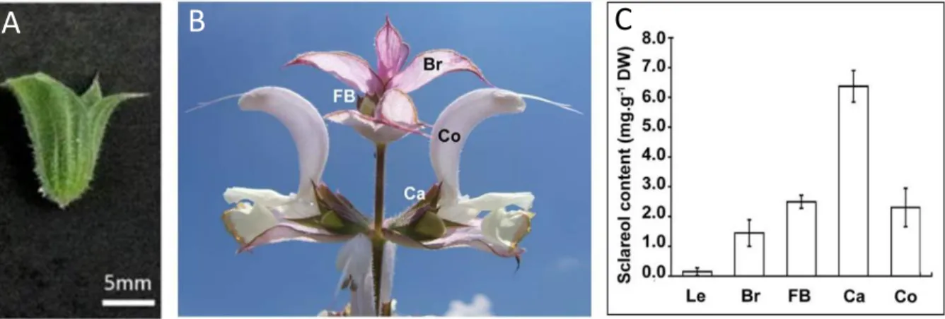
2.2. Sclareol biological function in clary sage
The biological function of sclareol in clary sage remains elusive. Sclareol is mainly produced in clary sage flowers, especially calyces (Caissard et al., 2012) (Figure 3). Calyces are tubular structures formed by the fusion of sepals and are generally considered to play a role in the protection of flower reproductive organs (Legrand et al., 2010). Some insects are known to pierce holes in the calyx to access nectar without pollination benefit for the plant, a behavior known as nectar robbing (Irwin et al., 2010). Sclareol has been shown to form epicuticular crystals at the surface of the calyx (Caissard et al., 2012). These crystals may protect the flower against florivory and nectar robbing, and could also reduce fungal spores adhesion (Legrand et al., 2010; Caissard et al., 2012).
Mic
h
el D
ro
17
The biological function of sclareol has been investigated in other plant species (Seo et al., 2012; Fujimoto et al., 2015). Notably, sclareol has been reported to display antimicrobial activity against different species of plant pathogenic bacteria and fungi (Seo et al., 2012; Fujimoto et al., 2015; Chain et al., 2015). Exogenous application of sclareol on tobacco, tomato and Arabidopsis thaliana plants was shown to induce resistance to bacterial wilt disease (Seo et al., 2012). In tomato and Arabidopsis thaliana, sclareol could also inhibit the penetration of the root-knot nematode in the roots, thereby conferring resistance to this pathogen (Fujimoto et al., 2015). Taken together, these results suggest that sclareol may be involved in the defense of clary sage flowers against pathogenic microorganisms.
Figure 3: Sclareol is mainly produced in clary sage calyces.
A: Clary sage calyx. B: Apex of a clary sage inflorescence (Caissard et al., 2012). C: Sclareol quantification in different flower organs of clary sage (Caissard et al., 2012). Legends: Br, bract; FB, flower bud; Co, corolla; Ca, calyx; Le, leaf.
2.3. Clary sage commercial use
Clary sage has been traditionally used as a medicinal plant for its antispasmodic, carminative and estrogenic properties (Lattoo et al., 2006). Notably, essential oil extracted from clary sage has been reported to have numerous biological effects, including anti-inflammatory, antioxidant, antimicrobial and cytotoxic activity (Kuźma et al., 2009b; Sharopov and Setzer, 2012; Kumar et al., 2013). The individual contribution of each of its components to essential oil medicinal properties is not known and synergy, antagonism or additive effects may be at play (Kuźma et al., 2009b). In particular, sclareol has been shown to display anti-bacterial and anti-proliferative activity (Mendoza et al., 2002; Sashidhara et al., 2007).
18
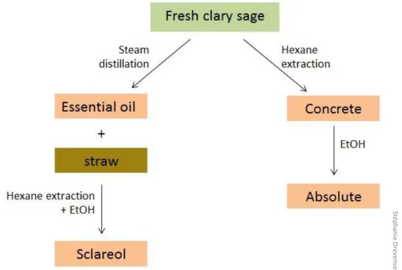
Today, clary sage is exploited at industrial scale mainly for its aromatic properties. It is commercially cultivated in different European countries, particularly in France, Hungary and Bulgaria, but also in North America and China (Lawrence, 1994; Lattoo et al., 2006; Caniard et al., 2012). Clary sage essential oil is used as a flavor for the elaboration of diverse processed food products (Lattoo et al., 2006; Wagner et al., 2012; Sharopov and Setzer, 2012). Most importantly, essential oil and sclareol extracted from clary sage are employed in perfume manufacture. Essential oil is used directly as a perfume component for its tenacious, herbaceous, sweaty and amber odor (Laville et al., 2012; Kumar et al., 2013), while sclareol is a common starting material for the hemisynthesis of the perfume component ambroxide (Yang et al., 2016). When grown for commercial use in flavor and fragrance industry, clary sage inflorescences are dried and submitted to steam distillation in order to collect essential oil. Sclareol is then obtained from remaining plant material by solid/liquid extraction with an organic solvent (Caissard et al., 2012; Laville et al., 2012; Leffingwell and Leffingwell, 2015) (Figure 4). In general, essential oil yield is between 0.1% and 0.3% (Wagner et al., 2012) and sclareol yield is between 0.5% and 1.5% (Caissard et al., 2012; ITEIPMAI, 7èmes Rencontres du Végétal, 2013).
Figure 4: Exploitation of clary sage in perfume industry.
St ép h an ie D re ve n se k
19
2.4. First advances in clary sage genetics and biotechnology
The identification of genes involved in specific biological processes is greatly facilitated when genomic data are available in the species of interest or in related species. The genome of clary sage is not sequenced yet, but genome sequences of Salvia miltiorrhiza and Salvia
splendens have been recently published (Xu et al., 2016; Dong et al., 2018). The study of
clary sage chromosomic material shows that it is a diploid species with a karyotype of 2n=22 (Ozdemir and Senel, 1999). Genome size was estimated around 600 Mb (Drevensek et al., unpublished data). Two sets of clary sage transcriptomic data are currently available. The first clary sage transcriptome was obtained from calyces and allowed the identification of putative candidate genes for different steps of terpenoid biosynthesis (Legrand et al., 2010). Another transcriptome has been generated from leaves and roots treated with the phytohormone methyljasmonate (Hao et al., 2015).
Biotechnological tools enabling the modification of gene expression level are highly valuable for the functional characterization of genes of interest. In many plants, the development of roots named “hairy roots” can be induced using the bacterium Agrobacterium rhizogenes, which is able to transfer human-engineered genetic material into plant genome. Hairy roots can then be separated from aerial parts and cultivated in a bioreactor (Ron et al., 2014). Several studies show that transgenic hairy roots can be generated from clary sage roots (Kuźma et al., 2009a; Vaccaro et al., 2014, 2017; Alfieri et al., 2018) (Figure 5). Stable lines of clary sage calli and cell cultures can also be obtained from seeds or stem explants, and were shown to produce sclareol (Banthorpe et al., 1990). Moreover, protocols enabling in vitro regeneration of whole clary sage plants from fragments of somatic tissue are available, starting from immature zygotic embryo cotyledons (Liu et al., 2000) or shoot tips (Kuźma et al., 2009b). Essential oil composition of clary sage plants regenerated from shoot tips was comparable to plants reproduced from seeds, but the yield was twice lower (Kuźma et al., 2009b). Therefore, the metabolic activity of regenerated clary sage plants may be different compared to plants reproduced from seeds, but further investigation is needed to confirm this hypothesis.
20 Figure 5: Clary sage hairy root culture (Alfieri et al., 2018).
2.5. Clary sage breeding in France
The growing demand for sclareol encouraged the development of clary sage breeding programs in countries where clary sage is cultivated, including Hungary, Bulgaria, Moldavia, Romania and France (source: Community Plant Variety Office). In France, the Interprofessional Technical Institute of Perfume, Medicinal, Aromatic and Industrial Plants (ITEIPMAI) has obtained several improved clary sage varieties through recurrent selection targeting sclareol yield and flowering time. Recurrent selection is a plant breeding approach involving successive cycles of three steps: intermating between best-performing populations, evaluation of traits of interest, and selection of best-performing individuals. Through this method, a clary sage variety with high sclareol yield was obtained: “Scalia”. Unfortunately, this variety displayed a late-flowering phenotype. This phenotype can be detrimental to sclareol yield because late-flowering plants are potentially more impacted by summer drought (Otto et al., 2017). Further recurrent selection cycles led to the development of the “Toscalia” variety, which is characterized by a high sclareol yield without any late-flowering phenotype. A third variety, “Claryssima”, is currently being developed using the same approach (source: ITEIPMAI).
21
3. Terpene biosynthesis and function in plants
3.1. Specialized metabolism and terpenoidsThroughout their lifetime, plants produce a wide variety of chemical compounds which perform different biological functions. Plant compounds have been historically divided into two classes according to their biological role. Metabolites which are essential to sustain normal growth are called primary metabolites. For example, this category comprises structural compounds like cell wall carbohydrates or membrane lipids, and compounds involved in energy metabolism or photosynthesis like organic acids or coenzymes. In addition to primary metabolites, plants produce various other compounds which are not indispensable for plant growth and development, but are involved in plant adaptation to biotic and abiotic environmental factors (Figueiredo et al., 2008; Tholl, 2015; Chezem and Clay, 2016). These compounds are therefore critical for plant survival in a fluctuating environment and are known as secondary or specialized metabolites. Unlike primary metabolites, which are generally present in all plant lineages, specialized metabolites are highly diverse between plant species. In a single plant species, the amount of specialized metabolites produced can also vary drastically according to environmental conditions.
Plant metabolites can also be classified according to early steps of their biosynthesis pathway. Terpenoids represent the largest class of plant metabolites with at least 50,000 compounds described (Liao et al., 2016). Some terpenoids play critical roles in plant growth and development. They are therefore considered as primary metabolites, like for example phytohormones of the gibberellin family and photosynthetic pigments of the carotenoid family. However, most plant terpenoids are specialized metabolites involved in diverse biological functions related to plant interaction with its environment (Lipko and Swiezewska, 2016).
The biosynthesis of all terpenoids starts with the condensation of the same five-carbon building blocks: isopentenyl pyrophosphate (IPP) and its isomer dimethylallyl pyrophosphate (DMAPP) (Tholl, 2015). The number of five-carbon units condensed defines subclasses among terpenoids: for instance, compounds derived from the condensation of 2, 3 or 4 five-carbon units belong to monoterpene (10C), sesquiterpene (15C) and diterpene (20C) subclasses, respectively. Sclareol belongs to the diterpene subclass.
22
3.2. The diverse biological functions of terpenes
Numerous terpenes are indispensable for plant cell function and correct plant growth and development. Primary metabolites belonging to the terpenoid family include carotenoids, which are essential for light harvesting during photosynthesis, and phytosterols, which are structural elements of plant plasma membranes (Figure 6). They also include important signaling metabolites, as several classes of important plant hormones are terpenoids: gibberellins, abscisic acid, strigolactones and brassinosteroids. Moreover, some protein post-translational modifications are related to terpenoid metabolism: protein prenylation consists in the transfer of a prenyl moiety to a cysteine, and the terpenoid dolichol is involved in protein glycosylation (Tholl, 2015).
Some terpenoids are involved in plant response to abiotic stress, although this is not their most common function. For example, in poplar, isoprene emissions provide protection against thermal stress and ozone-induced oxidative stress (Behnke et al., 2007, 2009). By contrast, a large number of plant terpenoids are key elements of plant constitutive and induced chemical defense systems against pathogenic microorganisms (fungi, bacteria) and insect herbivores (Figure 6). They can act as direct defense by displaying toxicity to plant pathogens or by acting as herbivore repellents (Moore et al., 2014). For example, numerous rice diterpenoid phytoalexins such as oryzalexins show antibiotic activity against the fungal pathogen Magnaporthe oryzae (Schmelz et al., 2014), and 17-hydroxygeranyllinalool diterpenoid glycosides produced by Nicotiana attenuata work as antifeedants against tobacco hornworm (Jassbi et al., 2008). Terpenoids can also act as indirect defense via the attraction of natural enemies of plant pathogens, i.e. predators or parasites (Moore et al., 2014). Such indirect action is illustrated by the response of maize roots to feeding by
Diabrotica virgifera larvae. Damaged maize roots produce β-caryophyllene, which in turn
strongly attracts an entomopathogenic nematode of Diabrotica virgifera larvae (Rasmann et al., 2005).
In addition to defense against pathogenic microorganisms and herbivores, terpenoids play other roles in plant-insect interactions. Notably, volatile scent terpenoids emitted by flowers are critical for pollinator attraction. The monoterpene 1,8-cineole is frequently present in the scent of orchids and strongly attract euglossine bees, a group of bees specifically pollinating orchids (Dodson et al., 1969). Plant-plant interactions can also be mediated by
23
terpenes. Some terpenoids allow parasitic plants to detect their host, induce defense responses in neighboring plants, or display toxicity to plant competitors (Tholl, 2015). For example, momilactone diterpenes produced by rice roots have a negative effect on the growth of barnyard grass, a plant competitor of rice (Xu et al., 2012).
Fi gu re 6: Exa m p le s ill u st ra ti n g th e d iv e rse b io lo gi cal f u n ct io n s o f te rp en e s in p la n ts. Camille Chalvin
24 3.3. Industrial exploitation of terpenes
Because of their various ecological functions, terpenes display distinct properties and biological activities which are valuable for industrial applications. In particular, a number of plant-derived terpenoids have been used for a long time as scents or flavors, and as active pharmacological ingredients (Tetali, 2018). Essential oils, which are mainly composed of terpenoids, are important natural ingredients of flavor and perfume industry, and their components are also individually employed for specific uses. For example, the sesquiterpene nootkatone is the main molecule responsible for the distinct aroma of grapefruit, and the monoterpenes limonene, linalool and 1,8-cineole are common ingredients of lemon or lime flavor (Hausch et al., 2015; Tetali, 2018). Pharmacologically active molecules are found in all subclasses of terpenes, including monoterpenes and diterpenes (Figure 7). The most famous plant diterpene possessing medicinal properties may be the cancer drug paclitaxel (Taxol®), initially isolated from yew (Mafu and Zerbe, 2018). The diterpene ingenol mebutate from
Euphorbia peplus also displays antitumor effects and has been recently approved in clinical
trials for skin cancer treatment (Mafu and Zerbe, 2018). In addition to cancer therapy, terpenes are used in many other medicinal applications; for example, the diterpene forskolin is a cardioprotective compound found in the roots of Coleus forskohlii (Mafu and Zerbe, 2018), and the sesquiterpene artemisinin produced by Artemisia annua is a reference for the treatment of malaria (Tetali, 2018).
In addition to scents and medicines, other plant-derived terpenoids are used in food, cosmetic and chemical industries (Tetali, 2018). Some terpenoids have important nutritional value, as illustrated by the role of carotenoids which represent an essential source of vitamin A for humans (Rodriguez-Concepcion et al., 2018). Carotenoids are also employed as antioxidants in cosmetic industry, notably in the elaboration of skin care products (Sathasivam and Ki, 2018). Terpenoid properties are useful in other sectors of chemical industry, like for instance polymer chemistry in the case of polyisoprene constituents of natural rubber produced by Hevea brasiliensis. Moreover, due to their low hygroscopy, high energy density, and good fluidity at low temperatures, biotechnologically-produced sesquiterpenes and diterpenes have been suggested as potential renewable alternatives for transportation fuels (Tippmann et al., 2013).
25
Figure 7: Pharmacologically active diterpenoids and their respective source plants (Mafu and Zerbe, 2018).
3.4. Terpene biosynthesis
The first step of terpenoid biosynthesis corresponds to the production of the five-carbon building blocks IPP and DMAPP. In plants, IPP and DMAPP are produced by two distinct metabolic pathways: the mevalonate pathway (MVA pathway) and the methylerythritol-phosphate pathway (MEP pathway). Then, linear intermediate compounds called prenyl intermediates are formed by condensation of IPP and DMAPP units (Tholl, 2015). Prenyl intermediates are finally converted into functional terpenes through a number of molecular modifications, for example cyclization and addition of functional groups like hydroxyl groups.
3.4.1. Production of five-carbon building blocks by MEP and MVA pathways
IPP and DMAPP are the universal five-carbon building blocks from which all terpenes are derived. The co-existence of two metabolic pathways for the production of IPP and DMAPP is an atypical feature of plants: most bacteria use only the MEP pathway, and animals, fungi and archae only have the MVA pathway (Hemmerlin et al., 2012; Lipko and Swiezewska,
26
2016). In plants, MVA and MEP pathways are compartmentalized: the MVA pathway is localized in the cytosol, whereas the MEP pathway is localized in plasts. It is now admitted that plants inherited the MEP pathway from a photosynthetic prokaryote through endosymbiosis (Vranová et al., 2013).
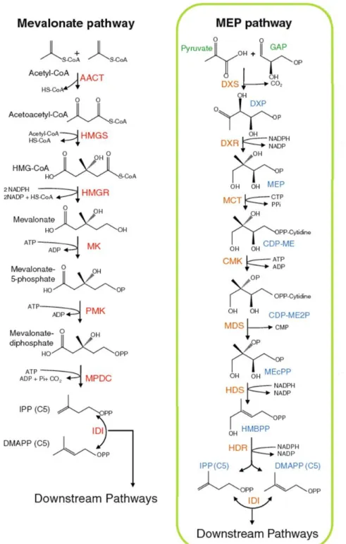
In plants, the MVA pathway involves 6 enzymatic steps (Tholl, 2015) (Figure 8). It starts with the condensation of 2 molecules of acetyl-coenzyme A (acetyl-CoA) by the acetoacetyl-CoA thiolase (AACT), which leads to the formation of acetoacetyl-CoA. The HMG-CoA synthase (HMGS) then catalyzes the condensation of acetoacetyl-CoA with another molecule of acetyl-CoA to generate hydroxymethylglutaryl-CoA (HMG-CoA). The third step of the pathway is a rate-limiting step where HMG-CoA is reduced to mevalonate (MVA) by the key enzyme HMG-CoA reductase (HMGR). MVA is then phosphorylated twice through the consecutive action of the mevalonate kinase (MK) and the phosphomevalonate kinase (PMK). The last step of the MVA pathway corresponds to the conversion of mevalonate-diphosphate into IPP, catalyzed by the mevalonate-mevalonate-diphosphate decarboxylase (MPDC). The MEP pathway found in plants consists of 7 enzymatic steps (Tholl, 2015) (Figure 8). The first step of the pathway is a rate-limiting step where the DXP-synthase (DXS) catalyzes the formation of 1-deoxy-D-xylulose-5-phosphate (DXP) from glyceraldehyde-3-phosphate and pyruvate. Plant DXS enzymes are divided in 2 classes which correspond to 2 distinct phylogenetic clades: DXS1 and DXS2 enzymes. DXS1 enzymes are expressed in photosynthetic and floral tissues and are thought to be mostly involved in primary metabolism, whereas DXS2 enzymes have more restricted spatiotemporal expression patterns and are generally involved in specialized metabolism (Tholl, 2015). The second step of the MEP pathway corresponds to the conversion of DXP into 2-methyl-D-erythritol-4-phosphate (MEP) through the action of the DXP-reductoisomerase (DXR). The MEP-cytidylyltransferase (MCT) then couples MEP to CDP to form 4-diphosphocytidyl-2-methyl-D-erythritol (CDP-ME), which is subsequently phosphorylated by the CDP-ME kinase (CMK). The obtained 4-diphosphocytidyl-2-methyl-D-erythritol-2-phosphate (CDP-ME2P) is cyclized by the MEcPP synthase (MDS) to form 2-methyl-D-erythritol-2,4-cyclodiphosphate (MEcPP). MEcPP is reduced into 4-hydroxy-3-methylbut-2-enyl-diphosphate (HMBPP) by the HMBPP-synthase (HDS) and HMBPP is finally converted into a mixture of IPP and DMAPP by the HMBPP-reductase (HDR) with a ratio of 5 to 6:1.
27
Figure 8: Production of five-carbon building blocks by MEP and MVA pathways (adapted from Tholl, 2015).
AACT, acetoacetyl-CoA thiolase; CDP-ME, diphosphocytidyl-2-methyl-D-erythritol; CDP-ME2P, 4-diphosphocytidyl-2-methyl-D-erythritol-2-phosphate; CMK, CDP-ME kinase; CoA, coenzyme A; DMAPP, dimethylallyl pyrophosphate; DXP, 1-deoxy-D-xylulose-5-phosphate; DXR, DXP-reductoisomerase; DXS, DXP-synthase; GAP, glyceraldehyde-3-phosphate; HDR, HMBPP-reductase; HDS, HMBPP-synthase; HMBPP, 4-hydroxy-3-methylbut-2-enyl-diphosphate; HMG-CoA, hydroxymethylglutaryl-CoA; HMGR, HMG-CoA reductase; HMGS, HMG-CoA synthase; IDI, IPP isomerase; IPP, isopentenyl pyrophosphate; MCT, MEP-cytidylyltransferase; MDS, MEcPP synthase; MEcPP, 2-methyl-D-erythritol-2,4-cyclodiphosphate; MEP, 2-methyl-D-erythritol-4-phosphate; MK, mevalonate kinase; MPDC, mevalonate-diphosphate decarboxylase; MVA, mevalonate; PMK, phospho-mevalonate kinase.
28
3.4.2. Condensation of five-carbon building blocks by prenyltransferases
The second step of terpenoid biosynthesis corresponds to the condensation of IPP and DMAPP units to form prenyl diphosphate intermediates, which are the percursors of all terpenes. These condensation reactions are catalyzed by enzymes called prenyl diphosphate synthases or prenyltransferases. First, an IPP molecule is condensed with a DMAPP molecule to produce a ten-carbon intermediate, and further addition of IPP units leads to the formation of prenyl diphosphate intermediates of various lengths (Kharel and Koyama, 2003). Prenyltransferase activity requires an appropriate ratio of IPP and its more reactive isomer DMAPP. Conversion of IPP into DMAPP is performed by the IPP isomerase (IDI). IPP isomerization is more important in the cytosol than in plastids because the MVA pathway produces only IPP, whereas the MEP pathway produces both five-carbon units (Tholl, 2015). Prenyltransferases are divided in two classes according to the stereochemistry of double bonds in the product of the condensation reaction: cis-prenyltransferases and trans-prenyltransferases produce all-cis and all-trans prenyl diphosphate intermediates, respectively (Kharel and Koyama, 2003). Condensation of 2, 3 or 4 five-carbon units in trans orientation leads to the formation of geranyl pyrophosphate (GPP, 10 carbons), (E,E)-farnesyl pyrophosphate (FPP, 15 carbons) and geranylgeranyl pyrophosphate (GGPP, 20 carbons), respectively (Figure 9). These trans-prenyl diphosphate intermediates are the main precursors of monoterpene, sesquiterpene and diterpene biosynthesis, respectively. They are more abundant than their all-cis isomers and constitute central metabolic branching points for the biosynthesis of terpenoids involved in primary and specialized metabolism (Tholl, 2015). For these reasons, most research studies conducted so far have been focused on trans-prenyltransferases.
GGPP, FPP and GPP are produced by geranylgeranyl pyrophosphate synthases (GGPP-synthases), farnesyl pyrophosphate synthases (FPP-synthases) and geranyl pyrophosphate synthases (GPP-synthases), respectively (Figure 9). Most GGPP and GPP molecules are produced in plasts, whereas FPP synthesis essentially occurs in the cytosol (Lu et al., 2016). GGPP-synthase and FPP-synthase genes are generally present in multiple copies in plant genomes, GGPP-synthase gene families being larger than FPP-synthase gene families (Tholl, 2015). In angiosperms, GGPP-synthases and FPP-synthases usually work as homodimers to produce respectively GGPP and FPP as major or unique product (Tholl, 2015). By contrast,
29
several GPP-synthases have been shown to work as heterodimers including a large catalytic subunit and a small regulatory subunit, like those described in Antirrhinum majus, Clarkia
breweri and Humulus lupulus (Tholl et al., 2004; Wang and Dixon, 2009). The large subunit is
closely related to GGPP-synthases and displays GGPP-synthase activity in vitro, whereas the small subunit shows only limited sequence similarity with GGPP-synthases (around 20%) and does not show any catalytic activity. It is admitted that in vivo, the small subunit binds to the large subunit and modifies its activity to make it produce GPP instead of GGPP (Burke and Croteau, 2002; Orlova et al., 2009). Homodimeric and heterotetrameric GPP-synthases have also been characterized (Burke et al., 1999; Burke and Croteau, 2002; Hsiao et al., 2008). Notably, the first GPP-synthase discovered in plants was identified in the Lamiaceae Mentha
piperita and works as a heterotetramer with two large catalytic subunits and two small
regulatory subunits similar to subunits forming heterodimeric GPP-synthases (Burke et al., 1999; Burke and Croteau, 2002). Some plant species have both heteromeric and homomeric GPP-synthases, for example Catharanthus roseus (Rai et al., 2013).
Figure 9: The role of prenyltransferases in terpene biosynthesis.
DMAPP, dimethylallyl pyrophosphate; FPP, farnesyl pyrophosphate; GPP, geranyl pyrophosphate; GGPP, geranylgeranyl pyrophosphate; IPP, isopentenyl pyrophosphate.
C amill e C h alv in
30
3.4.3. Terpene synthases
In the last step of terpenoid biosynthesis, prenyl diphosphate intermediates undergo various molecular modifications which eventually lead to functional terpenes. Possible modifications include cyclization or cleavage of the hydrocarbon skeleton, and functionalization reactions such as hydroxylation, peroxidation, methylation, acylation or glycosylation (Tholl, 2015). This step involves enzymes of the terpene synthase (TPS) superfamily and often requires other types of enzymes, like for example cytochrome P450 monoxygenases (Zerbe and Bohlmann, 2015; Bathe and Tissier, 2019).
Plant genomes generally contain numerous putatively functional TPS genes. Only 2 were found in the genome of the moss Physcomitrella patens, but genomes of vascular plants generally have more than 20 TPS genes, up to 113 in Eucalyptus grandis (Chen et al., 2011). The large number of TPS genes found in plant genomes, the promiscuous activity of these enzymes and their ability to acquire new catalytic properties by minor structural changes are believed to be responsible for the huge diversity of plant terpenoids (Tholl, 2015). According to phylogenetic studies, TPS genes are divided into 8 subfamilies: TPSa to TPSh. TPS enzymes can also be classified in 2 classes according to the mechanism of the enzymatic reaction they catalyze (Chen et al., 2011). This enzymatic reaction always involves the formation of a carbocation within the prenyl disphosphate substrate, which opens up the possibility of intramolecular rearrangements or attacks by nucleophilic compounds such as water. Class I TPS enzymes ionize their substrate by removing the diphosphate group, whereas class II TPS do it through a protonation reaction (Chen et al., 2011). In vitro, TPS enzymes are sometimes able to use prenyl diphosphate substrates of different lengths, but in vivo their activity is determined by the substrate pool present in the compartment where they are localized. Given the respective proportions of GPP, FPP and GGPP molecules in plasts and in the cytosol, plastidial TPS enzymes mainly produce monoterpenes and diterpenes, whereas cytosolic TPS enzymes mainly produce sesquiterpenes (Tholl, 2015). However, exceptions to this rule have been reported; for instance, plastidial sesquiterpene synthases have been characterized in tomato (Sallaud et al., 2009).
31
3.4.4. Focus on diterpene biosynthesis
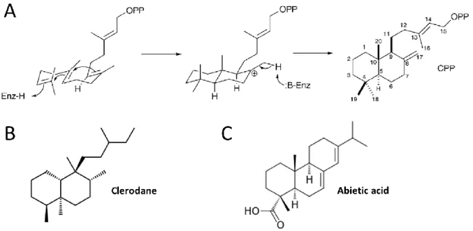
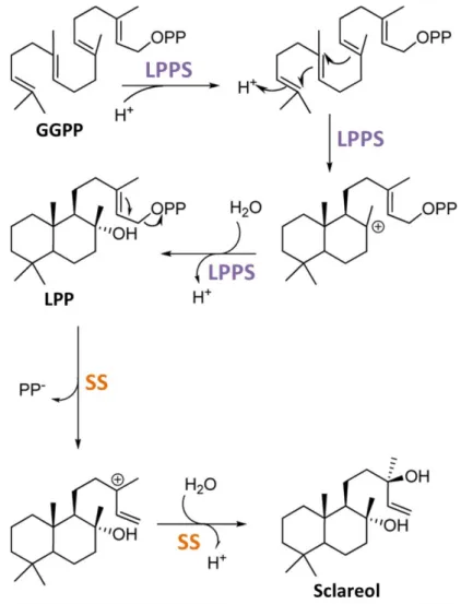
Diterpene biosynthesis usually occurs in chloroplasts, where most GGPP precursors are produced (Tholl, 2015). The various diterpene biosynthesis pathways are believed to have evolved from gibberellin biosynthesis (Peters, 2010). A majority of diterpenes, including sclareol, are labdane-type diterpenes. Formation of diterpenes with a hydrocarbon skeleton of the labdane type usually involves the cyclization of GGPP through the consecutive action of a class II diterpene synthase (Figure 10) and a class I diterpene synthase (Peters, 2010). The obtained bicyclic skeleton can be further modified, for example by the addition of hydroxyl groups mediated by diterpene synthases or cytochrome P450 monoxygenases (Zerbe and Bohlmann, 2015; Bathe and Tissier, 2019). Another cyclization reaction leads to tricyclic abietane diterpenes (Peters, 2010) (Figure 10).
Figure 10: Examples of labdane-related diterpenes.
A: Example of enzymatic reaction generating a labdane-type hydrocarbon skeleton (Peters, 2010). This protonation-initiated cyclization of GGPP is catalyzed by a class II diterpene synthase (Enz). Copalyl diphosphate (CPP) is an intermediate in the biosynthesis of many diterpenes (Peters, 2010). B: Chemical formula of a bicyclic labdane-related diterpene, clerodane.
32 3.5. Terpene biosynthesis regulation
3.5.1. Factors affecting terpene content
Specialized metabolite content varies at all scales, between species and populations, but also between individuals and between the different parts of the same plant (Moore et al., 2014). In a given species, populations displaying distinct metabolite profiles are referred to as chemotypes (Moore et al., 2014). Physiological variations, environmental conditions and genetic polymorphisms, along with the interactions of these three factors, are known to determine specialized metabolite content in plants (Figueiredo et al., 2008; Moore et al., 2014).
In a single plant, terpenoid production depends on the organ considered, on the developmental stage of this organ, and also on circadian and seasonal cycles (Moore et al., 2014). Important qualitative and quantitative variations are observed between the different organs of the plant. For example, the flowers of Lavandula pinnata produce more monoterpenes and less sesquiterpenes than leaves and stems. These differences are related to the important role played by volatile monoterpenoids in guiding pollinators to the flowers (Figueiredo et al., 2008). In Achillea millefolium, the amount of volatile terpenoids produced by flowers differs according to their developmental stage. The amount of sesquiterpene chamazulene present in the essential oil decreases with flower maturation, while the proportion of the monoterpenes 1,8-cineole and camphor increases (Figueiredo et al., 2008). Diurnal variations of light level impact MEP and MVA pathways in the opposite way: the MEP pathway is stimulated by exposure to light, whereas the MVA pathway is upregulated in the dark (Tholl, 2015). Seasonal variations in terpenoid content can also be observed in some species. In sea fennel (Crithmum maritimum), proportions of sabinene and γ-terpinene in the essential oil vary in opposite ways: sabinene is more abundant during the flowering season (between July and October), whereas γ-terpinene predominates during the rest of the year and peaks in April (Barroso et al., 1992) (Figure 11).
33
Figure 11: Seasonal variations of terpene production in Crithmum maritimum (Barroso et al., 1992).
Environmental conditions impacting specialized metabolite content include climatic factors (temperature, hygrometry), biotic factors (herbivores, microorganisms) and edaphic factors. Terpenoid production is often tightly related to weather conditions. Monoterpene emission by slash pine (Pinus elliotii) has been shown to increase with temperature (Tingey et al., 1980). Drought has contrasted effects on terpenoid production depending on the species considered: for example, hydric stress enhances terpenoid emissions in Ocimum basilicum, but decreases artemisinin production in Artemisia annua (Figueiredo et al., 2008). Since many terpenoids play a role in plant defense against pathogens, biotic factors significantly influence terpene levels in various plants. For instance, the sesquiterpene capsidiol accumulates in pepper stems upon infection with the oomycete Phytophthora capsici (Egea et al., 1996). Finally, soil physical and chemical structure is also considered as an important factor impacting specialized metabolite content in plants. Notably, the proportion of silt and sand as well as potassium oxide content influence the composition of Salvia desoleana essential oil (Rapposelli et al., 2015).
Genetic polymorphisms are responsible for variations in terpenoid content between plants belonging to the same species or to related species. In species of the genus Mentha, the dominant allele C has an important impact on essential oil composition: CC or Cc genotypes are associated with high carvone content, whereas the cc genotype promotes the
34
production of pulegone and menthol (Figueiredo et al., 2008). Genetic variations are shaped by the selective pressure exerted by environmental factors mentioned above. The action of natural selection is illustrated by the study of monoterpene production by Pinus ponderosa trees in the context of the development of bark beetles, which are the most destructive agent of conifer forest worldwide. Directional selection has been shown to take place for trees producing high amounts of limonene, a monoterpene toxic to the beetle (Trapp and Croteau, 2001).
3.5.2. Molecular mechanisms of terpene biosynthesis regulation
Phytohormone signaling pathways and master transcriptional regulators of plant growth and development control terpenoid biosynthesis in response to environmental and developmental signals. In Arabidopsis thaliana, the upregulation of the MEP pathway and carotenoid biosynthesis in light conditions was shown to be performed through the action of phytochrome interacting factors (Toledo-Ortiz et al., 2010) and the biosynthesis of floral sesquiterpenes is induced by master regulators of flower maturation in response to jasmonates, auxin and gibberellins (Tholl, 2015). Jasmonates stimulate terpenoid biosynthesis in many other plants (Goossens et al., 2016); for example, they induce artemisinin biosynthesis in Artemisia annua (Chen et al., 2017b).
Genes involved in the same terpene biosynthesis pathway are often organized as gene clusters in plant genomes, an organization which facilitates their transcriptional coregulation (Töpfer et al., 2017). Positive transcriptional regulators of terpene biosynthesis genes have been identified in several plant species and generally belong to WRKY, AP2/ERF, bHLH or basic leucine zipper (bZIP) transcription factor families (Lu et al., 2016). For example, AaWRKY1, AaERF1, AaERF2, AaORA1 and AabZIP1 transcription factors positively regulate the biosynthesis of the sesquiterpene artemisinin in Artemisia annua (Yu et al., 2011; Lu et al., 2013; Han et al., 2014; Zhang et al., 2015a), TcWRKY1 stimulates the production of the diterpene paclitaxel in Taxus chinensis (Li et al., 2013) and the bZIP transcription factor OsTGAP1 positively regulates the biosynthesis of diterpene phytoalexins in rice (Okada et al., 2009).
In plants, MVA and MEP pathways are compartmentalized. Therefore, they can be differentially regulated, and controlled regulatory crosstalks between terpenoid biosynthesis
35
pathways have been reported (Tholl, 2015). Notably, terpenoid biosynthesis has been shown to be regulated at the post-translational level by protein prenylation (Liao et al., 2016). For instance, the production of the MVA-derived sesquiterpene capsidiol in tobacco relies on MEP-dependent protein geranylgeranylation (Huchelmann et al., 2014) and in Catharanthus
roseus, MVA-derived farnesylated proteins regulate the expression of genes involved in
MEP-dependent monoterpenoid biosynthesis (Courdavault et al., 2005).
4. Glandular trichomes
4.1. Capitate and peltate glandular trichomes of the Lamiaceae
In Angiosperms, terpenoid production is often localized in specialized secretory structures. For example, in Hevea brasiliensis, polyisoprene components of natural rubber are produced in specialized ducts known as laticifers (Lange, 2015). Lamiaceae are generally characterized by the presence of epidermal secretory structures called glandular trichomes, which produce various compounds mainly involved in pollinator attraction or defense against herbivores (Werker, 1993). Terpenoids secreted by glandular trichomes are major components of the essential oil which can be extracted by hydrodistillation. Glandular trichomes of the Lamiaceae are multicellular and composed of 3 parts: a base, a stalk and a gland (or glandular head) (Figure 12). The gland is responsible for the secretion of specialized metabolites, the stalk is the structure bearing the gland, and the base connects the stalk to surrounding epidermal cells (Lange, 2015). Both vegetative and reproductive organs of the Lamiaceae harbor glandular trichomes already at early development stages (Werker, 1993).
36
Figure 12: Structure of a peltate glandular trichome (Lange, 2015)
Glandular trichomes of the Lamiaceae show high morphological diversity (Werker, 1993; Gul et al., 2019). Lamiaceae generally display at least two types of glandular trichomes: capitate glandular trichomes and peltate glandular trichomes (Werker, 1993). This classification is based on their distinct morphology: capitate trichomes have a long stalk topped by a small spherical secretory head, while peltate trichomes are sessile trichomes with a very short stalk and a large flattened glandular head. In addition to these morphological differences, capitate and peltate trichomes also differ in the way they secrete chemicals. Both glandular trichome types accumulate secreted chemicals under an elevated cuticle. In peltate glandular trichomes, the cuticle is thick and ruptures only upon contact with an insect or as a consequence of another mechanical stimulation. By contrast, compounds secreted by capitate glandular trichomes are regularly released to the outside; they are thought to be either exuded through the cuticle, or released after spontaneous breakage of the cuticle (Werker, 1993; Figueiredo et al., 2008; Tissier, 2012).
37 4.2. Clary sage glandular trichomes
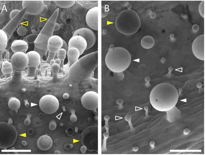
Like in most plants of the Lamiaceae family, both capitate and peltate glandular trichomes are found in clary sage, along with non-glandular trichomes (Werker et al., 1985; Schmiderer et al., 2008) (Figure 13). Several subtypes of capitate trichomes have been described according to their size: small capitate trichomes and large capitate trichomes are the most frequently observed, along with few intermediate trichomes. Small capitate heads measure between 10 and 20 µm, while large capitate heads measure between 50 and 100 µm (Figure
13). By contrast, the morphology of clary sage peltate trichomes is relatively uniform. Peltate
glands have a diameter of around 80-90 µm (Figure 13).
Figure 13: Glandular trichomes of clary sage calyces observed by scanning electron microscopy. A: Developing calyx (stage 1). B: Mature calyx (stage 5). Calyx development stages: see page 97. Full white arrows indicate large capitate glandular trichomes; empty white arrows indicate small capitate glandular trichomes; full yellow arrows indicate peltate trichomes; empty yellow arrows indicate non-glandular trichomes. Scale bars: 100 µm.