1
Acknowledgements ... 7
Summary ... 10
Résumé des travaux ... 12
Acknowledgements ... 14
Preface... 17
Abbreviations ... 20
Figures and tables ... 23
Introduction ... 25
General overview of the immune system and immune cell activities ... 25
1.1 Innate immunity ... 27
1.2 Cells at the interphase of the innate and adaptive immune systems ... 27
1.3 Adaptive immunity ... 27
A. B lymphocytes... 28
B. T lymphocytes ... 28
The cytotoxic function: the weapon of defense of the immune system ... 28
2.1 Natural killer cells ... 29
A. NK cell receptors ... 29
a. Natural cytotoxicity receptors ... 29
i. CD16 ... 30
ii. NKp46 ... 30
iii. NKp30 ... 30
iv. NKG2D ... 31
v. NKp44 ... 31
b. Inhibitory NK receptors (iNKR) ... 32
c. Costimulatory molecules ... 32
B. NK cell cytotoxicity: from target binding to target lysis ... 33
2
b. NK cell degranulation ... 34
A. Receptors, adhesion and co-stimulatory and inhibitory molecules involved in CD8+ T cell cytotoxicity ... 36
a. The T-cell receptor ... 37
b. The role of the integrin LFA-1 in cytotoxicity ... 39
i. LFA-1 activation ... 39
ii. Inside-out signaling ... 41
iii. Outside-in signaling ... 41
Costimulatory molecules ... 41
Inhibitory molecules ... 42
B. CD8+ T cell cytotoxicity: from naïve cells to professional killers ... 42
a. Differentiation into professional killers ... 43
CD8+ T cell cytotoxicity ... 44
2.3 Remodeling of the actin cytoskeleton governs the cytotoxicity of NK and CD8+ T cell cytotoxicity ... 45
2.4 Dynamic steps of cytotoxicity ... 46
A. Cell migration ... 46
B. The immunological synapse: the platform for cytotoxicity ... 47
a. Immunological synapse of CD8+ T cells ... 47
b. Immunological synapse of NK cells ... 49
Cytotoxicity in the clinical context ... 51
3.1 Cancers ... 51 3.2 Autoimmunity ... 51 3.3 Primary immunodeficiencies... 52 A. Hemophagocytic lymphohistiocytosis ... 52 a. Perforin deficiency (FHL2) ... 53 b. Munc13-4 deficiency (FHL3) ... 53 c. STX11 deficiency (FHL4) ... 54
3
d. Munc18-2 deficiency (FHL5) ... 54
e. Rab27a deficiency (Griscelli syndrome) ... 55
f. LYST deficiency (Chediak-Higashi syndrome) ... 55
B. Severe combined immunodeficiency ... 56
a. B cell immunodeficiencies ... 56
b. Innate immunodeficiencies ... 56
C. PIDs related to defects in the actin cytoskeleton ... 56
a. Wiskott Aldrich protein deficiency ... 57
b. WASp Interacting protein deficiency ... 59
c. ARHGEF1 deficiency ... 59 d. WDR1 deficiency ... 60 e. Coronin1A deficiency ... 61 f. Ccd42 deficiency ... 63 g. RhoA deficiency ... 63 h. RhoH deficiency ... 63 i. DOCK2 deficiency ... 64 j. DOCK8 deficiency ... 64 k. PSTPIP1 deficiency... 66 l. STK4 deficiency ... 66 m. RASGRP1 deficiency ... 67 n. MKL1 deficiency ... 68 o. Moesin deficiency ... 68 p. Rac2 deficiency ... 69 q. Myosin deficiency ... 69 r. ARPC1B deficiency ... 70
Methodologies to assess defects in cytotoxicity. ... 73
4
5.1 The basics of microscopy ... 74
A. Light sources to illuminate a sample ... 74
B. From an object to an image ... 75
5.2 The timeline of microscopy in biology ... 76
5.3 High content imaging: A game changer ... 79
A. High content imaging: microscopy at a large scale ... 80
a. HCI screens may be used to define gene function. ... 81
b. HCI screens can be effective in determining gene interactions. ... 81
c. HCI screening is a useful tool to assess alterations to cell morphology in response to chemical compounds. ... 82
d. HCI screens are a powerful tool to image disease models. ... 82
B. Instrumentation: High content imaging requires specialized devices ... 84
C. High content imaging analysis requires specialized software ... 86
a. CellProfiler ... 87
b. EBImage ... 87
D. Image analysis pipeline in high content imaging screens ... 88
a. Image analysis ... 89
b. Image quality control ... 89
c. Processing the extracted features ... 90
d. Dimensionality reduction ... 90
e. Single-cell data aggregation ... 91
f. Profile similarity measurement ... 91
g. Assay quality assessment ... 92
h. Downstream analysis and result interpretation ... 92
i. Linear dimensionality reduction: Principle component analysis ... 92
ii. Non-linear dimensionality reduction ... 93
5
b) Diffusion map ... 93
c) t-distributed stochastic neighborhood embedding ... 93
d) Uniform manifold approximation and projection ... 94
E. Statistics ... 96
Implementation of a high content imaging pipeline to study the immunological synapse of cytotoxic lymphocytes ... 99
Rationale ... 99
Objectives ... 100
Results ... 101
1. Setting up assays in the NK-92 cell line ... 102
2. Setting up assays in primary NK cells. ... 104
3. High content imaging of the immunological synapse ... 107
Materials and methods: optimizations on NK-92 and primary NK cells... 138
1. NK-92 cells ... 138
1.1 Migration ... 138
1.2 Conjugate formation kinetics ... 138
1.3 NK-92 – K562 conjugate assessment by microscopy ... 138
1.4 NK-92 cytotoxicity ... 139
1.5 TIRF microscopy ... 139
2. Primary NK cells... 139
2.1 Primary NK cell expansion ... 139
2.2 Migration ... 139
2.3 Conjugate formation kinetics ... 140
2.4 Primary NK– K562 conjugate assessment by microscopy ... 140
2.5 Primary NK cell cytotoxicity ... 140
Discussion and perspectives ... 141
6
HCI validates the role of the actin cytoskeleton in governing IS assembly ... 143
HCI coupled to robust statistics reveal important and sometimes unexpected IS morphological features ... 145
HCI could be indicative of a defect in the cytotoxic function ... 146
HCI of the immunological synapse reveals discrepancies in the regulation between cell lines and primary cells. ... 147
HCI reveals the lack of a standard “normal” or “disease” phenotype ... 148
HCI as a first step preceding functional assays ... 149
HCI of the immunological synapse as a useful tool in the field of personalized medicine .... 150
HCI could be the method study different functional steps of cytotoxicity ... 151
Limitations of HCI of the immunological synapse ... 153
References ... 155
Annexes... 183
7
Acknowledgements
This manuscript is the culmination of three years of doctoral studies, for which acknowledgments to various people are due. I apologize in advance if I have forgotten some people, who are nonetheless appreciated.
First, I would like to thank my supervisors: Drs Loïc Dupré and Kaan Boztug, without whom none of this would have been possible. Loïc, thank you for all your advice, help, support, the long hours and fruitful discussions, and for being a humane supervisor.
Drs Joerg Menche and Audrey Ferrand for their help and input in shaping the project.
My three reviewers Drs. Christophe Le Clainche, Delphine Muriaux and Fernando Sepulveda for taking the time to read this manuscript, advise and for agreeing to partake in the thesis jury, and Dr. Maha Ayyoub, the jury president.
A major thanks is due to Loan Vulliard, none of this would have been possible without your hard work, and the countless times you have helped me sort things out. Thank you for trying to make bioinformatics understandable for me, and for the much-appreciated occasional sarcasm.
The organoid platform of the IRSD, especially Aude Rubio, Muriel Quaranta-Nicaise and Dr. Isabelle Fernandes, for all the long hours spent acquiring, analyzing and transferring (and re-transferring) images, and for making me fall in love with the Opera Phenix.
The ladies of both the cytometry and microscopy platforms of the CPTP, for all your help not only during experiments but also your kind and encouraging words from the start until the very end. Drs Sophie Allart and Fatima L’faquihi-Olive, you are strong, knowledgeable and amazing boss ladies. Danièle Daviaud and Fatima, thank you for always being extremely sweet and helpful. Anne-Laure Iscache, Valérie Duplan-Eche and Lydia De La Fuente, I cannot forget to thank you for the times you have come to “rescue” me at the cytometers. “Maître” Astrid Canivet-Laffitte, I really appreciate every time you have helped me with ImageJ.
A huge thank you to all the colleagues in both Dupré /Lesourne and Boztug teams in Toulouse and Vienna: Dr. Renaud Lesourne, thank you for your feedback, advice and comprehension.
Nelly Rouquié, Cui Yang and Suzanne Mélique, you have been amazing ladies all the way. To the past members and now Drs Gaëtan Blaize and Jeremy Argenty, as well as Aurélie Mougel, it was a pleasure being in the same team with you. To Claire, our most recent newcomer, thank you for you for your
8
company along experiments, and of course for taking care of my cells along with Hélène. Dr. Hélène Daniels, thank you for the discussions, advice and feedback.
I cannot thank each member of the big Boztug team individually, but a huge thanks to every member for their sweetness, help, and for making the 9 months I spent in Vienna so memorable. Dr Artem Kalinichenko and Jakob Huemer, thank you for always being up for discussing anything NK. Dr. Wojciech Garncarz, you are a hero and no words can do you justice. Drs. Ana Krolo and Ozelm Yuce, you ladies are sweethearts. Ewa Lenartowicz, thank you for your kind and encouraging words, always. Dr. Johannes Pfeifenschneider, thank you for your sweetness and positive mood.
To the Valitutti team (Dr. Salvatore Valitutti, Dr. Eric Espinosa, Dr. Marie-Pierre Puissegur, recent Drs. Roman Jugelé and Yoan Eliasse, and Liza Filali Sabina Mueller), and the previous members of the Dupré team (Drs. Javier Rey-Barrosso and Raïssa Houmadi, and Alice Munaretto) thank you for making me feel welcome among you, your help, sweetness and support, and for making the beginning of this journey a bit easier. Every single one of you has helped me throughout this experience whether it was through advice, support or even words of encouragement over a coffee. To Dr. Delphine Guipouy, thank you for helping me with the French administration, sharing your knowledge with me, and answering my endless questions. Thank you infinitely Dr. Cat.
A special thank you to my colleagues Tala Shahin, Maximilian Rau, Laurène Pfajfer and Marianne Guisset, colleagues who also became great friends. Afterall, it is friends who make some days more bearable.
The colleagues at CPTP who have given feedback and input to help better shape this project through listening to my presentations and advising. Moreover, thanks to Drs Anne Quillet-Mary, Gaël Menasché and Abdelhadi Saoudi for their advice and feedback as part of my thesis committee. An immense thank you to Dr. Nabila Jabrane-Ferrat for all her advice, support and help. You are the best “godmother” I could have asked for. I would also like to thank the NJF team: Drs Hicham El Costa (thank you for your encouragement through your sarcasm), Jordi Gouilly and Qian Chen, and finally the wonderful Ana Espino, also known as “my hero”.
To my friends in Toulouse and away, and my family and friends back in Lebanon who have been there for me throughout this journey. From the bottom of my heart I would like to thank Celine, Ranine and Elio, you have become a family away from home.
To my small family. Whatever I say, words will never be enough. To my adorable grandma whom I love beyond words, and who has always believed in me. To my little brother Georges, thank you for
9
always being by my side and for all your wise words and encouragement, and for always encouraging me to have faith in myself. To my Georges (the other special Georges in my life), I could not have done this without you, without your endless support and patience. I cannot thank you enough for being there every single day on this journey.
And finally, the most important people in this journey (and my life): my parents. Without you I could not be here today. Thank you for all the sacrifices you have made for me to be who I am and where I am. Thank you for your unconditional love and for always believing in me and making sure I knew it. I cannot possibly thank you enough for everything you have done for me. If superheroes existed, my parents would be ones. Everything I am and everything I will ever be, I owe to you. I love you beyond words and I am eternally grateful for you. Mom, dad, this (and everything I am and ever will be) is for you.
10
Summary
High content cell imaging reveals actin cytoskeleton-mediated control of the immunological synapse
Cytotoxic lymphocytes rely on actin cytoskeleton remodeling to achieve their function. In particular cytotoxic T lymphocytes and NK cells assemble the immunological synapse (IS), a complex actin-rich structure that allows the interaction with target cells, such as infected cells or tumor cells, and permits the polarized delivery of lytic granules. Although actin cytoskeleton remodeling is known to be a driving force of IS assembly and dynamics, our understanding of the molecular control of actin remodeling sustaining IS dynamics remains fragmented. This PhD project consisted in developing a high-content imaging approach to unbiasedly define the metrics of IS from human T and NK lymphocytes and to characterize the requirements for actin cytoskeleton integrity in organizing the IS architecture.
For that purpose, the stimulation and staining of cell lines and primary cells in multiwell plates and acquisition of a unique set of >100.000 confocal images with a fully automatized high-content imager was optimized. The images were analyzed with two complementary CellProfiler analytical pipelines to characterize the morphological features associated with different treatments and disease status. We first extracted 16 morphological features pertaining to F-actin, LFA-1 or lytic molecules based on prior knowledge of IS assembly, and included features pertaining to the nucleus. We show that IS assembly in Jurkat and NK-92 cells is characterized by increased F-actin intensity and cell area. For Jurkat cells, we report an increase in LFA-1 intensity and surface area, and for NK-92 cells an increase in lytic granule detection at the IS plane. We then treated NK-92 cells with seven drugs known to affect different aspects of actin dynamics and investigated the associated effects on IS features. We report concentration dependent effects, not only on F-actin intensity, as expected, but also on lytic granule polarization. Furthermore, using a high-resolution morphological profiling based on >300 features, we show that each drug inflicts distinct alterations of IS morphology. In a next step, we applied our experimental pipeline to primary NK cells isolated from the blood of healthy donors. Distinct morphological features were characterized among the NK cells from different donors, highlighting the sensitivity of our approach, but also revealing an unsuspected variability of immune cell morphologies among donors. We then further applied our approach to primary CD8+ T cells from patients with a rare immunodeficiency due to mutations in the gene encoding the actin regulator ARPC1B. ARPC1B deficiency results in decreased F-actin intensity, as well as in lytic granule polarization. This prompted us to assess the ability of these cells to kill target cells, which was markedly reduced. These results
11
illustrate how the systematic analysis of the IS might be used to assist the exploration of fonctional defects of lymphocyte populations in pathological settings.
In conclusion, our study reveals that although assembly of the IS can be characterized by a few features such as F-actin intensity and cell spreading, capturing fine alterations of that complex structure that arise from cytoskeleton dysregulation requires a high-content analysis. The pipeline we developed through this project holds promises for the morphological profiling of lymphocytes from primary immunodeficiency patients whose genetic defect has not yet been identified. Moreover, the discriminative power of our high-content approach could be exploited to characterize the response of lymphocytes to various stimuli and to monitor lymphocyte activation in multiple immune-related pathologies and treatment settings.
12
Résumé des travaux
L'imagerie cellulaire à haut débit révèle le contrôle de la synapse immunologique par le cytosquelette d'actine
Les lymphocytes cytotoxiques dépendent du remodelage du cytosquelette d'actine pour atteindre leur fonction. En particulier, les lymphocytes T cytotoxiques et les cellules NK assemblent la synapse immunologique (SI), structure complexe riche en actine qui permet l'interaction avec des cellules cibles et la distribution polarisée de granules lytiques (GL). Bien que le remodelage du cytosquelette d'actine soit connu pour être une force motrice de l'assemblage et de la dynamique de la SI, la compréhension du contrôle moléculaire du remodelage de l'actine soutenant la dynamique de la SI reste fragmentée. Ce projet de thèse a consisté à développer une approche d'imagerie à haut débit pour définir de manière impartiale les métriques de la SI des lymphocytes T et NK et caractériser les exigences d'intégrité du cytosquelette d'actine dans l'organisation de l'architecture de la SI.
À cette fin, la stimulation et coloration des lignées cellulaires et cellules primaires dans des plaques à puits multiples et l'acquisition d'un ensemble unique d'images confocales avec un imageur à haut contenu entièrement automatisé ont été optimisées. Les images ont été analysées avec deux pipelines complémentaires avec CellProfiler pour définir les caractéristiques morphologiques associées aux différents traitements et à l'état de la maladie. Nous avons d'abord extrait 16 caractéristiques morphologiques se rapportant à la F-actine, LFA-1 ou aux GL, sur la base d'une connaissance préalable de l'assemblage de la SI, et inclus des caractéristiques relatives au noyau. Nous montrons que l'assemblage de la SI dans les lignées est caractérisé par une augmentation de l'intensité de la F-actine et la surface cellulaire. Pour les cellules Jurkat, nous rapportons une augmentation de l'intensité et de la surface LFA-1, et de la de la détection des LG sur le plan SI pour les cellules NK-92. Le traitement des cellules NK-92 avec 7 drogues affectant différents aspects de la dynamique de l'actine et l’étude des effets associés sur les caractéristiques de la SI montrent des effets dépendants de la concentration sur l'intensité de l’actine et sur la polarisation des GL. De plus, un profilage morphologique à haute résolution basé sur> 300 caractéristiques montre que chaque drogue inflige des altérations distinctes sur la morphologie de la SI. Nous avons appliqué ce pipeline à des cellules NK primaires isolées du sang de donneurs sains. Des caractéristiques morphologiques distinctes définissent les cellules NK de différents donneurs, soulignant la sensibilité de notre approche, mais révélant également une variabilité insoupçonnée des morphologies des cellules immunitaires parmi les donneurs. Nous avons appliqué notre approche aux cellules T CD8+ primaires de patients présentant une immunodéficience rare due à des mutations dans le gène codant pour le régulateur d'actine ARPC1B. La carence en ARPC1B entraîne une diminution de l'intensité de l'actine et de la polarisation des GL. Cela nous a incités à
13
évaluer la capacité lytique de ces cellules, qui a été considérablement réduite. Ces résultats illustrent comment l'analyse systématique de la SI pourrait être utilisée pour aider à l'exploration des défauts fonctionnels des populations de lymphocytes dans des contextes pathologiques.
En conclusion, notre étude révèle que, bien que l'assemblage de la SI puisse être caractérisé par quelques caractéristiques telles que l'intensité de l'actine et la propagation cellulaire, la capture de fines altérations résultant de la dérégulation du cytosquelette nécessite une analyse à haut débit. Le pipeline développé est prometteur pour le profilage morphologique des lymphocytes de patients atteints d'immunodéficiences primaires dont le défaut génétique n'a pas encore été identifié. De plus, le pouvoir discriminant de notre approche pourrait être exploité pour caractériser la réponse des lymphocytes à divers stimuli et surveiller leur activation dans de multiples pathologies et traitements.
14
Acknowledgements
This manuscript is the culmination of three years of doctoral studies, for which acknowledgments to various people are due. I apologize in advance if I have forgotten some people, who are nonetheless appreciated.
First, I would like to thank my supervisors: Drs Loïc Dupré and Kaan Boztug, without whom none of this would have been possible. Loïc, thank you for all your advice, help, support, the long hours and fruitful discussions, and for being a humane supervisor.
Drs Joerg Menche and Audrey Ferrand for their help and input in shaping the project.
My three reviewers Drs. Christophe Le Clainche, Delphine Muriaux and Fernando Sepulveda for taking the time to read this manuscript, advise and for agreeing to partake in the thesis jury.
A major thanks is due to Loan Vulliard, none of this would have been possible without your hard work, and the countless times you have helped me sort things out. Thank you for trying to make bioinformatics understandable for me, and for the much-appreciated occasional sarcasm.
The organoid platform of the IRSD, especially Aude Rubio, Muriel Quaranta-Nicaise and Dr. Isabelle Fernandes, for all the long hours spent acquiring, analyzing and transferring (and re-transferring) images, and for making me fall in love with the Opera Phenix.
The ladies of both the cytometry and microscopy platforms of the CPTP, for all your help not only during experiments but also your kind and encouraging words from the start until the very end. Drs Sophie Allart and Fatima L’faqihi-Olive, you are strong, knowledgeable and amazing boss ladies. Danièle Daviaud and Fatima, thank you for always being extremely sweet and helpful. Anne-Laure Iscache, Valerie Duplan-Eche and Lydia De La Fuente, I cannot forget to thank you for the times you have come to “rescue” me at the cytometers. “Maître” Astrid Canivet-Laffitte, I really appreciate every time you have helped me with ImageJ.
A huge thank you to all the colleagues in both Dupré /Lesourne and Boztug teams in Toulouse and Vienna: Dr. Renaud Lesourne, thank you for your feedback, advice and comprehension.
Nelly Rouquié, Cui Yang and Suzanne Mélique, you have been amazing ladies all the way. To the past members and now Drs Gaëtan Blaize and Jeremy Argenty, it was a pleasure being in the same team with you. To Claire, our most recent newcomer, thank you for you for your company along experiments, and of course for taking care of my cells along with Hélène. Dr. Hélène Daniels, thank you for the discussions, advice and feedback.
15
I cannot thank each member of the big Boztug team individually, but a huge thanks to every member for their sweetness, help, and for making the 9 months I spent in Vienna so memorable. Dr Artem Kalinichenko and Jakob Huemer, thank you for always being up for discussing anything NK. Dr. Wojciech Garncarz, you are a hero and no words can do you justice. Drs. Ana Krolo and Ozelm Yuce, you ladies are sweethearts. Ewa Lenartowicz, thank you for your kind and encouraging words, always. Dr. Johannes Pfeifenschneider, thank you for your sweetness and positive mood.
To the Valitutti team (Dr. Salvatore Valitutti, Dr. Eric Espinosa, Dr. Marie-Pierre Puissegur, recent Drs. Roman Jugelé and Yoan Eliasse, and Liza Filali Sabina Mueller), and the previous members of the Dupré team (Drs. Javier Rey-Barrosso and Raïssa Houmadi, and Alice Munaretto) thank you for making me feel welcome among you, your help, sweetness and support, and for making the beginning of this journey a bit easier. Every single one of you has helped me throughout this experience whether it was through advice, support or even words of encouragement over a coffee. To Dr. Delphine Guipouy, thank you for helping me with the French administration, sharing your knowledge with me, and answering my endless questions. Thank you infinitely Dr. Cat.
A special thank you to my colleagues Tala Shahin, Maximilian Rau, Laurène Pfajfer and Marianne Guisset, colleagues who also became great friends. Afterall, it is friends who make some days more bearable.
The colleagues at CPTP who have given feedback and input to help better shape this project through listening to my presentations and advising. Moreover, thanks to Drs Anne Quillet-Mary, Gaël Menasché and Abdelhadi Saoudi for their advice and feedback as part of my thesis committee. An immense thank you to Dr. Nabila Jabrane-Ferrat for all her advice, support and help. You are the best “godmother” I could have asked for. I would also like to thank the NJF team: Drs Hicham El Costa (thank you for your encouragement through your sarcasm), Jordi Gouilly and Qian Chen, and finally the wonderful Ana Espino, also known as “my hero”.
To my friends in Toulouse and away, and my family and friends back in Lebanon who have been there for me throughout this journey. From the bottom of my heart I would like to thank Celine, Ranine and Elio, you have become a family away from home.
To my small family. Whatever I say, words will never be enough. To my adorable grandma whom I love beyond words, and who has always believed in me. To my little brother Georges, thank you for always being by my side and for all your wise words and encouragement, and for always encouraging me to have faith in myself. To my Georges (the other special Georges in my life), I could not have
16
done this without you, without your endless support and patience. I cannot thank you enough for being there every single day on this journey.
And finally, the most important people in this journey (and my life): my parents. Without you I could not be here today. Thank you for all the sacrifices you have made for me to be who I am and where I am. Thank you for your unconditional love and for always believing in me and making sure I knew it. I cannot possibly thank you enough for everything you have done for me. If superheroes existed, my parents would be ones. Everything I am and everything I will ever be, I owe to you. I love you beyond words and I am eternally grateful for you. Mom, dad, this (and everything I am and ever will be) is for you.
17
Preface
Having come from a background in microbiology and virology, immunology was not always the most evident of topics for me, as I had done my masters in a lab that worked on mice, where I had mostly done Western blots and Real-time PCR. The decision to embark on a PhD in immunology was quite the challenge for me, but the idea of putting in place a screen of cytotoxic cells by high content imaging sure was appealing. I began my PhD in the Dupré lab in January 2017, in the Valitutti team.
The team is an expert on cytotoxic cells, and the Dupré team is an expert on the immunological synapse of T cells, especially in the context of primary immunodeficiencies. At the time I joined, the Dupré team was composed of a postdoc: Javier Rey-Barrosso, three PhD students: Delphine Guipouy, Raissa Houmadi and Laurène Pfajfer, and an engineer: Alice Munaretto.
Javier Rey-Barrosso was passionate about motility and was studying the different migration patterns of B cell lines on Collagen and Fibronectin, as well as their ability to remain and individual cells or cluster based on the matrix. He did so by seeding these cells in 96 well plates and imaging them by TIRF microscopy. He also studied the roles of actin branching and actoMyosin contractility by using two drugs: the ROCK1 inhibitor Y-27632 and the ARP2/3 inhibitor CK-869. He also did live imaging of chemotaxis of B cells over a CCL19 gradient. Javier loved to perform his experiments and allowed me to observe, while giving me thorough explanations. Indeed, he was my first exposure to chemotaxis, as well as the potential that multiwell plates offer in terms of efficiently imaging cells under different conditions.
Alice Munaretto was also working on migration, but as opposed to Javier who was working on a B cell line, she was working on cells from CLL patients, to assess the migratory behavior of specific cell populations pre- and post-treatment. To do so, she was using a protocol of barcoding to phenotype her cells and was assessing their migration in response to chemokines. She did so by trans well assays, whose readout is by flow cytometry. Observing these experiments with Alice was my first exposure to flow cytometry. Moreover, to minimize bias in sample readout, Alice would set up an automatic sample mixing and acquisition, which was the first time I saw “automated” experiments.
Delphine Guipouy was in her last year of PhD when I met her. She was working on a specific type of cytotoxic Tregs referred to as ova-Tr1 T cells, that are capable of specific elimination myeloid target cells via their granzyme and granulysin. Delphine was highly passionate about cytotoxic cells, and she showed me how to set cytotoxicity assays in 96 well plate formats and how to analyze the results acquired by Flow cytometry. She also showed me how to perform live microscopy assays to assess cytotoxicity in time. Delphine also spoke to me about a killing experiment she had done in
real-18
time, by using a high content microscope (The Operetta), where she could sort single CD8+ T cells and seed one cell per well in a 96 well plate, to observe their cytotoxicity over time. She also showed me how to perform imaging of the immunological synapse of cytotoxic cells incubated with target cells, on microscopy slides.
Raissa Houmadi was also in her 3rd year of PhD, and she was working on developing a super-resolution microscopy approach. Indeed, she revealed how LFA-1 is spatially organized in nanoclusters and how this assembly is regulated by WASP to ensure proper granule docking in CD8+ T cells.
Laurène Pfajfer was a first year PhD student who was doing a part of her PhD at the LBI-RUD in Vienna, where she had worked on two primary immunodeficiencies: WIP and WDR1 to show how these defects lead to defective immunological synapse assembly and cytotoxicity.
Delphine, Raissa and Laurène, as well as Loic’s background in WASp and other primary immunodeficiencies provided me the exposure I needed to primary immunodeficiencies, and the immunological synapse.
In the context of primary immunodeficiencies, NK cells are sometimes overlooked in cell biological assays due to their low numbers and the difficulty to expand them. I fell in love with NK cells only by working with the NK-92 cell line, but I admired these cells for their cytotoxic potential. The journey with these cells was not easy and being the first in the lab to have to grow them and use them in experiments was not an easy challenge. For that I would like to thank Delphine Guipouy, for her advice, feedback and help.
Despite the challenges that these cells presented, the difficulty to transduce the NK-92 cell line, not due to lack of trying, and their fragility and erratic behavior sometimes, I am glad have been able to set up the functional approaches routinely used in our lab on CD8+ T cells on the NK cells, as well as an approach that allows the high content imaging of the immunological synapse of these poorly adherent cells. Our lab is an expert of CD8+ T cells and their cytotoxicity, as well as the defects in the context of primary immunodeficiencies.
Taken together, the expertise of my host team has led me to optimize several assays established in the lab on the NK-92 cell line and eventually primary NK cells, as well as being the first in our lab to put in place the protocol for conjugation assay, which has later on been applied by colleagues to expanded CD8+ T cells.
Most importantly, the past three years have led me to develop the protocol of high content imaging of the immunological synapse of NK-92 and Jurkat cell lines, as well as primary NK cells and expanded
19
CD8+ T cells from patients suffering from a primary immunodeficiency caused by mutations in the ARPC1B gene of the ARP2/3 complex.
This project would not have been possible without the help of the smart, dedicated and through bioinformatician Loan Vulliard, whose expertise has helped take this project to the next level. I was lucky to have been able to work closely with Loan during my 9-month stay at the Center for Molecular Medicine in Vienna, as a guest scientist in the Boztug team.
Prior to the results section, I will briefly introduce the immune system, with a focus on cytotoxic cells and highlighting the role of the actin cytoskeleton and the immunological synapse in achieving the cytotoxic function. The second part will address dysregulations that can affect the cytotoxic functions, while focusing particularly on primary immunodeficiencies where the cytotoxic function is perturbed. The last part will comprise information of the various types of microscopy approaches, with a focus on high content imaging.
In the coming pages I hope to be able to highlight the importance of the cytotoxic function, as well the importance of the immunological synapse in this context. I also hope to convince the people reading this manuscript that high content imaging is the revolutionary step that they should invest in, and that, with some effort, even poorly adherent cells are amenable to being imaged with this method. Most importantly, I hope to shed light on the importance of morphology in predicting disease.
20
Abbreviations
ADP: Adenosine diphosphate APC: Antigen presenting cell ATP: Adenosine triphosphate BCR: B cell receptor
CTL: Cytotoxic T lymphocyte
CTLA: Cytotoxic T lymphocyte antigen Coro1A: Coronin 1A
DC: Dendritic cell
DIC: Differential interference contrast F-actin: Filamentous actin
FRAP: fluorescence recovery after photobleaching FRET: Förster resonance energy transfer
GBD: G protein binding domain GDP: Guanine diphosphate GFP: Green fluorescent protein GTP: guanine triphosphate Gzm: Granzyme
HCI: High content imaging
hESC: Human embryonic stem cells HLA: Human leukocyte antigen
HLH: Hemophagocytic lymphohistiocytosis ICAM: Intracellular adhesion molecule IFN-γ: Interferon gamma
21 Ig: Immunoglobulin
IL: Interleukin
ILC: Innate lymphoid cell IS: Immunological synapse KD: Knock-down
LAT: Linker for activation LED: Light emitting diode
LFA-1: Lymphocyte function associated antigen 1 ITAM: Immune tyrosine-based activating motif KIR: Killing inhibitory receptor
mAb: Monoclonal antibody
MHC: Major histocompatibility complex MSN: Moesin
MTOC: Microtubule organizing center
mp-value: multidimensional perturbation value MyH: Myosin heavy chain
NCR: Natural cytotoxicity receptor NK: Natural killer cell
NKT: Natural killer T cell ORF: Open reading frame
PALM: Photo-activated-localization microscopy PCA: Principal component analysis
PD-1: Programmed cell death protein 1 PID: Primary immunodeficiency
22
PSTPIP1: Proline-serine-threonine phosphatase-interacting protein 1 Rac2: Ras-related C3 botulinum toxin substrate 2
SCID: Severe combined immunodeficiency SMAC: Supramolecular activation cluster
STED: Stimulated emission depletion microscopy TCR: T cell receptor
Th: T helper cell
TIRF: Total internal reflection fluorescence TNF: Tumor necrosis factor
t-SNE: t-distributed stochastic neighborhood embedding UMAP: Uniform manifold approximation and projection VLA: Very late after activation
WAVE: WASp family-Verprolin homologous protein WASp: Wiskott-Aldrich Syndrome Protein
WDR1: WD repeat 1 gene WIP: WASp interacting protein
23
Figures and tables
Figure 1: Overview of the immune system
Figure 2: Natural killer cell surface receptors involved in cytolytic activity, and their association to signaling molecules
Figure 3: Schematic representation of T cell activation and differentiation in response to APC stimulation
Figure 4: The TCR and its association with CD3 Figure 5: Pathways of TCR signal transduction
Figure 6: Changes undergone by CD8+ T cells before/after infection
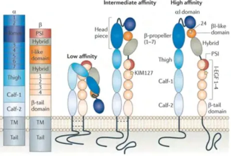
Figure 7: Schematic representation of the different components of the LFA-1 molecule in its different conformations
Figure 8: Overview of families of actin-binding proteins involved in the different processes pertaining to actin
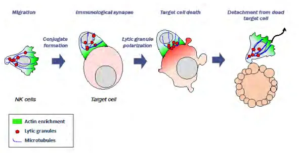
Figure 9: Dynamic steps of cytotoxicity
Figure 10: Top view model of the spatial organization of the immunological synapse
Figure 11: NK cells from STX11 deficient patients do not fail to polarize their perforin granules to the IS
Figure 12: NK cells from a normal donor and an STXBP2 deficient patient stained for F-actin and perforin reveal a normal granule polarization in patient cells
Figure 13: Wiskott–Aldrich syndrome protein activation, molecular partners, and cytoskeleton remodeling
Figure 14: The role of Coronin1A in the immunological synapse of NK cells
Figure 13: DOCK8 suppression leads to diminished F-actin and lytic granules at the IS Figure 16: Defective RASGRP1-deficient NK cell IS assembly
Figure 17: Crystal structure of the ARP2/3 complex from Baus taurus access protein data bank:1K8K Figure 18: PID-associated actin regulators
24 Figure 19: The timeline of microscopy
Figure 20: A typical high-content workflow pipeline
Figure 21: Schematic representation of the confocal light path of the Opera Phenix high content screening system.
Figure 22: Representative workflow for image-based cell profiling
Figure23: Visualization of 6000 handwritten digits from the MNIST data set by t-SNE Figure 24: Visualization of 6000 handwritten digits from the MNIST data set by UMAP Figure 24: Functional assays on the NK-92 cell line
Figure 25: Functional assays on primary NK cells
Table 1: HLH classification, causes and underlying defects
Table 2: Main criteria used in the assessment of a defective immunological synapse in cytotoxic lymphocytes by immunofluorescence.
25
Introduction
The following pages will comprise an introduction on the immune system with a focus on cytotoxic lymphocytes, their mechanism of action and receptors. In particular, the role of the actin cytoskeleton rearrangements and the immunological synapse in supporting cytotoxicity will be presented. Next, pathology-related defects in cytotoxicity will be addressed with a focus on primary immunodeficiencies, including a chapter on the methodologies used to assess defects in the cytotoxic function.
The second section of the introduction will focus on microscopy as a rapidly evolving collection of methodologies. In particular will be addressed how high content cell imaging applications provide novel opportunities to explore cell biology. This introduction will then provide a rationale for the implementation of high content cell imaging for the study of cytotoxic lymphocytes.
General overview of the immune system and immune cell activities
The human immune system is composed of a complex association of lymphoid organs, tissues, cells, humoral and soluble factors, which evolve simultaneously with the environment the body is exposed to, to ensure its protection against pathogens. Moreover, the immune system ensures the elimination cancer cells, as well as ensuring tissue reparation to maintain homeostasis. Also, it ensures symbiosis with the microorganisms of the microbiota.
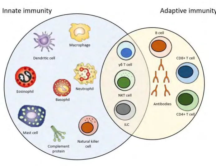
For long, the immune system has been divided into innate and adaptive immunity, due to the types of cells involved, as well the specificity and duration of their effector function. However, research is showing that cells of both types are highly interconnected. Therefore, an increasing number of immune cells is to be found at the interface between the innate and the adaptive immunity (Figure 1).
26
Figure 1: Overview of the immune system. The immune system is composed of the innate arm, the
27
1.1 Innate immunity
Innate immunity is a quick reaction which follows a non-specific recognition of various microorganisms, and can take as little as a few hours to be put in place2. The cells implicated are tissue resident natural killer cells, macrophages, mast cells, monocytes, neutrophils, basophils, eosinophils and dendritic cells (DC). However, only natural killer (NK) cells will be detailed in a section of this manuscript.
1.2 Cells at the interphase of the innate and adaptive immune systems
Innate lymphoid cells (ILC) are a new category of cells which were discovered in the past decade. Three groups of ILCs exist, based on the expression of transcription factors and cytokines that they produce3. NK cells were officially classified as the prototypical members of the group 1 innate lymphoid cells (ILC), which are defined by capacity to secrete interferon gamma (IFN-γ), but not type 2 cytokines4. Natural killer T (NKT) cells, which share surface markers of both natural killer and T cells. They express a semi-invariant receptor resembling that on T cells and which reacts with major histocompatibility complex (MHC) CD1d, as well as receptors present on NK cells such as CD1615. NKT cells are both cytotoxic cells and capable of releasing cytokines, such as IFN-γ, IL-4, IL-10 and IL-13. Tγδ cells are CD3+ T cells that express the gamma/delta alternative T cell receptor (TCR) (as opposed to the conventional alpha/beta). They are cells that produce cytokines and exert a cytotoxic activity. They recognize bacterial phosphoantigens and non-classical MHCI molecules, and in response can either exhibit cytotoxicity or secrete cytokines such as IFN-γ6–8.
1.3 Adaptive immunity
Adaptive immunity requires a longer time to take place than innate immunity, as it requires the action of antigen-specific cells, some of which would eventually become memory cells. Such a process requires several days to be put in place2. The players in this immune response are T and B lymphocytes. However, and as previously mentioned, innate and adaptive immune responses are interconnected. An adaptive immune response cannot be initiated without the dendritic cells, which are antigen presenting cells (APC). DCs will migrate into the lymphoid organs, where they will activate the T cells specific to their antigen. Through V(D)J rearrangements through their development, T cells would be endowed with unique and antigen specific TCR 9,10.
28
A. B lymphocytes
B cells are CD19+ lymphocytes. B lymphocytes recognize specific antigens by B cell receptors (BCR) or membrane bound immunoglobulins. BCRs recognize either soluble foreign antigens or ones bound to cell surface. Antigens are then internalized and processed in lysosomal/endosomal compartments, and subsequently expressed on the cell surface as antigenic peptides bound to MHC-II molecules for presentation to T helper cells (discussed in the next section). Activated B cells develop into plasma cells, which are antibody secreting cells.
B. T lymphocytes
T cells are CD3+ cells that can also be CD4+ or CD8+. CD4+ T cells are known as helper T (Th) cells and can be further subclassified into Th1, Th2 and Th17, based on the cytokines and interleukins they produce. Special subsets include follicular helper T cells (Tfh) and regulatory T cells (Treg). Their main role is aiding in B cell activation and antibody production. Follicular helper T cells are essential for B cells to mature into plasma cells and achieve proper immune functions11. Regulatory T cells suppress immune reactions and maintain immune homeostasis. CD8+ T cells were thought to be the only cytotoxic cells, prior to the identification of a subpopulation of cytotoxic CD4+ T cells12. CD8+ T cells are cytotoxic T cells (CTLs) and will be discussed in detail in the next section.
The cytotoxic function: the weapon of defense of the immune system
Natural killer cells and CD8+ T cells are two cell types belonging to the innate and the adaptive arms of the immune system respectively. Even though they, broadly speaking, appear to serve the same function, which is to eliminate target cell, their mechanism of action, cell surface receptors and regulation are not the same, and will be elaborated in detail in this section. The cytotoxic activity of NKT, Tγδ and cytotoxic CD4+ T cells will not be addressed.
Cytotoxicity is achieved when the cytotoxic cells kill their targets by the means of releasing lytic molecules through the process of degranulation. CD8+ T cells and NK cells can kill target cells by a tightly regulated secretion of cytotoxic granules containing pore-forming proteins perforin and/or granulysin and combinations of granzyme (Gzm) family effector proteases (in humans: Gzm A, B, K, M and H). However, a deep immune profiling of CTLs and NK cells has shown that NK cells have more abundant granulysin than CTLs13.
Lytic granules are delivered to the interface of the cytotoxic lymphocyte and target cell, and their exocytosis requires clearances in the actin cytoskeleton14. Released perforin monomers insert into the
29
target cell membrane and polymerize to form a pore through which granule contents including the effector protease enzymes are delivered, and subsequently cleave caspases to initiate cell death.
2.1 Natural killer cells
Natural killer cells are cytotoxic lymphocytes of the innate immune system, and which play a role in protection against viral infections and solid and hematological malignancies. They comprise about 10-15% of circulating lymphocytes. For a long time, NK cells were simply identified as cytotoxic cells that could kill without prior activation. They specifically target cells that lack MHC-I expression, without expressing clonally-distributed antigen receptors15. Moreover, they release cytokines that cause an inflammatory response which mediate the response of the adaptive immune system. Further details on the cytotoxic activity of NK cells will be addressed in the next chapter, “cytotoxicity”. Two major markers to characterize natural killer cells are CD16 and CD56. That being said, it should be noted that approximately 90% of natural killer cells are CD16bright CD56dim and those are the ones capable of generating the cytolytic response, also known as antibody-dependent cellular cytotoxicity (ADCC) 16. The other 10% are CD16dim CD56bright and are the ones that secrete cytokines. When it comes to expression of lytic molecules, among NK cells, CD56bright and CD56dim CD16+ NK cells showed clearly different cytotoxic molecule expression patterns: CD56bright NK cells had low Gzm B, but high Gzm K expression, whereas the CD56dim CD16+ NK cell subset expressed high Gzm B but lacked Gzm K13.
The cytotoxic activity of NK cells is tightly regulated by a complex interplay between stimulatory and inhibitory receptors and their ligands expressed on the target cells, which will be discussed in the coming section 17,18.
A. NK cell receptors
Natural killer cells receive signals through different receptors on target cells and use a multiple receptor recognition strategy and will kill cells that have an increased or a decreased expression of “self” proteins, and cells that express foreign proteins.
a. Natural cytotoxicity receptors
Natural cytotoxicity receptors (NCR) are key in recognizing virus infected and tumor cells and their clearance. The expression of these NCRs has been shown to correlate with the magnitude of the cytolytic activity of the NK cells.
30
i. CD16
CD16 is an Fc receptor (FcγIII) present on the CD56dim population of the natural killer cells. Signaling through CD16 delivers a powerful signal that enables the natural killer cells to kill target cells mainly by cytolysis. CD16 binds to the IgG portion of antibodies. In NK cells, CD16A is expressed and co-localizes with CD3ζ and Fc-εRI-γ, thus inducing the stimulatory signals. CD16 activation following binding to its ligand induces of the transcription of the genes encoding interleukin-2 receptor (IL-2R) and cytokines, and even though binding to anti-CD16 induces a strong cytolytic reaction, it does not lead to NK cell proliferation19,20. Moreover, it has been shown that the levels of expressions of CD16 may vary, and this variation correlates with the strength of serial killing21.
ii. NKp46
NKp46 (also known as CD335) is a member of the natural cytotoxicity receptor family, and is, along with NKG2D, one of the main receptors. In1997, a 46 kDa molecule was shown to play a role in NK mediated cytolytic activity, cytokine secretion as well as Calcium ion mobilization22. NKp46 is encoded in the leukocyte receptor complex on chromosome 1923,24. NKp46 has been shown to be expressed on subsets of natural killer cells, while it was not expressed in T and B cells25. NKp46 expression was shown to be decreased in post-transplant lymphoproliferative disease in all natural killer subsets, accompanied by an increase in programmed cell death protein 1 (PD-1), which may give a hint on the dysregulation in natural killer functions26. Cross-linking of NKp46 with a monoclonal antibody (mAb) resulted in an increase in cytolytic activity, cytokine secretion and Calcium ion mobilization. NKp46 molecules are coupled to intracytoplasmic transduction machinery through associating with CD3ζ and Fc-εRI-γ adaptor proteins that contain immune tyrosine-based activating motifs (ITAM).
iii. NKp30
NKp30 is encoded in the class III region of the MHC locus on chromosome 624. It has been demonstrated that NKp30 expression varies depending on the organ in which NK cells are examined: it is highest in spleen, lowest in liver and intermediate in blood, despite the absence of a general receptor down-regulation, thus suggesting either an organ-specific regulation of NKp30 receptors, or a preferential homing of NKp30+ NK positive cells27. Expression of the ligands of NKp30 on DCs is the main regulator of the supply of dendritic cells, as NKp30 can mediate the promotion or termination of dendritic cell maturation. One of the ligands of NKp30 is BAG6. Soluble BAG6 has been shown to be present in elevated levels in patients with hematological diseases 28,29. Galectin-3 has also been shown to be a soluble NKp30 target, and to play a role in tumor evasion from NK cells 30. B7-H6,
31
another soluble NKp30 ligand was shown to be associated with a down-regulation of NK cells in neuroblastoma patients31. Another NKp30 ligand is BAT6, a nuclear factor also released by tumor cells in exosomes rather than a soluble protein, which triggers cytokine release (tumor necrosis factor alpha (TNF-α), IFN-γ) and enhances NK-cell mediated killing 27,32. The cytoplasmic region of NKp30 lacks ITAM motifs.
iv. NKG2D
NK receptor member D of the lectinlike receptor family (NKG2D) is a natural killer receptor that binds to cells which have an upregulated expression of self-proteins. It is also expressed on CD8+ T cells, γδ T cells and Natural Killer T cells33,34. NKG2D binds to NKG2D-L on target cells, thus activating an effective anti-tumor response in early stages35. However, a sustained NKG2D-L expression and shedding of soluble ligands counteracts NKG2D-dependent NK cell activity in later stages. Contrary to NKG2A, NKG2D does not bind to CD94. The ligand for NKG2D is stress-inducible MICA, a distant homolog MHC I, and the expression of NKG2D on NK cells is increased by IL-1534,36. The structures of NKG2D–ligand complexes indicate that NKG2D binds diagonally over the α1 and α2 helices of the ligands, which bears a striking resemblance to the way T-cell receptors bind over MHC molecules. DAP10 is the membrane-bound signal transducing subunit of the NKG2D receptor37. It has been shown that one tumor evasion mechanism is the release of soluble NKp30 and NKG2D ligands, as well as a down-regulation of the expression of NCR ligands 38,39.
v. NKp44
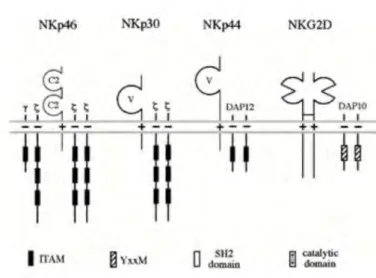
NKp44 is another member of the NCR family, and is about 44 kDa in size. The gene encoding NKp44 is encoded on chromosome 6. While NKp30 and NKp46 are expressed on resting and activated NK cells, NKp44 is exclusively expressed on activated NK cells in the presence of IL-240. NKp44 ligands include glycoproteins and proteoglycans expressed on the surface of target cells, nuclear proteins that can be exposed outside the cell, and molecules that can be either soluble or in vesicles41. In the same fashion as NKp46, monoclonal antibody cross-linking of NKp44 enhances the cytolytic activity of NK cells. NKp44, which does not contain any ITIM motif in the cytoplasmic region, associates with the adaptor molecules DAP12 and KARAP, which contain an ITAM motif in the cytoplasmic region40,42.
32
Figure 2: Natural killer cell surface receptors involved in cytolytic activity, and their association to signaling molecules24.
b. Inhibitory NK receptors (iNKR)
Regulation of the cytotoxicity of NK cells is also a crucial process and depends on killing inhibitory receptors (KIR), whereby the signals that NK cells receive completely bring their cytolytic activity to a halt. It has been shown that inhibiting the cytolytic activity of NK cells depends on the recognition of MHC-I molecules. Killer Ig-like KIR and NKG2A/B (heterodimer with CD94) are major human leukocyte antigen I (I) specific inhibitory receptors. The CD94/NKG2A/B recognize the HLA-E molecule (a non-classical MHC-Ib) and subsequently protect the target cells from cytolysis43. KIRs include but are not limited the p49, p50, p58, p70 and p14017,38. However, each KIR is expressed on a fraction of NK cells, thus allowing the NK cells to recognize a larger pool of MHC-I deficient cells. It has been shown that once an NK cell is in contact with an MHC-I expressing cell, KIRs accumulate at the contact area in a central area surrounded by LFA-1 and ICAM-1 to drive NK cell migration44. F-actin is a somewhat expendable player in this process, whereby it can accelerate KIR recruitment to the synapse but is not crucial for this process44.
c. Costimulatory molecules
2B4 (CD44) which binds to CD48, and NTBA are two members of the CD2 family which can play either an activating or inhibitory function, depending on the availability of downstream regulating element of the signaling pathway termed signaling lymphocyte activation molecule-associated protein (SAP). Both 2B4 and NTBA trigger NK cytolytic activity in cells that express high levels of NCRs45,46. NTBA promotes a strong production of IFN-γ and TNF-α46,47. 2B4 may also enhance the cytolytic activity of NK cells48. 2B4 does not associate with ITAM-bearing polypeptides but has functional
33
signaling motifs in the intracellular domain. The 2B4-CD48 interactions appear to take place at the center of the synapse44. Sivori and colleagues have shown that 2B4 can initiate cytolysis in the subset of NK cells that have a high level of NKp46 expression, without altering the expression of 2B4, but also that 2B4 may function as an inhibitory molecule at early stages of NK cell differentiation49,50. Other co-stimulatory molecules include CD80 and CD86, which have been shown to enhance the cytotoxic activity of NK cells51.
B. NK cell cytotoxicity: from target binding to target lysis
Natural killers were long compared to cytotoxic T cells. However, they are unique in the sense that they use specific NK receptors, and that their response is faster than that of CTLs. Being part of the innate immune system, natural killer cells do not need prior activation to achieve their cytotoxic function. Therefore, they do not express antigen-specific receptors, but rather the activation and inhibition receptors discussed in the previous chapter “NK cell receptors”. The cytotoxicity of NK cells can be broken down into several steps, leading to the formation of the immunological synapse (IS) and achievement of the cytotoxic function by release of lytic molecule content, and then detachment of scanning for another target cell.
a. NK receptors involved in immunological synapse formation
NK cell receptors can associate with ITAM bearing molecules. FcRγ and TCR ζ chains can form either homodimers or heterodimers which interact with CD16. NKp30 and NKp46 associate with FcRγ and TCR ζ, while NKG2D associates with DAP1052. After patrolling their environment, NK cells attach to a target cell through adhesion receptors, which include CD2, NKG2D and DNAM-1. By using tweezers and confocal microscopy, it has been demonstrated that upon NK cell contact with its target, NKG2D is recruited to the IS and forms a ring type of structure which marks a border in which lytic granules are secreted53. Moreover, stimulation through NKG2D was shown to lead to the formation of an F-actin ring at the periphery of the IS, and that NKG2D leads to a more stable synapse than CD1621,53. NKp44 associated with DAP12, a homodimer with a single ITAM motif. While signaling through CD16 is sufficient to cause NK cell – target conjugates and degranulation, stimulation of other receptors alone does not cause NK degranulation21,54,55. Other receptors only stimulate degranulation in pairs, suggesting a synergistic activation. CD16 binds to the Fc portions of antibodies and NK cells can therefore kill target cells coated with antibodies and eliminate them by ADCC. NKG2D signaling
34
leads to adhesion, granule polarization and degranulation56. DNAM-1, NKp80 and NKp65 also have activating potential. However, there is limited information of their signaling properties. The immunological synapse of NK cells will be touched upon further in the section pertaining to the steps of cytotoxicity.
b. NK cell degranulation
Degranulation of NK cells is not random process, and it has been shown that NK cells must converge their lytic granules to prevent bystander killing and achieve a more efficient elimination of target cells55. Moreover, it has been shown that receptor activation must occur in a specific sequence, as initial engagement of CD16 did not affect subsequent stimulation via NKG2D, whereas stimulation through CD16 was impaired after activation via NKG2D21. Once firmly tethered to its target, the NK cell undergoes a change in shape and becomes more flattened, and a lytic cleft is formed57. Following actin rearrangement as well as de novo actin synthesis, the MTOC undergoes reorientation to allow polarization of the lytic granules towards the synapse, and this is mediated by the formin homolog hDia, which in NK cells is not essential for actin branching58.
DNAM-1 has been found to physically associate with lymphocyte function associated antigen 1 (LFA-1), thus indicating that it plays a role in LFA-1 mediated signaling 59. Indeed, LFA-1 engagement was shown to mediate granule convergence, and to induce more targeted degranulation at the IS, along with CD16 than CD16 alone55. Therefore, the co-engagement of LFA-1 and CD16 leads to a monodirectional granule convergence, thereby reducing bystander killing rates and increasing the rates of specific killing. only a small fraction express the open conformation of LFA-1, and LFA-1 in NK cells requires activation to through the aforementioned adhesion receptors, which can induce inside-out signaling60. LFA-1 induces a downstream signaling cascade, changes in F-actin reorganization, as well as an arrest signal and symmetrical spreading61. Talin is also recruited to the sites of LFA-1 prior to granule polarization, and, via constitutive association of vinculin (an actin-binding protein that was shown to act as “leaky capper” of the barbed end of the actin filament) to talin and Arp2/3, recruits ARP2/362–64. Moreover, talin is essential for lytic granule polarization65. Furthermore, LFA-1 also leads to WASp recruitment, which in turn promotes Arp2/3 mediated actin polymerization62. Lytic granules must converge long the MTOC, and this step is independent of actin dynamics and of CD107a, and is rather mediated by dynein and does not imply a commitment for neither granule polarization nor for cytotoxicity66. However, formation of an organized synapse where LAMP-1 is retrieved at the center depends on the interaction of LFA-1 with the lipid bilayer44. Following lytic granule polarization, they dock at the IS membrane and fuse with the plasma membrane. The docking
35
is primarily mediated by Rab27a and Munc13-4, followed by priming, which occurs as a result of Munc13-4-mediated activation of Syntaxin 11 or bridging of the plasma membrane with lytic granule66–68. Fusion occurs as a result of the formation of a complex whose components are not all known, but include syntaxin11, VAMP 4 and VAMP 766.
Following lytic granule delivery to the IS, it has been shown that only a fraction of the lytic granules are exocytosed, and that granules are highly mobile, independently of F-actin dynamics, but may depend on Myosin II A(MyH9) to generate the necessary forces14,69. Indeed, MyH9 heavy chain knock-out impaired cytotoxicity, membrane fusion of lytic granules, and granzyme secretion70. It has also been shown that MyH9 constitutively physically interact with lytic granules, and lytic granule secretion was shown to be impaired in patients suffering from MyH9 mutations71. While Myosin IIa is not needed for the formation of an organized IS in NK cells, it is crucial for the final exocytosis step44. Following degranulation, the cell must undergo a termination process, followed by detachment. These steps are elaborated in details in66. Moreover, it has been shown that lytic granules require a pervasive actin meshwork of hypodense F-actin (which can be achieved through NKG2D and LFA-1 co-ligation) to pass through and that the actin clearances in which they pass are granule-sized53,72. However, actin dynamics are independent of the presence of granules, since actin clearances are observed prior to granule arrival. It has also been demonstrated by super resolution microscopy that the center of the NK IS contains thin F-actin filaments, as opposed to the previous belief that the IS formation leads to F-actin clearance from the center53,72.
2.2 Cytotoxic T cells: the killers of the adaptive immune system
CD8+ T cells have a classical αβ T-cell receptor TCR. Through a random process of genetic rearrangements, lymphocytes express on their surface unique TCR that have single antigen specificity, which is their capacity to recognize one type of peptide-MHC complex.
Progenitor lymphoid cells leave the bone marrow and reach the thymus to proceed to differentiate, where they undergo a negative and a positive selection. Following this selection, the remaining cells will be capable of recognizing APC, and not react against self-molecules. Naïve CD8+ T cells must be activated by CD4+ cells in order to produce lytic and effector molecules73. CD8+ lymphocytes recognize MHC I molecules presented on the surface of APCs, therefore most cells can be APC- as opposed to CD4+ T cells which recognize MHC II molecules on “professional” APC such as DC and B cells. Naïve T cells will give rise to several subpopulations, culminating in effector cells. Effector
36
CD8+ T cells express lytic molecules and have high migratory capacity. Following stimulation of their TCR, they undergo reorganization of their actin cytoskeleton, slow down their motility and establish firm adhesions with their targets leading to the assembly of the IS74.
The delivery of lytic granules from CD8+ T cells is a highly regulated process, and the signaling and cytotoxic activity of CD8+ T cells will be elaborated in detail in the section “CD8+ T cell Cytotoxicity”.
Figure 3: Schematic representation of T cell activation and differentiation in response to APC stimulation. Adapted from the thesis manuscript of Julie De Meester (Thesis, 2011).
A. Receptors, adhesion and co-stimulatory and inhibitory molecules involved in
CD8+ T cell cytotoxicity
37
The activity of cytotoxic T cells is governed by the interplay between several receptors and co-stimulatory molecules, which will be elaborated in the following pages.
a. The T-cell receptor
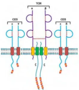
The αβ TCR on CTLs is composed of two transmembrane glycosylated polypeptide chains belonging to the immunoglobulin superfamily and linked through a disulfide bond. These glycoproteins contain a short intracytoplasmic tail composed of 4-12 amino acids and a hydrophobic transmembrane domain of 20 positively charged amino acids, thus allowing the TCR to achieve a stable association with the negatively charged transmembrane domain of the CD3 molecules (Figure 4)75.
Figure 4: The TCR and its association with CD3. The TCR is composed of the α and β chains and
forms a complex with CD3. From the thesis manuscript of Roxana El Khazen, 2016.
T cell activation requires sustained signaling by the pMHC specific to the TCR. However, the duration of the TCR-pMHC contact has been shown to be very short76,77. Therefore, it was proven that despite the rapidity of MHC interaction, sustained TCR signaling occurs via multiple rounds of TCR-MHC binding78,79. Interestingly, as little as 1 MHC molecule can induce calcium ion increase in T cells, and that T cell activation can be achieved by TCR recognizing as little as 10-15 MHC molecules, and CTLs can kill target cells expressing 1-10 MHC molecules80–83. This can be explained by the proposed model of “serial TCR engagement”, which suggests that one MHC molecule could trigger and internalize approximately 180 TCRs, thus leading to a sustained TCR signaling despite the small number of MHC molecules via a sequential high TCR occupancy84. However, another model referred to as “kinetic proofreading” suggests a prolonged TCR-MCH interaction, leading to modifications
38
which will be amplified only by highly specific interactions85. However, it is most likely that these two types of interactions are not mutually exclusive and may be dependent on the number of MHC molecules on the APC surface.
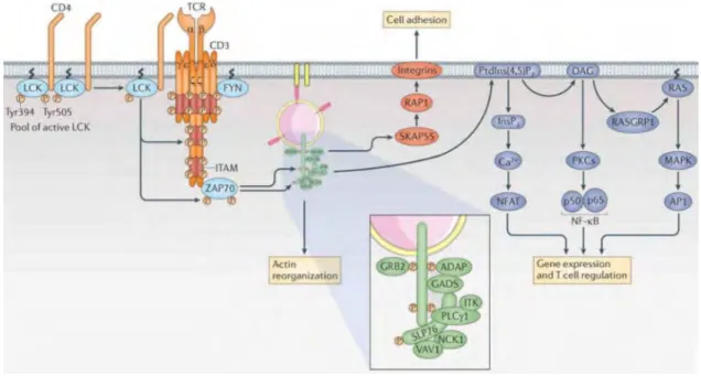
Briefly, the steps of TCR signaling include the translocation of LCK and Fyn, two members of the Src tyrosine kinase family, into the membrane following TCR engagement, which eases the phosphorylation of ITAM motifs. ZAP-70 also associates to ITAM motifs and activates Linker for activation of T cells (LAT) protein which will allow more signaling molecules to bind (Figure 5). However, TCR engagement is not enough and T cell activation requires signaling from co-stimulatory molecules. These include CD8 for CTLs, CD28 and the adhesion molecules CD2 and LFA-186.
39
b. The role of the integrin LFA-1 in cytotoxicity
Integrins are heterodimeric adhesion proteins, which allow leukocyte adhesion and play a role in their migration. Integrins play an essential role in infection control, where their absence leads to recurrent infections. They also have a role in immune homeostasis, and perturbations can lead to cancers and autoimmunity. Integrins have a crucial role in the recruitment of lymphocytes from the blood into infection sites, and for lymphocyte adhesion onto their target cells. Moreover, integrins make intracellular adhesion therefore playing a role in the activation of several signaling pathways.
Being heterodimeric proteins, they are formed of one of 18 α and one of 8 β subunits. They can be one of 24 different combinations88,89. The very late after activation (VLA) family of integrins which comprises VLA-1 to 5 has also been identified. Of the LFA-1 family, three members have been identified: LFA-1, 2 and 390. In this manuscript, only the integrin LFA-1 will be addressed.
LFA-1 is required for the migration of T and NK lymphocytes. It is composed of the CD11a (αL) and the CD18 (β2) subunits. LFA-1 plays a role in immunity, as its deficiency is characterized by recurrent bacterial infection91. LFA-1 is a transmembrane protein comprised of an extracellular domain, a transmembrane domain and a cytoplasmic domain. LFA-1 binds to intracellular adhesion molecule ICAM-1, among other ICAMs (ICAM-2 and 3, as well as JAM-1). In this manuscript, only the LFA-1 – ICAM-LFA-1 interaction will be addressed. LFA-LFA-1 binding to ICAM-LFA-1 is a temperature-dependent energy requiring process, which also requires the presence of magnesium ion and an intact cytoskeleton92.
ICAM-1 (CD54) is a transmembrane glycoprotein expressed on the surface of leukocytes and endothelial cells. ICAM-1 is required for the interaction of LFA-1 on immune cells with their target cells or DCs, or between the immune cells and their environment92–94. Moreover, full LFA-1 activation requires ICAM-1 on the target cell to provide a physical resistance to allow for F-actin flow95.
i. LFA-1 activation
Integrins in circulating lymphocytes are inactive and get activated through binding to chemokine receptors. LFA-1 activation is characterized by both affinity to ICAM-1, and avidity.
Affinity is defined as the capacity of a ligand to bind to its receptor, depending on the available number of molecules. FRET experiments have revealed that the constant of dissociation between LFA-1 and ICAM-1 to be 17.93±1.34 nM96.