(Received June 16/0ctober 10, 1986) - EJB 86 0608
An 11 450-base DNA fragment containing the gene for the extracellular active-site serine DD-peptidase of Streptomyces R61 was cloned in Streptomyces lividans using the high-copy-number plasmid pIJ702 as vector. Amplified expression of the excreted enzyme was observed. Producing clones were identified with the help of a specific antiserum directed against the pure DD-peptidase. The coding sequence of the gene was then located by hybridization with a specific nucleotide probe and sub-fragments were obtained from which the nucleotide sequence of the structural gene and the putative promoter and terminator regions were determined. The sequence suggests that the gene codes for a 406-amino-acid protein precursor. When compared with the excreted, mature DD-
peptidase, this precursor possesses a cleavable 3 1-amino-acid N-terminal extension which has the characteristics of a signal peptide, and a cleavable 26-amino-acid C-terminal extension. On the basis of the data of Joris et al. (following paper in this journal), the open reading frame coding for the synthesis of the DD-peptidase was established. Comparison of the primary structure of the Streptomyces R61 DD-peptidase with those of several active-site serine p-lactamases and penicillin-binding proteins of Escherichia coli shows homology in those sequences that comprise the active-site serine residue. When the comparison is broadened to the complete amino acid sequences, significant homology is observed only for the pair Streptomyces R61 DD-peptidaselEscherichja coli ampC p-lactamase (class C). Since the Streptomyces R61 DD-peptidase and p-lactamases of class A have very
similar three-dimensional structures [Kelly et al. (1986) Science (Wash. DC) 231, 1429- 1431 ; Samraoui et al. (1986) Nature (Land.) 320, 378-3380], it is concluded that these tertiary features are probably also shared by the F-lactamases of class C , i.e. that the Streptomyces R61 DD-peptidase and the p-lactamases of classes A and C are related in an evolutionary sense.
For the purpose of defining the interaction between
p-
lactam antibiotics and their bacterial enzyme targets at the molecular level, the active-site serine DD-peptidase of Streptomyces R61 is being actively investigated [1, 21. X-ray crystallography has led to the identification of the polypeptide chain folding at a resolution of 0.28 nm [3-
51. In parallel to this, the nucleotide sequence of the corresponding gene has been established. This paper describes experiments which led to (a) cloning of the gene for the Streptomyces R61 DD-peptidase; (b) determination of the nucleotide sequence; (c) identification of possible features related to the processing and excretion of the primary translation product; (d) comparison of the amino acid sequence of the protein with those of several active-site serine p-lactamases and penicillin- binding proteins.
Correspondence to J. Dusart, Service de Microbiologie, Institut de Chimie, B6, Universitt de Libge au Sart-Tilman, B-4000 Libge, Belgium
Note. This paper is part of a dissertation of C.P.F. presented as partial fulfilment for a Ph. D. thesis, at the University of Liege.
Abbreviations. Ac, acetyl; bp, base pair; kb, 1000 base pairs; PBP, penicillin-binding protein.
Enzymes. Bacterial alkaline phosphatase (EC 3.1.3.1); fi- lactarnases (EC 3.5.2.6); DD-peptidases (EC 3.4.16.-); lysozyme (EC 3.2.1.17); restriction endonucleases(EC 3.1.21.4); T4 DNA ligase (EC 6.5.1.1).
MATERIALS AND METHODS Bacterial strains and plasmids
Streptomyces R61 was from the Microbiology Department of the University of Liige. S. lividans TK24 (str-6; a strain cured of its natural phsmids) [6] and the non-conjugative high-copy-number plasmids pIJ702 [7] and pIJ385 [8] were from the John Innes Institute, Norwich, UK. Escherichia coli HBlOl [9] and plasmid pBR322 [lo] were also used.
Growth conditions and media
Growth of Streptomyces cultures was carried out at 28 "C with vigorous orbital shaking. The following media were used: YEME medium [ll]; Merck peptone medium and E9 broth [12]; Difco brain heart infusion; 2 xTY broth (Amersham handbook); and glycerol-casein medium [13]. R2YE agar [ l l ] was also used. E. coli HBlOl was grown at 37°C in Luria- Bertani or M9CA medium [14] with vigorous shaking. Enzymes, antiserum, proteins and antibiotics
The DD-peptidase of Streptomyces R61 was prepared as described in [15]. Rabbit anti-(R61 DD-peptidase) antiserum was prepared by Gamma S.A. (Tavier, Belgium). The enzymes used in the recombinant DNA techniques were from Bethesda
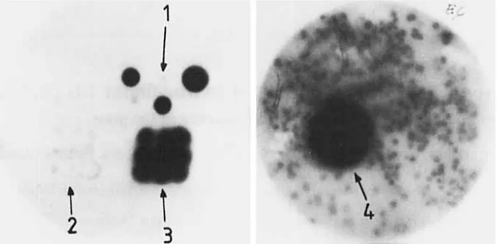
Fig. 1. Streptomyces R61 DD-peptidase-producing colonies us detected with the immunological test. (1) Three samples of the purified DD-
peptidase; (2) nine colonies of the Streptomyces lividans TK24 recipient strain; (3) nine colonies of the Streptomyces R61 donor strain; (4) one colony of the Streptomyces lividans CC1 recombinant
Research Laboratory (Gaithersburg, MD, USA); New England Biolabs (Beverly, MA, USA); Sigma Chemical Co. (St Louis, MO, USA); Boehringer, Mannheim, FRG; Amersham International, Amersham, UK. Radioactive 3-(4-
hydroxy-5-[125]iodophenyl)propionamide-protein A (product
IM144; specific radioactivity 35 mCi/mg) was from Amersham. Thiostrepton was a gift from Dr. R. B. Sykes (Squibb and Sons, New Brunswick, NJ, USA). The other antibiotics and bovine serum albumin were from Sigma.
Screening of Do-peptidase-excreting clones (immunological test)
Nitrocellulose filters (Millipore HATF) were (a) laid for 30 min on Streptomyces colonies grown on R2YE agar medi- um; (b) dried for 20 min at room temperature; (c) incubated for 1 h at room temperature and then for 2 h at 37°C with a 2.5% bovine serum albumin solution made in NaC1/Pi buffer pH 7.4 [NaCl/Pi buffer was a mixture of A (8 vol.), B (1 vol.) and C (1 vol.) where A = 1% NaC1, 0.025% KC1, 0.143% Na2HP04, 0.025% KH2P04; B = 0.1% CaClz . 2 H 2 0 ; and
C = 0.1% MgC12
.
6 H20]; (d) incubated at 37°C with a ~ l o o o dilution of the anti-(R61 DD-peptidase) antiserum made in NaC1/Pi buffer; e) washed five times with NaCl/Pi buffer at 20°C; (f) incubated with the 12sI-labelled protein A for 2 h at room temperature (10 pCi of the radioactive protein in 50 ml of NaCl/Pi buffer was sufficient to treat 10 filters at the same time); (g) incubated at room temperature, twice (for 30 min and 16 h, successively) with the NaCl/Pi buffer and then twice (for 30min each time) with the NaC1/Pi buffer supplemented with 0.1% Triton X-100; (h) dried for 1 h at room temperature; and (i) exposed for 3 - 4 days at - 70°Cto an X-ray film (Fuji Film Co. Ltd, Medical) in a Kodak X-omatic cassette with intensifying screen. Steps c
-
g were carried out with slow orbital shaking.Estimation of Do-peptidase activity in culture fluids
The tripeptide Ac,-L-Lys-D-Ala-D-Ala was used as sub- strate and the amount of released D-Ala was measured as described in [15].
Recombinant DNA techniques
Essentially, the procedures described in [I I] were used for the preparation of Streptomyces R61 chromosomal DNA, the
large-scale and mini (alkaline lysis) preparations of plasmid DNA, the preparation of protoplasts of S. lividans TK24 and the transformation experiments. However, lysis of Strepto- myces R61 with lysozyme was performed at 0°C for 150 min in the presence of 80 mM EDTA. Protoplasts (lo7 in 100-p1 samples), DNA (100 ng in 10-p1 samples) and poly(ethy1ene- glycol) (66 pl of a 28.5% solution) were mixed together. Before plating, the transformation mixtures were diluted with P buf- fer [I I].
Essentially, the procedures described in [ 141 were used for digestion with the various endonucleases, treatment with bacterial alkaline phosphatase, ligation, agarose gel electro- phoresis of digested and ligated DNAs and elution of separated DNA samples. However, restriction was carried out for 1 h at the optimal temperature of the enzyme using 1 pg DNA for two enzyme units. Digestion of restricted DNAs with the alkaline phosphatase was carried out in 50 mM Tris/ HC1 buffer pH 8 using 1 enzyme unit/pmol phosphate and the mixtures were incubated at 37°C for 1 h (in the case of 5' protruding ends) or at 56°C for 2 h (in the case of 3' pro- truding ends). Ligation mixtures contained 2 pg restricted genomic DNA, 1 pg restricted and alkaline-phosphatase- treated vector DNA and 1 unit of T4 DNA ligase; they were incubated at
4°C
for 16 h and then at 16°C for at least 3 h. All the digested DNAs were submitted to extraction with phenol/chloroform mixture and then with chloroform alone, precipitated with ethanol and stored at -28 "C in TE buffer [ill.D N A probe and hybridization experiments
Peptide T2 isolated from a trypsin digest of carboxy- methylated DD-peptidase [I 61 contained the sequence Asp-
Asp-Asn-Gly-Thr-Ile, on the basis of which a 17-nucleotide- long probe 3' CT&CT&TT$-CCN-TGN-TA 5' (N meaning A, G, C or T) was synthesized [17]. The polynucleotide was labelled with [ Y - ~ ~ P I A T P as described in [18]. The hybridiza- tion experiments were carried out at 37°C for 18 h using the Southern procedure [19, 201.
Nucleotide sequencing
The dideoxynucleotide chain-termination method [21] was used. Several lengths of sequence were proved to be difficult to read because of base compression, probably caused by the high G
+
C content of Streptomyces DNA, and were resolvedB
-
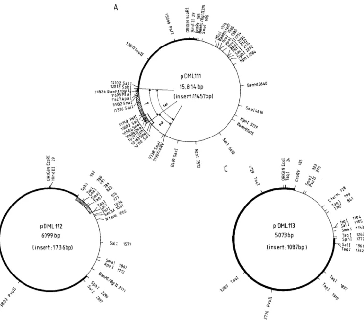
u W - 0 . ON C 4 z = ’9 2 -29 1 4 4 hlFig. 2. Restriction maps o f p D M L I l 1 ( A ) , p D M L I l 2 ( B ) andpDML113 ( C ) . ( A ) The 11.45-kb BglII insert (light line) in pBR322 (hcavy line). The SphI fragment (hatched box) and SalI fragment (open box) hybridize with the nucleotide probe (Fig. 3). (1) Segment cloned in pDML112 (Fig. 2 B ) ; (2) segment cloned in pDML113 (Fig. 2C); (3) segment cloned in pDML114 (see text). (B) The 1.75-kb SphI insert (light line) in pBR322 (heavy line). The SphI-SaZI insert (hatched box) hybridizes with the nucleotide probe. (C) The 1 .l-kb SphI-EcoRV insert (light line) in pBR322 (heavy line)
-
u W - 0 . ON C 4 z = ’9 2 -29 1 4 4 hlFig. 2. Restriction maps o f p D M L I l 1 ( A ) , p D M L I l 2 ( B ) andpDML113 ( C ) . ( A ) The 11.45-kb BglII insert (light line) in pBR322 (hcavy line). The SphI fragment (hatched box) and SalI fragment (open box) hybridize with the nucleotide probe (Fig. 3). (1) Segment cloned in pDML112 (Fig. 2 B ) ; (2) segment cloned in pDML113 (Fig. 2C); (3) segment cloned in pDML114 (see text). (B) The 1.75-kb SphI insert (light line) in pBR322 (heavy line). The SphI-SaZI insert (hatched box) hybridizes with the nucleotide probe. (C) The 1 .l-kb SphI-EcoRV insert (light line) in pBR322 (heavy line)
using the formamide procedure described in [8]. For each of the three possible reading frames, the codon usage was analysed with Staden’s program [22] using the neomycin phosphotransferase gene of Streptomyces fradiae as reference for codon usage [23]. Possible errors indicated by frame shifts were corrected by careful re-examination of the X-ray films (and using the aforementioned formamide procedure).
Comparison of the amino acid sequence of the DDpeptidase with those of p-lactamases and penicillin-binding proteins
Segments of varying span length (11, 15, 25 and 31 re- sidues, respectively) of a pair of proteins were analysed with the McLachlan’s procedure [24] which compares protein sequences on the basis of the relative amino acid substitution frequencies found among families of homologous proteins. Matrices expressing the probability ( P ) that similarity between pairs of segments occurred by chance were scored: blank
(1 10-2 < P
<
1); i ( 5 10-35
P5
1 x 10⁻²); 2(1 x 10-35
P5
5 x 10⁻³;3(1 x5 P
5
1 x 10⁻³)and4(05
P5
1 x The statistical significance of the global comparison between two proteins was estimated by calculating the &,p
value as described by Fitch [25]. McLachlan’s test and Fitch’s test were carried out with the help of programs developed by C. Wuilmart (Ldboratoire de Physiologie animale, Universitk Libre de Bruxelles, Belgium).
RESULTS
Cloning of the Do-peptidase gene
When applied to colonies (grown for 3 - 5 days at 20°C
on R2YE medium), the immunological test was positive in the case of Streptomyces R61 and negative in the case of S.
lividans TK24 (Fig. 1).
pIJ702 carries thiostrepton resistance (tsr) and melanin production (mel) genes. pIJ385 carries thiostrepton resistance
TTTIPhe 0
TTClPhe 12 TTAILeu 0 TTG/Leu 3
TCTISer 0 TATITyr 2 TGTICYS 0
TAAIOC 0 T G A ~ O P 1 TCC/Ser 1 1 TACITyr 12 TGCICys 2 TCAlSer 2
TCG/Ser 7 TAGIAM 0 TGG/Trp 2
DNA
Yo
1 BamHI pIJ702/BglII 3000 95 (rnel-) 3200
2 BgnI pIJ702/BgZII 15000 90 (mel-) 14300 3 SacI pIJ702/SacZ 50000 2 (mel-) 450 4 ClaI pIJ385/ClaI 2500 75 (tsr-) 3150 5 PstI pIJ385/PstI 2500 50 (aph-) 3400 24 500
Table 2. Excretion of the Do-peptidase by Streptomyces lividans CCI, Streptomyces lividans TK24 and Streptomyces R61 in various liquid media after 72 h of growth at 28°C
References describing the media are given in Materials and Methods Streptomyces Medium strains DD-Peptidase excreted CCl YEME Luria-Bertani Merck peptone 2xTY gl ycerol-casein brain heart E9 TK24 Merck peptone R61 gl ycerol-casein mg
.
I - ' 5.79 6.69 9.60 9.22 16.19 0.1 1 8.57 0 0.5 Tablelibrary 4). After a further 2.5-5 days, recombinants were identified on the basis of their white appearance (mel-; libraries 1, 2 and 3), sensitivity to thiostrepton (library 4) Table 4. Amino acidcomposition of the Streptomyces R61 DD-peptidase The number of residues per molecule as found by amino acid analysis [16], of the excreted protein and as deduced from the nucleotide sequence of the gene are given. The main discrepancies between the values are underlined
Amino acid Residues in DD-peptidase molecule
from protein from gene
Ala Arg Asx CYS Glx GlY His Ile Leu LYS Met Phe Pro Ser Thr TrP TYr Val 34 13 38 3 32 8 9 33 8 6 12 11 29 38 4 13 30 2 s - - 34 14 39 2 27 30 9 9 33 7 8 12 12 27 40 2 14 30
3. Codon usage for [he synthesis of the Streptomyces R61 Do-peptidase
1
m'r;;L;
l;1
CCT/Pro1
!l1
CGTIArg ~[
CCCIPro CGClArg
CTA/Leu CCA/Pro CAA/Gln CGA/Arg CTGILeu 23 CCGIPro 8 CAGjGln 19 CGClArg
ATA/Ile ATGIMet GTT/Val GTAIVal GTGIVal 15 ACTIThr 0 ACCIThr 23 ACAlThr 0 ACGIThr 20 GCTIAla 2 GCClAla 21 GCAIAla 4 GCGIAla 21 AATIAsn 0 AACIAsn 16 AAAlLys 0 AAG/LyS 7 G A T I A S ~ 1 G A A I G ~ U 2 G A G I G ~ U 8 GAClAsp 26 AGTISer 1 AGCISer 7 AGAIArg 1 AGGlArg 0 GGTIGly 6 GGClGly 21 G G A I G ~ Y 4 GGGIGIY 8
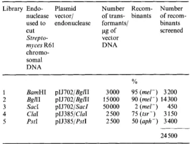
Fig. 3. Agarose gel electrophoresis of p D M L l l l digests ( A ) and hybridization with the nucleotide probe ( B ) . (A) Ehtidium bromide stain of gel. (B) Autoradiography of the Southern blot. The restriction endonucleases were (1) EcaRV; (2) PstI; (3) Sun; (4) SphI; ( 5 ) ApaI; (6) ApaI
+
HindIII; (7) HpaI+
HindIII; (8) KpnI+
HindIII; (9) MluI+
HindITI; (10) NcoI; (11) NcoI+
HindIII; (12) Sstl; (13) SstI+
HindIII; (14) BamHI; (15) BamHI+
HindIII. C for control: HindIII-restricted 1 DNAor sensitivity to neomycin (library 5) (by replica plating to original Streptomyces R61. The best producer, S . lividans antibiotic-containing R2YE) (Table 1). Of the 24500 re- CC1, was grown in several liquid media. The levels of DD- combinants screened for DD-peptidase excretion, eight had a peptidase activity in the culture fluids showed that gene positive immunological reaction (Fig. 1). All of them cloning had indeed resulted in amplification of the expressed originated from library 2. As judged from the size of the zones, protein but variations occurred depending on the medium they produced larger quantities of the DD-peptidase than the (Table 2).
0-
- -
'blpBR322lT w
-
G E a
a
-
-
- - -
Hlnd I!! blpBR3221 , 0: \ Taq 1 @ -I- -
-
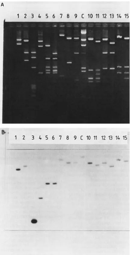
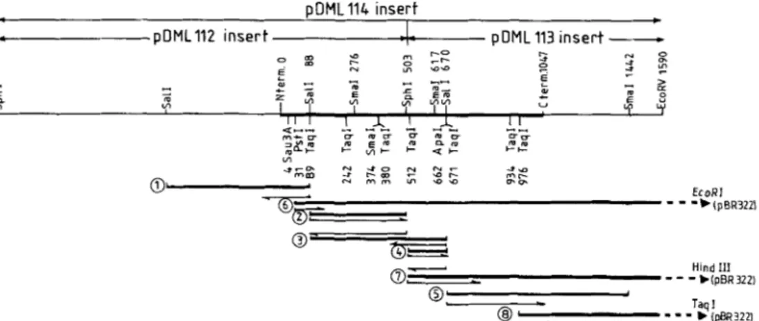
b 1pBR3221Fig. 4. Strategy of nucleotide sequencing. M131np11 was used to clone subfragments 1 (Sun, 602 bp) and 3 (Sun, 582 bp). M13tg130 was used to clone subfragments 2 (SaB-SphI, 41 5 bp), 4 (Sphl-Sd, 167 bp), 5 (Safl-SmuI, 772 bp), 6 (PstI-EcoRI, 1644 bp), 7 (SphI-HindIII, 1243 bp) and 8 (TuqI, 614 bp). M13tg131 was also used to clone subfragments 2 and 4
Localization of the Do-peptidase gene
Of the eight m-peptidase-producing clones isolated, five had acquired an 11.45-kb insert, two a 4.3-kb insert and one a 2.4-kb insert (as shown by agarose gel electrophoresis of mini-preparations). The 11.45-kb insert present in pDMLl10 of S. lividans CC1 was subcloned in the BamHI site of pBR322 and the resulting plasmid pDML111 was prepared in large quantities from E. coli HBlOl and purified on a CsCl gradient. Its restriction map is shown in Fig. 2A. Since there was no
BgAI site in the 11.45-kb insert, the 4.3-kb and 2.4-kb fragments in the other clones presumably were caused by post-cloning internal deletions, unless there are three different genes in R61.
Restriction fragments of pDMLl11 produced by several different endonucleases were separated by agarose gel electro- phoresis, transferred to nitrocellulose membranes (Southern procedure) and submitted to hybridization with the radio- active DNA probe. Of the two smallest hybridizing fragments detected (Fig. 3), the 1.75-kb SphI fragment was located on the left side of the original insert (and included about 200 bp of pBR322 between nucleotides 10277 and 12013; Fig. 2A) and the 0.6-kb SalI fragment overlapped the right end of the SphI fragment (nucleotides 10110- 10692; Fig. 2A).
The 1.75-kb SphI fragment was subcloned into pBR322, yielding pDMLll2 (Fig. 2B). pDMLll3 was digested with
SphI-EcoRV and the DNA segment overlapping the right end
of the 1.75-kb SphI fragment was subcloned into pBR322, yielding pDML113 (Fig. 2C). Finally, a 2.8-kb segment obtained by partial digestion of pDML111 with SphI,
followed by EcoRV restriction (and containing the combina- tion of inserts of pDML112 and pDML113) was also sub- cloned into pBR322, yielding pDML114 (see segments 1, 2 and 3 in Fig. 2A). The stretch responsible for the hybridiza- tion of pDML112 with the radioactive DNA probe was be- tween site SphI 562 and site SalI 977 (Fig. 2B).
Nucleotide sequencing of the DDpeptidase-encoding gene
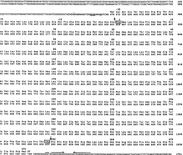
Fig. 4 illustrates the strategy used for the sequencing, Table 3 shows the codon usage in the synthesis of the DD-
peptidase and Fig. 5 gives the complete nucleotide sequence of the gene and its translation into amino acid sequence. This amino acid sequence was compared with the data presented in [16] and the zones of the gene coding for the N-terminal, active-site and C-terminal regions of the excreted DD-
peptidase were localized in subfragments 3 , 2 and 5, respec- tively.
The gene, from its translation startpoint (ATG for Met) to its stop codon (an opal TGA triplet), contained 1218 nucleotides and had the information for the synthesis of a 406-amino-acid polypeptide. In fact, the processed, extra- cellular DD-peptidase lacked segment Met-1 - Ala-31. It also lacked segment Thr-380
-
Asp-406 from the C-terminal re- gion. The amino acid sequence Ala-32 -Thr-380 as deduced from the nucleotide sequence of the corresponding portion of the gene was in excellent agreement with both the overall amino acid composition [26] (Table4) and the amino acid sequencing data [16] of the excreted DD-peptidase.DISCUSSION
All bacteria possess, bound to their plasma membrane, an assortment of active-site serine DD-peptidases which catalyse distinct carboxypeptidation and transpeptidation reactions during the last stages of wall peptidoglycan synthesis [l, 2, 271. These enzymes, mistaking a p-lactam antibiotic molecule for a normal substrate (i. e. a D-alanyl-D-alanine-terminated peptide), become immobilized in the form of a long-lived, serine-ester-linked acyl enzyme and thus behave as penicillin- binding proteins (PBPs) [28]. The genes that code for the PBPs lA, lB, 3 and 5 of E . coli have been analysed and sequenced [29 - 311. On the basis of these data and others [32 - 341, the primary sequences of the proteins have been deduced and the penicillin-binding serine residue has been identified.
This paper describes the cloning and sequencing of the gene that codes for a Streptomyces active-site serine DD-
peptidase/PBP which is unusual in being excreted during growth of the organism as a water-soluble enzyme [l]. In parallel with this work, the sequence of about 50% of the mature, excreted polypeptide chain has been determined after enzymatic and chemical cleavage [16].
In agreement with the current view on plasma membrane translocation of extracellular proteins [35], excretion of the
Streptomyces R61 DD-peptidase/PBP depends on the presence of a cleavable 31-amino-acid signal peptide which is synthe- sized as an N-terminal segment present on a larger precursor polypeptide. This segment has one positively charged amino acid, Arg at position 8; it possesses a stretch of hydrophobic residues in the middle, and processing by a presumed leader peptidase takes place by cleavage of an Ala-Ala peptide bond.
110 1 2 0 1 3 0 T h r V a l A r g G l n V a l M e t S e r H i s A r g Ser 6 1 y L e u T y r Asp T y r T h r A s n A s p M e t P h e A l a G l n T h r V a l P r o G l y P h e G l u Ser V a l ACC GTG COT CAG GTG ATG AGC CAC CGC ACT GGG CTG TAC GAC TAC ACC AAC GAC ATG TTC GCG CAG ACO GTC CCG GGC TTC GAG TCC GTC
1 4 0 1 5 0 I60
A r g A s n L y s V a l P h e S e r T y r G l n ASP Leu 110 T h r Leu S e r L e u L y s H i s G l y V a l T h r A s n A l a P r o G l y A I a A l a T y r Ser T y r S e r CGC AAC AAG GTC T T C AGC TAC CAG GAC CTG ATC ACC CTG TCC CTC AAG CAC GGG 6TC ACC AAC GCA CCG GGC GCG GCC TAT TCA TAC TCC
170 1 8 0 1 9 0
A s n T h r A s n P h e V a l V a l A l a G l y M e t L e u I l e G l u Lys L e u T h r G l y H l s S e r V a l A l a T h r G l u T y r G l n A a n A r g 110 P h e T h r P r o AAC ACG AAC TTC GTC GTC GCG GGC ATG CTC ATC GAG AAG CTC ACC GGC CAC TCG GTC GCC ACG GAG TAC CAG AAC CGC ATC TTC ACG CCG
2 0 0 2 1 0 2 2 0
9 1 8
0 0 8
00 8 L e u A s n L e u T h r ASP T h r P h e T y r V a l H l s P r o Asp T h r V a l 110 P r o G I y T h r H I S A l a A s n 61y T y r L e u T h r P r o A s p G l u A l a G I y CTG AAC CTG ACC GAC ACC TTC TAC GTG CAC CCC GAC ACC GTC ATC CCG GGC ACC CAC GCC AAC GGC TAC CTC ACG CCG GAC GAG GCC GGT 1188
2 3 0 2 4 0 2 5 0
G l y A l a L e u V a l ASP S e r T h r G I u G l n T h r V a l S t r Trp A l a G l n T h r G l y A I a A l a V a l I l a Ser S e r T h r G l n A s p L e u A s p T h r P h e GGG GCC CTG GTC GAC TCC ACC GAG CAG ACG GTG TCG TGG GCG CAB ACC GGG GCC GCG GTC ATC TCC AGC ACG CAG GAC CTG GAC ACG TTC
260 270 2 8 0
P h e S e r A I a L e u M 4 t S e r G l y G l n L e u M e t S e r A l a A l a G l n L e u A I a G l n M e t G l n G l n T r p T h r T h r V a l A a n S e r T h r G l n G l y T y r TTC TCC GCG TTG A T 6 AGC GGG CAG CTC ATG TCC GCC GCG CA6 CTC GCG CAG ATG CAG CAG TGG ACG ACG GTC AAC AGC ACC CAG 6GG TAC
290 300 3 1 0
G l y L e u G l y L e u A r g A r g A r g Asp Leu S e r C y s G I y 110 S e r V a l T y r G l y H l s T h r G l y T h r V a l G l n G l y T y r T y r T h r T y r A l a P h e GGC CTC GGC CTG CGC CGC CGT GAC CTG TCC TGC GGT ATC TCG GTG TAC GGC CAC ACG GGC ACC GTG CAG GGC TAC TAC ACG TAC GCC T T C
3 2 0 3 3 0 3 4 0
A l a S e r L y s Asp G l y G I n A 1 8 H i s V a l T h r A l a L e u A l a A s n T h r S e r A s n A s n V a l A s n V a l L e u A s n T h r M e t A l a A r g T h r L e u G l u GCC TCG AAG GAC GGC CAA GCG CAC GTC ACC GCG CTC GCC AAC ACC TCG AAC AAC GTG AAC GTG CTG AAC ACG ATG GCC CGC ACG CTG GAA
1278
1 3 6 8
1 4 5 8
1 5 4 8
4350 360 3 7 0
S i r A I a P h e c y s G I y L y s P r o T h r m G I y G I u A l a A I a G l n A r g Asp L e u L e u G 1 y Asp H i s A r g G l y A l a P r o G l u Asp I I r A l a P r o TCC GCG TTC TGC GGC AAG CCG ACG ACC GGC GAA GCT 6CG CAG CGC GAC CTC CTC GGC GAC CAC C 6 T GGA GCG CCA GAG GAC ATC GCG CCG 1638
3 7 5
-L
-
G l y 110 A l a A r g A s p O P
GGT ATC GCC CGC GAC TGA CGCGCGAAGGTGCGGCCGGACCCACCCGTCCGGCCGACCCCTCGCCGGACAGGACCTGGGCCGGGCTCATGACTTCTTCAGGTCGGACCGGTAG 1 7 5 1
Fig. 5 . Nucleotide sequence of the Streptomyces R61 Do-peptidase precursor-encoding gene and amino acid sequence of the expressed and excreted enzyme. The N-terminal alanine and C-terminal threonine of the mature, extracellular enzyme are in boxes. The processing sites are shown by vertical arrows. The putative ribosome binding site is underlined. The inverted repeats of the putative transcription termination signal are shown by horizontal arrows
At present, two other Streptomyces signal peptides have been described, namely those involved in membrane translocation of the j-N-glucosaminidase H in Streptomyces plicatus [36] and ORF 438 in Streptomyces antibioticus [37]. N o similarity
is seen between the three signal peptides. In the two latter cases, at least three arginine residues preceded the hydrophobic segment. The leader sequences of the agarase gene of Streptomyces coelicolor and the amylase of Strepto- myces limosus are also known [38].
Excretion of the Streptomyces R61 DD-peptidase involves (or is accompanied by) alteration of the carboxy-terminal portion of the enzyme precursor. Modification is by elimina- tion of a 26-amino-acid stretch through cleavage of a Thr-Gly peptide bond. This segment has a low hydrophobicity index and a high proportion of charged residues (4 Asp, 2 Glu, 3 Arg and 1 His). It thus differs from the membrane-anchoring peptide that occurs at the carboxy-terminal portion of the DD-
peptidaselPBP5 of Bacillus subtilis and Bacillus stearothermo-
philus [39]. But, the E. coli PBP5 also possesses a C-terminal
segment which plays a role in the anchoring of the protein to the plasma membrane, though it has no extended runs of hydrophobicity [40]. By analogy with this latter PBP, it may be hypothesized that, should it not be cleaved, the C-terminal segment of the Streptomyces DD-peptidase precursor would function as a stop-transfer sequence through which the enzyme would become membrane-bound. There is also no indication for the presence of a hydrophobic membrane- anchoring segment on the carboxy-terminal portion of the E. coli PBPs/DD-peptidases 1 A, 1 B and 3 [29- 311. PBPs 3, 5 and 6 have been shown to be synthesized as pre-proteins with amino-terminal signal peptides. The amino-terminal regions of PBPs 1 A and 1 B also have characteristics of a signal peptide but whether or not processing of the amino terminus occurs is not known [29].
Analysis of the gene that codes for the Streptomyces R61 DD-peptidase precursor reveals other interesting features. The 5’ GGAG-A-A 3’ segment, at position -13 to - 6 upstream of the translation start codon (ATG for Met) complements
- S. R61 (h) G V A D R A T G R A I T T T D R F R V G S V T X S F S A V V L L Q L V D E G K L D ti) d t k a g k e - v k f n s d k R F a y a S t s K a i n s a i L L e q V p y n K L n 1 4 5 ) Class A (j 1 d t g t n r T - ~ A y r p d e R F a f a S t i K a l t v g V L L Q q k S i e d L n ( 4 5 ) (k) d t g t n e T - i a y r p d q R F a f a S t y K a l a A g V L L Q q n s i d s L n ( 4 5 ) (1) d l n s g k i l e s f r p e e R F p m m S t f K v l l c g a v L s r V D a G q e q ( 4 5 ) DD-pep tidase 8-lactamases (m) d t g s g k T - ~ A y r a d e l F p m c S V f K t L S s a a v L r d l D r n g e f ( 4 6 ) (n) G y A D i A k k q p v T q q t l F e l C S V s K t F t g V l g g d a i a r G e i k (47) (P) k k A D i A a n k p v T p q t l F e l G S i s K t F t g (49) Class C ( 0 ) C k A D i A n n h p v T q q t l F e l G S V s K t F n g V l g g d r i a r G e i k ( 4 8 ) 6-lactamases (50) (q) v T p e t l F e i G S V s K ~~ (r) a d r a m l v f d p v r a k k R y s p a S t f K i p h t l f a L d a g a v r d e f (51) Oxa-2 8-lactamase Subtilisin (S) p g v s i q s t l p g g T y g a y n g t S m a t p h v A g a a a l i l s k h p t t (52,531
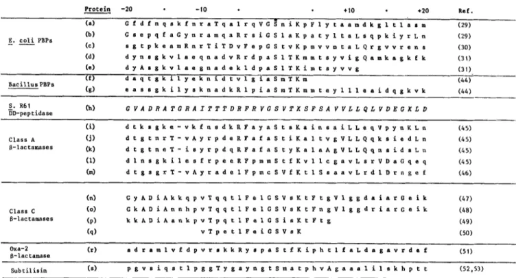
Fig. 6. Amino acid sequences around the active-site serine residue of the extracellular DDpeptidase of Streptomyces R61 ( h ) , various membrane- bound penicillin-binding proteins (PBPs) ( a - g ) ~ /I-lactamase ( i - r ) and subtilisin (s). Capital letters show identities with the Streptomyces
R61 DD-peptidase. (a, b, c, d and e) PBPs I A, 1 B, 3, 5 , and 6 of E. coli; (f and g) PBPs (DD-carboxypeptidases) of Bacillus stearothermophilus and Bacillus subtilis; (i, j, k, 1 and m) class A p-lactamases of Staphylococcus aureus, Bacillus lichenijormis, Bacillus cereus, Escherichia coli (RTEM plasmid) and Streptomyces albus G; (n, 0, p and q) class C 8-lactamases (chromosome ampC gene) of E. coli, Citrobacter freundii,
Enterobacter cloacae and Pseudomonas aeruginosa; (r) oxa-2 p-lactamase (E. coli)
six bases out of eight of the 3' end of the 16 S rRNA of S. lividuns 1411 and resembles a ribosome binding site [42]. In addition, the 31-nucleotide sequence that starts at position 9 downstream of the opal stop codon TGA, exhibits an inverted repeat of 12 bases with only one mismatch and thus may function as a terminator 142, 431. Up to 263 nucleotides have been sequenced upstream of the translation start codon. In vivo promoter probing, using the promoter-probe plasmid pIJ424 [8], has shown that the promoter region is included in this sequence (data not shown). However, since the transcrip- tion start is not known, the consensus -10 and -35 se- quences cannot be identified with certainty.
Comparison of the amino acid sequences of the Streptomyces R61 DD-peptidase with those of various active- site serine p-lactamases and DD-peptidase/PBPs shows that, remarkably, the sequence Ser*-Xaa2-Lys, where Ser* is the active-site serine, is conserved in all these proteins. Alignment of the 20-amino-acid sequences that flank the amino and carboxyl sides of the active-site serine (Fig. 6) leads to the following observations. (The gaps introduced in the sequences of several p-lactamases of class A are those proposed by Ambler [45] for optimal matching.)
a) The sequence Phe-Xaa3-Ser*-Xaa2-Lys which is present in all the p-lactamases of class A and C (but not in the oxa-2 p-lactamase) occurs also in the Streptomyces R61 DD-
peptidase. In addition to the triad Phe
...
Ser ... Lys, the Streptomyces R61 DD-peptidase and the E. coli and C. freundii P-lactamases of class C have identities at positions -20,-18, -17, -15, -9, -1, + I , +5, + 8 a n d +17. Similarily, the Streptomyces R61 DD-peptidase and the B. lichen iformis j-lactamase of class A have identities at positions -14,
-11, -5, f 9 , + l o , + 1 1 , + I 2 and +19. Strikingly, the
similarity between the Streptomyces R61 DD-peptidase and the p-lactamases of either class A or class C is greater than that between the p-lactamases of class A and C.
b) A phenylalanine at the fourth position on the amino side of the active-site serine (as it occurs in the p-lactamases of class A and C and in the Streptomyces R61 DD-peptidase) is found in the E. coli PBP3 and an arginine at the fifth
position on the amino side of the active-site serine (as it occurs in four p-lactamases of class A and in the Streptomyces R61 DD-peptidase) is found in the E . coli PBPs 1 B and 5 and in the B. subtilis DD-peptidase. In addition to the conserved Ser*- Xaa2-Lys sequence, the pair Streptomyces R61 DD-peptidasel E. coli PBPIA have five identities at positions -20, -8,
- 2, - 1 and
+
5 and the pair Streptomyces R61 DD-peptidasel E. coli PBP3 have eight identities at positions - 12, - 9, -7, -6, -4, -1, +11 and + I 2 (and an inverted diad Thr- Val or Val-Thr at positions+
1 and+
2).When, using the method of McLachlan, the comparison is broadened to the whole amino acid sequences, homology vanishes except between the P-lactamases of a given class (A or C), the pair PBPIA-PBPlB of E. coli and the pair Streptomyces R61 DD-peptidaselE. cok P-lactamase of class C. The
xf,
values are 5.8 for the pair Streptomyces R61 DD-peptidaselE. coli p-lactamase and 1.3 for the pair Streptomyces R61 DD-peptidaselBucillus licheniformis
p-
lactamase, indicating probabilities for relatedness due to evolutionary relationship of 98.2% and 74%, respectively. AxazPp
of 10, indicating a probability of 99.6%, is currently considered as a very strong index of such a relationship [25]. Restriction of the analysis to the N-terminal 150 residues, thus comprising the active-site serine area, does not increase thex.$p
values.---
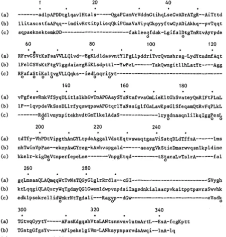
0 0 200 220 240 tdTfy-VhPDtVipgthAnGntpdeAggalVdstEqtvswaqtgaaViSstqDLdTffsA---lms nhTwinVpPae--eknyAwGYreg-kAvhvspgald---aeaygVkStieDmarwrqsnlkpldine kkelr-kigDeVtnperfepeLne---VnpgEtqd--- tStaraLvTslrA---fa1 260 280 goLmsaaQLAQmqqWtTvNsTQGyGlglrRrdls--cGI--- SVY gh ktLqqgiQLAQsryWqTgdmyQGlGwemldwpvnpdsiIngsdnkialaarpvkaitpptpavraSwvhk edklpsekrellidWmkrNtTgdali---Ragvp--dG~---eVadk 300 3 20 340 TGtvqGyytY---AFasKdgqahVtaLANtsnnvnvlntmArtL--EsA-fcgKptt TGatgGfgsYv----AFipekelgiVm-LANknypnparvd~wqi--lnA-lq ~Saasygtrn-diaiiwpgKgdpwlavLossrdkkd---akyddkLiaEatkwmKalnFig. I . Possible amino acid sequence alignment of the active-site serine extracellular DDpeptidase of Streptomyces R61 ( a ) , the class C j- lactamase of Escherichia coli (chromosome) ( b ) and the class A fl-lactamase of Bacillus licheniformis (c). Numbering starts from the N - terminus of the DD-peptidase and ignores the gaps postulated in the sequence to obtain optimal matching. Residues that are identical in the DD-peptidase and a given 8-lactamase (or both 8-lactamases) are in capital letters. Residues that are identical in the two 8-lactamases are marked by an open circle (0 j. Closed circles ( 0 ) indicate every 10th residue
Segments having a matrix score of 3 or 4 served as a basis for the possible alignments and gaps shown in Fig. 7. These alignments maximize the similarities between the enzymes and, on that basis, the pair Streptomyces R61 DD-peptidasel E. coli p-lactamase have 89 identities out of 315 residues effectively aligned. The figure drops to 47 (for 252 aligned residues) for the pair Streptomyces R61 DD-peptidaselB. lichenijormis p-lactamase and to 22 for the pair E. coli
fi-
IactamaselB. lichenijormis b-lactamase.Finally, an interesting feature of the comparison is that the triad His-Thr-Gly in the R61 Streptomyces DD-peptidase or the triad Lys-Thr-Gly in the B. lichenformis and E. coli p-lactamases occurs at the same position 298-300, i.e. at position 136- 139 on the amino side of the active-site serine. Moreover, the triad Lys-Thr-Gly is also conserved in the E.
coli PBPs 1 A, l B , 3 and 5 away from the active-site serine
(at positions 716-718, 698-670, 494-497 and 213-215, respectively; the active-site serine being at position 465, 510, 307 and 44, respectively).
In spite of a low level of relatedness in the primary
structures, it has been shown [5, 541 that the spatial arrange- ment of the secondary structure elements (helices and strands of p-sheet) of the Streptomyces R61 DD-peptidase and the B. lichenformis (Bacillus cereus) p-lactamase of class A is so great that these two enzymes are obviously homologous in an evolutionary sense. Since as shown above, the Streptomyces R61 DD-peptidase and the 8-lactamases of class C show higher similarities in their amino acid sequences, it may be concluded
that the D-lactamases of class C probably share all the tertiary structural features that are common to the fi-lactamases of class A and the Streptomyces R61 DD-peptidase.
We are grateful to Drs D. A. Hopwood, T. Kieser and M. J. Bibb, and the Streptomyces group at the John Innes Institute (Norwich, UK) for introducing us to the techniques of working with Streptomyces genetics and helpful discussions. We also thank Dr. B. G. Spratt (University of Sussex, UK) for his interest, Dr. C. Wuilmart (Universitk Libre de Bruxelles, Belgium) for his help in running McLachlan’s program and Fitch’s program and Mrs An Lesage (Uni- versity of Gent, Belgium), for her advice in the development of the immunological test. This work was supported by an Action concertee from the Belgian Government (convention 79/84-11), the Fonds de la Recherche Scientifrque Medicale, Brussels (contract 3.4507.83) and a Convention tripartite between the Rigion wallonne, Continental Pharma and the Universiy of Liege, J.D. is Chercheur qualifit and B. J. Chargi de recherches of the Fonds National de la Recherche Scientijiique (FNRS; Brussels) and C.P.F. is indebted to the Ministere de I’Emploi et du Travail for a Cadre special temporaire.
REFERENCES
1 . Ghuysen, J. M., Frere, J . M., Leyh-Bouille, M., Nguyen-Disttche, M., Coyette, J., Dusart, J., Joris, B., Duez, C., Dideberg, O., Charlier, P., Dive, G. & Lamotte-Brasseur, J. (1984) Scand. J . Infect. Dis. suppl. 42, 17 - 37.
2. Frere, J. M. & Joris, B. (1985) CRC Crit. Rev. Microbiol. 11,
6. Hopwood, D. A., Kieser, T., Wright, H. M. & Bibb, M. J. (1983) J . Gen. Microbiol. 129, 2257-2269.
7. Katz, E., Thompson, C . J. & Hopwood, D. A. (1983) J . Gen. Microbiol. 129, 2703 -2714.
8. Hopwood, D. A., Bibb, M. J., Chater, K. F., Kieser, T., Bruton, C. J., Kieser, H. M., Lydiate, D. J., Smith, C. P., Ward, J. M. & Schrempf, H. (1985) Genetic manipulation of Streptomyces. A
laboratory manual, The John Innes Foundation, Norwich. 9. Boyer, H. W. & Roulland-Dussoix, D. (1969) J. Mol. Biol. 41,
10. Bolivar, F., Rodriguez, R. L., Greene, P. J., Betlach, M. C., Heyneker, H. L., Boyer, H. W., Crosa, H. J. & Falkow, S. (1977) Gene 2,95-113.
11. Chater, K. F., Hopwood, D. A., Kieser, T. & Thompson, C. J. (1982) Curr. Top. Microbiol. Immunol. 96, 69 - 95.
12. Dehottay, Ph., Dusart, J., Duez, C., Lenzini, M. V., Martial, J. A,, Frkre, J. M., Ghuysen, J. M. & Kieser, T. (1986) Gene 42,
13. Leyh-Bouille, M., Coyette, J., Ghuysen, J. M., Idczak, J., Perkins, H. R. & Njeto, M. (1971) Biochemistry 10, 2163-2170. 14. Maniatis, T., Fritsch, E. F. & Sambrook, J. (1982) Molecular
cloning. A laboratory manual, Cold Spring Harbor Laboratory, Cold Spring Harbor, NY.
15. Frtre, J. M., Leyh-Bouille, M., Ghuysen, J. M., Nieto, M. & Perkins, H. R. (1976) Methods Enzymol. 45B, 610-636. 16. Joris, B., Jacques, Ph., Frkre, J. M., Ghuysen, J. M. & Van
Beeumen, J. (1987) Eur. J . Biochem. 162, 519-524.
17. Warner, B. D., Warner, M. E., Karns, G. A., Ku, L., Brown- Shimer, S. & Urdea, H. S. (1984) DNA 3,401 -411.
18. Maxam, A. M. &Gilbert, W. (1980) Methods Enzymol. 65,499- 560.
19. Wallace, R. B., Johnson, N. J., Hirose, T., Miyake, M.,
Kawashima, E. H. & Itakura, K. (1981) Nucleic Acids Res. 9 ,
D C ) 231,1429-1431.
459 - 472.
31 -36.
879 - 894.
20. Woods, D. (1984) FOCUS 6, 1
-
3.21. Sanger, F., Nicklen, S. & Coulson, A. R. (1977) Proc. Natl Acad.
22. Staden, R. (1984) Nucleic Acids Res. 12, 551 -567.
23. Thompson, C. J. & Gray, G. S. (1983) Proc. Nut1 Acad. Sci. USA
24. McLachlan, A. D. (1971) J. Mol. Biol. 61,409-424. 25. Fitch, W. M. (1970) J . Mol. Biol. 49, 1 - 14.
26. Frere, J. M., Ghuysen, J. M., Perkins, H. R. & Nieto, M. (1973) 27. Spratt, B. G. (1983) J . Gen. Microbiol. 129, 1247-1260.
28. Ghuysen, J. M., Frere, J. M., Leyh-Bouille, M., Nguyen-Disteche,
29. Broome-Smith, J. K., Edelman, A., Yousif, S. & Spratt, B. G. Sci. U S A 74, 5463 - 5467.
80, 5190-5194.
Biochem. J . 135,463-468.
M. & Coyette, J. (1986) Biochem. J . 235, 159-165.
(1985) Eur. J. Biochem. 147, 437-446.
33. Nicholas, R. A., Strominger, J. L., Suzuki, H. &Hirota, Y. (1985) J . Bacteriol. 164, 456-460.
34. Nicholas, R. A,, Ischino, F., Park, W., Matsuhashi, M. & Strominger, J. L. (1985) J. Biol. Chem. 260, 6394-6397. 35. Pugsley, A. P. & Schwartz, M. (1985) FEMS Microbiol. Rev. 32,
36. Robbins, P. W., Tumble, R. B., Wirth, D. F., Hering, C., Maley,
F., Maley, G. F., Das, R., Gibson, B. W., Royal, N. & Biemann, K. (1984) J . Biol. Chem. 259,7577-7583.
37. Bernan, V., Filpula, D., Herber, W., Bibb, M. & Katz, E. (1985) Gene 37,101-110.
38. Chang, S. (1986) Methods Enzymol., in the press.
39. Waxman, D. J. & Strominger, J. L. (1981) J . Biol. Chem. 256, 40. Pratt, J. M., Jackson, M. E. & Holland, I. B. (1986) EMBO J. 5, 41. Bibb, M. J. & Cohen, S. N. (1982) Mol. Gen. Genet. 187, 265-
277.
42. Hopwood, D. A., Bibb, M. J., Chater, K. F., Janssen, G. R.,
Malpartida, F. & Smith, C. P. (1986) in Regulation ofgene expression - 25 years on (Booth, E. R. & Higgins, C. F., eds) pp, 251 - 276. Cambridge University Press, Cambridge. 43. Bibb, Me. J., Bibb, Ma. J., Ward, J. M. & Cohen, S. N. (1985)
Mol. Gen. Genet. 199,26-36.
44. Waxman, D. J., Yocum, R. R. & Strominger, J. L. (1980) Phil. Trans. R. Soc. Lond. B289, 257-271.
45. Ambler, R. P. (1980) Phil. Trans. R. Soc. Lond. 8289, 321 -331. 46. Dehottay, Ph., Duez, C. & Dusart, J. (1986) in Biological, bio-
chemical and biomedical aspects of Actinomycetes (Szabo, G., Biro, S. & Goodfellow, M., eds.) pp. 121-123, Akadkmiai Kiad6, Budapest.
47. Jaurin, B. & Grundstrom, T. (1981) Proc. Nut1 Acad. Sci. USA 78,4897 - 4901.
48. Lindberg, F. & Normark, S. (1986) Eur. J . Biochem. 156,441
-
445.49. Joris, B., De Meester, F., Galleni, M., Reckinger, G., Coyette, J., Frere, J. M. & Van Beeumen, J. (1985) Biochem. J. 228, 241 - 248.
50. Knott-Hunziker, V., Petursson, S., Jayatilake, G. S., Waley, S. G., Jaurin, B. & Grundstrom, T. (1982) Biochem. J . 201,621 - 627.
51. Dale, J. W., Godwin, D., Mossakowska, D., Stephenson, P. & Wall, S. (1985) FEBS Lett. 191, 39-44.
52. Smith, E. L., Delange, R. J., Evans, W. H., London, M. & Markland, F. S. (1968) J . Mol. Biol. 243,2184-2191.
53. Stahl, M. L. & Ferrari, E. (1984) J. Bacteriol. 158, 411 -418. 54. Samraoui, B., Sutton, B. J., Todd, R. J., Artymiuk, P. J., Waley,
3-38.
2067 - 2077.
2399 - 2405.