Poly (D,L-lactic acid) macroporous guidance scaffolds seeded with
Schwann cells genetically modified to secrete a bi-functional
neurotrophin implanted in the completely transected adult rat
thoracic spinal cord
Andres Hurtado(1), Lawrence D.F. Moon(1), Véronique Maquet(3,4), Bas Blits(1), Robert Jérôme(3,4), Martin Oudega(1,2)
(1)
The Miami Project to Cure Paralysis, University of Miami School of Medicine, P.O. Box 016960, R-48, Miami, FL 33136, USA
(2)
Department of Neurological Surgery, University of Miami School of Medicine, Miami, USA
(3)
Center for Education and Research on Macromolecules (CERM), University of Liège, Sart-Tilman, 4000 Liège, Belgium
(4)
Interfacultary Center for Biomaterials, University of Liège, Sart-Tilman, 4000 Liège, Belgium
Abstract: Freeze-dried poly(D,L-lactic acid) macroporous scaffold filled with a fibrin
solution containing Schwann cells (SCs) lentivirally transduced to produce and secrete D15A, a bi-functional neurotrophin with brain-derived neurotrophic factor and neurotrophin-3 activity, and to express green fluorescent protein (GFP) were implanted in the completely transected adult rat thoracic spinal cord. Control rats were similarly injured and then implanted with scaffolds containing the fibrin solution with SCs lentivirally transduced to produce express GFP only or with the fibrin solution only. Transgene production and biological activity in vitro, SC survival within the scaffold in vitro and in vivo, scaffold integration, axonal regeneration and myelination, and hind limb motor function were analyzed at 1, 2, and 6 weeks after implantation. In vitro, lentivirally transduced SCs produced 87.5 ng/24h/106 cells of D15A as measured by neurotrophin-3 activity in ELISA. The secreted D15A was biologically active as evidenced by its promotion of neurite outgrowth of dorsal root ganglion neurons in culture. In vitro, SCs expressing GFP were present in the scaffolds for up to 6 h, the end of a typical surgery session. Implantation of SC-seeded scaffolds caused modest loss of spinal nervous tissue. Reactive astrocytes and chondroitin sulfate
glycosaminoglycans were present in spinal tissue adjacent to the scaffold. Vascularization of the scaffold was ongoing at 1 week post-implantation. There were no apparent differences in scaffold integration and blood vessel formation between groups. A decreasing number of implanted (GFP-positive) SCs were found within the scaffold during the first 3 days after implantation. Apoptosis was identified as one of the mechanisms of cell death. At 1 week and later time points after implantation, few of the implanted SCs were present in the scaffold. Neurofilament-positive axons were found in the scaffold. At 6 weeks post-grafting,
myelinated axons were observed within and at the external surface of the scaffold. Axons did not grow from the scaffold into the caudal cord. All groups demonstrated a similar
improvement of hind limb motor function. Our findings demonstrated that few seeded SCs survived in vivo, which could account for the modest axonal regeneration response into and across the scaffold. For the development of SC-seeded macroporous scaffolds that effectively promote axonal regeneration in the injured spinal cord, the survival and/or total number of SCs in the scaffold needs to be improved.
Keywords: CNS regeneration; Schwann cells; BDNF; NT-3; Poly (a-hydroxyacid); Biodegradable; Polymer; Spinal cord injury; Apoptosis; Caspase-3
1. Introduction
Injury to the adult mammalian spinal cord elicits a cascade of pathophysiological events that results in loss of nervous tissue, and, consequently, in partial or complete loss of neurological functions. For restoration of locomotor function following an injury, damaged axons need to regenerate across and beyond the injury site and form synaptic connections with neurons involved in motor function. Implantation of cells within the injured area may provide the regenerating axons with a substrate that will promote and support axonal growth towards the denervated neurons.
The efficacy of Schwann cells (SCs) to promote axonal regeneration and myelination in the injured adult rat spinal cord has been demonstrated repeatedly [1-4]. Following a complete transection and removal of several millimeters of the spinal cord, grafting a non-degradable polyacrylnitril/polyvinylchloride (PAN/ PVC) tubular scaffold filled with SCs that bridged both cord stumps, which were placed within the scaffold, resulted in axonal regeneration and myelination [1,3-5]. The use of a tubular scaffold for cell implantation in the injured cord may be beneficial to the overall axonal growth process by preventing scar formation, allowing accumulation of growth-promoting molecules, and serving as a protective casing for the implant. On the other hand, it can be anticipated that a non-degradable tubular scaffold becomes harmful during the later stages of the growth/recovery response by constricting the spinal cord and/or eliciting a foreign body response. The use of biodegradable scaffolds to implant axonal growth-promoting substrates may circumvent these potential problems. A widely used biodegradable material in tissue engineering is poly(D,L-lactic acid) (PLA). In vivo and in vitro, PLA and its breakdown products were demonstrated to be biocompatible with spinal cord tissue and with SCs [6]. Implantation of a PLA single-channel tubular scaffold containing SC into the completely transected adult rat spinal cord resulted at first in an axonal regeneration response comparable to that seen with a SC-filled non-degradable PAN/PVC channel [7]. However, in time axonal growth was obstructed due to the collapse of the PLA scaffold. Recently, Patist et al. [8] demonstrated that the implantation of a
macroporous PLA tubular scaffold in the transected rat spinal cord elicited a modest axonal regeneration response. These particular scaffolds were prepared by a thermally induced polymer-solvent phase separation process and contained longitudinally oriented macropores connected to each other by a network of micropores [8]. It was proposed that seeding these scaffolds with SCs prior to implantation would be beneficial for the overall axonal growth response [8].
Here, we have filled a freeze-dried PLA macroporous guidance scaffold with genetically modified rat SCs and analyzed cell survival, scaffold integration, and the axonal regeneration response following implantation into the completely transected adult rat thoracic spinal cord. Control rats were implanted with scaffolds filled with a fibrin solution with or without SCs genetically modified to express green fluorescent protein (GFP) for easy and reliable identification of the SCs. Experimental rats were grafted with scaffolds with the fibrin solution with SCs genetically modified to express GFP and, in addition, to produce and secrete a bi-functional neurotrophin (D15A), a molecule with brain-derived neurotrophic factor (BDNF) and neurotrophin-3 (NT-3) activity [9], to evaluate the effects of elevated levels of neurotrophins within the graft environment [3,10].
2. Materials and methods 2.1. Animals
housed according to NIH and USDA guidelines. The Institutional Animal Care and Use Committee of the University of Miami approved all animal procedures. Rats were
anesthetized using a mixture of ketamine (42.8mg/ml), xylazine (98.6mg/ml) and aceproma-zine (1.4mg/ml) at 0.06ml/100 g body weight, intramuscularly. The backs were shaved and aseptically prepared. Lacrilube ophthalmic ointment (Allergen Pharmaceuticals, Irvine, CA) was applied to the eyes to prevent drying and gentamicin (0.03 ml, Buck, Inc., Owings Mills, MO) was given intramuscularly. During surgery the rats were kept on a heating pad to maintain the body temperature at 37 ± 0.5°C.
2.2. Purification of Schwann cells
Highly purified SC cultures were obtained from sciatic nerves of adult female Fischer rats (Charles River Laboratories) as described previously [11]. Dissociated SCs were cultured on poly-L-lysine-coated tissue culture dishes in DF-10S medium supplemented with the
mitogens, bovine pituitary extract (2mg/ml), forskolin (0.8g/ml), and heregulin (2.5nM). The addition of heregulin is a modification of the original protocol. To determine the purity of the SCs used for seeding the polymer scaffolds, samples of the harvested cells were plated onto culture dishes, cultured for 3h, stained for S100, and then coverslipped with Citifluor (UKC Chemical Laboratory, Canterbury, England) with 100 mM Hoechst nuclear dye (Sigma, St. Louis, MO) to compare numbers of S100-positive cells with Hoechst-labeled cells. The purity of the SCs used for seeding the scaffolds was 95-98%.
2.3. Preparation of lentiviral vectors and transduction of SCs
The ViraPower Lentiviral Expression System (Invitrogen) was used to generate lentiviral vectors (LVs) encoding only GFP or both D15A and GFP. Briefly, cDNA of GFP (Clontech) and D15A (kind gift of Dr. Pantelis Tsoulfas, The Miami Project to Cure Paralysis) was amplified by PCR and subcloned into an expression/packaging plasmid. Expression was under control of the human cytomegalovirus promotor and the Woodchuck hepatitis virus Post-transcrip-tional Regulatory Element. Next, 293 T cells were co-transfected with the expression/packaging plasmid and three helper plasmids (pLP1, pLP2, and pLP/VSVG) and then cultured in DMEM supplemented with 10% fetal bovine serum and gentamycin
(Invitrogen). The medium of trans-fected 293 T cells, which contained the LVs, was collected and used to transduce SCs ex vivo at passage 2 at a multiplicity of infection of 30. Routinely, a transduction rate of >95% was achieved with LV-GFP. The transduced SCs were further cultured until passage 4 and then collected for seeding into the polymer scaffolds (see below). We used conditioned medium from these cultures to determine the amount of the secreted D15A molecule. The D15A molecule has an active NT-3 backbone with BDNF activity as well [9]. To determine the amount of secreted D15A, NT-3 levels were measured using ELISA according to manufacturer's instructions (Promega). The biological activity of the D15A molecule present in the medium of the cultures of transduced SCs was confirmed using a bioassay measuring neurite outgrowth from dorsal root ganglia (DRG; [12,13]). Rat embryo DRGs were aseptically removed, pooled in ice-cold L-15 Leibovitz medium with
L-glutamine (Gibco BRL), and then placed in 50 ml DMEM/10% fetal calf serum onto glass
coverslips coated with poly-L-ornithine (500mg/ml in 0.1-M boric acid, pH 8.3) and laminin (10mg/ml; Roche, Basel, Switzerland). The glass coverslips were placed in a 6-well plate (n = 3 per well) and kept overnight in an incubator at 37 °C, 5% CO2. Next, the DRG were
immersed for 24 h in conditioned medium. This medium was then refreshed and the cultures were grown for an additional 48 h at 37 °C, 5% CO2. The cultures were fixed in 4%
with anti-neurofilament antibodies to visualize the neurites.
2.4. Preparation of the poly(D,L-lactic acid) macroporous tubular scaffolds
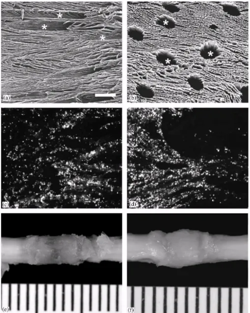
One gram of PLA (Resomer® R-206; Mn: 50,000, Boehringer-Ingelheim, Germany) and 50 mg of poly(D,L-lactide-b-ethylene oxide) 4k-4k copolymer (Mn: 10,000, CERM, Liege, Belgium) were dissolved in 10 ml dimethylcar-bonate (DMC, Acros) and filtered over 0.45 mm. Five milliliter of this polymer solution within a 50 ml-round bottom flask was then frozen with liquid nitrogen and freeze-dried by vacuum sublimation for 72 h at — 10°C, for 5h at 0°C, and finally at room temperature until it reached a constant weight. The fabrication and the in vitro degradation characteristics of the polymer scaffolds were described previously [8,14,15]. Using a metal trephine with an inner diameter of 2.6mm, 1cm long rods were cut from the polymer foam following the longitudinal direction of the pores. The rods were sterilized by UV exposure under a laminar flow for 15min. These sterilized rods were kept at -80°C until use. Just before surgery, the rods were thawed, cut into 4 mm long segments, seeded with SCs (see below) and kept in L15 medium with 1% fibrinogen (Sigma) and 0.1% gentamicin (Buck, Inc.) until grafting. As shown in Fig. 1a and b, the scaffolds have
longitudinal orientated macropores connected by micropores (diameter < 10 µm). The diameter of the macropores varied between 75 and 200 µm [8].
2.5. Seeding and survival of SCs into the macroporous tubular scaffolds
Scaffolds were first filled with a 1% fibrinogen (Type 1, human plasma; Sigma) in DMEM with 2% CaCl2, 0.2% gentamicin (Sigma), and 2% aprotinin (Sigma). This fibrin mixture was
drawn into scaffolds using a Vacutainer (Becton-Dickinson, Franklin Lakes, NJ). Then, 1 x 10⁶ SCs in 5 ml fibrin mixture was placed on the top of the scaffold, which was drawn gently inside by placing the scaffold onto Whatman filter paper (# 1). The cells were generally uniformly distributed within the pores of the scaffold although some small clusters of cells could also be discerned (Fig. 1c and d). The SC-seeded scaffold was placed into a tissue culture dish with L15 medium with 1% fibrinogen (Sigma) and 0.1% gentamicin (Buck, Inc.) until implantation. In vitro, SC survival was studied up to 6h after seeding. In vivo, SCs survival was evaluated by investigating the presence of GFP-positive cells within the scaffold at 1 and 3 days and at 1, 2, and 6 weeks after implantation into the transected spinal cord. The presence of apoptotic SCs within the scaffold was investigated at the same time points using immunostaining (see below) for active caspase-3, a marker for apoptotic cell death [16]. 2.6. Implantation of the SC-seeded scaffolds
After opening the skin and muscle layers, a laminectomy was performed at the T8-9 vertebral level and the dura mater carefully opened. The exposed T9-10 spinal cord was transected and 3 mm of tissue including visible spinal roots completely removed. After hemostasis was achieved, a 4 mm long scaffold with GFP-SCs or D 15A-GFP-SCs or the 1% fibrin solution alone was implanted in between the rostral and caudal spinal cord stumps. A layer of silicone sheeting (0.005'' thick) was placed on top of the implant and both cord stumps. The muscle layers were closed separately and the skin closed with metal wound clips. The rats received 10 ml Ringers' solution subcutaneously and were placed in warmed cages with food and water readily available. To prevent urinary tract infection, gentamicin was administered daily during the first 7 days post-surgery. The bladders were expressed manually two times a day until voluntary bladder release returned. In case urinary tract infection occurred, gentamicin administration was reinitiated for 7 days. In total, 13 rats received a GFP-SC implant (n = 2
for 1 week, n = 5 for 2 weeks, n = 6 for 6 weeks); 13 rats received a D15A-GFP-SC implant (n = 2 for 1 week, n = 5 for 2 weeks, n = 6 for 6 weeks); seven rats received a fibrin only implant (n = 2 for 1 week, n = 2 for 2 weeks, n = 3 for 6 weeks). Also, two rats received a SC-seeded scaffold for 1 day and 2 rats for 3 days to assess short-term cell survival. Panels 1e and 1f show the scaffold in vivo after 1 and 6 weeks, respectively.
Fig. 1. Macroporous poly (D,L-lactic acid) scaffolds can serve as vehicle for SC implantation. Scanning electron microscope photographs demonstrating a longitudinal section (a) and
cross section (b) of the scaffold. Horizontal cryostat sections counterstained with DAPI revealed that GFP-positive SCs were present within scaffold 1 h (c) and 6h (d) after seeding.
The lower panels show a dorsal view of the scaffold in vivo at 1 (e) and 6 (f) weeks after implantation. Scale bar in (a) represents 500 µm for panel (a) and 100 µm for panel (b-d). In
panels (e) and (f) the distance between the scale bars is 1mm. 2.7. Assessment of hind limb motor function
In rats that survived for 6 weeks, changes in hind limb function were assessed using the test developed by Basso, Beattie, and Bresnahan (BBB test; [17,18]), an open field test with a 21-point scale (21 is normal). The ;BBB test distinguishes between movements of individual