HAL Id: inserm-00827007
https://www.hal.inserm.fr/inserm-00827007
Submitted on 28 May 2013
HAL is a multi-disciplinary open access
archive for the deposit and dissemination of
sci-entific research documents, whether they are
pub-lished or not. The documents may come from
teaching and research institutions in France or
abroad, or from public or private research centers.
L’archive ouverte pluridisciplinaire HAL, est
destinée au dépôt et à la diffusion de documents
scientifiques de niveau recherche, publiés ou non,
émanant des établissements d’enseignement et de
recherche français ou étrangers, des laboratoires
publics ou privés.
99mTc-NTP 15-5 assessment of the early therapeutic
response of chondrosarcoma to zoledronic acid in the
Swarm rat orthotopic model.
Elisabeth Miot-Noirault, Emmanuelle David, Aurélien Vidal, Caroline
Peyrode, Sophie Besse, Marie-Mélanie Dauplat, Marie-Françoise Heymann,
François Gouin, Jean-Michel Chezal, Dominique Heymann, et al.
To cite this version:
Elisabeth Miot-Noirault, Emmanuelle David, Aurélien Vidal, Caroline Peyrode, Sophie Besse, et
al.. 99mTc-NTP 15-5 assessment of the early therapeutic response of chondrosarcoma to zoledronic
acid in the Swarm rat orthotopic model.. EJNMMI Research, SpringerOpen, 2013, 3 (1), pp.40.
�10.1186/2191-219X-3-40�. �inserm-00827007�
S H O R T C O M M U N I C A T I O N
Open Access
99m
Tc-NTP 15-5 assessment of the early
therapeutic response of chondrosarcoma to
zoledronic acid in the Swarm rat orthotopic
model
Elisabeth Miot-Noirault
1*, Emmanuelle David
2,3, Aurélien Vidal
1, Caroline Peyrode
1, Sophie Besse
1,
Marie-Mélanie Dauplat
4, Marie-Françoise Heymann
2,3,5, François Gouin
2,3,6, Jean-Michel Chezal
1,
Dominique Heymann
2,3and Françoise Rédini
2,3Abstract
Background: Since proteoglycans (PGs) appear as key partners in chondrosarcoma biology, PG-targeted imaging using the radiotracer99mTc-N-(triethylammonium)-3-propyl-[15]ane-N5 (99mTc-NTP 15-5) developed by our group was previously demonstrated to be a good single-photon emission computed tomography tracer for cartilage neoplasms. We therefore initiated this new preclinical study to evaluate the relevance of99mTc-NTP 15-5 imaging for the in vivo monitoring and quantitative assessment of chondrosarcoma response to zoledronic acid (ZOL) in the Swarm rat orthotopic model.
Findings: Rats bearing chondrosarcoma in the orthotopic paratibial location were treated by ZOL (100 μg/kg, subcutaneously) or phosphate-buffered saline, twice a week, from day 4 to day 48 post-tumor implantation.
99m
Tc-NTP 15-5 imaging was performed at regular intervals with the target-to-background ratio (TBR) determined. Tumor volume was monitored using a calliper, and histology was performed at the end of the study. From day 11 to day 48, mean TBR values ranged from 1.7 ± 0.6 to 2.3 ± 0.6 in ZOL-treated rats and from 2.1 ± 1.0 to 4.9 ± 0.9 in controls. Tumor growth inhibition was evidenced using a calliper from day 24 and associated to a decrease in PG content in treated tumor tissues (confirmed by histology).
Conclusions: This work demonstrated two proofs of concept: (1) biphosphonate therapy could be a promising therapeutic approach for chondrosarcoma; (2)99mTc-NTP 15-5 is expected to offer a novel imaging modality for the in vivo evaluation of the extracellular matrix features of chondrosarcoma, which could be useful for the follow-up and quantitative assessment of proteoglycan ‘downregulation’ associated to the response to therapeutic attempts. Keywords: Chondrosarcoma, Zoledronic acid,99mTc-NTP 15-5 radiotracer, Proteoglycans
Findings
Introduction
Considering the 10-year survival rate of chondrosarcoma (from 29% to 83% depending on the subtype and grad-ing), improving its clinical management is a challenge and novel therapeutic approaches are needed [1]. This implies that in vivo markers of the biologic phenotypic
features of chondrosarcoma behavior in the bone envir-onment could be used as quantitative criteria of grading, follow-up, and early evaluation of response to therapy.
Since proteoglycans (PGs) appear as key partners in chondrosarcoma biology, PG-targeted imaging using the radiotracer99m Tc-N-(triethylammonium)-3-propyl-[15]ane-N5 (99mTc-NTP 15-5) developed by our group was expected to be a good single-photon emission com-puted tomography (SPECT) tracer for the molecular im-aging of cartilage neoplasms in nuclear medicine [2,3].
* Correspondence:elisabeth.noirault@inserm.fr
1
INSERM UMR 990, Université d’Auvergne, BP 184, Rue Montalembert, Clermont-Ferrand, Cédex 63005, France
Full list of author information is available at the end of the article
© 2013 Miot-Noirault et al.; licensee Springer. This is an Open Access article distributed under the terms of the Creative Commons Attribution License (http://creativecommons.org/licenses/by/2.0), which permits unrestricted use, distribution, and reproduction in any medium, provided the original work is properly cited.
Resistance to chemotherapy is well known to make chondrosarcoma therapeutic management difficult [4]. The bisphosphonate zoledronic acid (ZOL) has demonstrated its therapeutic interest in a variety of tumors affecting the bones, such as osteosarcoma, Ewing's sarcoma, and chondrosarcoma [5-7].
This study was therefore initiated in the well-characterized Swarm rat chondrosarcoma (SRC) orthotopic model to determine the relevance of99mTc-NTP 15-5 im-aging for an early quantitative characterization of chon-drosarcoma response to ZOL.
Methods
Protocols were performed under the authorization of the French Directorate of Veterinary Services (Authorization No. C63-113-10) and were conducted under the supervi-sion of authorized investigators in accordance with the institution’s recommendations for the use of laboratory animals. * * * 0 1000 2000 3000 4000 5000 6000 7000 8000 4 11 16 24 36 48 T u moral v olume (mm 3)
Days post tumoral implantation ZOL-treated Controls
Figure 1Representative time course of the mean tumor volume ± standard deviation of ZOL-treated and control groups. Both groups have n = 12 animals. The asterisk indicates p < 0.05.
CT
ZOL-treated
A
B
C
D
E
F
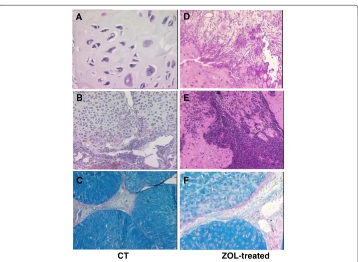
Figure 2Histological analysis of tumor tissue at the end of the study: control (A to C) and ZOL-treated (D to F) groups. The tumor slices were stained with hematoxylin-eosin (A, B, D, E) or with alcian blue (C, F). Magnification: ×50 (C), ×100 (B, D, E, F), and ×200 (A). CT, controls; ZOL, zoledronic acid.
Miot-Noirault et al. EJNMMI Research 2013, 3:40 Page 2 of 5
Model
Experiments were conducted on 24 male Sprague Dawley rats (Charles River, L'Arbresle, France). The SRC model was a generous gift from Dr. P.A. Guerne (Switzerland) as tissue fragments, which were frozen until use. Allograft transplantation of a 10-mm3SRC fragment was performed on the right hindlimb in the paratibial location of the anesthetized animals (isoflurane (Abbott, Rungis, France) in air (1.5%, 1 L/min)) associated with an intramuscular injection of 100 mg/kg of ketamine (Imalgene®, Rhone Merieux, Lyon, France), as published [3,6,8].
ZOL treatment
ZOL was kindly provided as a research-grade disodium salt by Novartis Pharma AG (Basel, Switzerland). Rats were randomly divided into two groups:
1. ZOL-treated: subcutaneous administration of 100 μg/kg twice a week from day 4 to day 48 after implantation (n = 12)
2. Control: PBS in the same conditions (n = 12)
Tumor growth assessment
Tumor volume was calculated from calliper measure-ment of the largest (L) and smallest (S) perpendicular tumor diameters, using the formula: V(mm3) = 0.5 × L ×
S2 [3]. Data were expressed as mean ± standard devi-ation. At each time point, the tumor volumes of ZOL and controls were compared by analysis of variance (ANOVA) with a level of significance set at p < 0.05.
99mTc-NTP 15-5 imaging 99m
Tc-NTP 15-5 imaging was performed at days 4 (before treatment), 11, 16, 24, 36, and 48 after implan-tation. NTP 15-5 was radiolabeled with 99mTc as described [3].
Acquisitions (10-min duration, 15% window at 140 keV) of each hindlimb of anesthetized animals positioned over a 10-cm collimator of a small-animal gamma camera (γImager, Biospace, Paris, France) were performed, 30 min after i.v. administration of 30 MBq of radiotracer. Fixed-size regions of interest (ROIs) were delineated over the tumor, adjacent muscles, and contralateral muscles, and average counts (cpm/mm2) were obtained. The use of ac-tivity profile for ROI placement ensured easy, reproducible positioning of the ROI for serial images.
At each time point and for each animal, the target-to-background ratio (TBR) was calculated as follows: TBR = Average count in the tumor (cpm/mm2) / Average count in
the contralateral muscle (cpm/mm2). Data were expressed as mean ± standard deviation.
Analysis of data with multiple comparisons was per-formed by repeated-measures ANOVA followed by Tukey’s post-test, with a level of significance set at p < 0.05.
Histology
At the end of the study, tumors fixed in 4% formalin were embedded in paraffin, sectioned (5 μm), and stained with hematoxylin-eosin and alcian blue (AB).
Results
ZOL inhibited tumor growth
As shown in Figure 1, ZOL inhibited tumor growth as compared to controls: From day 24 to day 48, a signifi-cant (p < 0.05) difference in tumor volume was observed between control and treated groups.
CT
ZOL
day 4 day 16 day 36
A
B
***
*
*
*
Figure 399mTc-NTP 15-5 imaging of control and ZOL-treated
groups. (A) Representative in vivo scintigraphic images of the tumor-bearing hindlimb obtained for controls and ZOL-treated animals at various stages of the study. (B) Semi-quantitative analysis of99mTc-NTP 15-5 imaging: results are expressed as mean TBR
values ± standard deviation. CT, controls; ZOL, zoledronic acid-treated; TBR, target-to-background ratio. The asterisk indicates p < 0.05.
Tumor regression patterns were clearly evidenced by histology (Figure 2): whereas controls showed nuclear atypia, anisokaryosis, mitosis, lobular arrangement of the tumor, and metachromasia (Figure 2A,B,C), ZOL-treated animals demonstrated fibrous and inflammatory remo-deling patterns of tumor regression (Figure 2D,E). Meta-chromasia, reflecting PG content, was also decreased in ZOL-treated animals as compared to controls (Figure 2F versus Figure 2C).
99mTc-NTP 15-5 monitoring of in vivo chondrosarcoma
response to ZOL
As illustrated in Figure 3A, for representative animals,
99m
Tc-NTP 15-5 accumulation was observed in the joints and at the sites of tumor implantation as early as day 4. As pathology evolved, a weaker radiotracer accu-mulation was observed in ZOL-treated tumors as com-pared to controls.
From day 11 to day 48, mean TBR values ranged from 1.7 ± 0.6 to 2.3 ± 0.6 in ZOL-treated rats and from 2.1 ± 1.0 to 4.9 ± 0.9 in controls (Figure 3B). Differences in TBR between treated rats and controls were statistically significant (p < 0.05) from day 24 to day 48. Repeated-measures ANOVA revealed both a significant ‘between-group effect’ as well as a within-subject effect (‘between-group * time).
Discussion
A few years ago, Heymann's group published the thera-peutic relevance of using ZOL in the SRC model [6]. Since then, others have reported the promising effects of bisphosphonates not only on chondrosarcoma prolifera-tion and invasion in vitro, but also in clinical practice [7,9,10]. In clinics, chondrosarcoma therapeutic response is still evaluated by conventional imaging by measuring anatomical tumor size reduction [11]. Unlike the late ef-fects of anticancer therapy on tumor size, molecular and metabolic changes are well recognized to be induced much earlier, before morphological changes occur [12].
Our previous study demonstrated 99mTc-NTP 15-5 to be a promising SPECT tracer for the molecular imaging of cartilage neoplasms in nuclear medicine [3]. 99mTc-NTP 15-5 appeared of particular interest since radiotracers currently available in clinics such as 201Tl, 99mTc-MIBI,
99m
Tc-tetrofosmin, 99mTc-DMSA(V), and 18F-FDG have evidenced limitations for imaging chondrosarcoma with low cellularity and high chondrogenic matrix [13].
The results reported in this new study bring additional data in favor of 99mTc-NTP 15-5 imaging for the in vivo follow-up of chondrosarcoma and more especially the semi-quantitative assessment of therapeutic response in vivo, with TBR values being significantly decreased in the treated group with respect to controls from day 24. Chondrosarcoma growth inhibition was confirmed by
calliper measurement from day 24 and characterized at the tissue level by histology: interestingly, AB staining evidenced a huge decrease in glycosaminoglycan content in ZOL as compared to controls. Such PG ‘downregulation’ reflects the tumor proliferation inhibition at the extracellu-lar matrix level and is expected to be at the origin of the de-creased accumulation of the99mTc-NTP 15-5 radiotracer in tumors responding to ZOL.
Conclusion
99m
Tc-NTP 15-5 is expected to offer a novel imaging modality for the semi-quantitative evaluation of PG in vivo as markers of the extracellular matrix features of chondrosarcoma, which could be useful for the follow-up and evaluation of the response to therapeutic at-tempts. Combining chondrosarcoma functional imaging at the PG level with relevant targeted therapies may rep-resent the opportunity to bridge the gap between pre-clinical and pre-clinical testing, which is of real need for improving the management of this pathology.
Competing interests
The authors declare that they have no competing interests. Authors’ contributions
EMN, CP, and SB carried out the experimental treatments and imaging studies. ED and FG carried out the development of the SRC orthotopic model. AV and JMC carried out the synthesis and radiolabeling of NTP 15-5. MMD carried out the histology. MFH participated in the histology analysis. DH and FR conceived of the study and participated in its design and in the manuscript. All authors read and approved the final manuscript.
Acknowledgements
The authors thank G. Hamery and P. Monmousseau from the Experimental Therapy Unit facility core (Nantes, France).
Funding
This work was supported by the Ligue contre le Cancer Auvergne, Auvergne Region for Contrat de Projets Etat Region/Regional Innovation Fund FRI2/OSEO and in collaboration with Cyclopharma.
Author details
1
INSERM UMR 990, Université d’Auvergne, BP 184, Rue Montalembert, Clermont-Ferrand, Cédex 63005, France.2INSERM UMR 957, Nantes 44000,
France.3Laboratoire de Physiopathologie de la Résorption Osseuse et
Thérapie des Tumeurs Osseuses Primitives, Faculté de Médecine, Nantes Atlantique Universités, Université de Nantes, Nantes 44000, France.
4
Département d’anatomo-pathologie, Centre Jean Perrin, Clermont-Ferrand 63001, France.5
Département d’anatomo-pathologie, Hôtel Dieu, CHU de Nantes, Nantes 44000, France.6Service d’orthopédie, Hôtel Dieu, CHU de Nantes, Nantes 44000, France.
Received: 4 March 2013 Accepted: 4 May 2013 Published: 20 May 2013
References
1. World Health Organization: Cartilage tumors. In Pathology and Genetics of Tumours of Soft Tissue and Bone. Edited by Fletcher CDM, Unni KK, Mertens F. Lyon: IARC; 2002:234–257 [Kleihues P, Sobin LH (Series Editors): World Health Organization Classification of Tumors].
2. Velasco CR, Colliec-Jouault S, Redini F, Heymann D, Padrines M: Proteoglycans on bone tumor development. Drug Discov Today 2010, 13–14:553–560.
3. Miot-Noirault E, Gouin F, Vidal A, Rapp M, Maublant J, Askienazy S, Chezal JM, Heymann D, Redini F, Moins N: First preclinical imaging of primary
Miot-Noirault et al. EJNMMI Research 2013, 3:40 Page 4 of 5
cartilage neoplasm and its local recurrence using99mTc-NTP 15-5
radiotracer. J Nucl Med 2009, 50:1541–1547.
4. Onishi AC, Hincker AM, Lee FY: Surmounting chemotherapy and radioresistance in chondrosarcoma: molecular mechanisms and therapeutic targets. Sarcoma 2011, 2011:381564.
5. Moriceau G, Ory B, Gobin B, Verrecchia F, Gouin F, Blanchard F, Redini F, Heymann D: Therapeutic approach of primary bone tumours by bisphosphonates. Curr Pharm Des 2010, 16:2981–2987.
6. Gouin F, Ory B, Rédini F, Heymann D: Zoledronic acid slows down rat primary chondrosarcoma development, recurrent tumor progression after intralesional curretage and increases overall survival. Int J Cancer 2006, 119:980–984.
7. Streitbuerger A, Henrichs M, Ahrens H, Lanvers-Kaminzky C, Gouin F, Gosheger G, Hardes J: Cytotoxic effect of clodronate and zoledronate on the chondrosarcoma cell lines HTB-94 and CAL-78. Int Orthop 2011, 35:1369–1373.
8. Grimaud E, Damiens C, Rousselle AV, Passuti N, Heymann D, Gouin F: Bone remodelling and tumor grade modifications induced by interactions between bone and swarm rat chondrosarcoma. Histol Histopathol 2002, 17:1103–1111.
9. Odri GA, Dumoucel S, Picarda G, Battaglia S, Lamoureux F, Corradini N, Rousseau J, Tirode F, Laud K, Delattre O, Gouin F, Heymann D, Redini F: Zoledronic acid as a new adjuvant therapeutic strategy for Ewing’s sarcoma patients. Cancer Res 2010, 70:7610–7619.
10. Montella L, Addeo R, Faiola V, Cennamo G, Guarrasi R, Capasso E, Mamone R, Caraglia M, Del Prete S: Zoledronic acid in metastatic chondrosarcoma and advanced sacrum chordoma: two case reports. J Exp Clin Cancer Res 2009, 28:7.
11. Fox E, Patel S, Wathen JK, Schuetze S, Chawla S, Harmon D, Reinke D, Chugh R, Benjamin RS, Helman LJ: Phase II study of sequential gemcitabine followed by docetaxel for recurrent Ewing sarcoma, osteosarcoma, or unresectable or locally recurrent chondrosarcoma: results of sarcoma alliance for research through collaboration study 003. Oncologist 2012, 17:321.
12. Chiti A, Oyen WJG: Imaging of therapy response in oncology. Q J Nucl Med 2011, 55:587–588.
13. Arsos G, Venizelos I, Karatzas N, Koukoulidis A, Karakatsanis C: Low-grade chondrosarcomas: a difficult target for radionuclide imaging. Case report and review of the literature. Eur J Radiol 2002, 43:66–72.
doi:10.1186/2191-219X-3-40
Cite this article as: Miot-Noirault et al.:99mTc-NTP 15-5 assessment of the early therapeutic response of chondrosarcoma to zoledronic acid in the Swarm rat orthotopic model. EJNMMI Research 2013 3:40.
Submit your manuscript to a
journal and benefi t from:
7 Convenient online submission 7 Rigorous peer review
7 Immediate publication on acceptance 7 Open access: articles freely available online 7 High visibility within the fi eld
7 Retaining the copyright to your article