LETTER TO THE EDITOR (Comment letter)
Species origin investigation of the St. Jude porcine lung epithelial cell line (SJPL) made available to researchers
David W. Silversides1,4, Nedzad Music1,4, Mario Jacques2,3,4, Richard Webby5, Carl A. Gagnon1,2,3,4*
1
Service de diagnostic; 2Centre de recherche en infectiologie porcine (CRIP); 3Groupe de recherche sur les maladies infectieuses du porc (GREMIP); 4Faculté de médecine vétérinaire, Université de Montréal, St-Hyacinthe, Québec, Canada. 5Department of Infectious Diseases, St. Jude Children’s Research Hospital, Memphis, TN, USA.
Running title: Species origin of the SJPL cell line.
*Address correspondence and reprint requests to Dr Carl A. Gagnon
Faculté de médecine vétérinaire Université de Montréal
3200 rue Sicotte,
St-Hyacinthe, Québec, Canada, J2S 7C6
Email: carl.a.gagnon@umontreal.ca
Phone: 450-773-8521 (8681) Fax: 450-778-8113
The SJPL cell line was reported and described as an immortalized porcine lung epithelial cell line suitable for influenza virus replication in Seo et al., J. Virol. 2001;75(19):9517-25 and has been available to researchers through ATCC (American Type Culture Collection) cell depositary services (deposit date: April 5, 2001; assigned accession number: PTA-3256) following a material transfer agreement (6). Recent findings indicate that the SJPL cell line is not of porcine origin but is more likely of monkey origin.
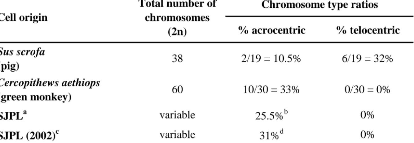
The percentage of acrosomic and telocentric chromosomes of the SJPL cell line obtained from ATCC was determined and the findings were compared to what would be expected for a normal pig karyotype as well as for a normal green monkey karyotype (Table 1). The domestic pig karyotype consists of 19 pairs of chromosomes (38 chromosomes in total), including 6 pairs of telocentric chromosomes and 2 pairs of chromosomes that can be classified as acrocentric (4). The African green monkey karyotype consists of 30 pairs of chromosomes (60 chromosomes in total), including 10 pairs of acrocentric chromosomes and no telocentric chromosomes (2). Acrocentric refers to the chromosomal configuration wherein the centromere is situated very close to one end of the chromosome, such that the short arm (p) is very small but still present, while telocentric refers to a chromosome configuration wherein the centromere is located at the terminal end of the chromosome, such that there is no short arm. Following karyotype analysis of SJPL cells obtained from ATCC, an absence of the characteristic porcine telocentric chromosomes, and a higher than expected ratio of acrocentric chromosomes per metaphase spread (25.5% for SJPL cells compared to 10.5% for porcine cells) were observed (Table 1). Overall, the acrocentric and telocentric chromosomes ratios were more closely related to monkey karyotype than porcine karyotype (Table 1). Furthermore, similar calculated acrocentric and
Research Hospital in 2002 (Table 1). Consequently, the obtained results raise questions concerning the porcine origin of the SJPL cells.
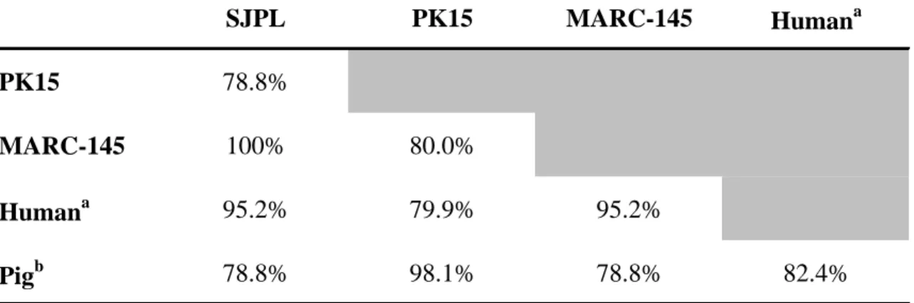
To further determine the species origin of the SJPL cell line, primers were designed to amplify by PCR a portion of the P53 gene to verify the genomic DNA sequences of the SJPL cells compared to genomic DNA sequences of other cells types such as PK15 cells (porcine origin) and MARC-145 cells (African green monkey origin). As shown in Table 2, the P53 genomic sequence of the SJPL obtained from ATCC was more related to the MARC-145 monkey cells and human nucleotide (nt) sequences (100% and 95.2% nt identities, respectively) compared to PK15 cells and pig nt sequences (78.8% nt identities for both). Based on chromosome analyses and genomic sequence analyses, the SJPL cells received from ATCC are not from pig origin, but are more genomically related to monkey origin.
Following the first report of the SJPL cells (6), other investigators have used these cells in their studies (1, 3, 5, 7, 8). In some of those reports, the SJPL cells were used as a model to study the viral and bacterial pathogenesis in regards to the respiratory tract environment of their susceptible host (1, 8). If the species origin of the SJPL cells is doubtful, then it is reasonable to question the respiratory tract origin of these cells. It is impossible for us to determine if the SJPL cells used in each published studies were obtained from ATCC or from another source and which cell passages were used and consequently, to assume that they are monkey cells. Nonetheless, karyotype analyses of the SJPL cells originating from ATCC and from St. Jude Children’s Research Hospital have both demonstrated the monkey origin of the cells (Table 1). Thus, in light of this recent finding, conclusions that have been drawn from previous studies using the SJPL cells should be carefully reconsidered.
MATERIALS AND METHODS
Chromosome analysis
Chromosome analysis was performed on SJPL cells using standard methods. Briefly, SJPL cells were seeded onto 60 X 15 mm petri dishes and grown under standard conditions. Mitotic arrest was induced by adding 150 l colcimid (KaryoMax solution, Gibco) and incubating the cells 30 min at 37˚C. Cells were then trypsinized and centrifuged for 10 min at 1000 RPM. A hypotonic shock was done by adding 10 ml KCl (0,075M) to the cell pellet and by incubating the cell suspensions for 15 min at 37˚C. Cell nuclei were pelleted and fixed by adding 10 ml of freshly made Carnoys’s solution (Methanol/glacial acetic acid, 3:1 v/v). The nuclei suspension was dropped onto microscope slides which were allowed to dry and mature for several days. Afterwards, slides were stained with KaryoMax Giemsa stain (Gibco). Slides were read for metaphase spreads using a Nikon Eclipse E800 microscope equipped with a Retiga 1300 (Q-Imaging) CCD camera and Simple PCI software for image analysis.
Sequence analysis
Genomic DNA extraction of cell lines was performed using the Qiagen QIAamp DNA Mini Kit (Qiagen, Valencia, CA, USA), according to the manufacturer’s instructions. PCR primers were designed to target exon 8, intron 8-9 and exon 9 of the TP53 gene (P53 gene in human). Based on sequence identity between pig and human sequences, the sense primer p53.A GGACGGAACAGCTTTGAGGTGCG) and the degenerate reverse primer p53.1 (5’-AGGGTGAAATA(T/C)TCICCATCCAGTG, where I = inosine) were designed and synthesized
human genomic DNA they should amplify a band of 303 bp. The extracted DNA was amplified by PCR with Taq DNA polymerase (New England Biolabs, Ipswich, MA, USA). The PCR consisted of an initial enzyme activation step at 95°C for 3 min, followed by 35 cycles of denaturation at 94°C for 1 min, annealing at 54°C for 45 sec, extension at 72°C for 1 min and 30 sec, and a final extension at 72°C for 5 min. The PCR products were purified using a commercial kit (QIAquick PCR purification kit; Qiagen) according to the manufacturer’s instruction. Sequencing reactions of amplified bands were performed using the di-deoxy method and Big Dye Terminator 3.1 (Applied biosystems) reagents, according to the manufacturer’s instructions. The same PCR primers were used for the sequencing reactions. Sequencing was performed on an ABI Prism 310 genetic analyzer. Identity comparisons were performed using MacDNAsis software (Hitachi).
REFERENCES
1. Auger, E., V. Deslandes, M. Ramjeet, I. Contreras, J. H. Nash, J. Harel, M. Gottschalk, M. Olivier, and M. Jacques. 2009. Host-pathogen interactions of Actinobacillus pleuropneumoniae with porcine lung and tracheal epithelial cells. Infect Immun 77:1426-41.
2. Finelli, P., R. Stanyon, R. Plesker, M. A. Ferguson-Smith, P. C. O'Brien, and J. Wienberg. 1999. Reciprocal chromosome painting shows that the great difference in diploid number between human and African green monkey is mostly due to non-Robertsonian fissions. Mamm Genome 10:713-8.
3. Gagnon, C. A., and M. Jacques. 2008. Porcine lung epithelial cell line and its use in production and detection of porcine reproductive and respiratory syndrome virus. patent International PCT/CA2008/001953.
4. Gustavsson, I. 1988. Standard karyotype of the domestic pig. Committee for the Standardized Karyotype of the Domestic Pig. Hereditas 109:151-7.
5. Herman, M., S. Haugerud, Y. S. Malik, and S. M. Goyal. 2005. Improved in vitro cultivation of swine influenza virus. Intern J Appl Res Vet Med 3:124-128.
6. Seo, S. H., O. Goloubeva, R. Webby, and R. G. Webster. 2001. Characterization of a porcine lung epithelial cell line suitable for influenza virus studies. J Virol 75:9517-25. 7. Seo, S. H., E. Hoffmann, and R. G. Webster. 2004. The NS1 gene of H5N1 influenza
viruses circumvents the host anti-viral cytokine responses. Virus Res 103:107-13.
anti-% acrocentric % telocentric Sus scrofa (pig) 38 2/19 = 10.5% 6/19 = 32% Cercopithews aethiops (green monkey) 60 10/30 = 33% 0/30 = 0% SJPLa variable 25.5%b 0% SJPL (2002)c variable 31%d 0%
aSJPL cell line obtained from ATCC and maintained at the Faculté de médecine vétérinaire, Université de Montréal.
b
Average of 8 analyzed metaphases. c
Data generated from a chromosome analysis performed at the St. Jude Children's Research Hospital in 2002.
d
Average of 3 analyzed metaphases.
Chromosome type ratios Total number of
chromosomes (2n) Cell origin
Table 1. Comparison of acrocentric and telocentric chromosomes in pig, green monkey genomes and in SJPL cell line methaphase spreads.
Table 2. Partial P53 genomic sequence identities of the SJPL cells with cells of porcine (PK15) and monkey (MARC-145) origins
SJPL PK15 MARC-145 Humana
PK15 78.8%
MARC-145 100% 80.0%
Humana 95.2% 79.9% 95.2%
Pigb 78.8% 98.1% 78.8% 82.4%
Two hundred and nine nucleotides base pair sequences including most of the exon 8, all of intron 8-9 and most of exon 9 were used for the analysis.
a
Human TP53 sequences taken from Ensembl GRCh37 (Feb. 2009).
b