HAL Id: hal-02631880
https://hal.inrae.fr/hal-02631880
Submitted on 27 May 2020
HAL is a multi-disciplinary open access
archive for the deposit and dissemination of
sci-entific research documents, whether they are
pub-lished or not. The documents may come from
teaching and research institutions in France or
abroad, or from public or private research centers.
L’archive ouverte pluridisciplinaire HAL, est
destinée au dépôt et à la diffusion de documents
scientifiques de niveau recherche, publiés ou non,
émanant des établissements d’enseignement et de
recherche français ou étrangers, des laboratoires
publics ou privés.
Copyright
features and is involved in the first steps of infection
Nabila Haddad, Rute G. Matos, Teresa Pinto, Pauline Rannou, Jean-Michel
Cappelier, Hervé Prevost, Cecília M. Arraiano
To cite this version:
Nabila Haddad, Rute G. Matos, Teresa Pinto, Pauline Rannou, Jean-Michel Cappelier, et al.. The
RNase R from Campylobacter jejuni has unique features and is involved in the first steps of infection.
Journal of Biological Chemistry, American Society for Biochemistry and Molecular Biology, 2014, 289
(40), pp.27814 - 27824. �10.1074/jbc.M114.561795�. �hal-02631880�
The RNase R from Campylobacter jejuni Has Unique Features
and Is Involved in the First Steps of Infection
*
□SReceived for publication, March 5, 2014, and in revised form, August 5, 2014 Published, JBC Papers in Press, August 6, 2014, DOI 10.1074/jbc.M114.561795
Nabila Haddad‡§1,2, Rute G. Matos¶1,3, Teresa Pinto¶4, Pauline Rannou‡§, Jean-Michel Cappelier‡§, Herve´ Pre´vost‡§, and Cecília M. Arraiano¶5
From the‡LUNAM Universite´, Oniris, University of Nantes, 44200 Nantes, France, the§UMR1014 Se´curite´ des Aliments et Microbiologie, INRA, 44322 Nantes, France, and the¶Instituto de Tecnologia Química e Biolo´gica, Universidade Nova de Lisboa, Avada Repu´blica, 2780-157 Oeiras, Portugal
Background:Members of the RNB family are involved in virulence; nothing is known about the Campylobacter jejuni homologue.
Results:Cj-RNase R is active in several conditions. It is also important for adhesion and invasion. Conclusion:RNase R is important for C. jejuni biology and infection.
Significance:RNase R could be targeted to reduce infection by this pathogen.
Bacterial pathogens must adapt/respond rapidly to changing environmental conditions. Ribonucleases (RNases) can be crucial factors contributing to the fast adaptation of RNA levels to differ-ent environmdiffer-ental demands. It has been demonstrated that the exoribonuclease polynucleotide phosphorylase (PNPase) facili-tates survival of Campylobacter jejuni in low temperatures and favors swimming, chick colonization, and cell adhesion/invasion. However, little is known about the mechanism of action of other ribonucleases in this microorganism. Members of the RNB family of enzymes have been shown to be involved in virulence of several pathogens. We have searched C. jejuni genome for homologues and found one candidate that displayed properties more similar to RNase R (Cj-RNR). We show here that Cj-RNR is important for the first steps of infection, the adhesion and invasion of C. jejuni to eukaryotic cells. Moreover, Cj-RNR proved to be active in a wide range of conditions. The results obtained lead us to conclude that Cj-RNR has an important role in the biology of this foodborne pathogen.
Campylobacter jejuniis a foodborne bacterial pathogen that is now considered the leading cause of human bacterial gastro-enteritis worldwide, with⬃400 million cases of campylobacte-riosis diagnosed each year (1). The symptoms of this disease include malaise, fever, severe abdominal pain, and diarrhea.
Post-infectious sequels can occur, including septicemia and neuropathies such as Guillain-Barre´ syndrome (2– 4). The prin-cipal reservoir of C. jejuni is the gut of avian species, which may contain up to 109cfu/g of feces (5). The fecal contamination of poultry meat during food processing is the main cause of
C. jejuniinfections (6). Despite its specific microaerobic growth requirements, C. jejuni is ubiquitous in the aerobic environ-ment, and it is capable of withstanding different stresses caused by growth or survival on a suboptimal carbon source, temper-ature changes, exposure to atmospheric oxygen, hypo- and hyper-osmotic stress, and desiccation. Moreover, during infec-tion, C. jejuni has to withstand other stresses, including changes in pH and the host innate immune response (7, 8).
Cellular levels of RNA are determined by their rate of synthe-sis and their rate of degradation. As such, ribonucleases are important in the adaptation of organisms to new environments. In the case of C. jejuni, little is known about the RNA processing pathways and associated ribonucleases. However, the exoribo-nuclease polynucleotide phosphorylase has been shown to be important for cell survival at low temperature and has an important role in swimming, cell adhesion/invasion ability, and chick colonization (9, 10). Moreover, the endoribonuclease RNase III was recently characterized and was shown to be active in an unexpectedly large range of conditions (11). This endoribonuclease may have an important role under a Mn2⫹
-rich environment, considering that Mn2⫹ is its preferred
co-factor (11). Similarly to what was observed in other organisms, RNase III from C. jejuni is involved in the processing of 30 S rRNA (11, 12) and participates in the maturation of CRISPR RNAs, which are key elements in the CRISPR (clustered regu-larly interspaced short palindromic repeats) system of bacterial adaptive immunity (12).
RNase R is a 3⬘ to 5⬘ hydrolytic enzyme that belongs to the RNB family of enzymes. This family of exoribonucleases is widely distributed in all domains of life and plays important functions in the cell. The E. coli genome codes for two proteins of this family, RNase II and RNase R. They present a similar mechanism of action; however, they behave differently regard-ing product released and the ability to degrade structured RNA
*This work was supported by grants from Fundac¸a˜o para a Cieˆncia e Tecnologia (FCT), including grant PEst-oE/EQB/LA0004/2013 and the projects PTDC/QUI-BIQ/111757/2009 and PTDC/BIA-MIC/4142/2012 both from FCT, and the European Commission Project FP7-KBBE-2011-1-289326.
□S
This article containssupplemental Fig. 1.
1Both authors contributed equally to this work.
2Supported by GENICAMP program. To whom correspondence may be
addressed: Oniris, UMR INRA1014 SECALIM, Rue de la Ge´raudie`re, 44322 Nantes, France. Tel.: 33-2-51785570; Fax: 33-2-51785520; E-mail: nabila.haddad@oniris-nantes.fr.
3Recipient of a postdoctoral fellowship (SFRH/BPD/75887/2011) funded by
the FCT.
4Recipient of a research grant funded by the FCT.
5To whom correspondence may be addressed: Instituto de Tecnologia
Química e Biolo´gica, Universidade Nova de Lisboa, Avada Repu´blica,
2780-157 Oeiras, Portugal. Tel.: 351-214469547; Fax: 351-214469549; E-mail: cecilia@itqb.unl.pt.
at INRA Institut National de la Recherche Agronomique on May 9, 2019
http://www.jbc.org/
(13). The crystal structure of RNase II showed that the protein is formed by a central RNB domain, which is responsible for the catalytic activity. This domain is flanked by two N-terminal cold shock domains and a C-terminal S1 domain, important for RNA binding (14, 15). All the other members of this family have the same domain organization, but may have extra domains at the N-terminal region. For example, RNase R has a helix-turn-helix motif, whereas in eukaryotes there is an extra PINc domain, which has endonucleolytic activity (16, 17). In the cat-alytic region, there are several highly conserved residues that are important for the activity of the protein. The role of these residues seems to be conserved both in eukaryotes and in pro-karyotes (18 –21).
In eukaryotes, the member of the RNB family of enzymes is called Dis3 and exists in three isoforms (Dis3/Rrp44, Dis3L1, and Dis3L2). These proteins locate in the cell differently. Dis3L2 prefers poly(U) RNAs and was recently shown to be involved in a new eukaryotic RNA degradation mechanism independent of the exosome (22). Dis3 and Dis3L2 were shown to be involved in important human diseases (23). In some path-ogenic organisms, proteins from this family were shown to be crucial for the establishment of virulence (24).
Considering that C. jejuni is a foodborne bacterial pathogen with impact on human health, it is important to understand the mechanisms behind the infection process. In the genome of
C. jejuni, we found a homologue of the RNB family of enzymes, which, based on sequence alignment, seems to be more similar to RNase R (from here on designated by Cj-RNR).6Taking into account the involvement of RNase R-like proteins in the estab-lishment of virulence in other pathogens, Cj-RNR seems to be an excellent candidate for studies in C. jejuni.
The aim of this work was to access the role of RNase R in growth and virulence of C. jejuni. We have also cloned, expressed, purified, and characterized the activity of this pro-tein at different conditions. The importance of RNase R in
C. jejuni virulence and in its adaptation to different environ-ments is discussed.
EXPERIMENTAL PROCEDURES
Bacterial Strains and Culture Conditions—The strains used in this study are listed in Table 1. C. jejuni 81–176 and ⌬rnr mutant strains were routinely cultured on Karmali agar plates (Oxoid, Basingstoke Hampshire, UK) or in brain heart infusion broth (Merck), and kanamycin at 50g/ml was added for the culture of⌬rnr mutant strain. Culture on plates and broth were incubated at 42 °C for 48 and 24 h, respectively, under a microaerophilic atmosphere in jars flushed with a gas mixture
of 10% CO2, 5% O2, and 85% N2. E. coli strains were cultivated overnight at 37 °C in Luria-Bertani medium (Sigma, Steinheim, Germany). When necessary, 100g/ml ampicillin was added to the growth medium.
Construction of the C. jejuni⌬rnr Mutant Strain—The
pro-tocol to obtain C. jejuni 81–176 rnr mutant strain was adapted from Ref. 25. Briefly, a DNA sequence coding a putative RNase R (CJJ81176_0659) was identified in the genome of C. jejuni 81–176 using National Center for Biotechnology Information (NCBI) BLAST features and the EMBOSS alignment (1935 bp). The C. jejuni 81–176⌬rnr deletion mutant (⌬rnr::aphA3) was generated by homologous recombination using a PCR-ampli-fied nonpolar cassette carrying the aphA3 kanamycin resis-tance gene flanked by⬃300-bp regions that flank either side of the rnr gene. Overlapping extension PCR protocol was used to amplify the deletion cassette. To prepare the three PCR prod-ucts used as templates in overlapping extension PCR reaction, PCR was performed with Phusion high-fidelity DNA polymer-ase (Finnzymes, Thermo Fisher Scientific, Illkirch, France) and with three different primer sets: MO576 (217–199 upstream of
rnr coding sequence) and MO577 (nucleotides 38 –56 of rnr coding sequence) were used for the upstream region (273 bp), MO582 and MO583 were used to amplify the kanamycin resis-tance cassette (aphA3) (1388 bp), and MO578 (1892–1911 of
rnrcoding sequence) and MO579 (347–366 downstream of rnr coding sequence) were used for the downstream region (410 bp). The sequences of all primers used in this study are pre-sented (see Table 3). The rnr fragments were amplified using genomic DNA isolated from C. jejuni 81–176 by using the DNeasy blood and tissue kit (Qiagen, Courtaboeuf, France), while the kanamycin resistance cassette was amplified from pBF14 (26). The reverse primer for the upstream fragment (MO577) and the forward primer for the downstream fragment (MO578) included 22 bp of DNA sequences (see Table 3, underlined) at the 5⬘ end that are complementary to MO582 and MO583, respectively, to allow joining of the kanamycin resistance cassette to the upstream and downstream fragments. These three fragments were purified from agarose gels (with QIAquick gel extraction kit, Qiagen) and then used in overlap-ping extension PCR (using MO576/579) to obtain the deletion cassette, which consisted of the upstream (273 bp) and down-stream (410 bp) sequences of the target gene rnr with a kana-mycin resistance cassette (1388 bp) inserted between them. This deletion cassette of 2071 bp was sequenced and then cloned into pGEM-T vector (Promega), which is a suicide vec-tor for C. jejuni, leading to the recombinant plasmid pNH07. 5 l of pNH07 was used to transform C. jejuni 81–176-compe-tent cells. Transformants were selected by incubation in Columbia plates containing 5% sheep blood and 50g/ml kana-mycin. The insertion of the cassette and deletion of the rnr gene were confirmed by PCR using primer set MO587/588.
C. jejuni Growth Experiments—Growth experiments were performed at optimal temperature (37 °C) and close to the min-imal temperature of growth (32 °C) of C. jejuni to compare the behavior of the parental and rnr mutant strains. C. jejuni 81–176 parental and derivative strains were obtained from ⫺80 °C stocks and cultured for 48 h at 42 °C under microaero-philic conditions on Karmali agar plates. Cells were transferred
6The abbreviations used are: Cj-RNR, C. jejuni homologue of RNB family; RNR,
RNase R; nt, nucleotide(s). TABLE 1
Strains used in this study
Strain
Antibiotic
resistance Reference Genetic
E. coliDH5␣ Invitrogen recA1 endA1 BL21 StarTM(DE3) Invitrogen rne131 C. jejuni81–176 Korlath et al. (51)
⌬rnr::aphA3 Kanamycin This study C. jejuni⌬rnr
at INRA Institut National de la Recherche Agronomique on May 9, 2019
http://www.jbc.org/
to 20 ml of brain heart infusion medium and incubated with shaking for 24 h under microaerophilic conditions to obtain a starter culture. 100 ml of brain heart infusion medium was then inoculated with a 1/100 dilution of starter culture and incu-bated microaerobically at 32 and 37 °C with agitation (110 rpm). Viable counts were measured by serial dilution and plat-ing on Karmali agar at regular time intervals to estimate the total number of cfu/ml. The growth rate was calculated using the formula ln(Nt/N0)/t, where N0corresponds to the viable count in cfu/ml at time 0, Ntcorresponds to the viable count in
cfu/ml at time t, and t corresponds to the interval time between each points. Each experiment was performed four times inde-pendently, and for each experiment, the individual samples were plated in triplicate on Karmali agar plates.
Adhesion and Invasion Assays Using Ht-29 Epithelial Cells—
Bacterial adhesion and invasion into Ht-29 cells were studied using the gentamicin protection assay, as described previously (27). Briefly, microplate wells were seeded with 2⫻ 105Ht-29
cells and incubated for 5 days at 37 °C in a humidified, 5% CO2
incubator. After washing, the Ht-29 monolayers were infected with a suspension of⬃2–5 ⫻ 107cfu. To measure cell adhesion,
the infected monolayers incubated for 1 h at 37 °C in a humid-ified 5% CO2incubator were washed, and then Ht-29 adherent
Campylobacterwere enumerated on Karmali agar. To evaluate bacterial invasion, the infected monolayers were incubated during 3 h, gentamicin (250g/ml) was added, and the mono-layers were incubated for 2 more hours. After washes and Ht-29 cell lyses, intracellular Campylobacter was enumerated on Kar-mali agar. Each experiment was done in duplicate and per-formed at least three times independently.
Construction of the Plasmid Expressing RNase R from C. jejuni—
The C. jejuni 81–176 RNase R gene (rnr) (GenBankTMaccession
number YP_001000332) was amplified from genomic DNA using the primers MO625 and MO591 (see Table 3) (NdeI and
BamHI sites, respectively, are underlined in the table). For
rnr amplification by PCR, genomic DNA, 200 Mof each
primer, and 1 unit of Phusion high-fidelity DNA polymerase (Finnzymes) were used. The resulting product (1944 bp) was purified (QIAquick PCR purification kit, Qiagen) and cloned into the pGEM-T plasmid (Promega) to generate
pGEMT-rnr. To overexpress the Cj-RNR protein, the rnr gene dou-ble-digested with NdeI and BamHI was subcloned from pGEMT-rnr into the pET19b expression vector previously digested with the same enzymes (Novagen, Merck). Plasmids and primers used are presented in Tables 2 and 3, respec-tively. The resultant pETrnr-His was confirmed by DNA sequencing (Beckman Coulter Genomics).
Overexpression and Purification of Recombinant RNase R from C. jejuni—The plasmid harboring the histidine-tagged Cj-RNR protein (pETrnr-His) was introduced by transformation into E. coli BL21(DE3) Star to allow the production of the recombinant protein. Cells were grown at 37 °C in 100 ml LB medium supplemented with 100g/ml ampicillin to an A600of
0.5, induced by the addition of 0.5 mMisopropyl-1-thio--D
-galactopyranoside, and grown at 32 °C for 4 h. Cells were pel-leted by centrifugation and stored at⫺80 °C. Purification was performed by immobilized metal affinity chromatography using HiTrap chelating HP columns (GE Healthcare) and A¨ KTA FPLC system (GE Healthcare) following a protocol described previously (19). The purity of the protein was verified by SDS-PAGE using an 8% gel and visualized by Coomassie Blue staining. Proteins were quantified using the Bradford method (28), and 50% (v/v) glycerol was added to the final frac-tions prior to storage at⫺20 °C.
In Vitro Transcription—Templates for tRNASer and 5 S
rRNA were generated by PCR using the primers Cjej3 and Cjej4 and Cjej7 and Cjej8, respectively (Table 3). The phage T7 RNA polymerase promoter sequence was included in the forward
TABLE 2
Plasmids used in this study
Plasmid Antibiotic resistance Reference Comments
pGEM-T Ampicillin Promega Commercial vector
pET19b Ampicillin Novagen Commercial expression vector
pNH07 Ampicillin; kanamycin This study pGEM-T vector containing the rnr deletion cassette pGEMT-rnr Ampicillin This study Encodes Cj-RNR
pETrnr-His Ampicillin This study Encodes His-Cj-RNR TABLE 3
Primers used in this study
Primers Sequence (5ⴕ–3ⴕ) Comments
MO576 ATGAGCGTGTCCTTGAGAG Primers used to amplify the beginning of C. jejuni rnr gene (fragment of 273 bp); the underlined region of MO577 is complementary to MO582
MO577 CTTACCTATCACCTCAAATGGTTTGCTTACTTCGTGAGAGC
MO578 CTGGATGAATTGTTTTAGTACCATGCTAAAATCACCCAAAGA Primers used to amplify the end of C. jejuni rnr gene (fragment of 410 bp); the underlined region of MO578 is complementary to MO583
MO579 GCAAAATGAGGAGAGAAAAT
MO582 ACCATTTGAGGTGATAGGTAAG Primers used to amplify kanamycin resistance cassette from pBF14(26) MO583 GGTACTAAAACAATTCATCCAG
MO587 ATGAGCGTGTCCTTGAGAG Primers used to check C. jejuni⌬rnr mutant strain MO588 GTCTTTGGGTGATTTTAGCA
MO625 CATATGAAAGAATTTTTAAA Construction of pET RNR (NdeI restriction site underlined) MO591 GGATCCTAGAAAGTAGGGTTTGAAGC Construction of pET RNR (BamHI restriction site underlined) Cjej3 TGGCCGGGAGAAAGGGATTCG tRNASer
transcription Cjej4 GAAATTAATACGACTCACTATACGGGAGATGGCTGAG tRNASer
transcription Cjej7 GAAATTAATACGACTCACTATAATGTCCGTGATTATAC 5 S rRNA transcription Cjej8 AAGTCCGCAATGAGC 5 S rRNA transcription 16ss CCCGACACCAACCACU RNA substrate 30ss CCCGACACCAACCACUAAAAAAAAAAAAAA RNA substrate
16ss-comp AGUGGUUGGUGUCGGG RNA oligonucleotide complementary to 16ss and 30ss 24ss AATATGGCTCATAGGCGCAGAGGG DNA substrate
at INRA Institut National de la Recherche Agronomique on May 9, 2019
http://www.jbc.org/
primer sequences and is underlined. In vitro transcription was carried out using the purified amplicons as template in the pres-ence of an excess of [␣-32P]UTP over unlabeled UTP with the Riboprobe in vitro transcription system (Promega) and T7 RNA polymerase. The reaction mixture was incubated 2 h at 37 °C. To remove all trace of DNA, 1 unit of DNase (Promega) was added and incubated for 30 min at 37 °C. The unincorporated radioactive nucleotides were removed using a G50 column as instructed (GE Healthcare). The transcripts were purified by electrophoresis on an 8Murea, 10% polyacrylamide gel. The gel
slice was crushed, and the RNA was eluted overnight at room temperature into elution buffer (0.5Mammonium acetate, pH
5.2, 1 mMEDTA, 2.5% (v/v) phenol, pH 4.3). The RNA was
precipitated using ethanol and resuspended in RNase-free water. The yield of the labeled substrates (cpm/l) was deter-mined by scintillation counting.
RNase R Activity Assays—The activity assays were performed using different substrates: a poly(A) oligomer of 35 nt, a 16-mer oligoribonucleotide, a 30-mer oligoribonucleotide, a 24-mer DNA substrate, tRNASer, and 5 S rRNA (transcribed in vitro as
described in the above section) (Table 3). To obtain the double-stranded substrates 16 –30ds and 16 –16ds, the 30-mer and the 16-mer oligoribonucleotides, respectively, were hybridized to the complementary unlabeled 16-mer oligoribonucleotide (5 ⬘-AGTGGTTGGTGTCGGG-3⬘) (Table 3). The hybridization was performed in a 1:1 (mol:mol) ratio by incubation for 5 min at 100 °C followed by 45 min at 37 °C. All the synthetic RNA molecules were labeled at its 5⬘ end with [␥-32P]ATP using T4
polynucleotide kinase. The RNA oligomers were then purified using a G25 column (GE Healthcare) to remove the unincorpo-rated nucleotides. After optimization of the buffer, the activity assays were performed in a final volume of 20l with an activity buffer composed of 10 mMTris-HCl, pH 7.5, 5 mMKCl, 0.5 mM
MgCl2, and 0.1 mMDTT and 10,000 cpm of substrate. The
reactions were started by the addition of the enzyme at the concentrations indicated in the figures and further incubated at 37 °C. As a control, the reaction was also performed in the absence of the enzyme. Samples were withdrawn at the time points indicated in the respective figures, and the reactions were stopped by the addition of formamide-containing dye supplemented with 10 mM EDTA. Reaction products were
resolved in an 8Murea, 20% polyacrylamide gel as indicated in
the respective figure legends. Signals were visualized by phos-phorimaging and analyzed using ImageQuant software (Molec-ular Dynamics).
For the buffer optimization, RNase activity assays were car-ried out at various pH levels (pH 5.2, 5.4, 6.1, 6.5, 7.0, 7.5, 8.0, 8.5, and 9.0), at various KCl or NaCl concentrations, using dif-ferent divalent ions, and at difdif-ferent temperatures of incubation (4, 30, 37, and 42 °C). The exoribonucleolytic activity of the enzyme was determined by measuring and quantifying the disap-pearance of the substrate in several distinct experiments in which the protein concentration was adjusted so that, under those con-ditions, less than 25% of substrate was degraded. Each value obtained represents the mean for these independent assays.
Sequence Alignment and Construction of the Phylogenetic Tree—RNase R sequences of Escherichia coli (UniProt ID: P21499), Salmonella enterica (UniProt ID: E7V4H1),
Staphylo-coccus aureus(UniProt ID: Q7WRI2), Mycoplasma genitalium (UniProt ID: P47350), Streptococcus pneumoniae (UniProt ID: C6GKN7), Bacillus subtilis (UniProt ID: O32231), C. jejuni (NCBI reference sequence: YP_001000332.1), and Helicobacter
pylori(UniProt ID: P56123) were aligned using Clustal Omega (29, 30). The phylogenetic tree was built using Phylogeny.fr (31, 32).
RESULTS AND DISCUSSION
Analysis of C. jejuni RNase R—RNase R-like proteins are very heterogeneous in their size and composition. In some organ-isms, they present the organization described for E. coli protein, with a helix-turn-helix domain at the N-terminal region fol-lowed by two cold shock domains, a central RNB domain, a C-terminal S1 domain, and a lysine-rich region (Fig. 1A). How-ever, the N- and C-terminal regions are very variable. For instance, in C. jejuni there is no helix-turn-helix or Lys-rich region, and in archaea the existence of an RNase R that only has the RNB domain was described (33). Despite these differences, the RNB domain (characteristic from this family of enzymes) is very conserved, with all the residues involved in catalysis found in all members of the family (supplemental Fig. 1). We have constructed a phylogenetic tree based on the RNase R sequence of Gram-positive and negative bacteria (Fig. 1B). As expected,
Cj-RNR is more similar to the RNase R from H. pylori, which is also an Epsilonproteobacteria. Moreover, it is possible to see a clear separation in two groups: Gram-positive and Gram-neg-ative bacteria (Fig. 1B).
Cj-RNR Activity at Different Conditions—To characterize the activity of Cj-RNR protein, we have first determined the opti-mal conditions for the catalysis. For this purpose, we have analyzed the activity of this protein at different conditions. We have cloned the rnr gene from C. jejuni in an expression vector. We tested several conditions for protein expression in different expression strains, and finally, we expressed Cj-RNR at 32 °C in a BL21 StarTM(DE3) (Invitrogen). We then
purified the protein by histidine affinity chromatography as described under “Experimental Procedures.”
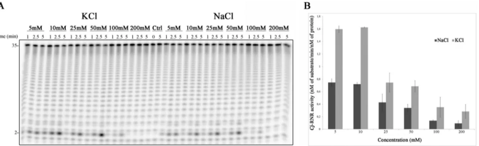
We started by analyzing the activity of the protein using two different types of salts, KCl and NaCl, using six different con-centrations, 5, 10, 25 50, 100, and 200 mM. The results show that
Cj-RNR is active with both KCl and NaCl and prefers low
con-FIGURE 1. A, schematic representation of the domain organization of RNase R proteins from E. coli and C. jejuni. N, N terminus; C, C terminus; HTH, helix-turn-helix. B, phylogenetic tree of RNase R from E. coli, S. enterica, C. jejuni,
S. aureus, S. pneumoniae, M. genitalium, B. subtilis, and H. pylori.
at INRA Institut National de la Recherche Agronomique on May 9, 2019
http://www.jbc.org/
centrations of monovalent cations (Fig. 2A). We have quanti-fied the activity of the protein in these conditions. In fact, Cj-RNR has a maximum activity when lower concentrations are used and prefers KCl to NaCl (Fig. 2B). In contrast, it was described that E. coli RNase II and RNase R are more active with higher KCl concentrations (50 –500 mM) (34).
We have also determined the effect of pH on the activity of
Cj-RNR. With this purpose, we have performed activity assays at different pH values between 5.4 and 9.0. The E. coli proteins, RNase II and RNase R, present an optimal activity at a pH range between 7.5 and 9.5 (34). Cj-RNR activity is higher at a pH of 7.5, although it is active in a wide range of pH from 6.5 to 9.0 (Fig. 3). Based on these results, we used a buffer containing 5 mMKCl and with a pH of 7.5 in subsequent experiments.
Enzymes from the RNB family require a divalent ion for catalysis, normally Mg2⫹. However, they are also active in the presence of other ions. For that reason, we tested Cj-RNR activ-ity in the presence of Mg2⫹, Mn2⫹, Ca2⫹, Zn2⫹, Ni2⫹, Cu2⫹, and Co2⫹. As it is possible to observe, Cj-RNR is active in the
presence of Mg2⫹, Mn2⫹, Ca2⫹, and Ni2⫹(Fig. 4A). For the first
two ions, the enzyme seems to have the same activity, whereas for Ca2⫹and Ni2⫹, there is a reduction in the activity, especially
with regard to the degradation of the smaller fragments (Fig.
4A). We then tested different Mg2⫹and Mn2⫹concentrations.
The results show that Cj-RNR prefers lower concentration of divalent ions (from 0.1 to 1 mM) (Fig. 4, B and C). We then
determined the activity of Cj-RNR in those conditions. We con-firmed that it is more active in the presence of lower concen-trations of Mg2⫹and Mn2⫹. However, in contrast to what was
described for other proteins of this family, Cj-RNR prefers Mn2⫹and not Mg2⫹ (Fig. 4D). Manganese has emerged as a very important metal in virulence. Moreover, macrophages have poor magnesium and acidic and low oxygen environment (35). Campylobacter can survive within macrophages for a period of 24 –30 h, and it is within the macrophages that man-ganese is thought to be important for C. jejuni (36). Manman-ganese and magnesium contents in C. jejuni are not yet known. How-ever, they were determined in E. coli, where they were shown to change accordingly to growth phase; their levels decline as growth progresses (37). One of the major challenges that bac-teria have when growing in oxygenated environments is to effi-ciently resist or repair damages caused by reactive oxygen spe-cies. One strategy to reduce oxidative damage involves limiting intracellular iron content. In these situations, bacteria appear to have an absolute requirement for Mn2⫹(38). The differences in
the activity of Cj-RNR in the presence of both Mg2⫹and Mn2⫹
FIGURE 2. Salt dependence of Cj-RNR. A, 1 nMrecombinant protein was incubated with 10 nMpoly(A) at 37 °C for 5 min in a reaction buffer with different salt concentrations as indicated. Samples were taken during the reaction at the time points indicated. B, determination of the activity of Cj-RNR. Error bars indicate mean⫾ S.D.
FIGURE 3. pH dependence of Cj-RNR. A, 1 nMrecombinant protein were incubated with 10 nMpoly(A) at 37 °C for 5 min in a reaction buffer with different pH, ranging from 5.4 to 9. Samples were taken during the reaction at the time points indicated. Ctrl, control. B, determination of the activity of Cj-RNR. Error bars indicate mean⫾ S.D.
at INRA Institut National de la Recherche Agronomique on May 9, 2019
http://www.jbc.org/
may allow C. jejuni to manipulate the enzymatic activity of RNase R according to the environment, thus altering gene expression. This feature can be an important advantage for
C. jejunito adapt and survive within macrophages. Addition-ally, it was shown that the endoribonuclease RNase III from
C. jejunimay also have an important role under a Mn2⫹-rich
environment (11).
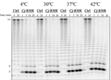
The optimal temperature of growth for C. jejuni is between 37 and 42 °C, the human and avian body temperatures, respec-tively (39). Although not able to grow below 30 °C, C. jejuni is able to survive at refrigerated temperatures (40). It was already shown that the exoribonuclease polynucleotide phosphorylase was important for the survival at refrigerated temperatures (9). Recently, it was also demonstrated in vitro that the endoribo-nuclease RNase III is active in an unexpectedly large range of temperatures from 4 to 42 °C (11). Taking this into account, we decided to analyze the activity of Cj-RNR at various tempera-tures. The activity assays were performed at four distinct tem-peratures: 4, 30, 37, and 42 °C. The results demonstrate that, in these conditions, this protein is active in all the temperatures
tested, even at 4 °C (Fig. 5), where we would expect a drastic decrease in its activity.
RNA Cleavage by Cj-RNR—Taking into account that Cj-RNR is active in a broad range of conditions, for the following exper-iments we have used a buffer with pH 7.5, 5 mMKCl, 0.1 mM
MgCl2, and all the assays were performed at 37 °C. The activity
of Cj-RNR was evaluated using three different single-stranded substrates (poly(A), 30ss and 16ss) and two different double-stranded RNAs (16 –30ds and 16 –16ds). Regarding the degra-dation of ssRNA, it is possible to see that the substrate is pro-cessively degraded until a 2-nt fragment is released, similarly to what was described for RNase R-like proteins (Fig. 6A). It is also possible to observe the presence of an intermediate product with 4 nt of length (Fig. 6). A mixture of two different degrada-tion products with 2 and 4 nt of length was also observed in
E. coliRNase R protein. Because there is no crystal structure available for any RNase R protein, it was postulated that, simi-larly to what occurs in RNase II, some 4-nt fragments are released, although they are still partially “clamped” in the active site. Others are degraded to a 2-nt fragment (21). We tested the
FIGURE 4. Divalent metal ion dependence of Cj-RNR. A, 25 nMrecombinant protein were incubated with 10 nMpoly(A) at 37 °C for 10 min in a reaction buffer with different divalent metal ions, which are indicated in the figure. Ctrl, control. B and C, 1 nMrecombinant protein was incubated with 10 nMpoly(A) at 37 °C for 5 min in a reaction buffer with different Mg2⫹(B) and Mn2⫹(C) concentrations. Samples were taken during the reaction at the time points indicated. D, determination of the activity of Cj-RNR. Error bars indicate mean⫾ S.D.
at INRA Institut National de la Recherche Agronomique on May 9, 2019
http://www.jbc.org/
activity of Cj-RNR protein using two structured substrates: 16 –16ds and 16 –30ds (see “Experimental Procedures” for a description of these substrates). When the substrate tested was
the 16 –30ds, we were able to see that, once again, the protein behaved like RNase R, being able to overcome the double-stranded structures (Fig. 6B). RNase R from E. coli requires a 3⬘ single-stranded region to cleave structured substrates (41). In contrast, Cj-RNR does not have such a strict requirement. In the conditions tested (10 nMRNA and 25 nMCj-RNR), it is able
to digest the perfect double-stranded 16 –16ds substrate (Fig. 6B); however, the degradation of this substrate is less efficient when compared with the degradation of 16 –30ds (Fig. 6B). We have tested the E. coli RNase R (Ec-RNR) in the same conditions and observed that it is not able to degrade the 16 –16ds (data not show). E. coli RNase R has at the C terminus a region rich in lysines (Fig. 1A), which was shown to be involved in the degra-dation of double-stranded substrates, probably by helping to unwind the two strands (42). This region is absent in Cj-RNR. Together with the evidence that Cj-RNR is able to degrade the 16 –16ds RNA, although with less efficiency, it seems that the mechanism by which the dsRNA is degraded by RNase R in
C. jejuniis different from the one in E. coli; however, these dif-ferences seem to be minimal. In E. coli, one of the existing deg-radation pathways implies that the RNA molecules to be
FIGURE 5. Exoribonucleolytic activity of Cj-RNR at different
tempera-tures. 25 nMrecombinant protein was incubated with 10 nMpoly(A) at 4, 30, 37, and 42 °C for 10 min. Samples were taken during the reaction at the time points indicated. Ctrl, control.
FIGURE 6. Exoribonucleolytic activity of Cj-RNR using ssRNA (A) or dsRNA (B) molecules. The recombinant protein was incubated with 10 nMRNA substrates at 37 °C for 20 min. Samples were taken during the reaction at the time points indicated. The protein concentration used is indicated in the figure. Ctrl, control.
at INRA Institut National de la Recherche Agronomique on May 9, 2019
http://www.jbc.org/
degraded are tagged with poly(A) tails synthetized by poly(A) polymerase I (PAP I). These extra nucleotides serve as platform for the binding of RNases such as RNase R because these pro-teins require a single-stranded tail to proceed with the degra-dation of structured substrates. In C. jejuni, until date, there is no data on the poly(A) polymerase I protein, although a puta-tive gene that could code for a poly(A) polymerase is found in its genome. Considering that Cj-RNR, to some extent, degrades perfect double-stranded RNAs, we can speculate that perhaps polyadenylation may play a different role in this organism.
We also determined the activity of Cj-RNR with different substrates and at various temperatures. To do it, we adjusted the reaction conditions, as described under “Experimental Pro-cedures,” to ensure that less than 25% of the substrate was being degraded. The results show that there is no preference for a specific substrate, except at 42 °C, where the enzyme seems to prefer poly(A) (Fig. 7). Moreover, this experiment allowed us to determine that, in fact, there are some differences in the activity of the protein at different temperatures; at 4 and 30 °C, the activity is half that the determined at 37 and 42 °C (Fig. 7). These differences were not visible by eye in the poly-acrylamide gels presented before due to the conditions used (Fig. 5).
Cj-RNR Is Able to Cleave DNA Substrates—It is known that RNase II, the prototype of this family of enzymes, is able to bind to DNA molecules; however, it is not able to cleave them because there are specific requirements for a ribose in the sec-ond or fourth nucleotides counting from the 3⬘-end (43, 44). When RNase R was initially characterized, it was shown that is able to cleave DNA, albeit with a reduced efficiency and using higher protein concentrations (34). We decided to test the activity of Cj-RNR also with a DNA substrate and compare it with the E. coli counterparts. As found previously, E. coli RNase II is not able to cleave DNA, whereas E. coli RNase R can degrade a small percentage of the substrate (Fig. 8). When we tested Cj-RNR activity, we were able to see that it can cleave DNA in a distributive way. Moreover, it is more efficient in degrading DNA substrates when compared with the E. coli counterpart (Fig. 8). These results confirm that Cj-RNR
behaves like an RNase R-like protein. Interestingly, the exori-bonuclease polynucleotide phosphorylase from B. subtilis, in the presence of Mn2⫹and low level of inorganic phosphate (Pi),
is able to degrade ssDNA. This activity was postulated to be important for DNA repair pathways (45). Why RNase R-like proteins are able to cleave DNA is still unknown, and structural studies need to be performed to understand the mechanism involved.
tRNAs Are Substrates for Cj-RNR—It was described that RNase R is able to degrade defective tRNAs or rRNA and mRNAs containing repetitive extragenic palindromic (REP) sequences (13). M. genitalium RNase R was shown to maturate tRNAs (46). E. coli RNase R is able to efficiently cleave 23 S and 16 S rRNAs but acts poorly on 5 S rRNA and tRNA (34). We have tested the activity of Cj-RNR using 5 S rRNA and a tRNA molecule (we chose tRNASer). We transcribed both RNAs and
performed the degradation assays as described under “Experi-mental Procedures.” The results obtained showed that, simi-larly to what was shown in E. coli, Cj-RNR acts poorly over 5 S rRNA; however, it is able to degrade tRNA molecules (Fig. 9). tRNA molecules contain very small 3⬘ single-stranded over-hangs, which explains why E. coli RNase R is not able to effi-ciently cleave this substrate (34). As we discussed previously,
Cj-RNR can cleave structured substrates in the absence of a 3⬘ single-stranded region, although with less efficiency (Fig. 6B). This characteristic may be useful to confer to this protein the ability to degrade tRNAs in C. jejuni.
Growth and Viability of Parental and⌬rnr Strains—In some
bacteria, RNase R-like proteins are essential (13). However, as reported previously for H. pylori, it was possible to construct a
C. jejunistrain deficient in RNase R, which shows that, in this organism, RNase R is not essential (47). This meant we were able to analyze the effect of rnr deletion in growth and viability. We tested this at two different temperatures, at 37 °C (the opti-mal temperature of growth is between 37 and 42 °C), and at 32 °C, which is considered as the minimal temperature of growth of C. jejuni (48).
FIGURE 7. Determination of the activity of Cj-RNR at different
tempera-tures. The activity of the protein was determined at 4, 30, 37, and 42 °C as
described under “Experimental Procedures” using three different synthetic substrates: poly(A), 16ss, and the double-stranded 16 –30ds. All the activity assays were performed in triplicate. Error bars indicate mean⫾ S.D.
FIGURE 8. Exoribonucleolytic activity of Cj-RNR with a DNA molecule
showing comparison with E. coli RNase II (Ec RNB) and RNase R (Ec RNR).
10 nMof each protein was incubated with 30 nMDNA at 37 °C for 60 min. Samples were taken during the reaction at the time points indicated. Ctrl, control.
at INRA Institut National de la Recherche Agronomique on May 9, 2019
http://www.jbc.org/
If we compare the growth of the two strains at optimal tem-perature, 37 °C, we observe that the lag phase of the wild-type strain is more prolonged when compared with the mutant strain (Fig. 10A). Moreover, when we calculated the maximum rate growth, we were able to see that the mutant strain grows faster when compared with the wild type (0.49 and 0.3 h⫺1, respectively).
At minimal temperature of growth, 32 °C, it is possible to observe the “Phoenix” effect described by Kelly et al. (49). This phenomenon is characterized by a decrease in viability after inoculation followed by an increase in survival to levels similar or higher to the initial ones. Both strains show an exponential phase of 78 h (Fig. 10B). In this phase, wild-type cells grow better than the mutant ones, although the viability was shown to be similar. This phase is followed by a decline phase, which reflects the high mortality due to stress induced by tempera-ture. The bacterial cells that survived then start to regrow, and we can see that the⌬rnr strain recovers viability sooner than the wild-type strain.
In conclusion, Cj-RNR is not an essential protein, and its deletion does not cause significant changes in growth and via-bility when compared with the wild-type strain. However, at lower temperatures (32 °C), the mutant strain recovers better than the wild type.
Involvement of RNase R in C. jejuni Adhesion and Invasion of Eukaryotic Cells—RNase R-like proteins have been involved in the establishment of virulence in several pathogenic organisms (24). In C. jejuni, it was already shown that polynucleotide phosphorylase, another exoribonuclease, has an important role
FIGURE 9. Exoribonucleolytic activity of Cj-RNR with tRNASerand rRNA
molecules. 50 nMof the recombinant protein was incubated with 10 nM
tRNASerand rRNA at 37 °C for 30 min. Samples were taken during the reaction
at the time points indicated. Ctrl, control.
FIGURE 10. Growth (left panel) and viability (right panel) of C. jejuni 81–176 wild-type strain (in blue) and⌬rnr mutant strain (in red) at optimal
temperature of growth (37 °C) (A) and at minimal temperature of growth (32 °C) (B). The data represent the mean of the experiments under each
condition, with standard deviations shown. OD, optical density.
at INRA Institut National de la Recherche Agronomique on May 9, 2019
http://www.jbc.org/
in adhesion and invasion (10). Moreover, we showed that Cj-RNR is active in a large range of conditions, which may be important for the adaptation of C. jejuni to different environ-ments during the infection process. For that reason, we decided to address the role of Cj-RNR in adhesion and invasion. This was done using wild-type and⌬rnr strains of C. jejuni 81–176 and a eukaryotic cell line of intestinal origin, Ht-29. Several bacterial concentrations were tested, while the concentration of eukaryotic cells was set as 2⫻ 105cells/well (multiplicity of
infection of 100). Experiments were done three times, and the results correspond to the mean value calculated from the three experiments.
The results show that the wild-type strain is three times more adherent and six times more invasive than the mutant strain (Fig. 11). The results are in agreement with what has been reported for Shigella flexneri and E. coli EIEC, in which the⌬rnr mutant strains were shown to be less invasive (50). These results indicate that RNase R, similarly to what was shown for polynucleotide phosphorylase (10), is an important protein for the first steps of infection of C. jejuni.
Conclusions—In this study, we have undertaken a functional and biochemical analysis of Cj-RNR. We demonstrated that
Cj-RNR is active in a wide range of conditions and determined the optimal conditions for its activity. We also demonstrated that Cj-RNR behaves like an RNase R-like protein regarding its ability to degrade structured RNAs. However, the mechanism of action seems to be different because it is able to degrade perfect double-stranded structures, although with less effi-ciency. Our results also showed that Cj-RNR is capable of acting on a variety of RNA molecules, namely highly structured RNAs such as tRNAs. Finally, we saw that, although not essential,
Cj-RNR is important for C. jejuni adhesion and invasion ability. This demonstrates that RNase R plays an important role in the first steps of C. jejuni invasion, probably by regulating virulence factors.
Acknowledgments—We thank Andreia Aires for technical support at Instituto de Tecnologia Química e Biolo´gica (ITQB) and Christopher M. Burns for discussion about construction of the plasmid overex-pressing C. jejuni RNase R. RNA research in UMR1014 SECALIM was funded by the Re´gion Pays de la Loire through the program grant GENICAMP.
REFERENCES
1. Ruiz-Palacios, G. M. (2007) The health burden of Campylobacter infec-tion and the impact of antimicrobial resistance: playing chicken. Clin.
Infect. Dis. 44,701–703
2. Nachamkin, I., Allos, B. M., and Ho, T. (1998) Campylobacter species and Guillain-Barre syndrome. Clin. Microbiol. Rev. 11, 555–567
3. Riddle, M. S., Gutierrez, R. L., Verdu, E. F., and Porter, C. K. (2012) The chronic gastrointestinal consequences associated with Campylobacter.
Curr. Gastroenterol. Rep. 14,395– 405
4. Nyati, K. K., and Nyati, R. (2013) Role of Campylobacter jejuni infection in the pathogenesis of Guillain-Barre syndrome: an update. Biomed. Res. Int.
2013,852195
5. Newell, D. G., and Fearnley, C. (2003) Sources of Campylobacter coloni-zation in broiler chickens. Appl. Environ. Microbiol. 69, 4343– 4351 6. Ketley, J. M. (1997) Pathogenesis of enteric infection by Campylobacter.
Microbiology 143,5–21
7. Murphy, C., Carroll, C., and Jordan, K. N. (2006) Environmental survival mechanisms of the foodborne pathogen Campylobacter jejuni. J. Appl.
Microbiol. 100,623– 632
8. Park, S. F. (2005) Campylobacter jejuni stress responses during survival in the food chain and colonization. in Campylobacter Molecular and
Cellu-lar Biology(Ketley, J. M., and Konkel, M. E., eds), pp. 311–330, Horizon Bioscience, Norfolk, UK
9. Haddad, N., Burns, C. M., Bolla, J. M., Pre´vost, H., Fe´de´righi, M., Drider, D., and Cappelier, J. M. (2009) Long-term survival of Campylobacter jejuni at low temperatures is dependent on polynucleotide phosphorylase activ-ity. Appl. Environ. Microbiol. 75, 7310 –7318
10. Haddad, N., Tresse, O., Rivoal, K., Chevret, D., Nonglaton, Q., Burns, C. M., Pre´vost, H., and Cappelier, J. M. (2012) Polynucleotide phosphor-ylase has an impact on cell biology of Campylobacter jejuni. Front. Cell.
Infect. Microbiol. 2,30
11. Haddad, N., Saramago, M., Matos, R. G., Pre´vost, H., and Arraiano, C. M. (2013) Characterization of the biochemical properties of Campylobacter
jejuniRNase III. Biosci. Rep. 33, e00082
12. Dugar, G., Herbig, A., Fo¨rstner, K. U., Heidrich, N., Reinhardt, R., Nieselt, K., and Sharma, C. M. (2013) High-resolution transcriptome maps reveal strain-specific regulatory features of multiple Campylobacter jejuni iso-lates. PLoS Genet. 9, e1003495
13. Arraiano, C. M., Andrade, J. M., Domingues, S., Guinote, I. B., Malecki, M., Matos, R. G., Moreira, R. N., Pobre, V., Reis, F. P., Saramago, M., Silva, I. J., and Viegas, S. C. (2010) The critical role of RNA processing and degrada-tion in the control of gene expression. FEMS Microbiol. Rev. 34, 883–923 14. Amblar, M., Barbas, A., Fialho, A. M., and Arraiano, C. M. (2006) Charac-terization of the functional domains of Escherichia coli RNase II. J. Mol.
Biol. 360,921–933
15. Fraza˜o, C., McVey, C. E., Amblar, M., Barbas, A., Vonrhein, C., Arraiano, C. M., and Carrondo, M. A. (2006) Unravelling the dynamics of RNA degradation by ribonuclease II and its RNA-bound complex. Nature 443, 110 –114
16. Lebreton, A., Tomecki, R., Dziembowski, A., and Se´raphin, B. (2008) En-donucleolytic RNA cleavage by a eukaryotic exosome. Nature 456, 993–996
17. Schaeffer, D., Tsanova, B., Barbas, A., Reis, F. P., Dastidar, E. G., Sanchez-Rotunno, M., Arraiano, C. M., and van Hoof, A. (2009) The exosome contains domains with specific endoribonuclease, exoribonuclease and cytoplasmic mRNA decay activities. Nat. Struct. Mol. Biol. 16, 56 – 62 18. Amblar, M., and Arraiano, C. M. (2005) A single mutation in Escherichia
coliribonuclease II inactivates the enzyme without affecting RNA binding.
FEBS J. 272,363–374 FIGURE 11. Adhesion and invasion ability of C. jejuni 81–176 wild-type
and⌬rnr strains. The results are expressed as the ratio between wild-type
and⌬rnr strains. Error bars indicate mean ⫾ S.D.
at INRA Institut National de la Recherche Agronomique on May 9, 2019
http://www.jbc.org/
19. Barbas, A., Matos, R. G., Amblar, M., Lo´pez-Vin˜as, E., Gomez-Puertas, P., and Arraiano, C. M. (2008) New insights into the mechanism of RNA degradation by ribonuclease II: identification of the residue responsible for setting the RNase II end product. J. Biol. Chem. 283, 13070 –13076 20. Dziembowski, A., Lorentzen, E., Conti, E., and Se´raphin, B. (2007) A single
subunit, Dis3, is essentially responsible for yeast exosome core activity.
Nat. Struct. Mol. Biol. 14,15–22
21. Matos, R. G., Barbas, A., and Arraiano, C. M. (2009) RNase R mutants elucidate the catalysis of structured RNA: RNA-binding domains select the RNAs targeted for degradation. Biochem. J. 423, 291–301
22. Malecki, M., Viegas, S. C., Carneiro, T., Golik, P., Dressaire, C., Ferreira, M. G., and Arraiano, C. M. (2013) The exoribonuclease Dis3L2 defines a novel eukaryotic RNA degradation pathway. EMBO J. 32, 1842–1854 23. Reis, F. P., Pobre, V., Silva, I. J., Malecki, M., and Arraiano, C. M. (2013)
The RNase II/RNB family of exoribonucleases: putting the ‘Dis’ in disease.
Wiley Interdiscip. Rev. RNA 4,607– 615
24. Matos, R. G., Ba´rria, C., Pobre, V., Andrade, J. M., and Arraiano, C. M. (2012) Exoribonucleases as modulators of virulence in pathogenic bacte-ria. Front. Cell Infect. Microbiol. 2, 65
25. Hansen, C. R., Khatiwara, A., Ziprin, R., and Kwon, Y. M. (2007) Rapid construction of Campylobacter jejuni deletion mutants. Lett. Appl.
Micro-biol. 45,599 – 603
26. Wyszyn´ska, A., Tomczyk, K., and Jagusztyn-Krynicka, E. K. (2007) Com-parison of the localization and post-translational modification of
Campy-lobacter coliCjaC and its homolog from Campylobacter jejuni, Cj0734c/ HisJ. Acta Biochim. Pol. 54, 143–150
27. Haddad, N., Maillart, G., Gare´naux, A., Jugiau, F., Federighi, M., and Cap-pelier, J. M. (2010) Adhesion ability of Campylobacter jejuni to Ht-29 cells increases with the augmentation of oxidant agent concentration. Curr.
Microbiol. 61,500 –505
28. Bradford, M. M. (1976) A rapid and sensitive method for the quantitation of microgram quantities of protein utilizing the principle of protein-dye binding. Anal. Biochem. 72, 248 –254
29. Sievers, F., Wilm, A., Dineen, D., Gibson, T. J., Karplus, K., Li, W., Lopez, R., McWilliam, H., Remmert, M., So¨ding, J., Thompson, J. D., and Higgins, D. G. (2011) Fast, scalable generation of high-quality protein multiple sequence alignments using Clustal Omega. Mol. Syst. Biol. 7, 539 30. Goujon, M., McWilliam, H., Li, W., Valentin, F., Squizzato, S., Paern, J.,
and Lopez, R. (2010) A new bioinformatics analysis tools framework at EMBL-EBI. Nucleic Acids Res. 38, W695–W699
31. Dereeper, A., Guignon, V., Blanc, G., Audic, S., Buffet, S., Chevenet, F., Dufayard, J. F., Guindon, S., Lefort, V., Lescot, M., Claverie, J. M., and Gascuel, O. (2008) Phylogeny.fr: robust phylogenetic analysis for the non-specialist. Nucleic Acids Res. 36, W465–W469
32. Dereeper, A., Audic, S., Claverie, J. M., and Blanc, G. (2010) BLAST-EXPLORER helps you building datasets for phylogenetic analysis. BMC
Evol. Biol. 10,8
33. Matos, R. G., Lo´pez-Vin˜as, E., Gome´z-Puertas, P., and Arraiano, C. M. (2012) The only exoribonuclease present in Haloferax volcanii has an unique response to temperature changes. Biochim. Biophys. Acta 1820, 1543–1552
34. Cheng, Z. F., and Deutscher, M. P. (2002) Purification and characteriza-tion of the Escherichia coli exoribonuclease RNase R: comparison with RNase II. J. Biol. Chem. 277, 21624 –21629
35. Papp-Wallace, K. M., and Maguire, M. E. (2006) Manganese transport and
the role of manganese in virulence. Annu. Rev. Microbiol. 60, 187–209 36. Sikic´ Pogacar, M., Rubesa Mihaljevic´, R., Klancnik, A., Brumini, G.,
Abram, M., and Smole Mozina, S. (2009) Survival of stress exposed
Cam-pylobacter jejuniin the murine macrophage J774 cell line. Int. J. Food
Microbiol. 129,68 –73
37. Me´dicis, E. D., Paquette, J., Gauthier, J. J., and Shapcott, D. (1986) Magne-sium and manganese content of halophilic bacteria. Appl. Environ.
Micro-biol. 52,567–573
38. Jakubovics, N. S., and Jenkinson, H. F. (2001) Out of the iron age: new insights into the critical role of manganese homeostasis in bacteria.
Mi-crobiology 147,1709 –1718
39. Lee, A., Smith, S. C., and Coloe, P. J. (1998) Survival and growth of
Cam-pylobacter jejuniafter artificial inoculation onto chicken skin as a function of temperature and packaging conditions. J. Food Prot. 61, 1609 –1614 40. Chan, K. F., Le Tran, H., Kanenaka, R. Y., and Kathariou, S. (2001) Survival
of clinical and poultry-derived isolates of Campylobacter jejuni at a low temperature (4° C). Appl. Environ. Microbiol. 67, 4186 – 4191
41. Vincent, H. A., and Deutscher, M. P. (2006) Substrate recognition and catalysis by the exoribonuclease RNase R. J. Biol. Chem. 281, 29769 –29775
42. Matos, R. G., Barbas, A., Go´mez-Puertas, P., and Arraiano, C. M. (2011) Swapping the domains of exoribonucleases RNase II and RNase R: con-ferring upon RNase II the ability to degrade ds RNA. Proteins 79, 1853–1867
43. Barbas, A., Matos, R. G., Amblar, M., Lo´pez-Vin˜as, E., Gomez-Puertas, P., and Arraiano, C. M. (2009) Determination of key residues for catalysis and RNA cleavage specificity: one mutation turns RNase II into a “super-en-zyme.” J. Biol. Chem. 284, 20486 –20498
44. Cannistraro, V. J., and Kennell, D. (1994) The processive reaction mech-anism of ribonuclease II. J. Mol. Biol. 243, 930 –943
45. Cardenas, P. P., Carrasco, B., Sanchez, H., Deikus, G., Bechhofer, D. H., and Alonso, J. C. (2009) Bacillus subtilis polynucleotide phosphorylase 3⬘-to-5⬘ DNase activity is involved in DNA repair. Nucleic Acids Res. 37, 4157– 4169
46. Alluri, R. K., and Li, Z. (2012) Novel one-step mechanism for tRNA 3⬘-end maturation by the exoribonuclease RNase R of Mycoplasma genitalium.
J. Biol. Chem. 287,23427–23433
47. Tsao, M. Y., Lin, T. L., Hsieh, P. F., and Wang, J. T. (2009) The 3⬘-to-5⬘ exoribonuclease (encoded by HP1248) of Helicobacter pylori regulates motility and apoptosis-inducing genes. J. Bacteriol. 191, 2691–2702 48. Hazeleger, W. C., Wouters, J. A., Rombouts, F. M., and Abee, T. (1998)
Physiological activity of Campylobacter jejuni far below the minimal growth temperature. Appl. Environ. Microbiol. 64, 3917–3922
49. Kelly, A. F., Martínez-Rodriguez, A., Bovill, R. A., and Mackey, B. M. (2003) Description of a “phoenix” phenomenon in the growth of
Campy-lobacter jejuniat temperatures close to the minimum for growth. Appl.
Environ. Microbiol. 69,4975– 4978
50. Tobe, T., Sasakawa, C., Okada, N., Honma, Y., and Yoshikawa, M. (1992)
vacB, a novel chromosomal gene required for expression of virulence genes on the large plasmid of Shigella flexneri. J. Bacteriol. 174, 6359 – 6367
51. Korlath, J. A., Osterholm, M. T., Judy, L. A., Forfang, J. C., and Robinson, R. A. (1985) A point-source outbreak of campylobacteriosis associated with consumption of raw milk. J. Infect. Dis. 152, 592–596
at INRA Institut National de la Recherche Agronomique on May 9, 2019
http://www.jbc.org/
Hervé Prévost and Cecília M. Arraiano
Nabila Haddad, Rute G. Matos, Teresa Pinto, Pauline Rannou, Jean-Michel Cappelier,
the First Steps of Infection
doi: 10.1074/jbc.M114.561795 originally published online August 6, 2014 2014, 289:27814-27824.
J. Biol. Chem.
10.1074/jbc.M114.561795
Access the most updated version of this article at doi: Alerts:
When a correction for this article is posted
•
When this article is cited
•
to choose from all of JBC's e-mail alerts
Click here
Supplemental material:
http://www.jbc.org/content/suppl/2014/08/06/M114.561795.DC1 http://www.jbc.org/content/289/40/27814.full.html#ref-list-1This article cites 50 references, 17 of which can be accessed free at
at INRA Institut National de la Recherche Agronomique on May 9, 2019
http://www.jbc.org/