En vue de l'obtention du
DOCTORAT DE L'UNIVERSITÉ DE TOULOUSE
Délivré par :
Institut National Polytechnique de Toulouse (INP Toulouse)
Discipline ou spécialité :
Pathologie, Toxicologie, Génétique et Nutrition
Présentée et soutenue par :
Mme BRANKICA ALEKSIC
le lundi 5 décembre 2016
Titre :
Unité de recherche :
Ecole doctorale :
Mycotoxins and indoor environment: Aerosolization of mycotoxins during
development of toxigenic species and development of tools for monitoring
in habitats
Sciences Ecologiques, Vétérinaires, Agronomiques et Bioingénieries (SEVAB)
Toxicologie Alimentaire (ToxAlim)
Directeur(s) de Thèse :
M. JEAN-DENIS BAILLY M. ENRIC ROBINE
Rapporteurs :
M. DOMINIQUE LISON, UNIVERSITE CATHOLIQUE DE LOUVAIN M. MANFRED GAREIS, LUDWIG-MAXIMILIANS-UNIVERSITAT MUNCHEN
Membre(s) du jury :
1 Mme ISABELLE OSWALD, INRA TOULOUSE, Président
2 M. DENIS CAILLAUD, CHU CLERMONT-FERRAND, Membre
2 M. ENRIC ROBINE, CENTRE SCIENT. ET TECHNIQUE DU BATIMENT, Membre
2
“I strongly believe in the rule of compensation. True awards are always in proportion with
work and sacrifices.”
Nikola Tesla, Serbian inventor
3
Acknowledgements
I have always appreciated the value of education, not only for the opportunities in life that it opens, but for the extension of knowledge it provides from one person to another. As many believe, there is no greater gift than the gift of knowledge.
I consider myself fortunate to be able to do my PhD while working with many talented people who have enriched my education. People that shared with me their endless enthusiasm, patience, and kindness. I would like to write few words to the persons that helped me to travel this long but fruitful journey.
First and foremost (if the words can come even close to explain all my gratitude) I would like to gratefully acknowledge my PhD supervisor Prof. Dr. Jean-Denis BAILLY from National veterinary school of Toulouse (ENVT). You have provided me continuous support, excellent scientific guidance, various advices and encouragement during these three years. In particular, I would like to express my appreciation for your support during the chaotic “fourth quarter” and for always opening your door whenever I needed help. Above all I am thankful for invaluable guiding me back to right paths whenever I strayed and expressing great amount of patience during these three years. I am also very thankful for precious knowledge, network and professional experiences that I have collected while presenting our team worldwide, and I sincerely thank you for providing me opportunities to have that. If I asked for the best team to spend my PhD adventure I am sure I will be still doing it here. I had found my scientific home and it has been my great privilege to be your student.
I am sincerely grateful to Prof. Dr. Isabelle OSWALD for accepting me as a PhD student and receiving me in her team, Biosynthesis & Toxicity of Mycotoxins (Toxalim). My gratitude goes for all your support you bestow to young scientists by giving us great conditions to work, develop and growth, personally and professionally. Even as a foreigner, I have always felt as an appreciated member of this amazing team.
I am also very thankful for the valuable contribution I have received from Dr. Enric ROBINE (CSTB), co-supervisor of my thesis; and also Dr. Marjorie DRAGHI (CSTB). You have been important guides on nearly every aspect of this project. I would like to express my special thanks for incredible support I received throughout my time in Division for biological agents and airborne contaminants (CSTB). You have accepted me, taught me and helped me to successfully finish all
4
projects. It was enormous experience for me, and I am very grateful to have that opportunity. I wish to thank you as well for all suggestions and advices for my work. Marjorie, I will remember all the random conversations, your help and advices always when I needed them. Finally, Enric, thank you for all breathtaking SEM photos.
Especially, I would like to express my most sincere gratitude to Dr. Sylviane BAILLY (ENVT) for your enormous help with many aspects of my research. For your time, energy, and resources you generously shared with me. For insights, valuable advices and for introducing me to this project, with important guidance and with great patience in the early stages of my PhD. Thank you for letting me step into your “microscopic” world, it has been always huge fun. I will treasure all memories we had together during these past years, sincere laughter, interesting discussions, life advices, my “redecorating” of our office with your support… Above all, I am thankful for always looking out for me, for even being my boss, nevertheless being my family at the same time. I wish to express my deepest appreciation to our laboratory assistant, Arllete QUERIN (ENVT). Dear Arlette, I will always be grateful for taking time to teach me and to share your knowledge with me, but most of all thank you for your big warm heart. Many thanks for helping me learn about the magic of French language. I sincerely thank you for support, joyful discussions, coffees and lunch word puzzles.
My most sincere thanks go to Philippe PINTON and Joelle LAFFITTE, for your invaluable guidance, advices and patience during my work in your lab. Thank you for introducing me to the domain of Cell cytotoxicity, I appreciate all new knowledge I gathered. Above that I warmly thank you for all your kindness. Moreover, I express my appreciation to Imourana ALASSANE-KPEMBI and Delphine PAYROS. Thank you all for being there to help at all times when I needed it and for all interesting chatters we had, and thanks Delphine for all fun we had during Mycotoxin Workshop. I extend my thanks to each person in the team 5 of Toxalim, Biosynthesis and Toxicity of Mycotoxins, INRA. Olivier PUEL, Alain DELOLY, Sophie LORBER, Soraya TADRIS, Thais HAUTBERGUE, as well as Selma SNINI, Pascal GOURBEYRE, Sophal CHEAT, Joanna TANNOUS and dear Anwar EL MAHGUBI. Thank you all for all your help and support.
Who run the world….? To my dearest women power in THE Team 5. Isaura CACERES, Laura COSTES, Amaranta CARVAJAL-CAMPOS, Rhoda El KHOURY, Joya MAKHLOUF, Alix PIERRON, Christelle EL HAJJ ASSAF and Vanessa GASS DA SILVEIRA. Thank you all for everything we shared during these years. It is sure that without you my thesis wouldn’t be even half fun as it is… I will
5
treasure all memories we collected during this time. I would like to thank you for all crazy laughing sessions, random conversations, evenings, concerts… And your love as well. Amy, thank you for all fun evenings and for introducing me Equator in such a nice way, thank you for your care and your happy smile. Rhody, special thanks for the delicious cookies of all kind and our cooking sessions. Alix thank you for laser game and especially social games evenings, I enjoyed it so much. Thanks for fun we had at the Night of Researchers. Joya, thank you so much for time we spend together in lab and always being there for me. Christelle, you are my photo sister, I loved all our evening together that we memorized on your super smart phone, that way I will treasure them forever (Instagram will for sure ). Vanessa, thank you for all life advices, many laughter and our cool trips around France.
One special thanks goes to two amazing beauties in our girl group. Laura, thank you for all fun, French lessons and endless behind-thescenes help. For your support, care and enormous encouragements. Thank you for always be around in these few challenging months. I will always treasure your sketch of lab Bianca.
And finally, to you… Isa, you became a family to me. I can’t even express all my gratitude for caring so much about me, for pushing me to finish my thesis and for all happiness and positive energy you are sending me daily. Thank you for sharing so many great ideas and your endless creativity. Especially thank you for your contagious smile and positivity. My life is bright because of several stars in my life, you are one of them.
Additionally, I would like to thank Marlene LACROIX (AXIOM) for your help, time and advices you offered to me, especially analytical ones and for your enormous help on developing method. I would like to thank Master 2 student Joya MAKHLOUF and DUT intern Sophie PLAZA, who helped with some experiments.
My sincere gratitude goes to Stephane Moularat (CSTB) as well. For tirelessly answering my countless questions, for introducing me to some new and interesting scientific domains of VOCs, for being great guide, for your support and cheerful coffee sessions during my stay in CSTB. I extend my gratitude to Sebastien and Christelle for great help with the data and all experiments performed in CSTB.
I would like to express my sincere appreciation and gratefulness to Prof. J.J. Pestka and co-workers (Michigan State University, USA) and Prof. J. B. Nielsen with co-workers (Technical University of Denmark) for valuable collaborations. Moreover, I express my gratitude to Prof. J.J. Pestka for providing us with macrocyclic trichothecenes standards and Prof. J.B. Nielsen for generous gift of
6
I would like to extend my gratitude to several organizations for financial support and my PhD grant:
CSTB (Centre scientifique et technique du bâtiment - Scientific and Technical Center for
construction, Marne-la- Valle)
ADEME (Agence d l'Environnement et de la Maîtrise de l'Energie - The French Environment and
Energy Management Agency, Paris)
PRIMEQUAL (project DSC-BIO/2013-121) ANSES (project AeroStachyTox).
My deepest appreciation goes to my friends I made in France. To you Marija, Sheyla, Adri, Sanja, Ubavka, Natasa, Andres, Atilio, Vedran,… for being part of my story in this beautiful country. I am very grateful for all memories, love and kindness you gave me.
Finally I would like to thank my family for your constant support and unconditional love. Without you this journey would have been impossible. I would love to dedicate this thesis to my grandparents whom I am hoping would have been proud of my achievements throughout the life, firstly as a person and then as a scientist as well.
A very special thanks to my dearest… My sister Olgica, my brother Draško, my dad Milan, my mom Snezana and my aunt Suzana, for their love, support and encouragement which never faltered.
Thank you all for always believing that I could complete this journey. You are everything that is generous and good in my world.
Goga, the most sincere gratefulness goes to you, my baby sister. Thank you for always being there
through my struggles and happiness. Your love, kindness, care, positivism, happy laughers and unconditional support throughout these years have definitely been among the highlights of this journey. Thank you for being such an amazing person and thank you for being the best sister there is. I love you.
Brankica Aleksić
14. 10. 2016.
Toulouse, France
7
List of publications and communications
Article - published:Aleksic B., Bailly S., Draghi M., Pestka JJ., Oswald IP., Robine E., Bailly JD., Lacroix MZ. Production
of four macrocyclic trichothecenes by Stachybotrys chartarum during its development on different building materials as measured by UPLC-MS/MS. Building and Environment 106 (2016) 265-273
Article - submitted:
Aleksic B., Draghi M., Ritoux S., Bailly S., Lacroix M., Oswald IP., Bailly JD., Robine E. Aerosolization
of mycotoxins after development of toxinogenic fungi on wallpaper. Submitted to Indoor Air
Lectures in international and national congresses:
o Aleksic B., Draghi M., Ritoux S., Bailly S., Lacroix M., Oswald IP., Bailly JD., Robine E. Evaluation of the exposure to the mycotoxins aerosolized after development of toxigenic moulds on wallpaper. The 14th international conference on indoor air quality and climate
‘Indoor Air’, Ghent, 3rd – 8th July 2016
o Aleksic B., Draghi M., Ritoux S., Bailly S., Lacroix M., Oswald IP., Robine E, Bailly JD. Transfer of mycotoxins from mouldy wallpaper to indoor air. 38th mycotoxin workshop, Berlin, 2nd -4th May 2016
o Aleksic B., Draghi M., Ritoux S., Bailly S., Lacroix M., Oswald IP., Bailly J.D., Robine E. Exposure to toxic aerosols related to mycotoxinogenesis on building materials used in indoor furnishings. European Aerosol Conference, Milano, 6th -11th September 2015
o Aleksic B., Bailly S., Draghi M., Lacroix M., Oswald IP., Robine E., Bailly JD. Mycotoxin production during the development of toxigenic fungal species on materials used in indoor furnishing. 37th mycotoxin workshop, Bratislava, 1st -3rd June 2015
o Aleksic B., Bailly S., Draghi M., Oswald IP., Bailly JD., Lacroix M. Développement d’une méthode analytique permettant l’analyse de la toxinogénèse de Stachybotrys chartarum sur matériaux, Journée AFSEP, Toulouse, 13th November 2014
o Draghi M., Aleksic B., Lacroix M., Moularat S., Robine E. Surveillance des environnements intérieurs vis-à-vis du développement de moisissures et du risque mycotoxique. Journées Mycotoxines, Montpellier, 5th -6th June 2014
Posters in international and national congresses:
o Draghi M., Aleksic B., Bailly S., Lacroix M., Ritoux S., Bailly J.D., Robine E. Study of mycotoxins aerosolized during the shaking of hays contaminated with Stachybotrys
chartarum. European Aerosol Conference, Tours, 4th – 9th September 2016
o Aleksic B., Bailly S., Draghi M., Lacroix M., Oswald IP., Robine E., Bailly JD. Production de mycotoxines sur les matériaux de construction par trois espèces fongiques toxinogènes - contaminants communs des environnements intérieurs. Journées Mycotoxines, Toulouse, 15th -16th March 2016
8
o Lacroix M., Aleksic B., Bailly S., Draghi M., Robine E., Bailly JD. Développement et validation d’une méthode de dosage de mycotoxines par UPLC/MS/MS dans les matériaux d’environnement intérieur. 11ème congrès francophone des sciences séparatives, Paris, 31st March – 2nd April 2015.
Lectures in annual conferences for Ph.D students:
o Aleksic B. Mycotoxines et environnement intérieur: aérosolisation lors de développement d’espèces toxinogènes et développement d’outils de surveillance des habitats. Journée annuelle des doctorants CSTB, Paris, 4th October 2016
o Aleksic B. Étude de la mycotoxinogénèse sur les produits du bâtiment: de l’exposition aux aérosols toxiques aux procédures de décontamination. Journée annuelle des doctorants CSTB, Paris, October 2015 (poster)
o Aleksic B. Etude du développement d’espèces toxinogènes et de l’aérosolisation des mycotoxines produites: Application à l’élaboration d’outils de surveillance. Journées des doctorants AIR Ademe, Paris, 7th July 2015
o Aleksic B., Bailly S., Draghi M., Lacroix M., Oswald I.P., Robine E., Bailly JD. Production de mycotoxines lors du développement d’espèces fongiques toxinogènes sur des matériaux utilisés dans l’aménagement intérieur des habitations. Journées des doctorants Toxalim, Toulouse, 26th June 2015
o Aleksic B. Production de mycotoxines lors du développement d’espèces fongiques toxinogènes sur des matériaux utilisés dans l’aménagement intérieur des habitations. 8ème Colloque 2AD-ADEME, Angers, 3rd -4th February 2015
o Aleksic B. Étude de la mycotoxinogénèse sur les produits du bâtiment : de l’exposition aux aérosols toxiques aux procédures de décontamination. Journée annuelle des doctorants CSTB, Paris, 20th November 2014 (poster)
o Aleksic B. Etude du développement d’espèces toxinogènes et de l’aérosolisation des mycotoxines produites appliquée à l’élaboration d’outils de surveillance et de remédiation. Journées des doctorants AIR ADEME, Paris, 27th June 2014
o Aleksic B. Aérosolisation lors de développement d’espèces toxinogènes et développement d’outils de surveillance des habitats. 7ème Colloque 2AD-ADEME, Angers, 3rd -5th February
2014 (poster)
Scientific expositions:
o European researchers' night - La nuit européenne des chercheurs 2015, Cité de l’Espace, Toulouse, 25th September 2015
o Événement éducatif "Journée de la science" - Muséum d’histoire naturelle, Toulouse, June 2013
Award:
o
Best presentation by the scientific jury of Journée AFSEP (Association francophone des sciences séparatives), Toulouse, November 20149
List of figures
Figure 1: Life cycle of fungi (asexual and sexual reproduction of Aspergillus nidulans)... 21
Figure 2: Life cycle of fungi (asexual and sexual reproduction of Penicillium) ... 22
Figure 3: Methods to detect fungi in indoor environments ... 27
Figure 4: Andersen multistage; Slit-to-agar; Casella, SAS, Centrifugal sampler ... 29
Figure 5: Variety of fungal species growing on MEA ... 33
Figure 6: Fiberglass samples (supplemented with 0.5 mL or 1 mL of NS) contaminated with 2 different strains, NCPT 54 strain (TOX+) and E26a strain (TOX-) of Aspergillus versicolor after cultivation at 25 °C for 10 days ... 82
Figure 7: Sterigmatocystin production by 14 Aspergillus versicolor strains tested after 10 days of cultivation at 25 °C on PDA ... 84
Figure 8: Production of MPA by 9 strains of Penicillium brevicompactum after 10 days of culture on PDA at 25 °C ... 86
Figure 9: Appearance of 6 strains of Penicillium brevicompactum after 10 days of culture on PDA at 25°C ... 86
Figure 10: Appearance of TOX+ and TOX- strains of Penicillium brevicompactum after growing on fiberglass supplemented with 0.5 mL and 1 mL NS ... 87
Figure 11: Aspect of the S. chartarum ST81 and ST82 strains after cultivation on fiberglass at 25 °C ... 88
Figure 12: UPLC-MS/MS System used in this study ... 91
Figure 13: Structures of analysed macrocyclic trichothecenes ... 92
Figure 14: Structures of sterigmatocystin and mycophenolic acid, and their paired internal standards ... 92
Figure 15: MRM chromatograms of all six mycotoxins of interest ... 96
Figure 16: STG production by toxinogenic strain of Aspergillus versicolor NCPT 54 after 10 days of culture on different materials at 25 °C followed by observation about fungal growth ... 112
Figure 17: Development of Penicillium brevicompactum Pb25 strain on different building materials. ... 113
Figure 18 : Production of MPA by the toxinogenic strain of Penicillium brevicompactum Pb25, 10 days of incubation on different materials at 25 °C ... 114
Figure 19: Experimental setup dedicated to collect the VOCs emitted during fungal development ... 118
Figure 20: Schema of the experimental assembly (sampling chain) ... 118
Figure 21: The GC–MS analytical chain ... 119
Figure 22: Obtained chromatogram for TOX+ strain of P. brevicompactum (Pb25 parent) emissions’ while growing on WP ... 120
Figure 23: Chromatogram for TOX+ strain of P. brevicompactum (Pb25 parent) emissions’ while growing on WP, between 60 and 65 minute of sampling ... 121
Figure 24: Chromatogram for TOX+ strain of A. versicolor (NCPT54) emissions’ while growing on Fir (red), compared to paired control (fir without fungal inoculum) (pink) ... 121
Figure 25: Analysis of mass spectra of chosen peak, thanks to NIST spectral library... 122
Figure 26: Analysis of mass spectra of chosen peak, thanks to NIST spectral library... 122
Figure 27: Schema and photography of the whole experimental assembly used for aerosolization of mycotoxins from wallpaper... 141
Figure 28: Blowing device and its placement ... 142
Figure 29: Illustration of the cascade impactor and domain of particle’s size investigated in different stages of Andersen collector and its correlation with human respiratory system ... 143
Figure 30: Number of particles aerosolized from P. brevicompactum culture, after different solicitations applied ... 144
Figure 31: Granulometric profiles of Penicillium brevicompactum aerosolized from PDA culture, for different air solicitations ... 145
Figure 32: Evolution of the overall particle concentration generated during the three successive aeraulic solicitations of 1 m/s, on culture of Penicillium brevicompactum ... 146
10
Figure 33: Granulometric profiles of aerosols generated from Penicillum brevicompactum during
solicitations of 1 m/s ... 147
Figure 34: Evolution of the particulate concentration generated by the different air velocity applied to a culture of Aspergillus versicolor on PDA ... 147
Figure 35: Granulometric profile of the fungal aerosols emitted from a culture of Aspergillus versicolor on PDA generated after aerodynamic solicitations of 6 m/s ... 148
Figure 36: Evolution of the particulate concentration in successive solicitations of 8.4 m/s on a culture of Stachybotrys chartarum ... 149
Figure 37: Profile size of the aerosol produced from a culture of S. chartarum on MEA according to the applied air speeds ... 149
Figure 38: Effects of different solvents on cell viability measured by Luminescent Cell Viability Assay ... 176
Figure 39: Effects of different solvents on proliferation of A549 lung cells ... 176
Figure 40: Structure of MTS tetrazolium and formazan product ... 178
Figure 41: Cytotoxicity of STG on human lung cells A549 ... 179
Figure 42: Cytotoxicity of STG on human intestinal cells Caco-2 ... 179
Figure 43: Cytotoxicity of MPA on human lung cells A549 ... 180
Figure 44 : Cytotoxicity of MPA on human intestinal cells Caco-2 ... 180
Figure 45: Cytotoxicity of VerA on human lung cells A549 ... 181
Figure 46: Cytotoxicity of VerA on human intestinal cells Caco-2 ... 181
Figure 47: Cytotoxicity of MCT mixture on human lung cells A549 ... 182
Figure 48: Cytotoxicity of MCT mixture on human intestinal cells Caco-2 ... 182
Figure 49: Schema of different sampling steps and obtainment of samples ... 188
Figure 50: Flask system: wallpaper (A), painted fiberglass wallpaper (B) and fir (C) before and wallpaper (D) after fungal development ... 189
Figure 51: Decontamination of wallpaper with diluted bleach until disappearance of visible contamination ... 190
Figure 52: Development of A.versicolor NCPT54 strain on different building materials, observations made by naked eye (left), under stereo-microscope (middle) and by SEM (right). A: wallpaper. B: painted fiberglass wallpaper. C: fir ... 191
Figure 53: Edge of fir, highly colonized with development in "brushes" ... 192
Figure 54: Observations of different building materials after treatment with bleach, made with naked eye (left), under stereo-microscope (middle) and by SEM (right). A: wallpaper. B: painted fiberglass wallpaper. C: fir ... 193
Figure 55: Residual A.versicolor contamination on edges of painted fiberglass wallpaper (left) and fir (right) after treatment with bleach ... 194
Figure 56: Observations of different building materials after re-incubation, made by naked eye (left), stereo-microscope (middle) and by SEM (right). A: growth on wallpaper. B: growth on painted fiberglass wallpaper. C: growth on fir ... 196
Figure 57: Development of A.versicolor during re-incubation on edges of painted fiberglass wallpaper (left) and fir (right) ... 196
Figure 58: Total spore count for A.versicolor developed on three different materials at three different stages of experiment... 197
Figure 59: CFU count for A.versicolor developed on three different materials at three different stages of experiment ... 198
11
List of tables
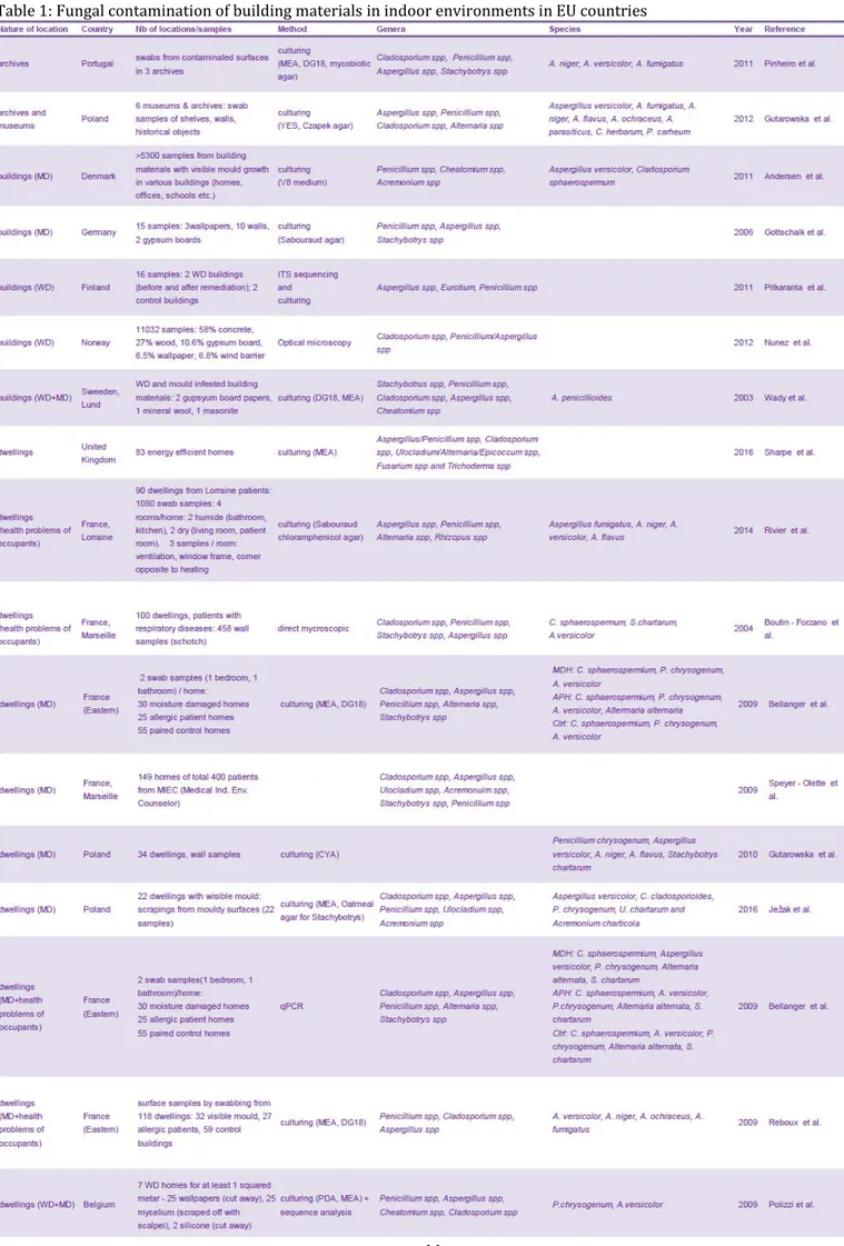
Table 1: Fungal contamination of building materials in indoor environments in EU countries .... 44
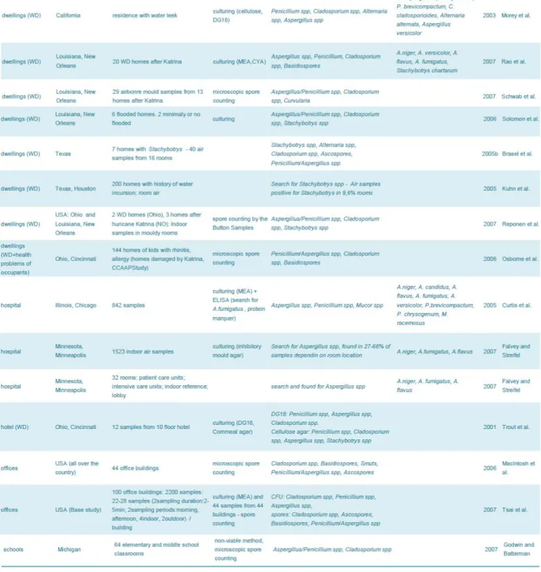
Table 2: Fungal contamination of building materials in indoor environments in USA ... 46
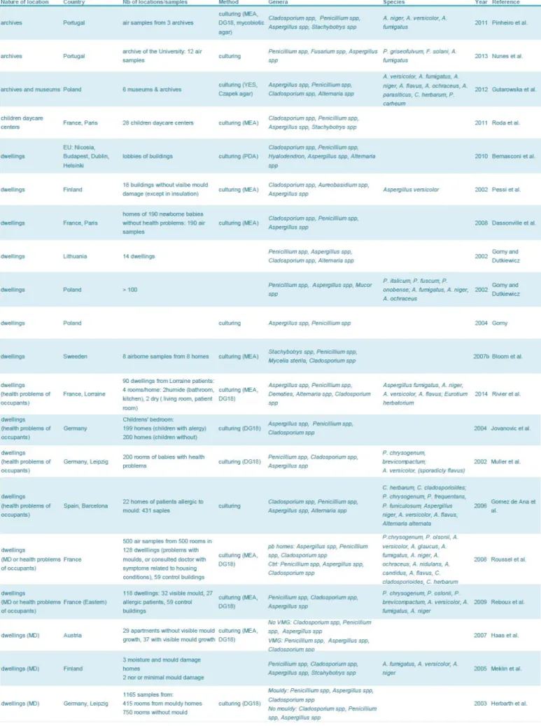
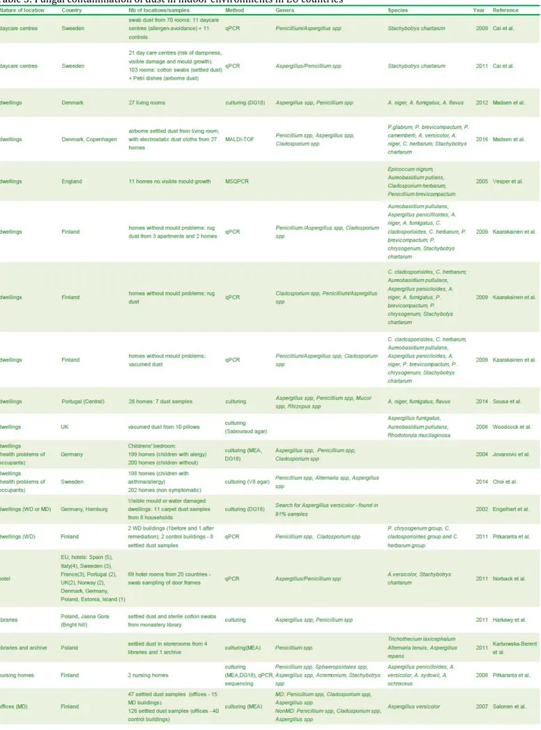
Table 3: Fungal contamination of air in indoor environments in EU countries ... 47
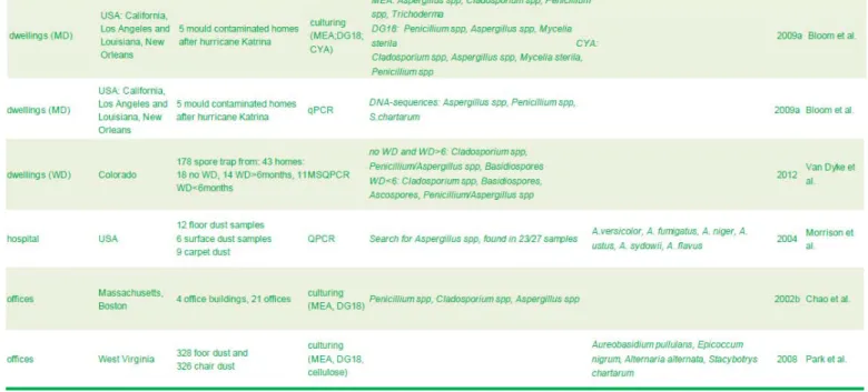
Table 4: Fungal contamination of air in indoor environments in USA ... 51
Table 5: Fungal contamination of dust in indoor environments in EU countries ... 54
Table 6: Fungal contamination of dust in indoor environments in USA ... 56
Table 7: Toxin contamination of building materials in indoor environments in USA and EU countries ... 67
Table 8 : Toxin contamination of air in indoor environments in USA and EU countries ... 68
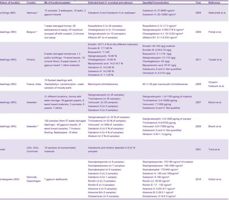
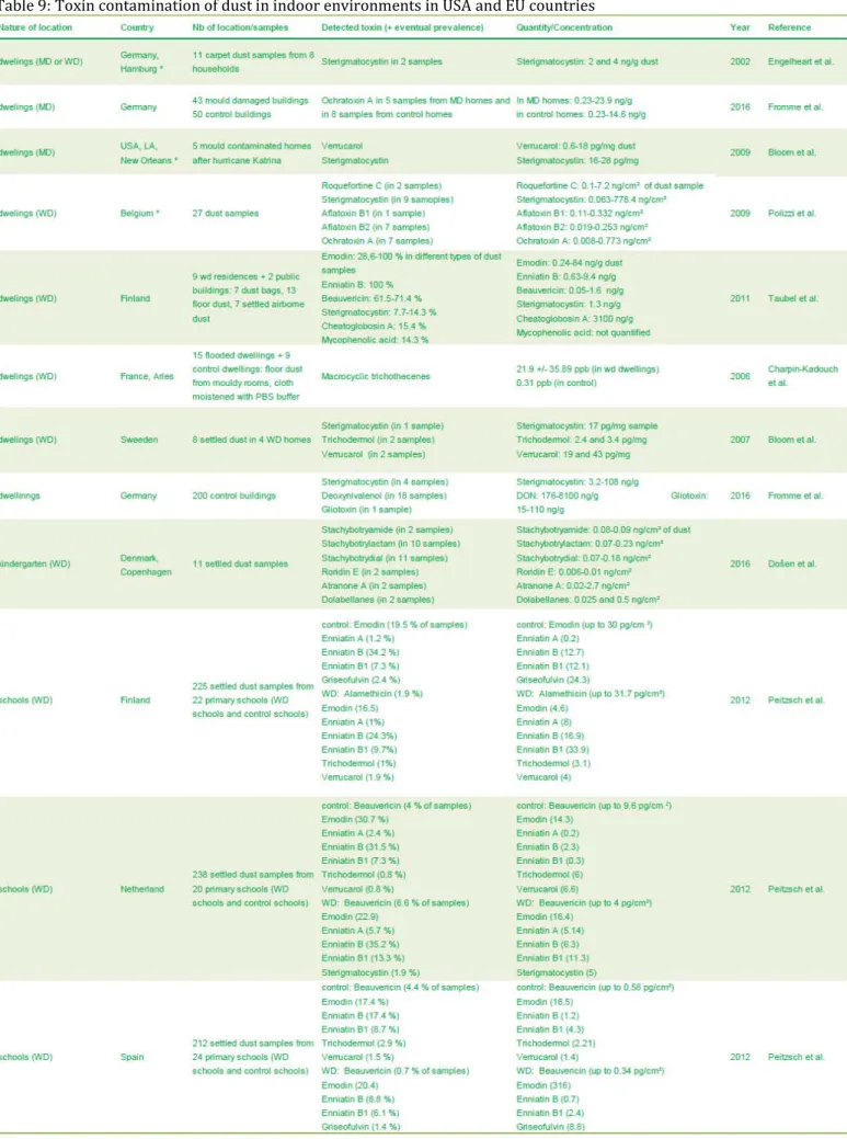
Table 9: Toxin contamination of dust in indoor environments in USA and EU countries ... 69
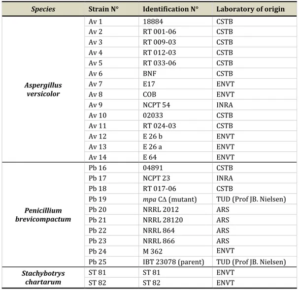
Table 10: Identification and origin of the different fungal strains tested in this study ... 81
Table 11: Sterigmatocystin production by A. versicolor strains NCPT54 (TOX+) and E26a (TOX-) after 10 days of incubation on FG ... 85
Table 12: Toxin production by S. chartarum strains quantified by ELISA ... 88
Table 13: Parameters of the electrospray source in positive mode ESI+ used for ionization of mycotoxins of interest ... 92
Table 14: AcN / H2O gradient used in developed method ... 92
Table 15 : MRM transitions, cone voltages and collision energies used for toxin detection ... 93
Table 16: UPLC-MS/MS analysis of toxins produced by Stachybotrys chartarum ST82 using different solvents for extraction ... 96
Table 17 : Extraction recovery of the pair Internal Standard/Analyte of interest ... 98
Table 18 : Matrix factors observed for STG, O-mSTG, MPA, VerA and RL2 ... 98
Table 19: Comparison of MF between UPLC-MS/MS and TLC method for MPA ... 99
Table 20: Results of mycotoxins calibration standards used to assess linearity ... 99
Table 21: Validation results and chosen internal standards for each mycotoxin of interest ... 100
Table 22: Global extraction recoveries observed for STG, O-mSTG, MPA, VerA and RL2 ... 100
Table 23: Pairs TOX+ and TOX- strains used for VOCs analysis for three species of interest ... 117
Table 24: Characteristics of the analytical chain and analysis parameters ... 120
Table 25: MVOCs emitted during development of P. brevicompactum strains on FG ... 123
Table 26: MVOCs emitted during development of P. brevicompactum strains according to the growth material ... 125
Table 27: MVOCs emitted during development of three P. brevicompactum strains according to the growth material ... 129
Table 28: MVOCs emitted during development of A. versicolor strains on FG ... 134
Table 29: MVOCs emitted during development of S. chartarum strains on FG ... 135
12
List of abbreviations
AcN ANSES
acetonitrile
French agency for food, environmental and occupational health & safety
ANOVA analysis of variance
ATP aw
adenosine thiotriphosphate water activity
CFUs colony forming units
CHCl3 chloroform
CV coefficient of variation
DG18 dichloran glycerol
DMSO dimethyl sulfoxide
DNA deoxyribonucleic acid
ELISA enzyme-linked immunosorbent assay
ERMI environmental relative mouldiness index
ESI electrospray ionization source
ESI+ positive electrospray ionization mode
EtOH ethanol
EU European Union
FG FISH
fiberglass
fluorescence in situ hybridization
FM epifluorescence microscopy
FWP painted fiberglass wallpaper
GC gas chromatography
HPLC high performance liquid chromatographicy
IC50 concentration inhibiting 50 % of cell viability
IS LC
internal standard liquid chromatographicy
LM light microscopy
LOD limit of detection
LOQ
MALDI-TOF MS
limit of quantification
Matrix assisted laser desorption/ionization time of flight mass spectrometry
MCT macrocyclic trichothecenes
MEA malt extract agar
MeOH methanol
MF matrix factor
MPA mycophenolic acid
MPA-d3 mycophenolic acid - d3
MRM multiple reaction monitoring
MS mass spectrometry
MSC microbial safety cabinet
MSQPCR mould specific quantitative PCR
MVOC(s) microbial volatile organic compound(s)
NADP(H) nicotinamide adenine dinucleotide phosphate
13
o-mSTG o-methyl sterigmatocystin
PBS phosphate buffer saline
PCA principal component analysis
PCR polymerase chain reaction
PD petri dish
PDA potato dextrose agar
QC quality control
RH relative humidity
RL2 roridin L2
RSD relative standard deviation
RT retention time
SBS sick building syndrome
SD standard deviation
SEM scanning electron microscopy
SG satratoxin G
SH satratoxin H
STG sterigmatocystin
TLC thin layer chromatography
TOX- non-toxinogenic strain or weakly toxinogenic strain
TOX+ toxinogenic strain
UPLC ultra performance liquid chromatography
USA United States of America
VerA verrucarin A
VerJ verrucarin J
VOC(s) volatile organic compound(s)
VWP vinyl wallpaper
14
Table of contents
List of publications and communications……….……….7
List of figures………..………..…………9
List of tables………..………..………11
List of abbreviation………..……..…12
PART ONE. Bibliographic review………..16
CONTEXT OF THE STUDY ... 17
1.1 MOULD AND MYCOTOXINS IN HABITATS –GENERAL INTRODUCTION ... 18
1.1.1 Generalities on fungal development ... 20
1.1.2 Fungal development in indoor environments ... 22
1.2 BIOMARKERS OF FUNGAL CONTAMINATION ... 26
1.2.1 Environmental sampling techniques ... 27
1.2.2 Methods to characterise fungal population ... 31
1.2.3 Methods to assess global fungal development ... 37
1.3 FUNGAL CONTAMINATION OF INDOOR ENVIRONMENTS ... 43
1.3.1 Comparison of mould contaminations observed in EU and USA - Conclusion ... 57
1.4 HEALTH PROBLEMS RELATED WITH EXPOSURE TO MOULD CONTAMINATIONS ... 59
1.4.1 Types of health problems ... 59
1.4.2 Implication of fungal metabolites on health problems ... 64
AIMS OF THE STUDY ... 71
PART TWO. Experimental work ………..72
1 CHAPTER ONE. FUNGAL DEVELOPMENT AND TOXINOGENESIS AFTER CONTAMINATION OF INDOOR MATERIALS OF INTEREST WITH CHOSEN FUNGAL STRAINS ... 73
1.1 INTRODUCTION ... 74
1.2 SELECTION OF STRAINS OF INTEREST FOR THIS STUDY ... 80
1.2.1 Materials and methods ... 80
1.2.2 Results and discussion ... 84
1.2.3 Conclusion ... 88
1.3 DEVELOPMENT OF AN ANALYTICAL METHOD FOR MYCOTOXINS’ ANALYSIS BY UPLC-MS/MS ... 90
1.3.1 Materials and methods ... 90
1.3.2 Results and discussion ... 95
1.3.3 Conclusion ... 101
1.4 CHARACTERIZATION OF TOXINOGENESIS OF THREE FUNGAL SPECIES AFTER DEVELOPMENT ON DIFFERENT INDOOR MATERIALS OF INTEREST ... 101
1.4.1 Production of four macrocyclic trichothecenes by Stachybotrys chartarum during its development on different building materials as measured by UPLC-MS/MS. 2016. Building and Environment 106, 265– 273 ………101
1.4.2 Supplementary data ... 111
1.4.3 Conclusion ... 114
2 CHAPTER TWO. DEVELOPMENT OF A MONITORING TOOL FOR INDOOR ENVIRONMENTS BY VOCS ANALYSIS RELATED TO MYCOTOXIN PRODUCTION ... 115
2.1 INTRODUCTION ... 116
2.2 MATERIALS AND METHODS –DEVELOPMENT OF METHOD ... 117
2.2.1 Samples ... 117
2.2.2 Experimental assembly and VOC sampling ... 118
2.2.3 Chemical analysis of samples by GC-MS ... 119
2.2.4 Analysis of chromatograms ... 120
2.3 RESULTS ... 123
15
2.3.2 Study of chemical emissions from Pb16 strain (04891) on various materials ... 129
2.3.3 Determination of global footprint for production of mycophenolic acid ... 133
2.3.4 Identification of MVOCs issued from the fungal growth of A. versicolor and S. chartarum ... 134
2.3.5 Determination of global footprint for production of mycotoxins ... 136
2.4 DISCUSSION AND CONCLUSION ... 136
3CHAPTER THREE. AEROSOLIZATION OF MYCOTOXINS FROM CONTAMINATED BUILDING MATERIAL ... 139
3.1 INTRODUCTION ... 140
3.2 DEVELOPMENT OF THE EXPERIMENTAL SYSTEM ... 141
3.2.1 Description of the experimental system ... 141
3.2.2 Characterization of aerosolization conditions ... 144
3.3 AEROSOLIZATION OF MYCOTOXINS AFTER DEVELOPMENT OF TOXINOGENIC FUNGI ON WALLPAPER.BRANKICA ALEKSIC, MARJORIE DRAGHI,SEBASTIEN RITOUX,SYLVIANE BAILLY,MARLENE LACROIX, ISABELLE P.OSWALD,JEAN-DENIS BAILLY,ENRIC ROBINE.SUBMITTED TO INDOOR AIR ... 150
3.4 CONCLUSION ... 15172
4 CHAPTER FOUR. TOXICITY OF STERIGMATOCYSTIN, MYCOPHENOLIC ACID AND MACROCYCLIC TRICHOTHECENES ON HUMAN PULMONARY CELLS ... 173
4.1 INTRODUCTION ... 174
4.2 PRELIMINARY RESULTS: METHODOLOGICAL DEVELOPMENTS ... 175
4.2.1 Choice of method ... 175
4.2.2 Choice of solvent ... 176
4.3 MATERIALS AND METHODS ... 177
4.3.1 Mycotoxins ... 177
4.3.2 Cell lines and culture conditions ... 177
4.3.3 Cytotoxicity assay (MTS test) ... 177
4.3.4 Statistical analysis ... 178
4.4 RESULTS AND DISCUSSION ... 179
4.4.1 Sterigmatocystin ... 179
4.4.2 Mycophenolic acid ... 180
4.4.3 Macrocyclic trichothecenes ... 181
4.4.4 Determination of IC50 values ... 182
4.5 CONCLUSION ... 184
5 CHAPTER FIVE. EVALUATION OF MOST USED PROCEDURE OF DECONTAMINATION IN HOMES ON PERSISTENCE OF FUNGAL CONTAMINATION……….185
5.1 INTRODUCTION ... 186
5.2 MATERIALS AND METHODS ... 188
5.2.1 Fungal strain ... 188 5.2.2 Building materials ... 189 5.2.3 Contamination procedure ... 189 5.2.4 Decontamination procedure... 190 5.2.5 Re-incubation procedure ... 190 5.3 RESULTS ... 191
5.3.1 Examination of fungal development under stereo-microscope and SEM ... 191
5.3.2 Evaluation of fungal development by mycological methods ... 197
5.4 DISCUSSION AND CONCLUSION ... 199
GENERAL CONCLUSION AND PERSPECTIVES ... 201
Annex 1………..………..208
Annex 2………..………..211
Annex 3 ………..……….222
16
PART ONE.
17
Context of the study
With new life style today, people spend most of their time inside the buildings and indoor air quality progressively rose as an important concern for human health. There are many described air pollutants, such as: physical, chemical or microbiological pollutants.
Among these, fungi are frequent contaminants with possible important impact on health. Indeed, some species are well known for their ability to produce toxic secondary metabolites named mycotoxins.
These toxic compounds are frequent food contaminants and therefore, their deleterious effect following ingestion have been widely studied and led to the setup of regulations at international level. By contrast, the possible presence of mycotoxins in case of development of toxinogenic moulds on non-alimentary substrates is less documented. However, the frequent identification of potent toxinogenic species in indoor environments, especially following water damages, rose the question of the possible consequences for inhabitants in case of contact with contaminated materials or if toxins can be aerosolized from support to the air and subsequently inhaled. Our overall objective was to generate data allowing a better risk assessment regarding mycotoxins in indoor environment.
For that, we characterized successively the production of mycotoxins on different building materials, their possible aerosolization and their toxicity on pulmonary cells.
In parallel, we also studied the possibility to use specific volatile organic compounds to identify an active toxinogenesis; as well as the efficacy of a common remediation procedure on the persistence of moulds.
All these results will be presented in distinct chapters following a general introduction on bibliographic data available on moulds and mycotoxins in indoor environments.
18
1.1 Mould and Mycotoxins in habitats – General introduction
Today, in industrialized countries, people spend most of their time inside the buildings (work, school, home, sports etc.). Therefore indoor air quality presents an important aspect of human health. Many air pollutants can contaminate indoor environments such as: physical, chemical or microbiological pollutants.
Fungi are members of a complex community of biological indoor agents, along with bacteria, viruses and protists, such as amoebae; pollen and other plant material; and even arthropods, such as house dust-mites and ants (Nevalainen et al., 2015). Fungi are omnipresent in nature and more than 100 000 species have been identified. They are found in air, water, soil, plants and many other substrates. However, only a few hundred of them are known to cause diseases (Douwes et
al., 2003). Fungi can also be found in indoor environments. They are able to grow on almost all
natural and synthetic materials, especially if water activity is sufficient (Haleem Khan and Mohan Karuppayil, 2012). The minimum requirements for temperature, pH, light, and availability of nutrients are usually present in buildings, and therefore, moisture is the key factor regulating microbial growth.
Several hundreds of fungal species can be found in indoor environments (Gutarowska and Akowska, 2002). Cladosporium, Penicilium, Aspergillus, Alternaria and Stachybotrys are the most found genera in dwellings and other buildings (Verdier et al., 2014).
Fungal development in a building can represent a source of air pollution, more precisely as spores, fragments of hyphae and spores, but also microbial metabolites such as microbial volatile organic compounds (MVOCs) or toxins can be transferred to air following aeraulic solicitations of contaminated supports. Due to its possible health effects, mould contamination of homes is a public health concern.
In a study performed by Hulin et al. (2013), it was estimated that more than 35% of French dwellings were contaminated with moulds. Other available surveys confirm the importance of this problematic. In 2009, the survey on housing conditions in Ile de France, reported that 21% of households surveyed reported traces of moisture on the walls of their homes, humidity and this presented the leading cause of discomfort (IAU, 2009). Similarly, according to ESMHA investigation, an epidemiological study in the Ile-de-France, performed in 2010, 25% of homes surveyed had at least a trace (patch) of dampness or mould presence. Thus, according to the index of contamination build by CSTB, 750 000 housing might be concerned by the presence of visible mould development. It is probably that this number underestimated the reality considering the
19
fact that the most vulnerable households weren’t in condition to be sampled (Host et al., 2010). Moreover, a national campaign by the Observatory of the Quality of Indoor Air revealed that more than 610 000 homes had mouldy surfaces of more than 1 m² (Moularat et al., 2008a). The percentage of contaminated homes revealed to be more important in urban areas, probably due to overpopulation. According Host et al. (2010) in Île-de-France, the presence of visible mould was partially linked to overcrowding. They concluded that 63% of dwellings with visible mould were over-occupied, against 15% of dwellings being unoccupied. The results of Rocchi et al. (2015) confirmed the link between occupancy rate and the presence of mould in homes in France. Nielsen (2002) estimated that proportion of dwellings with mould contamination in Northern Europe and North America varied from 20 to 40% affected buildings. Report for specific countries showed comparable frequency:
- 30-45% in the United Kingdom - 20-25% in Netherlands
- 20-30% in Finland in USA - up to 40% in USA
- up to 30% in Canada.
Mould growth only occurs when water conditions are favourable, therefore mainly in water-damaged and humid constructions. Accordingly the major part of the problems in Scandinavia and North America are due to insufficient aeration (Nielsen, 2002). In the USA about 40% buildings suffer from poor indoor air quality (Gutarowska and Piotrowska, 2007).
Such a high frequency of mould contamination goes with direct impact on health of inhabitants or workers. As an example, studies performed in offices in Canada revealed that 30–69% office workers display symptoms typical for sick building syndrome (Miller et al., 1988). Studies concerning Czech Republic and Romania revealed that more than 30% of all surveyed buildings suffered from health implications of fungal growth. In the USA, Mudarri and Fisk (2007) estimated that more than 4.5 million cases of asthma result from exposure to damp and mould damaged buildings. Such health problems have important social and economic consequences since annual economic cost of asthma was estimated at approximately $3.5 billion in USA (Verdier et al., 2014). It was also estimated that fungi are responsible for over 80% of total building material degradation (Gutarowska and Piotrowska, 2007).
The interest in mould exposure in different indoor environments has increased over the last 20 years. Probably the main reason is that exposures to those biological pollutants in occupational and residential environments are associated with a wide range of adverse health effects with
20
major public health impact, including irritations, inflammations, infectious diseases, acute toxic effects, allergies and other respiratory diseases (inhalation exposure), skin diseases (dermal exposure) and even cancer (Douwes et al., 2003). Moreover in new buildings, increased insulation allowing energy saving, together with poor ventilation and multiplication of steam making apparatus (coffee machine, smoothing iron…) lead to more favourable environment for mould development.
Overall significance of this problem led to publishing guidelines for indoor air quality related to humidity and mould by The World Health Organisation (World Health Organization, 2009). To further describe the problems related to mould contamination of indoor environment, we will now present parameters influencing their development, markers that can be used to monitor their presence, available data on indoor contamination with a special focus of the situation in Europe, compared to USA and finally, we will describe their possible deleterious effects on health of inhabitants.
1.1.1 Generalities on fungal development
A fungus is any member of the group of eukaryotic organisms that includes microorganisms such as yeasts and moulds, as well as macroscopic fungi known as mushrooms. These organisms are classified in a kingdom Fungi which belongs to the domain Eukarya (Cahagnier, 1998).
In this work the terms “mould” or “fungi” will be related to microscopic fungi. They belong to the division Eumycota (Silar and Malagnac, 2013). Fungi are not capable of photosynthesis; they are heterotrophic and use complex organic compounds as sources of energy and carbon (Boullard, 1997). For this, they degrade complex organic matter thanks to the excretion of enzymes and acids in the external medium and they absorb the digested components, all this taking place through the permeable wall of their vegetative system (Silar, 2016). Like bacteria, fungi play an essential role in ecosystems because they are decomposers and participate in the cycling of nutrients by breaking down organic materials to simple molecules. Fungi are most often saprophytic and develop on the inert organic material. However, some can also act as parasites and develop on the living organisms (Fusarium in cereal plants or Aspergillus fumigatus in animals). Some others are symbiotic and live together in mutual benefit with other organisms (Neotyphodium in fescue). “Perfect fungi” reproduce both sexually and asexually, while imperfect fungi reproduce only asexually (by mitosis) (Figure 1 (Casselton and Zolan, 2002) and Figure 2 (Samiksha, 2016)). In both sexual and asexual reproduction, fungi produce spores that disperse from the parent
21
organism by either floating on the wind or transported by humans, animals or insects. The most common mode of asexual reproduction is through the formation of asexual spores. Spores allow fungi to expand their distribution and colonize new environments. They may be released from the parent thallus either outside or within a special reproductive “bag” called a sporangium.
Figure 1: Life cycle of fungi (asexual and sexual reproduction of Aspergillus nidulans)
The germination will be triggered by the combined presence of water, light intensity, temperature or the types of nutrients available on substrate. In a general matter, fungi develop more easily on substrates with high carbohydrate/proteins ratio. However, their enzymatic abilities make them able to colonize many substrates, with various compositions. The spores will germinate and then give birth to a first undifferentiated filament, called hyphae, which will lengthen to form a group called mycelium. This set of filaments, more or less branched constitutes the thallus of fungus. In the presence of favourable conditions for sporulation, the mycelium will generate more specialized structures that will produce asexual spores (conidia) or, more rarely, sexual spores. Each mould produces a great number of conidia that are dissemination and resistance structures. The size, shape and colour of conidia vary greatly from one species to another. These elements are one of the parameters used for morphologic identification (Cahagnier, 1998; Pitt and Hocking, 2009; Samson et al., 2010).
22
Figure 2: Life cycle of fungi (asexual and sexual reproduction of Penicillium)
1.1.2 Fungal development in indoor environments
Broad spectre of fungal species is found in outdoor environments and can be transferred to indoor by people movements, animals, insects or airflows. There is no sterile indoor space (except for special facilities such as surgical block) and the presence of biological contaminants at low concentrations is normal. The contamination problem occurs when concentrations of biological agent(s) arise or increase above certain level.
Probably the first reference about the destructive influence of fungal flora on human dwellings and clothes is found in the 3rd Book of the Bible, Leviticus (Bible, n.d.). Prehistoric times (confirmed by the analysis of rock paintings from Paleolithic caves), archaeological investigations and conservation studies revealed that the destruction of inorganic and organic materials was mainly connected with fungal and actinomycetal biodeterioration activities (Górny, 2004). Colonization abilities of microorganisms present in indoor environment are determined by the physical and chemical characteristics of building materials. The main factors influencing mould
23
colonization of indoor environments are moisture, available nutrients and temperature (Haleem Khan and Mohan Karuppayil, 2012). This latter parameter, usually ranging from 18–25°C, promotes the growth of mesophilic fungi and can’t be used to control microorganisms’ development. .
1.1.2.1 Humidity as a major factor for mould growth
Among the most important factors, the moisture content in the substrate, together with the nutritional substances derived from building materials, initiate the development of microbial colonies. Therefore, the growth of microorganisms depends to the highest degree on the availability of water. Availability of water is usually expressed in terms of water activity (aw). It
corresponds to the ratio of the water vapour pressure exerted by the material to the vapour pressure of pure water at the same temperature, i.e. 1/100 of the equilibrium relative humidity (ERH) for a defined temperature. It is important to note that water availability and temperature are interdependent and, for example, increasing temperature has been found to lead to a reduction in the aw requirement of moulds (Grant et al., 1989). Several studies have shown that
aw equal to 0.65 is the lowest value necessary to initiate the microbial growth on the material
when enough nutrition substances are available (Górny, 2004). Microbial activity and ability to colonize new surfaces increase as the water activity approaches 1, i.e., when water is freely available. Based on aw parameter, fungal and actinomycetal microorganisms can be categorized
according to their ability to initiate the growth on building materials and the order in which they appear on material’s surface as primary, secondary, and tertiary colonizers. Combined classification for several fungal and actinomycetal species is summarized by Górny (2004): - Primary colonizers (aw<0.85): Alternaria citri, Eurotium amstelodami, E. repens, Aspergillus
candidus, A. niger, A. penicillioides, A. restrictus, A. versicolor, Paecilomyces variotii, Penicillium aurantiogriseum, P. brevicompactum, P. chrysogenum, P. commune, P. expansum, P. griseofulvum, Wallemia sebi;
- Secondary colonizers (aw=0.85-0.90): Aspergillus flavus, Cladosporium cladosporioides, C.
herbarum, C. sphaerospermum, Mucor circinelloides, Rhizopus oryzae;
- Tertiary colonizers (aw>0.90): Alternaria alternata, Aspergillus fumigatus, Epicoccum spp.,
Exophiala spp., Fusarium moniliforme, Mucor plumbeus, Phoma herbarum, Phialophora spp., Rhizopus spp., Stachybotrys chartarum, Trichoderma spp., Ulocladium consortiale, Rhodotorula spp., Sporobolomyces spp., Actinomycetes.
24
Microbial investigations on building materials can be also expressed in terms of relative humidity (RH). It appears that building materials become sensitive to microbial colonization when the RH reaches 70% for wooden materials, 85% for gypsum-board and around 90-95% for cementitious and concrete materials. Study conducted by Johansson et al. (2013) provided different ranges of critical RH according to the nature of material. They also highlighted the influence of the temperature, incubation time and criteria used for mould growth assessment on the results of such testing. Adan (1994) showed a significant increase in the development of P. chrysogenum during testing on gypsum substrates when changing RH from 86 to 97%. Several authors highlighted that increased humidity favours the germination, the proliferation, and the diversity of mould on building materials. Several studies also showed that RH measurements could be used as a microbial contamination indicator for construction materials in water-damaged buildings. Pasanen et al. (2000) stated that RH of a material describes the water availability for microorganisms better than the moisture content does. Some authors have even developed mathematical models for predicting contamination by moulds, which use RH as a major parameter (Chase et al., 2016; Lugauskas et al., 2003; Thelandersson and Isaksson, 2013).
Some major characteristics of most building materials are high porosity and surface roughness. Thanks to the high porosity, materials have particular abilities for water absorption. When the environment provides high moisture levels, porous materials become supplies of water for microorganisms and offer them a larger growth subsurface (Hoang et al., 2010). In addition, surface roughness and porosity could favour the attachment of nutrients carried by dust and resulting from the activity in buildings (Verdier et al., 2014).
Water damage of homes can occur due to flooding or storm damage, leaks in plumbing, damage of air conditioners, dehumidifiers, humidifiers; as well as because of ice damming on building roofs (Haleem Khan and Mohan Karuppayil, 2012). Each time when the water appears on the surfaces of construction materials or penetrates them through holes and cavities, it can provoke microbial contamination. American investigations reveal that 27-56% of homes have problems with visible fungal contamination of surfaces, and/or bad quality of indoor air. In Europe, this percentage ranges from 12–80% (summarized in Górny, 2004). Microbial contamination of buildings is very often connected with environmental disasters. One of the latest examples is Katrina hurricane in the USA in 2005 (Chew et al., 2006). In those cases, long terms effects provoke serious health outcomes, influencing mostly the families whose dwellings had not been rebuilt, drained or protected against moulds.
25
1.1.2.2 Impact of material composition and surface biodeterioration
Concerning the nutrition requirements, moulds are very elastic and have several adaptation possibilities. They are able to grow on almost all natural and synthetic materials, especially if they are hygroscopic or wet. These microorganisms reach basic nutrients, rich in carbon and nitrogen, thanks to the decomposition of organic materials. The majority of fungi present in indoor environment are saprophytic, which means that they gather nutrients from dead materials such as: wood, paper, paints, glues, soil, dust, food chips, etc. Moreover, they are able to frequently colonize surfaces consisting of inorganic moist material (glass, fibreglass, metal or concrete) if they are covered with dust, air contaminants or even finger-marks, creating invisible layer of biofilm (Górny, 2004; Samet and Spengler, 2003). Nevertheless, the works of Hoang et al. (2010) and Gutarowska (2010) indicate that cellulose-based materials are more sensitive to contamination than inorganic materials (gypsum, mortar, concrete, etc.) because cellulose can be metabolised by some fungi (Gutarowska, 2010; Hoang et al., 2010).
Wood is highly vulnerable to fungal contamination. Cladosporium and Penicillium (Penicillium
brevicompactum and Penicillium expansum) are reported to infest wooden building materials. Kiln
dried wood surfaces are more susceptible to fungi (Sailer et al., 2010). Acetylated wooden furniture, wood polyethylene composites, plywood and modified wood products are susceptible to infestation by Aspergillus, Trichoderma and Penicillium (Doherty et al., 2011; Thacker, 2004). Inner wall materials used in buildings, such as gypsum board which is hygroscopic, highly favours the growth of Stachybotrys chartarum (Haleem Khan and Mohan Karuppayil, 2012). Also, paper and glue used for indoor surfaces are very good substrates for growth of most of indoor fungi. Fiberglass insulation and ceiling tiles support the growth of a number of fungi, among them A.
versicolor, Alternaria, Cladosporium, and Penicillium species were frequently isolated (Erkara et al., 2008). Aspergillus and Penicillium grow superficially on painted surfaces. Acrylic painted
surfaces can be attacked by Alternaria, Cladosporium, and Aspergillus (Shirakawa et al., 2011). Air filters and ventilation ducts can also be colonized by fungi (Noris et al., 2011).
The physiological, biochemical and morphological properties of fungi and their frequent presence make them the most important cause of biodegradation of building materials. While growing on material, fungi produce various substrate-specific enzymes that destroy and/or disintegrate organic materials. Bio-corrosion occurs due to the microbial excretion of organic and inorganic acids. They include a variety of acids as oxalic, citric, gluconic and others formed during respiration. Moreover, biogenic organic acids produced by moulds are considered as one of major damaging agents leading to biodeterioration of stone, rocks, minerals (Gutarowska and Piotrowska, 2007). Such degradation process of homes has major economic importance. Only in
26
Germany the costs related to mould damages in buildings are estimated to more than 200 million Euro per year (Sedlbauer, 2001).
Among fungi, there are many organisms with strong cellulolytic (e.g., Trichoderma, Botrytis,
Chaetomium, Alternaria, Stemphylium), proteolytic (e.g., Mucor, Chaetomium, Aureobasidium, Gymnoascus, Trichoderma, Verticillium and Epicoccum), and lipolytic properties (e.g., fungi from
the previous group plus Paecilomyces) (Górny, 2004).
pH range of 5–6.5 observed on most building materials allows the growth of most of the fungi (Hoang et al., 2010; Vacher et al., 2010). Cementitious materials, which are alkaline (pH around 12 - 13) are relatively insensitive to colonisation at early ages. However, over time, the carbonation process reduces the pH of these materials to values around 9, which allows fungal growth. These materials thus become the target of significant contamination (Verdier et al., 2014). Sufficient light and oxygen are also critical for the growth of fungi in indoor environments (Airaksinen et al., 2004; Haleem Khan and Mohan Karuppayil, 2012; Voisey, 2010).
Degraded building materials then represent sources of small particles easily aerosolized to indoor air, with numerous adsorbed substances, some of them being biologically active.
1.2 Biomarkers of fungal contamination
Evaluation of fungal contamination in indoor air samples to estimate health risks for occupants has been widely reported (Pasanen, 2001; Portnoy et al., 2004). Indeed, the identification of a direct relationship between moulds’ exposure and health problems in habitants requires a precise measurement of fungal colonization of indoor environment and several techniques can be used to estimate fungal presence.
Culture-based methods are the traditional approach in mycology, but other methods such as chemical, immunological and PCR-based methods are now also frequently used for examination of indoor environments. All these techniques have advantages and limits, so it seems that combining them should probably allow producing wider picture about fungal flora in indoors (Niemeier et al., 2006; Pitkäranta et al., 2008; Reboux et al., 2009). Méheust et al. (2014) proposed schema below (Figure 3) to summarize methods to detect fungi in indoor environments.
27
Figure 3: Methods to detect fungi in indoor environments
MVOCs - microbial volatile organic compounds. GC-MS - gas chromatography/mass spectrometry. LC-MS - liquid chromatography/mass spectrometry. ELISA – enzyme-linked immunosorbent assay
A presentation of the possible interest of these different methods will be done in following part of this manuscript. However, the first and major step in estimating the presence of indoor moulds is sampling.
1.2.1 Environmental sampling techniques
The consequences of exposure to fungal particles on health are related to several parameters, such as species of microorganisms, exposure pathway (inhalation or contact with skin/eyes) and environmental conditions, quantitative importance of microbial growth, aerosolization of contaminants, etc. (Verdier et al., 2014). Many authors highlighted that air sampling is not sufficient to describe the entire microflora present inside buildings, especially in water-damaged buildings (Andersen et al., 2011; Lappalainen et al., 2001). Identifying microbial flora on building materials by surface sampling, has been shown to provide additional information about the potential sources of airborne microbial contaminants (Verdier et al., 2014). In addition, species producing mucilaginous spores, that remain attached to substrates, require the use of surface sampling methods to determine full microbial biodiversity. Although microbial contamination present on surfaces is not directly correlated with health troubles of the occupants, French Agency for Food, Environmental and Occupational Health & Safety recommends sampling such communities on building materials, in addition to air sampling, in order to evaluate microbial proliferation indoors (ANSES, 2016).
28
1.2.1.1 Surface sampling
Surface sampling is used to determine nature of the microbial presence on environmental surfaces to measure the relative degree of contamination and identify the types of present fungi (Cabral, 2010). It can also assess the effectiveness of remediation and clean-up of indoor environments ( Méheust et al., 2014). The samples provide the hyphae fragments and the reproductive structures, spores, which may help for identification (Haleem Khan and Mohan Karuppayil, 2012). Different methods exist for sampling microbial populations on materials: swab, bulk, adhesive, contact plate, etc. (Verdier et al., 2014), but the collecting process has not been well standardised yet, which makes it very difficult to compare results (Méheust et al., 2014). Moreover, although many of these methods have been tested to evaluate their collecting efficiency on porous and non-absorbent surfaces (glass, steel, plastic, etc.), few studies have concerned construction materials such as concrete, coatings, mortar, and gypsum board, which are porous, rough and more or less dusty materials. The “Mould in the home” working group of the French Agency for Food, Environmental and Occupational Health & Safety has issued methodological recommendations for sampling on surfaces of building materials and suggests the use of at least two of the following surface sampling methods: swab, bulk sampling, adhesive tape and agar contact (imprint methods)(ANSES, 2016). Swab, adhesive and contact plate sampling, along with bulk sampling, are techniques commonly used on the surface of building materials to collect microorganisms and microbial contaminants prior to analysis.
Many authors emphasise the need for standardisation of the protocols for microorganism sampling on construction materials (ANSES, 2016; Bellanger et al., 2009; Brown et al., 2007; Hyvärinen et al., 2002; Santucci et al., 2007). At present, results can be influenced by the operator and many other factors, including the sampling technique itself and its different steps (sampling location, size of sampled surface, pressure applied, conservation of strains, etc.), the analysis method (observation, chemical, molecular, etc.) and/or the chosen culture medium. Moreover, the number of microorganisms collected from a surface is likely to depend on the species and the stage reached in the adhesion and biofilm formation process. This aspect has also been little studied to date (Verdier et al., 2014).
1.2.1.2 Air sampling
Since moulds are associated with various respiratory disorders, air sampling is usually performed in indoor environments. This way of sampling is useful for determining whether the air in homes or workplaces is normal or not in the sense of microbiological composition. Air samples are also taken in hospitals as part of surveillance programs, especially in facilities where fungal presence
29
can have direct and important health consequences (surgical blocks, areas with immune-compromised patients…) (Méheust et al., 2014).
The concentration of spores in the indoor air is a reliable indicator of the air quality and, indirectly, of some of the health hazards. Assessment of mould contamination of indoor air involves estimation of total number of fungi, classification of mould species and their relative proportion (Gutarowska and Piotrowska, 2007).
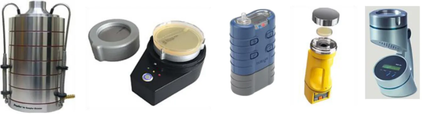
Air sampling for fungi can be done by three standard methods including: impactor, liquid impinger, and air filtration methods (Godish and Godish, 2007).
In the impactor method, the air stream passes through a slit into a culture medium and adhesive microscopic slide or tape strip is used to collect the sample (Zhen et al., 2009). The most common impactor samplers (Figure 4) that are used are: Single stage impactor, Andersen, multistage impactor, Burkard, slit samplers, rotorod, casella, SAS, sierra marple impactor and centrifugal samplers. The air flow rate can be from 2 to 180 L/min (Engelhart et al., 2007).
Figure 4: Andersen multistage; Slit-to-agar; Casella, SAS, Centrifugal sampler (from left to right)
The use of impingers is a flexible method for producing samples for a range of laboratory techniques (Méheust et al., 2014). Liquid impingers collect the samples directly into the fluid and the microorganisms are retained in the liquid until they are cultivated on media or evaluated by techniques like biochemical or immunoassays (Haleem Khan and Mohan Karuppayil, 2012). The most common impinger devices that are used are: multistage, AGT– 30, shipe sampler, midget, and micro impingers. The airflow rate can be from 0.1 to 55 L/min and the sampling time varies from minutes to hours. Centrifugal samplers such as Reuter centrifugal sampler, aerojet cyclone are devices with 40–100 L/min air flow rate (Gralton et al., 2011).
The use of high- volume centrifugal samplers is increasing since the air flow rate is often greater than 300 L/min. Collected solution is used for the cultivation of microbes or examination with analytical techniques. Filtration methods allow long-term measurements. Sampling volumes are
30
usually adapted to measure the fungal concentration. The design of the air samplers is known to influence their efficiencies for measuring airborne microbial concentrations (Yao and Mainelis, 2007). In air filtration sampling most common filters that are used are: glass, polycarbonate cellulose ester and Teflon filters. The air flow rate for this sampling is 1–1000 L/min (Haleem Khan and Mohan Karuppayil, 2012).
1.2.1.3 Dust sampling
Dust is analysed to evaluate presence of fungi or fungal agents that have accumulated with time. This method provides an indication of the microbial agents that may have been airborne. The term ‘‘settled dust’’ is usually used to describe the particulate matter that collects on horizontal surfaces, primarily floors. Over a defined time period, vacuum cleaners or suction devices are used to collect dust from a given surface of carpets or hard floors in homes (usually 1–2m2). This
method is commonly performed following moisture damage and/or health complaints in workplace investigations. Furthermore, efficiency of the air treatment in hospitals can be practically indicated by analysing dust in ventilation ducts. One of the main advantages of dust sampling is that this matrix may be analysed by different techniques. Nevertheless, variables such as the type of carpet, vacuum cleaner capture velocity and relative humidity can affect how well dust is removed from the floor (Macher, 2001). Moreover, the inhalable fraction of the dust and the length of the dust accumulation are unknown. Passive airborne dust collection methods, such as electrostatic dust fall collectors, may thus be a low-cost means of assessing long-term fungal exposure in standardizing the time and the surface of dust accumulation (Frankel et al., 2012; Madsen et al., 2012; Normand et al., 2011).
As conclusion, sampling strategies is of major importance since can influence the nature of information given by analysis. The method used may depend on the type of sampled environment. Surface and air sampling are generally used in hospitals to confirm that the care environment is safe, especially in Europe. They are also useful in houses to quantify the exposure and evaluate the biodiversity. Dust is more often sampled in homes because it is an indicator of the past fungal exposure. Even if methods differ slightly depending on the level of fungal contamination, air sampling is the most commonly used technique in indoor environments, as it provides a better characterization of the airway exposure (Méheust et al., 2014).