Universite de Sherbrooke
Effects of DP and CRTH2 activation on osteoblast function
Mihai Nedelcescu
Departement de Pharmacologic
Memoire presente a la Faculte de Medecine
en vue de l'obtention du grade de
Maitre en Science (M.Sc.) en Pharmacologic, 2010
Jury: Artur Fernandes - Medecine Guylain Boulay - Pharmacologic Guillaume Grenier - FMSS © Mihai Nedelcescu 2011
Library and Archives Canada Published Heritage Branch Biblioth&que et Archives Canada Direction du Patrimoine de l'6dition 395 Wellington Street Ottawa ON K1A 0N4 Canada 395, rue Wellington Ottawa ON K1A 0N4 Canada
Your file Votre r6f6rence ISBN: 978-0-494-83726-9 Our file Notre r6f6rence ISBN: 978-0-494-83726-9
NOTICE:
The author has granted a
non-exclusive license allowing Library and Archives Canada to reproduce, publish, archive, preserve, conserve, communicate to the public by
telecommunication or on the Internet, loan, distrbute and sell theses
worldwide, for commercial or non-commercial purposes, in microform, paper, electronic and/or any other formats.
AVIS:
L'auteur a accord§ une licence non exclusive permettant £ la Biblioth§que et Archives Canada de reproduire, publier, archiver, sauvegarder, conserver, transmettre au public par telecommunication ou par I'lnternet, preter, distribuer et vendre des theses partout dans le monde, £ des fins commercials ou autres, sur support microforme, papier, 6lectronique et/ou autres formats.
The author retains copyright ownership and moral rights in this thesis. Neither the thesis nor substantial extracts from it may be printed or otherwise reproduced without the author's permission.
L'auteur conserve la propriety du droit d'auteur et des droits moraux qui protege cette these. Ni la th6se ni des extraits substantiels de celle-ci ne doivent etre imprimis ou autrement
reproduits sans son autorisation.
In compliance with the Canadian Privacy Act some supporting forms may have been removed from this thesis.
While these forms may be included in the document page count, their removal does not represent any loss of content from the thesis.
Conform&ment d la loi canadienne sur la protection de la vie privee, quelques formulaires secondares ont ete enleves de cette these.
Bien que ces formulaires aient inclus dans la pagination, il n'y aura aucun contenu manquant.
Resume
"Les effets de l'activation des recepteurs DP and CRTH2 sur la
fonc-tion des osteoblastes"
Par
Mihai Nedelcescu
Departement de Pharmacologic
Memoire presente a la Faculte de medecine et des sciences de la sante en vue de l'obtention du grade de Maitre en science (M.Sc.) en Pharmacologic, 2010, Universite
de Sherbrooke, Sherbrooke, Quebec, Canada, J1H 5N4.
La modulation de PG par l'inhibition ou la stimulation de leur production peut etre un facteur a considerer dans la gestion des differentes conditions pathologiques osseusses. Sur la base de resultats recents de nos laboratoires ainsi que sur la litera-ture, nous emettons l'hypothese que la prostaglandine D2 (PGD2) est un important agent anabolisant pour les osteoblastes. Nos resultats montrent que la PGD2 diminue la proliferation des osteoblastes agissant probablement par 1'intermediaire du recep-teur CRTH2. Curieusement, alors que le DK-PGD2 (agoniste specifique CRTH2) a ete utilise seul ou avec le Naproxene, bien que la proliferation diminue avec la dose, l'effet semblait etre restaure au niveau de controle avec les concentrations plus elevees de DK-PGD2. Ainsi, nous envisageons l'hypothese de l'existence d'autres mecanismes de compensation. La PGD2 n'a pas eu aucun effet utilisee seule ou lorsqu'elle est utilisee avec le Naproxene, mais semblait reduire la differenciation osteoblastique lorsqu'elle est utilisee avec Diclofenac a une concentration plus elevee seulement. Lorsque la vi-tamine D a ete ajoute a toutes les conditions, PGD2 a eu un effet inhibiteur sur la differenciation (dose-reponse). Lors d'un essai de competition avec PGD2 et des an-tagonistes DP/CRTH2, le blocage des recepteurs DP n'a pas donne aucun effet, et en bloquant le recepteur CRTH2 on a observe une diminution signifiante a une forte concentration de PGD2. L'effet est semblable au test fait avec PGD2 et antagoniste PPARy ce qui suggere que celui-ci pourrait avoir un role compensatoire qui a renverse l'activation du DP. La PGD2 a un effet legerement positif sur la mineralisation des osteoblastes, mais pas par le biais de ses recepteurs. Lorsque nous avons utilise la PGD2, en presence d'un antagoniste PPARy, la calcification diminue de maniere signi-ficative, indiquant que l'effet positif de la PGD2 sur la calcification fonctionne plutot a travers ce recepteur.
Abstract
"Effects of DP and CRTH2 activation on osteoblast function"
Modulation of PGs by inhibition or stimulation is a promising approach for the management of pain and inflammation in patients with rheumatic disease. Based on recent results from our laboratories as well as on the literature, we hypothesise that Prostaglandin D2 (PGD2) is an important anabolic agent for osteoblasts. Our re-sults show that the PGD2 decreases the osteoblasts proliferation acting probably through the CRTH2 receptor. Surprisingly, when DK-PGD2 was used alone or with Naproxen, although the proliferation decreased with the dose, it seemed to be re-stored to the control level at higher concentrations of DK-PGD2. Thus, we hypothe-sise the existence of other compensatory mechanisms. The PGD2 had no relevant ef-fect alone or when used with Naproxen, but seemed to decrease the osteoblast differ-entiation when used with Diclofenac at a higher concentration only. When vitamin D was added to all conditions, PGD2 had an inhibitoiy effect on the differentiation (dose-response), but this could not be replicated when Naproxen was used. In a test with Diclofenac, we can assume a decreasing trend-line for differentiation when aug-menting the PGD2 dose, but the effect is not statistically relevant. In a competition test with PGD2 and DP/CRTH2 antagonists, blocking DP receptor yielded no effect on differentiation, and blocking the CRTH2 receptor showed a relevant decrease at high concentration of PGD2. The effect was similar in a test with PGD2 and PPARy
an-tagonist suggesting that it might have a compensatory, positive effect that reversed
DP activation. The PGD2 has a slight positive effect on the osteoblast matrix
minerali-sation (with Naproxen), but not through its receptors since use of DP/CRTH2
an-tagonists did not abrogate this. In a competition test with PGD2 and DP/CRTH2
an-tagonists we had no response. When we used the PGD2 in the presence of PPARy
an-tagonist, the calcification decreased significantly, indicating that the positive effect of PGD2 on calcification works rather through this receptor.
Table of contents
List of figures VI
List of Tables VIII
List of terms and abbreviations IX
Resume XI
Abstract Xll
I. Introduction i
1.1. Bone structure and architecture 2
1.1.1. Macroscopical structure 2
1.1.2. Microscopic structure 3
L1.3. Bone matrix and minerals 4
A. Collagen 5
B. Non-collagenous proteins 5
C. Bone Gla-containing proteins (BGP) - osteocalcin 5
D. Sialoproteins 6 E. Alkaline phosphatases 6 F. Bone minerals 7 L1.4. Bone cells 7 A. Osteoblasts 7 B. Osteoclasts 9 C. Osteocytes 11 1.2. Bone modelling 11
1.3. Regulation of bone remodelling 13
L3.1. Factors acting on osteoblasts and osteoclasts 13
Ill
I.4.1 Prostaglandin Synthesis 18
1.4.2. Synthesis enzymes: PGHS 19
1.4.3. Prostaglandins - general effects and roles. 23
1.4.4. Prostaglandin receptors. 25
A. Tromboxane, TP receptors 27
B. PGF2CX and FP receptors 28
C. PGE2 and EP receptors 29
D. PGI2 receptors 30
E. PGD2 receptors: DP and CRTH2. PPARy. 31
F. PGD2 - Generalities 33
1.5. NSAlD's 34
1.6. Prostaglandins and bone 36
1.7. Bone pathology 38
L7.1. Osteoporosis 39
1.7.2. Rickets and Osteomalacia 40
1.7.3. Osteopetrosis 41 1.7.4. Hypophosphatasia 41 1.7.5. Paget's Disease 42 1.7.6. Rheumatoid arthritis 42 1.7.7. Cancers 43 Rationale 45 Objectives 46
11. Materials and methods 47
11.1. Materials 47
IV
11.3- Cell proliferation 48
11.4. Measurement of alkaline phosphatase activity 48
11.5. Calcium measurements 49
11.6. Data analysis 50
111. Results 51
111.1. Proliferation of the cultured human osteoblastic cells. 51
111.1.1. Time-course proliferation assay 51
111.1.2. Overall effect of exogenous PGD2 / agonists on proliferation 52 111.1.2. The effect of different concentrations of exogenous PGD2 on hOB
proliferation 54
111.1.3. The effect of different concentrations of BW245C (DP specific
recep-tor agonist) on hOB proliferation 57
111.1.4. The effect of different concentrations of DK-PGD2 (CRTH2 specific
agonist) on hOB proliferation 60
111.2. Differentiation of the cultured human osteoblastic cells 63 111.2.1. Effect of different concentrations of exogenous PGD2 on the
differ-entiation of human osteoblasts 63
111.2.2. Effect of different concentrations of exogenous PGD2 on the differ-entiation of human osteoblasts in presence of DP-receptor and
CRTH2-receptor antagonists 66
Hl.2.3. Effect of different concentrations of exogenous PGD2 on the differ-entiation of human osteoblasts in presence of T0070907, a selective
antagonist of the human PPARy nuclear receptor 68
Ul.2.4. Effect of different concentrations of exogenous PGD2 on the differ-entiation of human osteoblasts in the presence of Vitamin D 69 111.3. Matrix mineralisation (calcification) from the cultured human osteoblastic
cells 73
Hl.3.1. Effect of different concentrations of exogenous PGD2 on the calcifi-cation of human osteoblasts. Silver staining von Kossa method. 74
V
111.3-2- Effect of exogenous PGD2 in presence of DP-receptor and CRTH2-receptor antagonists on the calcification of human osteoblasts. Silver
staining von Kossa method. 75
III.3.3. Effect of different concentrations of exogenous PGD2 on the calcifi-cation of human osteoblasts. QuantiChrom™ colorimetric assay. 76 Hl.3.4. Effect of exogenous PGD2 in presence of DP-receptor and
CRTH2-receptor antagonists on the calcification of human osteoblasts.
Quan-tiChrom™ colorimetric assay. 77
Hl.3.5. Effect of different concentrations of exogenous PGD2 on the cal-cium production of human osteoblasts in presence of T0070907, a se-lective antagonist of the human PPARy nuclear receptor.
Quanti-Chrom™ colorimetric assay. 78
IV. Discussion 80
lV.i. PGD2 81
1V.2. Primary culture of osteoblasts 82
Vl.3. Osteoblast proliferation 83 IV.4. Differentiation 86 Vl.5. Matrix mineralisation 89 V. Conclusions 91 Perspectives 93 Acknowledgments 94 References 95
List of figures
Fig. 1: Bone macroscopical structure 3
Fig. 2: Bone cells 8
Fig. 3: Differentiation and Activation of osteoclasts. 10
Fig. 4: Osteoblasts action 14
Fig. 5: Biosynthesis of eicosanoids starting from polyunsaturated fatty acids 19 Fig. 6: Conversion of free arachidonic acid to prostaglandins and other eicosanoids is
initi-ated by oxidative enzymes of the cyclooxygenase 20
Fig. 7: The lipoxygenase pathway. 21
Fig. 8: Prostaglandin synthesis and actions in a generic cell. 24
Fig. 9: TxA2-induced signaling via TP. 27
Fig. 10: PGFza-induced signaling via FP. 28
Fig. 11: Activation of distinct EPs by PGE2 induces several signaling pathways and cytokine
release 29
Fig. 12: Binding ofPGh to IP induces cell-specific signaling 30
Fig. 13: PGD2 and PGD2 metabolite-induced signaling. 31
Fig. 14: CRTH2 induced signaling 32
Fig. 15: Possible effects of dietary lipids on the bone 37
Fig. 16: "Fatty bone" - Femoral head presenting rough (destroyed) articular surface and
large, spongiform trabeculae filled with fat. 40
Fig. 17: Fracture on pathological bone 43
Fig. 18: Effect of the PGD2, BW245C (DP specific receptor agonist) and DK-PGD2 (CRTH2 specific agonist) on human osteoblast cell proliferation determined by incorporation of
[.3H]thymidine (2 [iCi/ml) - time course 53
Fig. 19: Effect of the PGD2, BW245C (DP specific receptor agonist) and DK-PGD2 (CRTH2 specific agonist) on human osteoblast cell proliferation determined by incorporation of [3H]thymidine (2 |J Ci/ml) in the presence of Naproxen 54 Fig. 20: The effect of different concentrations of exogenous PGD2 on human osteoblast cell proliferation determined by incorporation of [3H]thymidine (2 |JCi/ml) 55
Fig. 21: The effect of different concentrations of exogenous PGD2 on human osteoblast cell proliferation determined by incorporation of [3H]thymidine (2 }JCi/ml) in the presence of
Naproxen 56
Fig. 22: The effect of different concentrations of exogenous PGD2 on human osteoblast cell proliferation determined by incorporation of [3H]thymidine (2 \iCi/ml) in the presence of
Diclofenac. 57
Fig. 23: Effect of the BW245C on human osteoblast cell proliferation determined by incor-poration of [3H]thymidine (2 \iCi/ml) at different concentrations 58 Fig. 24: Effect of the BW245C on human osteoblast cell proliferation determined by incor-poration of [3H]thymidine (2 \iCi/ml) at different concentrations in presence of
Vll
Fig. 25: Effect of the BW245C on human osteoblast cell proliferation determined by incor-poration of [3H]thymidine (2 [iCi/ml) at different concentrations in presence of
Diclofenac. 60
Fig. 26: Effect of different concentrations ofDK-PGD2 on human osteoblast cell prolifera-tion determined by incorporaprolifera-tion of [3H]thymidine (2 \lCi/ml) 61 Fig. 27: Effect of different concentrations ofDK-PGD2 on human osteoblast cell prolifera-tion determined by incorporaprolifera-tion of [3H]thymidine (2 [iCi/ml) in presence of
Naproxen 62
Fig. 28: Effect of different concentrations of DK-PGD2 on human osteoblast cell prolifera-tion determined by incorporaprolifera-tion of [3H]thymidine (2 \iCi/ml) in presence of
Diclofenac 63
Fig. 29: Effect of different concentrations of exogenous PGD2 on the differentiation of
hu-man osteoblasts 65
Fig. 30: Effect of different concentrations of exogenous PGD2 on the differentiation of
hu-man osteoblast in presence of Naproxen 66
Fig. 31: Effect of different concentrations of exogenous PGD2 on the differentiation of
hu-man osteoblast in presence of Diclofenac 67
Fig. 32: Effect of different concentrations of exogenous PGD2 on the differentiation of hu-man osteoblasts in presence of DP and CRTH2 antagonists 68 Fig. 33: Effect of different concentrations of exogenous PGD2 on the differentiation of hu-man osteoblasts in presence ofT0070907, a selective antagonist of the huhu-man PPARy
nu-clear receptor. 69
Fig. 34: Effect of different concentrations of exogenous PGD2 on the differentiation of
hu-man osteoblasts in the presence ofVitD. 70
Fig. 35: Effect of different concentrations of exogenous PGD2 on the differentiation of hu-man osteoblasts in the presence ofVitD and Naproxen 71 Fig. 36: Effect of different concentrations of exogenous PGD2 on the differentiation of hu-man osteoblasts in the presence ofVitD and Diclofenac 72
Fig. 37: Effect of different concentrations ofPGD2 on human osteoblast calcification in
presence of Naproxen 74
Fig. 38: Effect of DP/CRTH2 receptor antagonists (in the presence ofPGD2) on human
os-teoblast calcification in presence of Naproxen 75
Fig. 39: Effect of different concentrations ofPGD2 on human osteoblast calcification in
presence of Naproxen - colorimetric 76
Fig. 40: Effect ofDP/CRTH2 antagonists on human osteoblast calcification in presence of
PGD2 (10-9M) 77
Fig. 41: Effect ofPGD2 in conjunction with T0070907, PPARy antagonist on human
List of Tables
Table 1. Prostaglandin receptors - signalling
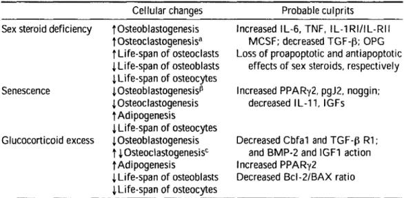
Table 2. Cellular Changes and Their Culprits in the Three Most Common Types of Osteoporosis
List of terms and abbreviations
15d-PGJ2 15-deoxy-A12 ^-Prostaglandin ]zAlkP Alkaline phosphatase
cAMP Cyclic Adenosine monophosphate
BSA Bovine serum albumin
BW 245C (4S)-(3-[(3R,S)-3-cyclohexyl-3-hydroxypropyl]-2,5-dioxo)-4-imidazolidineheptanoic acid
BW A868C 3-[(2-cyclohexyl-2-hydroxyethyl)amino]-2,5-dioxo-l-(phenylmethyl)-4-imidazolidine-heptanoic acid
Cbfal Cbfal/Runx2 is a key transcription factor associated with os-teoblast differentiation.
COX Cyclooxygenase
CRTH2 Chemoattractant homologous receptor expressed on Th2 cells
Diclofenac Benzeneacetic acid, 2-[(2,6-dichlorophenyl)amino]- monoso-dium salt. Nonsteroidal anti-inflammatory drug (NSA1D)
DK-PGDz 13,14-dihydro-15-keto Prostaglandine D2
DMEM Dulbecco's Modified Eagle's Medium
BMD Bone mineral density
DP PGD2 receptor
FBS Fetal bovine serum
GPCR G protein-coupled receptors
hOB Human osteoblasts
H-PGDS Hematopoietic Prostaglandin D synthetase
1GF-1 Insulin-like growth factor-1
1L-1 lnterleukine-1
IPs Inositol 1,4,5-trisphosphate
L-PGDS Lipocalin Prostaglandine D synthetase
M-CSF Macrophage colony-stimulating factor
Naproxen 2-Naphthaleneacetic acid, 6-methoxy-a-methyl-, (S)-. Non-steroidal anti-inflammatory drug (NSA1D)
X
NFkB Nuclear factor kappa-light-chain-enhancer of activated B cells
ODF
Osteoclast differentiation factorOPG
OsteoprotegerinePGD
2 Prostaglandin D2PGE
2 Prostaglandin E2PIP
2 Phosphatidylinositol-4,5-bisphosphatePLA
2 Phospholipase A2RA
Rheumatoid arthritisPTH
ParathormonePTX
Pertussis toxinRANK
Receptor activator of NFkBRANKL
Receptor activator of NFkB (RANK) ligandTNF
-(X Tumour necrosis factor-alphaTRAP
Tartrate-resistant acid phosphataseVEGF
Vascular endothelial growth factor1. Introduction
The bone matrix is continuously regenerated through the process of bone turnover when the "old" bone is replaced with "new" functional bone in the remodel-ling process. The lack of equilibrium between these two processes, the lysis and the deposition, is triggering the rheumatic disease.
Until recently the mainstay in their treatment has been the use of general measures without specificity. Such drugs as prednisone and other corticosteroids were used in the treatment of most of the diseases to suppress the inflammatory process and a usually over-active immune system. The effect was nonspecific and the side effects were often life-threatening.
Prostaglandins (PGs) are active biologic substances that are involved in a wide range of physiologic processes and their imbalance could be a factor in pathologies. The overproduction of PGs is responsible for pathologic inflammation, trauma and injury in rheumatologic and non-rheumatologic diseases. Restoring the PG balance could bring back the homeostasis in a more physiological way by acting over signal-ling mechanisms of the PG receptors as soon as these pathways are deciphered.
The new therapies include potentially safer (although not more effective with pain control) cyclooxygenase-2 specific nonsteroidal anti-inflammatory drugs (NSAlDs), leflunomide, tumour necrosis factor (TNF) inhibitors, etanercept and in-fliximab.
In this work, we will describe the bone modelling, remodelling, the factors in-volved, and we will underline the role of the PGD2 receptors on the bone turnover.
1.1. Bone structure and architecture
2
Bone is a specialised connective tissues that serves three important functions in the body:
1. mechanical support in locomotion 2. protective function of vital organs
3. metabolic function as a reserve of calcium and phosphate.
Bones are extremely dense connective tissue that, in various shapes, constitute the skeleton. They are the hardest structures in the body, maintaining nevertheless a degree of elasticity because of their structure and composition. Hollow tube like, they provide great resistance and durability against axial compression forces having at the same time a very low weight. The ultimate tensile strength of bone approaches that of cast iron, and its capacity to absorb and release energy is twice that of oak, yet the weight of bone is only one third that of steel (Martin, 1989; Lee et al., 2002).
Bone is enclosed, except for the articular regions, in a fibrous outer membrane called the periosteum. Periosteum is composed of two layers, an outer fibrous layer and a deeper elastic layer containing osteoblasts that are capable of proliferating rapidly when a fracture occurs. In the interior of the long bones is a cylindrical cavity filled with bone marrow and lined with a membrane composed of highly vascular tissue called the endosteum (Sambrook, 2001).
1.1.1. Macroscopical structure
At the macroscopic level there are two major types of bone: compact or corti-cal bone and trabecular or cancellous bone. Corticorti-cal bone is located in the diaphyses of long bones and on the surfaces of flat bones. There is also a thin cortical shell at the epiphyses and metaphyses of long bones. Trabecular bone is limited to the epiphyseal and metaphyseal regions of long bones and is present within the cortical coverings in the smaller flat and short bones.
The morphology of cortical and cancellous bone is arranged to accommodate the stresses and strains during weight bearing. The functional differences are a
conse-4 (Rubin et al., 1995), indicating that the woven bone response is a strategic means of rapidly responding to changes in functional activity.
Lamellar or mature bone is found in both cortical and trabecular bone. The structural subunits, the lamellae, run parallel to the trabeculae or, as is the case in cor-tical bone, are arranged in osteons, which are composed of up to 20 concentric lamel-lar plates forming a cylinder with a diameter of 200-300|im. A central capillamel-lary runs through the osteon, and up to seven concentric rings of osteocytes are incorporated into its wall (Albright J, 1987).
1.1.3. Bone matrix and minerals
Calcified bone contains about 25% organic matrix, including cells (2-5%), 5% water, and 70% hydroxyapatite [Cai0(PO4)6(OHh]. The osteoid, the freshly synthe-sised matrix prior to its mineralisation consists primarily of fibers (approximately 94%) of collagen type 1 oriented in a preferential direction. It is now accepted that the backbone of the gene for type 1 collagen is identical in all connective tissues through-out the body. Therefore, it is most probable that the genetic specificity of the bone extracellular matrix resides within the non-collagenous constituents of the tissue. These proteins, however, are not spatially isolated within bone but are intimately as-sociated with the bone collagen, forming a composite structure fulfilling all the bio-chemical, biomechanical and homeostatic requirements demanded of the bone ma-trix throughout life (Cowin, 2004).
The non-collagenous proteins (10% to 15% of the bone protein content), some of them unique to bone such as osteocalcin, are embedded in the extracellular matrix and may have important signalling functions (bone morphogeneic proteins, growth factors, cytokines, adhesion molecules) or play a role during the mineralisation proc-ess (osteopontin, osteonectin, matrix-gla protein). These highly anionic complexes have a high ion-binding capacity and are thought to play an important role in the cal-cification process.
5
A. Collagen
The collagen in bone is type 1, which is the same in skin and tendon. Except for collagen V, no other forms of collagen are found in the bone matrix. The insoluble fi-brils of collagen in bone are formed from the individual soluble tropocollagen mole-cules and are stabilised by intermolecular cross-links derived from aldehyde forms of hydroxylysine and lysine. These intermolecular cross-linking compounds of collagen are only present in its mature form and are specific for bone and cartilage. Therefore, it is believed that they represent a sensitive and specific marker for bone resorption. Urinary hydroxyproline, commonly used as marker for bone resorption in the past, is not specific to bone (Marc C. Hochberg, 2003).
B. Non-collagenous proteins
The proteoglycans consist of a central protein core to which are bound poly-saccharide chains - glycosaminoglycans - which are strongly polyanionic due to car-boxyl and sulphate groups. Studies in vitro and histochemical localisation in situ showed that proteoglycans are found in close association with collagen fibers and that they affect both the rate of fiber growth and the diameter of collagen fibers; it is pos-sible that they influence the collagen scaffolding (Marc C. Hochberg, 2003).
C. Bone Gla-containing proteins (BGP) - osteocalcin
The name osteocalcin derives from the abundance of this protein in osseous tissue (10-20% of the non-collagenous protein) and its affinity for Ca2+. Osteocalcin has also been called "the vitamin K-dependent protein of bone". Serum osteocalcin increases in situations where the bone formation rate is elevated or where bone turn-over is increased. Osteocalcin binds tightly to hydroxyapatite and is believed to have a function in the assembly of mineralised bone, perhaps by participating in the regula-tion of hydroxyapatite crystal growth. The synthesis of osteocalcin is stimulated sev-eralfold when 1,25-dihydroxyvitamin D3 is added in osteoblast culture or in vivo (Marc C. Hochberg, 2003).
6
D. Sialoproteins
Two sialoproteins, osteopontin and bone sialoprotein (BSP), previously called sialoproteins I and 11, are both cell adhesion molecules, mediating cell attachment of a
number of cell types in vitro, including bone cells. Both proteins, however, behave dif-ferently in vitro and probably have a different function in vivo. The synthesis of BSP is inhibited by 1,25-dihydroxyvitamin D3, whereas it is stimulated by dexamethasone
added to the cultured osteoblasts. In contrast, the synthesis of osteopontin by osteo-blasts is stimulated by 1,25-dihydroxyvitamin D3 (Atkins et al., 2007).
E. Alkaline phosphatases
The classic vertebrate alkaline phosphatases are a group of isozymic membrane-bound glycoproteins with molecular weights of 100-200 kDa. The wide organ and tissue distribution of alkaline phosphatase activity suggests some type of generalised function. That this glycoprotein is primarily located in the plasma mem-brane implies either a carrier or a signal transducer function. Several possible actions of alkaline phosphatase in biomineralisation have been proposed:
• increasing local concentrations of inorganic phosphate (Pi);
• local destruction of mineral crystal growth inhibitors via expression of phosphohydrolase activity;
• Pi-transporter; • Ca-binding protein;
• Ca-pump (Ca2+-ATPase) in cells or vesicle membranes;
• regulator of cellular division or differentiation, by acting as a tyrosine-specific phosphoprotein phosphatase.
Although there are supportive data for each, no singular function appears to be the principal action of the enzyme (Rodan, 1998).
7
F. Bone minerals
Bone mineral is generically referred to as hydroxyapatite [Cai0(PO4)6(OH)2], a plate-like crystal 20-80 nm in length and 2-5 nm thick. Because it is four times smaller than naturally occurring apatites and less perfect in structure, it is more reactive and soluble and facilitates chemical turnover. The crystals are oriented in the same direc-tion as the collagen fibers.
In 10 to 15 days after the organic matrix has been synthesised, the mineralisa-tion process starts. During the 10 to 15 days, the mineral content increases to 70% of its final amount, whereas deposition of the final 30% takes several months. Other gly-coproteins such as matrix-gla protein and glycosaminoglycans appear to play a role in the inhibition of excessive mineralisation (Sommerfeldt & Rubin, 2001).
1.1.4. Bone cells
Three distinctly different cell types can be found within bone: the matrix-producing osteoblast, the tissue-resorbing osteoclast, and the osteocyte, the last ac-counting for 90% of all cells in the adult skeleton. Osteocytes can be viewed as highly specialised and fully differentiated osteoblasts; similarly, osteoblasts have recently been described as sophisticated fibroblasts (Huang et al., 2009). Fibroblasts, osteo-blasts, osteocytes, and adipocytes derive from pluripotent mesenchymal stem cells, whereas osteoclasts are of hematopoietic descent and their precursors are located in the monocytic fraction of the bone marrow.
A. Osteoblasts
Osteoblasts are mono-nucleated cells that originate from mesenchymal stro-mal cells. Since osteoblasts arise from mesenchystro-mal cells, there are critical develop-mental mechanisms appropriately activated to control the cell cycle and ensure that the phenotype of the osteoblast differs from that of other cells arising from the same origin, such as chondrocytes and adipocytes.
Osteoblasts are recruited to a site of bone formation where they are responsi-ble for synthesising, secreting, organising, and mineralising the bone matrix, or
os-9
Contributors to osteoblast growth and differentiation include the bone mor-phogenic proteins, cell growth factors and cytokines (1GF-1, TGF-b, others), hormones (PTH, GH, insulin, glucocorticoids, 1,25 (OH)2-vitamin D3), and biomechanical forces
(Watkins et al., 2001).
The lifespan of an osteoblast reaches up to 8 weeks in humans (Parfitt et al., 2000), during which time it lays down 0.5-1.5 pm osteoid per day (Owen, 1972). Glu-cocorticoid use reduces the osteoblast lifespan, increasing its apoptosis.
There are four commonly accepted stages in the osteoblast life span: the preosteoblast, which demonstrates alkaline phosphatase (ALP) activity and is located within bone; the bone matrix protein producer or the mature osteoblast; the osteo-cyte transformed osteoblast; the post-proliferative osteoblasts or the bone-lining cells.
B. Osteoclasts
Osteoclasts are large multi-nucleated bone-resorbing cells that form at skeletal sites from the fusion of hemopoietic precursors of the monocyte-macrophage lineage that arrive via the circulatory system. The M-CSF stimulation is necessary for the dif-ferentiation, proliferation and survival of the cells of the macrophage lineage.
The hemopoietic precursors express also a receptor known as receptor activa-tor of NFkB (RANK). By activating this recepactiva-tor, the transcriptional facactiva-tor NFkB translocates to the nucleus and appears responsible for expression of genes that lead to the osteoclast phenotype. This receptor interacts with a ligand found on cells of the osteoblast/stromal lineage termed osteoclast differentiation factor (ODF, also known as RANKL) and with TRANCE (TNF-related activation-induced cytokine). The inter-action of RANK with ODF keys the generation of the mature and active osteoclast (Watkins et al., 2001). A soluble receptor, osteoprotegerin (OPG, which leads to an-other alias for ODF - OPGL), has been shown to block the ODF/RANK interaction. Therefore, the ratio of ODF/OPG is an important regulatory mechanism in bone re-sorption (Fig. 3).
The mature osteoclast is a multinucleate cell, containing up to 20 nuclei, stain-ing for tartrate-resistant acid phosphatase (TRAP).
II
C. Osteocytes
Derived from osteoblasts yet distinctly different in morphology and function, osteocytes are the most abundant cells in bone. They are smaller in size than osteo-blasts, contain less cell organelles such as ribosomes and endoplasmatic reticula, and have an increased nucleus to cytoplasm ratio. There is a higher number of filopodia, or cytoplasmatic extensions, which serve to interconnect the osteocytes and to con-nect them with the bone-lining cells, creating a three-dimensional syncitium (Curtis et al., 1985). Considering that it is osteocytes that are the principal cell in adult bone and that neither osteoclasts nor osteoblasts are evident in any significant numbers in a skeleton with low turnover, this osteocyte construct may actually orchestrate the spa-tial and temporal recruitment of the cells that form and resorb bone (Burger & Klein-Nulend, 1999).
The osteocytes remain connected with other similar cells but also with bone-lining cells, inactive osteoblasts (Miller and Jee, 1987) at the bone's surface, creating an extensive network of intercellular communication. There is accumulating evidence for a functional role of these cellular connections in sensing the need for and direct-ing the site of new bone formation (Donahue et al., 1995) (Mosley, 2000) .The death of osteocytes by apoptosis in oestrogen deficiency, in corticosteroid therapy, in advanc-ing age, or after damage to bone, is associated with a loss of bone strength before any bone loss (Seeman, 2008).
1.2. Bone modelling
The bone matrix is secreted by osteoblasts that lie at the surface of the existing matrix and deposit fresh layers of bone onto it. Some of the osteoblasts remain free at the surface, while others gradually become embedded in their own secretion. This freshly formed material (consisting chiefly of type 1 collagen) is called osteoid. It is rapidly converted into hard bone matrix by the deposition of calcium phosphate crys-tals in it. Once imprisoned in hard matrix, the original bone-forming cell, now called an osteocyte, has no opportunity to divide, although it continues to secrete further matrix in small quantities around itself. Since the networks of osteocytes do not
se-12
crete or erode substantial quantities of matrix, they probably play a part in controlling the activities of the cells that do. Hence, the first step in remodelling is unlikely to be bone resorption.
Osteoclasts must first be formed and then be told where to go and how much bone to resorb. These instructions are likely to arise from signals produced by the de-formation or death of osteocytes, which define the location and amount of resorption needed (Seeman, 2008). While bone matrix is deposited by osteoblasts, it is eroded by osteoclasts. The precursor cells are released as monocytes into the bloodstream and collect at sites of bone resorption, where they fuse to form the multi-nucleated osteo-clasts, which then cling to surfaces of the bone matrix and erode it (Fujikawa et al., 1996). Osteoclasts are capable of tunnelling deep into the substance of compact bone, forming cavities that are then invaded by other cells. A blood capillary grows down the center of such a tunnel, and the walls of the tunnel become lined with a layer of os-teoblasts. To produce the plywood-like structure of compact bone, these osteoblasts lay down concentric layers of new bone, which gradually fill the cavity, leaving only a narrow canal surrounding the new blood vessel. Many of the osteoblasts become trapped in the bone matrix and survive as concentric rings of osteocytes. At the same time as some tunnels are filling up with bone, others are being bored by osteoclasts, cutting through older concentric systems.
The area causing matrix resorption is composed of three different domains: the attachment zone, ruffled border and the remainder. At the contact zone with the bone, proton pumps lower the pH to values between 2 and 4, activating the secreted enzymes such as tartrate-resistant acid phosphatase (Blair et al., 1989). In the attach-ment zone, a highly organised actin filaattach-ment network, form dot-shaped, F-actin rich adhesion sites (Akisaka et al., 2006), so-called podosomes (Chabadel et al., 2007) and the actin ring (Luxenburg et al., 2006).
Vacuolar-type proton ATPase (V-ATPase) in osteoclasts is a ruffled border-associated enzyme responsible for the proton secretion, the acidity being the main factor in the Ca solubilisation (Blair et al, 1989; Okumura et al., 2006). The PH on the resorbtion pit reaches a value of 2-4, activating the secreted enzymes such as tartrate-resistant acid phosphatase (Sommerfeldt & Rubin, 2001). This resorptive phase is then
13
followed by a bone formation phase where osteoblasts fill the lacuna with osteoid. The latter is subsequently mineralised to form new bone matrix (Sambrook, 2001).
1.3. Regulation of bone remodelling
The skeletal system is involved in the body's homeostasis, it is an active system. To achieve this, there must be a constant interaction responding to hormones, to the physical demand, to stress, to the need of repairing.
To all these needs, the response is achieved trough the balance of two major processes: bone formation and bone resorption, the former controlled by the osteo-blasts and the latter by osteoclasts. The endocrine, autocrine and paracrine systems also have a role in this interplay, acting on the mentioned cells on different stages of their development or as conjugated factors of their activities. They add to the roles of the bone specific functions like the homeostatic, hematopoietic and mechanical func-tions. The regulation of bone remodelling is both systemic and local (Hadjidakis & Androulakis, 2006).
1.3.1. Factors acting on osteoblasts and osteoclasts
Remodelling is a continuous, dynamic activity driven by humoral and biofunc-tional cues, the result being that about 25% of trabecular bone and about 3% of corti-cal bone are removed and replaced each year (Parfitt, 1994) (Fig 4). As we age, the bal-ance between osteoblastic formation and osteoclastic resorption becomes asynchro-nous: bone loss occurs and results in the clinical disease (Parfitt et al., 1995).
The plasma concentration of calcium is one of the homeostatic triggers of bone remodelling, acting through a feedback mechanism which includes the liver, the kidneys and the parathyroid glands. The serum ions of calcium activate the humoral system changing the balance for 1,25-dihydroxy vitamin D3, androgen, calcitonin, es-trogen, glucocorticoids, GH, PTH, and thyroid hormone (Raisz, 1999; Bilezikian et al, 1996).
15
The decrease of plasma oestrogen may lead to increased levels of of cytokines, 1L-1, 1L-6, TNF-a, (Pacifici, 1998) but also can trigger osteocytes apoptosis (Tomkin-son et al., 1997). An inflammation - local or systemic - could trigger also the release of cytokines and limphokines, leading to osteolysis.
The osteoclasts are formed as a response to several osteotropic factors like 1,25-dihydroxyvitamin D3 [l,25(OH)2D3], interleukin (lL)-6, PGE2 (Takahashi et al.,
1988) which induce the expression of NFKB ligand (RANKL, a member of the TNF
family) in osteoblasts. The binding of RANKL to the RANK receptor found on the sur-face of the osteoclast progenitors (monocytes) activates the osteoclastogenesis. The PTH induction of RANKL is conditioned by the presence of cyclooxigenase-2 (COX-2) via cAMP production (Okada et al., 2000). The activation of RANKL by the 1L-1,1L-6, 1L-17 and l,25(OH)2D3 requires the presence of COX-2 as well. (Li et al., 2006).
The osteoprotegerin (OPG), a soluble "decoy receptor", regulates the RANK effect by competing for the same ligand, thus inhibiting the RANK osteolytic effect, but it is down-regulated by PTH (Lee & Lorenzo, 1999). These are not the only par-ticipants involved, different publications mentioning TNF, annexin-11, TGF-£, etc (Mundy, 1996; Boyce et al., 1999).
The damaged area of the bone is signalled by the apoptotic osteocytes to the lining cells. The target is confined to minimise the bone loss, creating a Basic Multi-cellular Unit (BMU) (Hauge et al., 2001). It is possible that the trigger is the collagen digestion around the mineralised bone and the exposure of the calcified area. The bone lining cells may be responsible for this action as they express collagenase mRNA. (Chambers et al., 1985). More than that, Parathyroid hormone (PTH) stimulates colla-genase production and secretion by osteoblastic cells and it appears to be involved in clearing the osteoblast-lining surfaces of the bone to permit access to the osteoclasts (Chiusaroli et al., 2003). The PTH binds to the PTH receptor 1 (PTHR1) found on the osteoblasts, leading to an increase of RANKL, activating the osteoclastogenesis (Fu et al., 2002). Some authors report that PTH acts directly on the osteoclasts receptors, although there are not sufficient studies to accustom this theory yet (Dempster et al., 2005).
i6
It is accepted today that the parathyroid hormone (PTH) has dual effect, ana-bolic and cataana-bolic, depending on the signalling cascade (Datta, et al., 2009) or de-pending on the way it is administered. Intermittent administration has an anabolic effect (Horwitz et al, 2003; Pettway et al., 2005; Pettway et al., 2008), whereas con-tinuous administration induces bone resorption (Kaji, 2007).
Calcitonin is a peptide hormone synthesised and secreted by thyroid parafol-licular C cells. It is regulated by extracellular calcium levels and by gastrointestinal hormones such as gastrin. Calcitonin receptors are present on osteoclasts, preosteo-clasts. The hormone blocks bone resorption probably via mature osteoclasts by en-hancement of adenylate cyclase and cAMP or as a mitogen acting on bone cells.
The mechanical load is also a trigger of bone remodelling. The signal is the de-formation of the bone, followed by the local release of cytokines and also of AA (ara-chidonic acid) (Cissel et al., 1996) and consecutively of PGE2 leading to the bone
re-sorption in this first step (Rodan, 1998). The bone deposition under the release of an-drogens and BMPs follows.
There are many other proposed mechanisms of acting on the bone turnover including a bidirectional regulation between osteoclasts and osteoblasts through a signalling system (Zhao et al., 2006), and a nervous/neuroendocrine system regulation based on leptin. Leptin is a polypeptide hormone that influences body weight, satiety and lipid metabolism. It plays a role in the central hypothalamic modulation of bone formation, as well as locally within the skeleton, by enhancing differentiation of bone marrow stroma into osteoblasts and by inhibiting its differentiation into osteoclasts and adipocytes (Ducy et al., 2000) (Takeda et al., 2002) thus proving that the bone re-modelling is a complex process yet to be discovered.
17
1.4. Eicosanoids and Prostanoids
Although prostaglandins were the first biologically active eicosanoids to be identified, it is now known that the essential fatty acids are converted into a number of different types of eicosanoids. Eicosanoid is a term meaning a 20 carbon fatty acid derivative.
Arachidonate and some other 20 carbon polyunsaturated fatty acids give rise to eicosanoids, physiologically and pharmacologically active compounds known as prostaglandins (PG), thromboxanes (TX), leukotrienes (LT), and lipoxins (LX). Physio-logically, they are considered to act as local hormones generated in situ, rapidly me-tabolised, functioning through G-protein-linked receptors to elicit their biochemical effects in the immediate vicinity.
Both von Euler in Sweden and Goldblatt in England discovered marked stimu-lation of smooth muscle by seminal plasma. Von Euler (1935) then showed that lipid extracts of ram vesicular glands contained the activity and this was associated with a fatty acid fraction. The factor that showed this effect was named prostaglandin and it was thought to possess a variety of physiological and pharmacological properties. The name prostaglandin (and the related prostanoic acid structure) derives from the fact that early researchers believed that the prostate gland was the site of their synthesis.
In 1947, Bergstrom started to purify these extracts and soon showed that the active principle was associated with a fraction containing unsaturated hydroxy acids. In 1956, with the help of an improved test system (smooth muscle stimulation in the rabbit duodenum), Bergstrom isolated two prostaglandins in crystalline form (PGEi and PGFia). Their structure, as well as that of a number of other prostaglandins, was elucidated by a combination of degradative, mass spectrometric, X-ray crystallo-graphy and NMR studies. The nomenclature is based on the fully saturated 20 carbon acid with C8 to C12 closed to form a 5-membered ring, called prostanoic acid. Thus PGEi is designated 9-keto-ll a,15 a-dihydroxyprost-13-enoic acid. The 13,14 double bond has a trans configuration; all the other double bonds are cis. In the Fig. 6 one can see the difference between the 'E', which have a keto group at position 9 and 'F' series,
i8
which have a hydroxyl group at the same position; 'a' refers to the stereochemistry of
the hydroxyl, and the suffix 1, 2 or 3 is related to the number of double bonds con-tained in the prostaglandin structure (Michael I. Gurr, 2002).
PGA, PGB and PGC are ketones, PGD and PGE are hydroxyl ketones and PGF a is a diol. PG1 has a different structure, given the second ring attached to the cy-clopentane structure.
1.4.1 Prostaglandin Synthesis
Arachidonate (AA) is usually derived from the sn-2 position of glycerophos-pholipids in the plasma membrane by the action of phospholipase A2 (Fig. 5). It is re-leased from phosphatidylcholine (PC) and phosphatidylethanolamine (PE) by phos-pholipase A2, or from phosphatidylinositol (PI) by phosphos-pholipase C (PLC) pathway. The arachidonate can also come from the diet. AA is the substrate for the synthesis of the PG2, TX2 series (prostanoids) by the cyclooxygenase pathway (Fig. 6), or the LT4
and LX4 series by the lipoxygenase pathway (Fig. 7), with the two pathways competing
for the arachidonate substrate (Tang et al., 2006).
The control of the AA release depends mainly of the presence of type IV cyto-solic PLA2 which can be translocated to the nuclear envelope, endoplasmic reticulum (ER) and Golgi apparatus.
The metabolism of arachidonate substrate by cyclooxygenase pathway gives cyclic endoperoxides from which the classical prostaglandins or thromboxanes and prostacyclin can be synthesised. A third pathway occurs via cytochrome P450 oxy-genation where atomic oxygen is introduced leading to fatty acid hydroxylation or epoxidation of double bonds. Whereas both the lipoxygenase and cyclooxygenase re-actions arise from the formation of a fatty acid radical, in P450-oxygenation, activa-tion of atmospheric molecular oxygen is involved. After this, one oxygen is transferred to the fatty acid substrate and one is reduced forming water (Porubsky et al., 2008; Moreno, 2009).
22
There are two major isoforms of the enzyme and crystal structures for both have been obtained: PGHS-1 or COX-1 and PGHS-2 or COX-2 (discovered by Daniel L. Simmons in 1991). The cyclooxygenases are haemoproteins and they present both cy-clooxygenase and peroxidase activity (Garavito and Mulichak, 2003). Also, they use a variety of substrates like linoleates (a and y) giving rise to different endoperoxides.
There is a 60% aminoacid homology between the two COX isoforms, but there is a difference in localisation, COX-2 acting at the nuclear envelope and COX-1 mainly in the ER and close to the cell membrane (Versteeg et al., 1999).
The product of PGHS is an endoperoxide, subsequently converted to prosta-glandins D, E and F, to a thromboxane (TXA2) and prostacyclin (PGh).
Today, we acknowledge the existence of the two isoforms, one with constitu-tive expression and the other inducible. COX enzymes are integral membrane pro-teins that sit within one leaflet of the lipid bilayer of intracellular phospholipid mem-branes of the nuclear envelope and ER. The cyclooxygenase active site is located in a channel formed in the center of enzyme, allowing the hydrophobic fatty acid sub-strate access without leaving the membrane. The peroxidation function is located on the outside of the enzyme and appears to be similar in both enzymes.
It has been proposed that cyclooxygenase COX-1 and COX-2 serve different physiologic functions largely because of the striking differences in their tissue expres-sion and regulation. COX-1 displays the characteristics of a "housekeeping" gene and is constitutively expressed in almost all tissues. COX-1 appears to be responsible for the production of prostaglandins (PG) that are important for homeostatic functions, such as maintaining the integrity of the gastric mucosa, mediating normal platelet function, and regulating renal blood flow. In contrast, COX-2 is the product of an "immediate-early" gene that is rapidly inducible and tightly regulated. Under basal conditions, COX-2 expression is highly restricted; however, COX-2 is dramatically up-regulated during inflammation. For example, synovial tissues in patients with rheu-matoid arthritis (RA) express increased levels of COX-2. In vitro experiments on endo-thelial cells, chondrocytes, osteoblasts, synoviocytes and monocytes/macrophages have revealed increased COX-2 expression after stimulation with proinflammatory cytokines, such as interleukin 1 (IL-1) and tumour necrosis factor-alpha (TNF-alpha).
23
COX-2 is also increased in some types of human cancers, particularly colon cancer. Mechanisms underlying the association between COX-2 over-expression and tu-mourigenic potential may include resistance to apoptosis (Garavito et al., 2002).
A PGHS-1 variant, COX-3 (Chandrasekharan et al., 2002) have been under in-vestigation after its presence has been confirmed at the cartilage level (Gosset et al., 2006).
The Prostaglandin Synthase action is the concluding step into the formation of a specific prostaglandin. All known PG's have at least one PGS who generates the spe-cific PG from the PGH2 substrate.
1.4.3. Prostaglandins - general effects and roles.
The prostaglandins are involved in modulations of different systems, from the CNS (central nervous system) to gastrointestinal (Gl) and immune systems. Moreover, they are involved in a plethora of pathological states ranging from inflammation to cancer.
They are considered potent pro-inflammatory mediators and they play an im-portant role in nociception and pain, since the COX-2 and mPGES-1 (microsomal prostaglandin E2 synthase-1) expression are induced in the CNS via the pro-inflammatory cytokines like 1L-1|3 and TNFcx (Zeilhofer, 2007). They are involved in the contraction and the relaxation of the smooth muscles, sleep mechanisms, fever induction, renal tubular reabsorption, apoptotic regulation, cell differentiation.
Fig. 8 presents a general cellular structure activated by possible triggering mechanisms like mechanical trauma, inflammation (cytokines). The inducible form of Prostaglandin Synthase (COX-2) can also contribute to the increased expression of PG's. The synthesised prostaglandins are carried by the prostaglandin transporter (PGT) to exert actions on specific receptors. Of course, their action is perhaps ex-tended to some non-specific receptors - like the nuclear hormone receptor PPAR.
25
Concentrations of major active prostaglandin products in blood are less than 0.1 nM and because of their rapid catabolism they can only act as local hormones modifying biological events close to their sites of synthesis.
Degradation occurs via 3 and U) oxidation, de-methylation or by oxidation of the C-15 hydroxyl group to a ketone by 15-hydroxy-PG dehydrogenase (15-OH-PGDH) present in tissues (peroxizomes) (Gosset et al., 2006) (Tai et al., 2002). On the other hand, TNF-cx, lL-($ decrease the 15-OH-PGDH mRNA, impeding the keto-transformation of the PG's, thus prolonging their effects.
A prostaglandin transporter (PGT), which plays a primary role in mediating prostanoid transport and metabolic clearance, has been identified (Chi & Schuster, 2010). The PGT plays a role in the uptake of newly released prostanoids, thus acting as a carrier across the plasma membrane before intracellular oxidation (Chi et al., 2006). Moreover, PGT can facilitate intracellular actions of circulating as well as intracellu-larly produced prostanoids. The PGT preferentially transports PGE2, PGEi, PGF2«, PGD2, with high affinity and to a lesser extent, TXB2 and PGI2 (Funk, 2001).
1.4.4. Prostaglandin receptors.
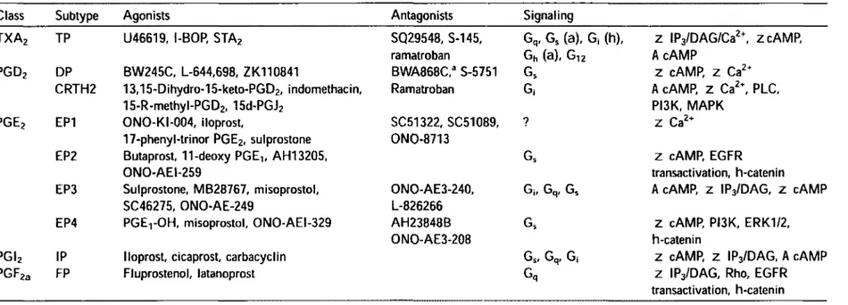
There are eight types of the prostanoid receptors (Table 1) conserved in mam-mals from mouse to human. They are the PGD receptor (DPI), four subtypes of the PGE receptor (EP1, EP2, EP3, EP4), the PGF receptor (FP), PG1 receptor (IP) and the TXA receptor (TP). They all are G-protein coupled rhodopsin-type receptors with seven transmembrane domains, and each is encoded by different genes. In addition, there are several splice variants of the EP3, FP, and TP receptors, but they differ only in their C-terminal tails.
For PGD2 there is a distinct type of receptor, chemoattractant receptor-homologous molecule expressed on T helper type 2 (Th2) cells (CRTH2). This recep-tor was originally cloned as an orphan receprecep-tor expressed in Th2 lymphocytes, and has recently been shown to bind PGD2 with an affinity as high as that of DP, although the binding profile to other PGD analogs differs from that of DP. The CRTH2
recep-Class Subtype Agonists Antagonists Signaling
TXA2 TP U46619, l-BOP, STA2 SQ29548, S-145, Gq, Gs (a), G| (h), z IP3/DAG/Ca2\ zcAMP,
ramatroban Gh (a), Gi2 AcAMP
PGD2 DP BW245C, L-644,698, ZK110841 BWA868C,3 S-5751 Gs z cAMP, z Ca2+
CRTH2 13,15-Dihydro-15-keto-PGD2, indomethacin, Ramatroban G, A cAMP, Z Ca2t, PLC,
15-R-methyl-PGD2, 15d-PGJ2 PI3K, MAPK
PGE2 EP1 ONO-KI-004, iloprost, SC51322, SC51089, ? Z Ca2+
17-phenyl-trinor PGE2, sulprostone ONO-8713
EP2 Butaprost, 11-deoxy PGE1( AH13205, Gs z cAMP, EGFR
ONO-AEI-259 transactivation, h-catenin
EP3 Sulprostone, MB28767, misoprostol, ONO-AE3-240, Gj, Gq, Gs A cAMP, z IPj/DAG, z cAMP
SC46275, ONO-AE-249 L-826266
EP4 PGEtOH, misoprostol, ONO-AEI-329 AH23848B Gs z cAMP, PI3K, ERK1/2,
ONO-AE3-2Q8 h-catenin
PGI2 IP Iloprost, cicaprost, carbacyclin Gs, Gq, Gj z cAMP, z IPj/DAG, A cAMP
PGF2a FP Fluprostenol, latanoprost Gq z IP3/DAG, Rho, EGFR
transactivation, h-catenin
a Partial agonist.
Table 1: Prostaglandin receptors - signaling (Hata and Breyer, 2004).
33
F. PGD2 - Generalities
The prostaglandin D2 has an important role in the regulation of different body functions like sleep regulation, allergic responses, asthma and pregnancy.
The role of PGD2 in wake-sleep mechanisms has been studied for a long time, and published works connected the PGD2 with the non rapid eye movement and sleep (Hayaishi, 2002), with pathological states triggered by systemic or infectious diseases (Jordan et al., 2004), with narcolepsy (Jordan et al., 2005), and perhaps with more complex mechanisms involving melatonine and GABA systems in the brain (Urade & Mohri, 2006).
The PGD2, being released from basophiles and mast cells (Schleimer et al., 1984), is an important mediator in allergy (Naclerio et al., 1983) having a bronchocon-strictor effect in allergic asthma (Hardy et al., 1984). This effect is probably regulated through the CRTH2 receptor (Boehme et al., 2009a; Boehme et al., 2009b). PGD2 is a coronary constrictor in anaphylaxis (Weinerowski et al., 1985) and is a mediator in skin dermatitis, probably through CRTH2, (Oiwa et al., 2008).
The transformation of PGH2 into PGD2 is realised through the catalytic activ-ity of specific synthetase, called the PGDS, which has two known forms: lipocalin-type (L-PGDS) and the hematopoietic-lipocalin-type (H-PGDS) (Urade & Eguchi, 2002).
Cyclopentenone prostaglandins (PGs), such as 15-deoxy-12,13-didehydro-14,15-didehydro-PGj2 (15d-A<1214)-PDj2), 12,13-didehydro-PGj2
(A
12-PGj2), are the products resulting from the PGD2 dehydration. The PGJ2 synthesis was initially related to the presence of serum albumin (Keelan et al., 2003), but studies showed that the conver-sion could be done through an albumin-independent mechanism (Shibata et al., 2002), and through the intermediaries like PGJ2 and 15d-PGD2 to the final compounds 15d-PGJ2 and A12-PGJ2.The biological responses of PGD2 could be in fact the result of the combined effects at different levels of the PGD2 and its dehydrated products. The ultimate me-tabolite of PGD2, 15deoxy-A1214-PGj2, binds specifically to a nuclear receptor, the gamma isoform of the peroxisome proliferator-activated receptor (PPARy), thereby
34
promoting adipogenesis (Negishi & Katoh, 2002) and triggering anti-inflammatory responses. It also binds to the PGD2 receptors, DPI and CRTH2 (Scher & Pillinger,
2005). The intranuclear target of the 15d-PGJ2, the PPAR's, are transcription factors
that regulate gene expression of enzymes associated with lipid homeostasis, inflam-mation, cell proliferation, and malignancy. The 15d-PGj2 could intervene in other in-tracellular cascades in a PPAR-independent manner, through a mechanism of covalent binding to proteins from the NF-KB system (the IKB kinase-lKK). It blocks the
activa-tion of NF-KB and the extracellular signal-regulated kinase (Erk) signalling pathway
(Chawla et al., 2001) and also binds to H-Ras and increases cell proliferation (Oliva et al., 2003).
1.5. NSAID's
Non-steroidal anti-inflammatory drugs - NSAlDs, are drugs with analgesic, antipyretic and anti-inflammatory effects. The term "non-steroidal" is used to distin-guish these drugs from steroids, which have a similar eicosanoid-blocking, anti-inflammatory action. NSAlDs are non-narcotic. The most prominent members of this group of drugs are aspirin, ibuprofen, and naproxen partly because they are avail-able over-the-counter in many areas.
Prostaglandins are potent hyperalgesic mediators which modulate multiple sites along the nociceptive pathway and enhance both transduction (peripheral sensi-tising effect) and transmission (central sensisensi-tising effect) of nociceptive information. Inhibition of the formation of prostaglandins at peripheral and central sites by NSAlDs thus leads to the normalisation of the increased pain threshold associated with inflammation.
The structure of the COX proteins consists of three distinct domains: an N-terminal epidermal growth factor domain, a membrane-binding motif, and a C-terminal catalytic domain that contains the COX and peroxidase active sites. The COX active site lies at the end of a hydrophobic channel that runs from the membrane-binding surface of the enzyme into the interior of the molecule (Dann-hardt & Kiefer, 2001).
35
NSAlDs act at the COX active site in several ways:
• Aspirin irreversibly inactivates both COX-1 and COX-2 by acetylating an active-site serine. This covalent modification interferes with the binding of arachi-donic acid at the COX active site.
. By contrast, reversible competitive inhibitors of both COX1 and COX2 iso-forms (e.g., mefenamate, ibuprofen) compete with arachidonic acid for the COX ac-tive site.
• A third class of NSAlDs (e.g., flurbiprofen, indomethacin) causes a slow, time-dependent reversible inhibition of COX-1 and COX-2 which results from the formation of a salt bridge between the carboxylate of the drug and the arginine 120, followed by conformational changes. (Kalgutkar et al., 2000)
Most of the NSAlDs are non-selective inhibitors acting at the active site of both enzymes, COX-1 and COX-2, but selective COX-2 inhibitors have been available for use in arthritis public use starting from 1998 (under the U.S. Food and Drug Ad-ministration approval). Apart from its involvement in inflammatory processes, COX-2 seems to play a role in angiogenesis, colon cancer and Alzheimer's disease. This is based on the fact that it is expressed during these diseases. Lately, acetaminophen is believed to act on the COX-3 izoenzyme (Chandrasekharan et al., 2002) decreasing fever and pain. Of course, the specificity has a certain degree, interactions could occur in a crossover way, the COX-1 inhibitors interacting with the COX-2 sites (Hinz et al., 2006). Studies have shown that the use of NSAlDs is associated with a reduced inci-dence of colon cancer (COX-2) and a reduced risk of Alzheimer development (COX-2). The NSAlDs have an important role in controlling pain associated with rheumatoid arthritis (Simmons et al., 2004).
36
1.6. Prostaglandins and bone
Prostaglandins have multiple effects on bone cells, and sometimes opposite effects in different species. Their role is therefore difficult to discern. They are power-ful bone and cartilage resorbing agents in certain in vitro studies, yet they are potent anabolic (bone forming) agents when administered in vivo. Experimental investiga-tions proved their influence on inflammatory processes, and, above all, on the bone destruction in the pathogenesis of rheumatoid arthritis and osteomyelitis.
Prostaglandins very probably induce the local intercellular communication and regulate the metabolism of bone and cartilage. They are supposed to be local media-tors of mechanical stress, electric phenomena, as well as of hormonal control mecha-nisms in cytobiologic and cytochemical reactions within the skeletal system.
Prostanoids produced by bone include PGE2, PGF2a, and 6-keto-PGFia, the metabolite of PGI2, as well as PGD2 and thromboxane.
The first thing worth mentioning is the involvement of dietary fatty acids into the bone metabolism. The poly-unsaturated fatty acids (PUFA, n-6 and n-3) seem to modulate hOB metabolism by influencing the OPG/RANKL system responsible for the PGE2 interactions (Coetzee et al., 2007).
Osteoblasts and osteoclasts, the most important contributors to bone remod-elling, produce PGs, which are shown to modulate the cell function in normal bone metabolism and in bone healing (Gajraj, 2003). Several receptors have a proven exis-tence on the hOB (Sarrazin et al., 2001), and the hOB themselves produce these auta-coids (Hackett et al., 2006). Moreover, in fluids containing albumin, PGE2 and PGD2 are slowly dehydrated within the cyclopentane ring to the cyclopentenone prosta-glandins PGA2 and PGJ2, respectively. PGJ2 is metabolised further to yield
15-deoxy-A12-14 PG]2 (15d-PGj2). The cyclopentenone PGs, which are not thought to act via the
classical PG receptors, are active when given exogenously and can have opposite ef-fects from some of the primary PGs (Negishi & Katoh, 2002).
The PGE2 is the most abundant PG produced by the OB, producing through the EP2 and EP4 receptors, either anabolic or catabolic effects on the bone (Fig. 15). A
3«
fication (possible by its metabolite, 15d-PGj2) (Koshihara & Kawamura, 1989) and has probable proliferative (Tsushita et al., 1992) or indirect anti-proliferative properties (Haberl et al., 1998). In human primary OB, PGD2 activates the DP receptor thereby decreasing the osteoprotegerin expression, but in the same cells, through the CRTH2 receptor, it decreases the RANKL production. The production of PGD2 is stimulated
by TNF-a, 1L-1, PTH, VEGF (Gallant et al., 2005) and also by mechanical strain (per-haps through the system of A12 PGJ2/PPAR) (Siddhivarn et al., 2006).
The PGD2 has anabolic or catabolic effects on the bone, carrying a
controver-sial role in bone homeostasis. PGD2 stimulates calcification of hOB (Koshihara &
Ka-wamura, 1989) and has proliferative effects (Tsushita et al., 1992) or anti-proliferative effects through its metabolites (Haberl et al., 1998). PGD2 stimulates the
osteoclasto-genesis (via 1L-6) (Durand et al., 2008; H. Tokudaa, 1999) probably by inhibiting the OPG production (Samadfam et al., 2006).
The PG1 receptor, IP, has been identified also on the cultured hOB (Fortier et al., 2001), (Sarrazin et al., 2001) and its proven role seems to be inducing the expres-sion of COX-2 enzyme (Sakuma et al., 2004).
The TP and FP receptors presence on human osteoblasts have been also proven (Sarrazin et al., 2001) (Samadfam et al., 2006). The FP acts as an inducer of COX-2 on rodent cloned osteoblastic cells, inhibits the collagen synthesis (Pilbeam et al., 1995) and increase the OPG accumulation on MG-63 cells, but not on hOB (Sa-madfam et al., 2006).
1.7. Bone pathology
The bone pathologies are the diseases resulting from disorders of bone forma-tion and/or resorpforma-tion, by changing the rates of bone turnover. These include the metabolic bone disease and those related to cancer and inflammatory processes.