O
pen
A
rchive
T
oulouse
A
rchive
O
uverte
(OATAO)
OATAO is an open access repository that collects the work of some Toulouse
researchers and makes it freely available over the web where possible.
This is
an author'sversion published in:
https://oatao.univ-toulouse.fr/23069Official URL : https://doi.org/10.1302/0301-620X.99B9.BJJ-2016-1043.R2
To cite this version :
Any correspondence concerning this service should be sent to the repository administrator: [email protected]
Laumonerie, Pierre and Reina, Nicolas and Ancelin, David and Delclaux, Stéphanie and
Tibbo, Meagan E. and Bonnevialle, Nicolas and Mansat, Pierre Mid-term outcomes of 77
modular radial head prostheses. (2017) The Bone & Joint Journal, 99-B (9). 1197-1203. ISSN
2049-4394
OATAO
Open Archive Toulouse Archive Ouverte
P. Laumonerie, N. Reina, D. Ancelin, S. Delclaux, M. E. Tibbo, N. Bonnevialle, P. Mansat
From Hôpital Pierre
-Paul Riquet, Toulouse, France • P. Laumonerie, MD, Orthopaedic Surgeon • N. Reina, MD, PhD, Orthopaedic Surgeon • D. Ancelin, MD, Orthopaedic Surgeon • S. Delclaux, MD, Orthopaedic Surgeon • M. E. Tibbo, MD, Orthopaedic Surgeon • N. Bonnevialle, MD, PhD, Orthopaedic Surgeon • P. Mansat, MD, PhD, Orthopaedic Surgeon, Oepartment of Orthopaedics Hôpital Pierre-Paul Riquet, Place du Docteur Baylac, Toulouse 31059, France Correspondence should be sent to P. Laumonerie; email: [email protected]
Mid-term outcomes of
77
modular radial
head prostheses
Aims
Radial head arthroplasty (RHA) may be used in the treatment of non-reconstructable radial head fractures. The aim of this study was to evaluate the mid-term clinical and radiographie results of RHA.
Patients and Methods
Between 2002 and 2014, 77 RHAs were implanted in 54 men and 23 women with either acute injuries (54) or with traumatic sequelae (23) of a fracture of the radial head. Four designs of RHA were used, including the Guepar (Small Bone Innovations (SBi)/Stryker; 36),
Evolutive (Aston Medical; 24), rHead RECON (SBi/Stryker; ten) or rHead STANDARD (SBi/
Stryker; 7) prostheses. The mean follow-up was 74.0 months (standard deviation (sol 38.6;
24 to 141). The indication forfurther surgery, range of movement, mean Mayo Elbow Performance (MEP) score, quick Disabilities of the Ann, Shoulder and Hand (quickDASH) score, osteolysis and positioning of the implant were also assessed according to the design,
and acute or delayed use. Results
The mean MEP and quickDASH scores were 90.2 (so 14; 45 to 100), and 14.0 points (so 12;
1.2 to 52.5), respectively. There were no significant differences between RHA perfonned in acute or delayed fashion. There were 30 re-operations (19 with, and 11 without removal of the implant) during the first three post-operative years. Painful loosening was the primary indication for removal in 14 patients. Short-stemmed prostheses (16 mm to 22 mm in length) were also associated with an increased risk of painful loosening (odds ratio 3.54
(1.02 to 12.2), p = 0.045). Radiocapitellar instability was the primary indication for re-operation with retention of the implant (5). The overall survival of the RHA, free from re-operation, was 60.8% (so 5.7%) at ten years.
Conclusion
Bipolar and press-fit RHA gives unsatisfactory mid-term outcomes in the treatment of acute fractures of the radial head or their sequelae. The outcome may vary according to the design of the implant. The rate of re-operation during the first three years is predictive of the long-term survival in tight-fitting RHAs.
Fractures of the proximal radius represent about one third of all fractures involving the elbow and are the most common fractures affecring this joint.1 Patients whose radial head cannot be reconstructed and who undergo excision of the radial head develop progressive valgus instability, potential radial ascent, and secondary ulnocarpal
symptoms with alteration in the k.inemarics of the elbow and forearm to a self-perpetuaring cycle of degenerative changes.2·6 ln the presence
of associated ligamentous injury, good functional results have been reported with radial head arthroplasty (RHA).7·11 This procedure allows maintenance of the integrity of the four columns
of the elbow in patients with very comminuted fractures of the radial head which cannot be treated by open reduction and internai fixation (ORIF).12"14 RHA produces satisfactory ou t-comes. However, it bas recently been reported that a tight-fitting RHA may have inferior mid -term survival than a loose-fitting RHA.12,15·24 High rates of complications have also been reported after this procedure.25·30 There is lim -ited information about the mid-and long-term outcomes comparing the functional results of different designs of RHA, due in part to the small effect sizes and varied indications for use in the available studies.
Between 2002 and 2014, four different models of tight-fitting RHA were used in our department to treat acute, non-reconstructable fractures of the radial head or their post-traumatic sequelae; the GUEPAR (Small Bone Innova-tions (SBi)/Stryker, Morrisville, Pennsylvania), the Evolu-tive (Aston Medical, Saint-Etienne, France), the rHead RECON and the rHead STANDARD prosthesis (both SBi/ Stryker).
Our primary aim in this study was to investigate and compare the mid-term survivorships of press-fit and bipo-lar RHAs.
Patients and Methods
This is a retrospective, single-centre study performed at an academic department of orthopaedic surgery. Inclusion cri-teria were: patients undergoing surgery for a non recon-structable fracture of the radial head or the sequelae of trauma, including malunion, pseudarthrosis, necrosis, fail-ure of fixation, for whom a RHA was performed between 2002 and 2014 with a minimum follow-up of two years or follow-up until removal of the implant. Patients with follow-up of less than two years and patients aged < 16 years of age were excluded.
A total of 94 patients underwent RHA during this time; four were excluded due to a short follow-up and 13 were lost to follow-up. A total of 77 patients were included in the study. There were 54 men and 23 women. Their mean age was 52 years (20 to 82). The dominant hand was involved in 42 patients. 54 involved acute fractures and 23 the
sequelae of trauma. There were 36 Guepar (Fig. 1), 24 Evolutive (Fig. 2), ten rHead RECON and seven rHead STANDARD prostheses (Fig. 3). The characteristics of these prostheses are shown in Table I. In our department, a call for tenders was performed for each model of RHA; one type was preselected to be used for all these procedures for a limited period of time. Our preference changed three times, giving a total of four different prostheses during this time. The RHA which was used in each patient was dependent on our preference at the time of surgery. No ran-domisation of the RHAs was performed, as only one choice was available at the time of surgery for each patient.
Initial evaluation of the fractures showed 65 Mason type III radial head fractures, two Mason type II fractures and ten radial neck fractures.31 There were 27 isolated frac-tures, 27 “terrible triad” fracfrac-tures,14 four Essex-Lopresti injuries,32 four distal metaphyseal-epiphyseal fractures of the radius or ulna, and 11 patients had an associated tran-solecranon fracture-dislocation of the elbow.
Fig. 1
Anteroposterior radiograph of the elbow suggesting overstuffing of a Guepar prosthesis (Small Bone Inno-vations/Stryker, Morrisville, Pennsylvania) with widen-ing of the lateral part of the ulno-humeral joint).
Fig. 2
Lateral radiograph of the elbow showing a radial head arthroplasty (Evolutive, Aston Medical, Saint-Etienne, France) with periprosthetic osteolysis.
Fig. 3
The ratio of the length of the head (R) divided by total length of the implant (T) of a short-stemmed prosthesis (rHead RECON; Small Bone Innovations/Stryker, Morrisville, Pennsylvania).
A lateral approach to the elbow was used in 66 patients, and a posterolateral approach in 11, when there was an associated fracture of the olecranon. Particular attention was paid to preservation of the radial collateral ligament if it was intact. The annular ligament was incised longitudi-nally (i.e. transverse to its fibres). The capitellum was rou-tinely carefully examined for the presence of cartilage lesions. Nine olecranon fractures were fixed with a plate and two by tension-band wiring. Four fractures of the coro-noid process were fixed using retrograde screw fixation with intra-articular control of the reduction.
The radial neck was divided so as to preserve as much bone as possible. The radial neck was systematically con-served for all short-stemmed implants. The medullary canal of the radius was reamed and the prosthesis was introduced such that it did not pass the superior surface of the radial notch of the ulna. The elbow was then put through a full arc of flexion and the position was checked on extension and on anteroposterior (AP) and lateral fluoroscopic views. Low-viscosity, antibiotic impregnated cement (Palacos Genta; Heraeus Medical, Wehrheim, Germany) was used for fixation of the 60 smooth-stemmed components. The 17 rough-stemmed components were either press-fitted (two) or fixed with cement (15) if the stability when press-fitted was felt to be insufficient according to the manufac-turers’ specifications (Aston Medical and SBi/Stryker). The final radial head components were impacted onto the neck of long-stemmed prostheses, and directly onto the stem of rHead prostheses. The radial collateral ligament was re-attached to the lateral epicondyle using trans-osseous sutures or suture anchors in 39 patients. In the remaining patients, the annular ligament and tendon layer were simply repaired. The stability of the elbow was then re-assessed. The ulnar collateral ligament was re-attached to the medial epicondyle in three patients.
In 31 patients, in whom the lateral collateral ligament (LCL) was repaired, the elbow was immobilised in a long-arm dorsal-volar splint with the wrist in pronation, for 15 days post-operatively. The wrist was left free in 38 patients, in whom the LCL was not repaired. In eight patients, in whom the elbow remained unstable despite LCL reconstruction, a static external fixator was retained
for two to three weeks. A hinged brace was used to allow extension up to -30° between the second and third post-operative week. Active mobilisation of the elbow and phys-iotherapy started about six weeks post-operatively.
All patients were assessed by an independent reviewer at the time of final review or at removal of the implant. The range of movement (ROM) of both elbows and wrists was recorded. AP and lateral radiographs of the elbow were undertaken in maximal extension and 90° of flexion for the 59 patients who retained the radial head prosthesis at the time of last follow-up. The pre- and post-operative clinical and radiographic data, and operative details were noted from the medical records for all 77 patients in the series. This information allowed analysis of the cause and timing of re-operation, with or without retention of the RHA.
Clinical analysis. Analysis was possible for 58 patients in whom the RHA was retained at the time of the final review. The maximum ROM was measured using a goniometer. The ratio, expressed as a percentage of the force of flexion and extension of both elbows, was measured using a Kinedyn dynamometer (Smith & Nephew, Memphis, Ten-nessee). Function was assessed using the Mayo Elbow Per-formance Score (MEPS)33 and the Quick Disabilities of the Arm, Shoulder and Hand (QuickDASH) score.34
Radiographic analysis. Radiographic results were available for all 77 patients. AP and lateral post-operative graphs were used to assess alignment according to radio-capitellar congruence in both planes, overstuffing (associated or not associated with an asymmetry to the humero-ulnar interval, also called the river delta sign).19 Assessment included the position of the stem for the stemmed components which was considered to be valgus or varus when the distal extremity apposed the lateral or medial aspect of the radial cortex, respectively, signs of periprosthetic osteolysis (Fig. 2), heterotopic ossification according to the Brooker classification,35,36 and capitellar wear. These were noted at each post-operative review.
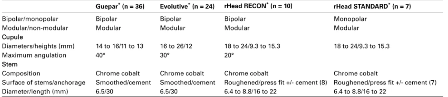
Statistical analysis. The primary objective was a descriptive analysis of the mid-term clinical and radiological outcomes. Results were described according to the mean, standard deviation (SD), maximum and minimum values. Fisher’s exact test and Kruskal-Wallis tests were used to compare Table I. Characteristics of the components of the radial head arthroplasties
Guepar* (n = 36) Evolutive* (n = 24) rHead RECON* (n = 10) rHead STANDARD* (n = 7)
Bipolar/monopolar Bipolar Bipolar Bipolar Monopolar
Modular/non-modular Modular Modular Modular Modular
Cupule
Diameters/heights (mm) 14 to 16/11 to 13 16 to 26/12 18 to 24/9.3 to 15.3 18 to 24/9.3 to 15.3
Maximum angulation 40° 30° 20°
Stem
Composition Chrome cobalt Chrome cobalt Chrome cobalt Chrome cobalt
Surface of stems/anchorage Smoothed/cement Smoothed/cement Roughened/press fit +/- cement (8) Roughened/press fit +/- cement (7)
Diameter/length (mm) 6.5/30 6.5/30 6.4 to 8.8/16 to 22 6.4 to 8.8/16 to 22
*Guepar/rHead RECON/rHead STANDARD (Small Bone Innovations (SBi)/Stryker, Morrisville, Pennsylvania); Evolutive (Aston Medical, Saint-Etienne, France)
the clinical and radiographic outcomes according to the type of RHA. Fisher’s exact test was used to compare the rates of complications and re-operation according to four models for both acute and delayed use. The Mann-Whitney U test, also known as the Wilcoxon rank-sum test was used to compare the quickDASH and MEPS, and the ROM and force with respect to the healthy contralateral side for both acute and delayed use. Odds ratios (OR) were used to assess the link between the size of the stem (short; rHead RECON and STANDARD and long; Guepar, Evolutive), stemmed RHA and painful loosening. Survival analysis was performed using the Kaplan-Meier method, with failure including all causes of further surgery as the endpoint. Comparisons between survival rates were calculated using the log rank (Mantel Cox) method. Confidence intervals (CI) were fixed at 95%. Statistical significance was set at p < 0.05. The Bonferroni weighting system was used for sub-group comparisons. Results
The mean follow-up for the entire cohort was 74.0 months (SD 38.6; 24 to 141). The mean follow-up for the different types of RHA was: 110.4 months (SD 28.5; 66 to 141) for the Guepar, 36.7 months (SD 17.9; 24 to 57) for the Evolu-tive, 62.8 months (SD 11.8; 69 to 59) for the rHead RECON and 53.2 months (SD 8.1; 36 to 62) for the rHead
STANDARD prostheses. The remaining patients were cen-sored due to removal of the implant before this time.
Clinical results. The mean quickDASH score and MEPS are shown in Table II, as are the mean ROMs and the mean forces of flexion and extension of the elbows.
Radiographic results. The radiographic results are summa-rised in Table II. Grade 0 Brooker heterotopic ossification was found in 48 patients (62.33%), Grade I in 18 (23.38%), Grade II in four (5.19%) and Grade III in seven patients (9.09%).
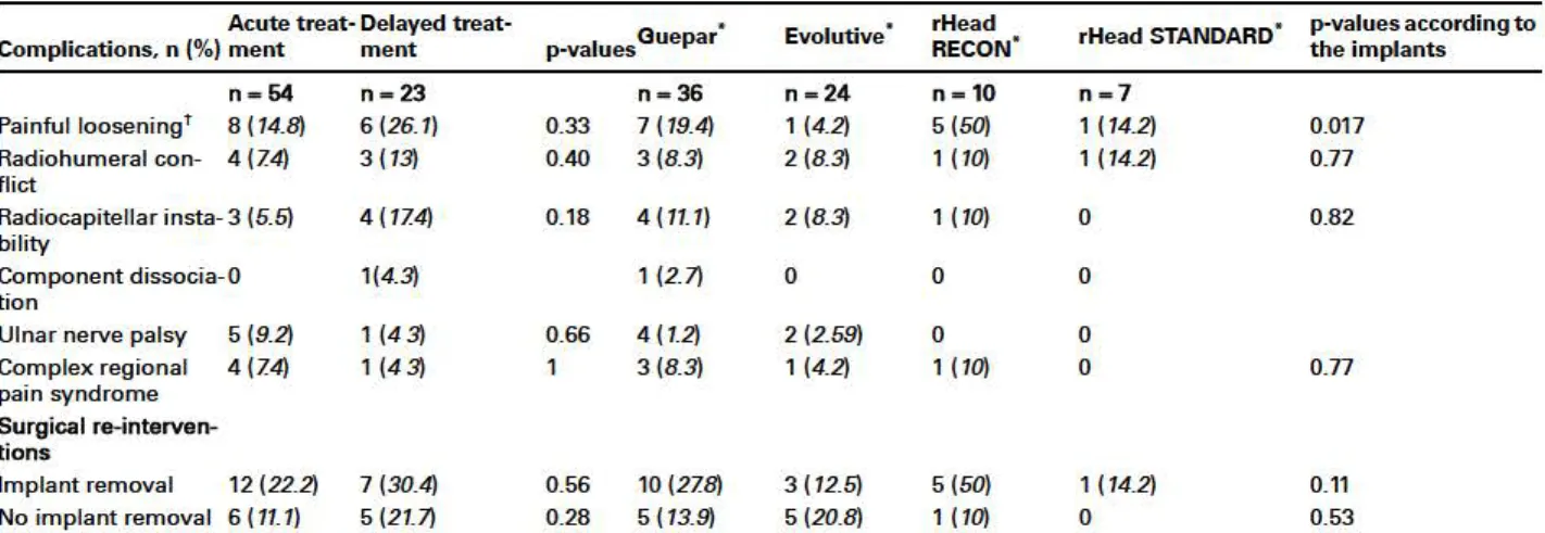
Reasons for re-operations. A total of 40 complications were encountered and 30 patients (38.9%) required a re-operation at a mean follow-up of 14.75 months (SD 11; 0.2 to 36). A total of 11 patients (14.28%) had a re-operation with retention of the implant at a mean of 4.57 months (SD 4.13; 0.2 to 13). A total of 19 implants (24.7%) were removed at a mean of 21 months (SD 9; 6 to 36). These results are shown in Table III.
We reviewed the pre-operative findings including proxi-mal radial forearm pain as described by O’Driscoll and Herald37 and the operation notes including records of loos-ening to identify whether painful loosloos-ening was the cause for re-operation in each patient. Sub-group analysis revealed that using an implant with a short stem signifi-cantly increased the risk of painful loosening compared Table II. Description of clinical and radiographic outcomes by design of radial head arthroplasty and acute or delayed use
Acute
treatment Delayed treatment p-values Guepar* Evolutive* rHead RECON* rHead STANDARD* p-values according to the implants
Clinical results, mean (SD) n = 42 n = 16 n = 26 n = 21 n = 5 n = 6
QuickDASH score (points) 13.1 (10.24) 16.25 (11.27) 0.86 12.3 (11.6) 13.9 (9.5) 18.2 (11.1) 17.5 (13) 0.98
MEPS (points) 91.5 (12) 86.8 (16.16) 0.06 93 5 (15.2) 88 (16.19) 85.3 (11) 88.5 (16.6) 1.38 Range of movement (°) Flexion 132.1° (16.49°) 128.1° (18.97°) 0.37 132.5° (18.1°) 135.3° (16.1°) 126.1° (17.3°) 120.5° (20.3°) 0.30 Extension - 12 9° (11.03°) -16.9° (13.88°) 0.28 -16.19° (15.5°) - 14.9° (12.2°) -9 3° (8.1°) - 11.9° (12.04°) 0.16 Supination 67.8° (7.66°) 65° (9.97°) 0.30 67.7° (7.1°) 65.7° (6.6°) 67.2° (9 3°) 68.1° (13.1°) 0.88 Pronation 76° (7.71°) 75° (9.12°) 0.74 77° (8.7°) 74.1° (9.8°) 76.8° (5.4°) 75.9° (9.1°) 0.44
Force compared with contralateral side (%)
Flexion 87.2 (19.25) 90 (19.35) 0.41 91.1 (21) 86.6 (17.1) 95.9 (15.8) 92.1 (20.1) 0.81
Extension 93.6 (15.79) 95.1 (14.72) 0.94 -16.9 (15.5) - 9.1 (12.2) -13 (8.1) - 9.3 (16 3) 0.88
Radiographic results, n (%) n = 54 n = 23 n = 36 n = 24 n = 10 n = 7
Osteolysis 22 (40.7) 16 (69.5) 0.22 14 (38.9) 12 (50) 8 (80) 4 (57.1) 0.13
Around the stem† 21 (38.9) 13 (56.5) 0.21 13 (36.1) 8 (33.3) 8 (80) 5 (71.4) 0.03
Under the head (collar)† 13 (24.1) 4 (17.4) 0.76 8 (22.2) 1 (4.2) 4 (40) 4 (57.1) 0.009
Malposition 25 (46.3) 13 (56.52) 0.46 20 (55.6) 7 (29.1) 8 (80) 3 (42.9) 0.18 Overstuffing 23 (42.6) 10 (43.5) 1 20 (55.6) 6 (25) 4 (40) 3 (42.9) 0.13 Stem position Centered† 25 (46.3) 10 (43.5) 1 17 (47.2) 14 (58 3) 2 (20) 2 (28.6) 0.04 Varus 20 (37) 10 (43.5) 0.61 17 (47.2) 9 (37.5) 2 (20) 2 (28.6) 0.40 Valgus† 9 (16.7) 3 (13) 1 2 (5.6) 1 (4.2) 6 (60) 3(42.9) 2 x10⁻⁵ Capitellar erosion† 19 (35.2) 10 (43.5) 0.60 15 (41.7) 5 (20.8) 6 (60) 3 (42.9) 0.04
*Guepar/rHead RECON/rHead STANDARD (Small Bone Innovations (SBi)/Stryker, Morrisville, Pennsylvania); Evolutive (Aston Medical, Saint-Etienne, France)
†statistically significant result (p < 0.05)
p-values were calculated using Fisher’s exact test, Kruskal-Wallis tests, and the Mann-Whitney U test QuickDASH, Quick Disabilities of the Arm, Shoulder and Hand; MEPS, Mayo Elbow Performance Score
Table Ill. Description of complications and re-operations by the type of radial head arthroplasty and acute or delayed use Acute treat• Delayed treat• Complications, n (%) ment ment n- 64 Painful looseningr 8 (14.8) Radiohumeral con- 4 (Z4) flict
Radiocapitellar insta-3 (5.5) bility
Component dissocia-0 tien
Ulnar nerve palsy 5 (9.2) Complex regional 4 (Z4) pain syndrome
Surgical ra-interv
en-tions n- 23 6 (26.1) 3 (13) 4 ( 114) 1(4.3) 1 (4 3) 1 (4 3) p•values Ouepar. n- 36 0.33 7 (19.4) 0.40 3 (8.3) 0.18 4 (11.1) 1 (2.7) 0.66 4 (1.2) 3 (8.3) Evolutive
.
rHead RECON" n- 24 n- 10 1 (4.2) 5(50) 2 (8.3) 1 ( 10) 2 (8.3) 1 (10) 0 0 2(2.59) 0 1 (4.2) 1 ( 10) rHead STANDARD" n-7 1 (14.2) 1 (14.2) 0 0 0 0 p-values according to the implants 0.017 0.77 0.82 0.77 Implant removal 12 (22.2) 7 (30.4) 0.56 10 (2Z8) 3 ( 12.5) 5 (50) 1 ( 14.2) 0.11 No implant removal 6 ( 11.1) 5 (21.7) 0.28 5 ( 13.9) 5 (20.8) 1 ( 10) 0 0.53*Guepar/rHead RECON/rHead STANDARD (Small Bene Innovations (SBi)/Stryker, Morrisville, Pennsylvania); Evolutive (Aston Medical, Saint-Etienne, France)
.,
>-
~
::,"'
Cl) >-~
::, E ::, (.)p-values were calculated using Fisher's exact test îstatistically significant result (p < 0.05)
1.0 0.8 0.6 0.4 0.2 0.0 0 2 3 4 5 6 7 8 9 10 11 12 Time (yrs) 1.0 0.8
.,
>-
~
0.6 ::,"'
Cl) >-~
::, 0.4 E ::, (.) 0.2 0.0 0 ...rGuepar ...r Evolutive ...r rHead RECON ...rrHeadSTANDARD 2 3 4 5 6 7 8 9 10 11 12 Time (yrs) Fig.4Kaplan-Meier curves showing overall (left) and sub-group (right) survival (without re-operation) rates of the types of four radial head arthroplasty and acute or delayed use; event = re-operation with or without removal of the implant). (Guepar/rHead RECON/rHead STANDARD (Small Bene Inno -vations (SBi)/Stryker, Morrisville, Pennsylvania); Evolutive (Aston Medical, Saint-Etienne, France)).
with long-stemmed implants (OR 3.54; 1.02 to 12.2;
p = 0.045).
Survivorship analysis. The overall survival for the 77 RHAs was 60.8% (SD 5.7%) at ten years. Five-year survival rates were 75% (SD 7.2%), 66.7% (SD 9.6%), 40.0% (SD 15.5%) and 85.7% (SD 13.2%) for the Guepar, Evolutive, rHead RECON, and rHead STANDARD prostheses, respectively. There was no statistically significant difference between the survival of the four designs (Log Rank (Mantel Cox), p = 0.42) (Fig. 4).
Discussion
This study shows unsatisfactory mid-term results, and does not corroborate excellent outcomes of RHAs published recently in the literature.16•18•19•21
We speculate that outcomes in the present series would
have been worse if ORIF had been performed, as conserva-tive treatment of comminuted radial head fractures leads to an increased risk of early failure of fixation and pseudar-throsis. 38 Despite good mean quickDASH scores and MEPSs of 14.0 and 90.2 points respectively, we report a
high rate of re-operation of 38.9%, including 11 (14.3%) with retention of the implant and 19 revisions (24.67%).
The rate of complications and failures of RHA per-formed in a delayed fashion were high. We confirmed that, when performed acutely, RHA results in improved clinical and radiographic outcomes compared with those per-formed in a delayed fashion, although the difference was not statistically significant due to the small sample size.23,27,29 The three primary reasons for failure were pain-ful loosening (14; 18.2%), radiocapitellar instability (six; 7.5 %), and humeroradial conflict (five; 17.5%). Painful loosening was the most common indication for removal of the implant, although its rate varied significantly among the different designs of RHA (p = 0.017) (Fig. 4). Short-stemmed implants (rHeadRECON and STANDARD) were significantly more prone to loosening compared with those with a long stem (Guepar, Evolutive) (OR 3.54; 1.02 to 12.2; p = 0.045). The rHead prostheses have shorter stems than Guepar or Evolutive designs, but their acetabular components are of identical height to the two others in the series (Table I). According to Shukla et al39 the risk of insta-bility is dependent on the ratio of the length of the radial head of the RHA divided by the total length of the implant. When this ratio is > 0.4, the risk of instability is signifi-cantly higher due to increased micromotion of the stem. The increased ratio in rHead short-stemmed RHAs could explain the significantly increased rate of loosening and osteolysis that we found. For all short-stemmed implants, intra-operative press-fit was found to be insufficient, and cement was required, except in two patients, to obtain a satisfactory fixation. Since a layer of cement could be added, it follows that the diameter of these prostheses was smaller than the maximal and sub-maximal diameter needed. Moon et al40 found that implants of sub-maximal size had micromotion (> 250 micrometers) that exceeded the threshold needed for bone ingrowth and initial stability. Lastly, the level of comfort with the surgical technique could play a role in the high failure rate. Malpositioning (overstuffing) theoretically contributes to the risk of micro-motion of the stem by increasing the extramedullary portion of the implant.39,41-43 We speculate that the increased rate of malposition which we found was due to difficulties in obtaining stable fixation. This may predis-pose the surgeon to favour stability over positioning.
The rate of capitellar wear in our series varied with the design of the implant (p = 0.04). The rates of early capitel-lar wear for the Guepar and monopocapitel-lar rHead STAND-ARD designs were > 40%. This could be explained by hypermobility of the acetabular component and repeated posterolateral subluxation of Guepar RHAs, and higher radiocapitellar contact pressures with rHead STANDARD RHAs.44-47 It has recently been reported that monopolar implants are preferable to bipolar implants in patients with associated ligamentous injury because they allow for supe-rior radiocapitellar stability.44-47 The implant selected for each patient did not depend on the integrity of the soft
tissues. Only one design of RHA was available at the time of each operation for all the patients in this series. We rec-ognise that this is a weakness of the study as the bipolar implant is clearly recommended only when there is malalignment of the proximal radius with respect to the capitellum.
Our study identified two distinct follow-up periods after RHA. Within the first three years there was early drop in survival and during this time re-operations with and with-out removal of the implant were undertaken at a mean of 15.4 months post-operatively. Subsequent survival rates stabilised with an increased life expectancy of the implants which survived for more than three years (Fig. 4).
The limitations of this study relate to its retrospective, single-centre nature and sample size. The retrospective design inherently leads to more loss of data and bias. The small sample size did not allow us to find a statistically sig-nificant difference in outcome between the different types of design. We considered only tight-fitting RHAs and did not include loose-fitting designs. We analysed a hetero-geneous set of uni- and bipolar prostheses and a variety of associated lesions that were not accounted for by compar-ative analysis in the follow-up period. The differences in the sizes of the groups, with, for instance, seven with a monop-olar design and 70 with a bipmonop-olar design, did not allow for reliable comparative sub-group analysis. Surgeon training in elbow surgery, particularly in RHA was variable; we speculate that this may have also influenced the results.27 The analysis of the position of the stem on AP radiographs may have depended on the ROM of the elbow; 16 radio-graphic analyses were performed in patients with incom-plete supination or extension. Follow-up was < 30 months in eight patients with the Evolutive design. These patients were therefore only included in analyses of outcome during the first three post-operative years and the true rate of com-plications may have been lower. Similarly, the true overall survival (mean time 14.75 months, SD 11; 0.2 to 36 and mean time to removal 21 months, SD 9; 6 to 36) may be higher.
In conclusion, the mid-term outcomes of bipolar and press-fit RHAs are unsatisfactory, with a high rate of re-operation during the first three post-operative years. Fixed RHAs may be prone to painful loosening, especially those implants with short stems. A comparative study would be necessary to further assess the risk of painful loosening in loose- compared with tight-fitting RHAs.
Take home message:
- There were high rates of re-operation during the first three years after implantation.
- Fixed RHAs may be prone to painful loosening.
- Short-stemmed implants may be prone to painful loosening.
Author contributions:
P. Laumonerie: Conception and design, Acquisition, analysis and interpretation of data, Critically revising the article, Reviewed submitted version of manu-script, Statistical analysis.
N. Reina: Critically revising the article, Reviewed submined version of manu
-script, Statistical analysis.
D. Ance in: Analysis and interpretation of data, Reviewed submined version of manuscript, Statistical analysis.
S. Delclaux: Analysis and interpretation of data, Critically revising the article,
Reviewed submined version of manuscript.
M. E. Tibbo: Analysis and interpretation of data, Critically revising the article,
Reviewed submined version of manuscript.
N. Bonnevialle: Critically revising the article, Reviewed submitted version of manuscript.
P. Mansat: Conception and design, Analysis and interpretation of data, Critically
revising the article, Reviewed submined version of manuscript, Approved the
final version of the manuscript on beha f of ail authors, Administrative/tec hni-cal/material support, Study supervision.
N. Reina is a paid Consultant for BBraun. N. Bonnevialle is a paid consultant for DePuy, GlaxoSmithKline, Sanofi-ventis and Tomier. Mansat: is a paid consult -ant for DePuy, Synthes, Tornier, and Zimmer.
The author or one or more of the authors have received or will receive benefits
for persona! or professional use from a commercial party related directly or
indirectly to the subject of this article. ln addition, benefits have been or will be
directed to a research fund, foundation, educational institution, or other non
-profit organisation with which one or more of the authors are associated.
References
1. Kaas L van Riet RP, Vroemen JPAM, Eygendaal D. The epidemiology of radial head fractures. J Shau/der Elbaw Surg2010;19:52~23.
2. Herbertsson P. Josefsson PO, Hasse ri us R, et al. Fractures of the radial head and
neck treated with radial head excision. J Bane Joint Surg {AmJ2004;86-A:1925-1930.
3. Ikeda M, Oka Y. Function after early radial head resection for fracture: a retrospec -tive evaluation of 15 patients followed for 3-18 years. Acta Orthap Scand 2000;71:191-194.
4. Ikeda M. Sugiyama K. Kang C, Takagaki T. Oka Y. Comminuted fractures of the radial head. Comparison of resection and internai fixation. J Bane Joint Surg {Am} 2005;87-A:76-84.
5. Schiffern A. Bettwieser SP, Porucznik CA. Crim JR. Tashjian RZ. Proximal radial drift following radial head resection. J ShaulderE/baw Surg2011;20:426-433.
6. van Riel RP, Morrey BF. Delayed valgus instability and proximal migration of the radius after radial head prosthesis failure. J Shau/der Elbow Su,g 2010;19:7-10.
7. Boulas HJ. Morrey BF. Biomechanical evaluation of the e bow following radial head fracture. Comparison of open reduction and internai fixation vs. excision. silastic replacement. and non-operative management. Chir Main 1998;17:314-320.
8. Jensen SL. Olsen BS, Ssjbjerg JO. Bbow joint kinematics after excision of the radial head. J Shau/der Elbaw Surg 1999;8:238-241.
9. Jensen SL Olsen BS, Tyrdal S, Ssjbjerg JO, Sneppen O. Elbow joint laxity after experimental radial head excision and lateral collateral ligament rupture: efficacy of
prosthetic replacement and ligament repair. J Shau/der Elbaw Surg2005;14:78-84. 10. Pomianowski S. Morrey BF, Neale PG, et al. Contr bution of monoblockand bipo
-lar radial head prostheses to valgus stability of the e bow. J Bone Joint Surg {Am} 2001;83-A:1829-1834.
11. Yu SV, Yan HD, Ruan HJ. Wang W. Fan CV. Comparative study of radial head resection and prosthetic replacement in surgical release of stiff elbows. /nt Orthap 2015;39:73-79.
12. Moro JK. Werier J. MacDenmid JC. Panerson SD, King GJ. Arthroplasty with a metal radial head for unreconstructible fractures of the radial head. J Bane Joint Surg {AmJ2001;83-A:1201-1211.
13. Pike JM, Athwal GS, Faber KJ, King GJW. Radial head fractures-an update J Hand Surg Am 2009;34 557-565.
14. Ring D, Jupiter JB. Zilberfarb J. Posterior dislocation of the elbowwith fractures of the radial head and coronoid. J Bane Joint Surg {AmJ2002;84-A 547-551.
15. Shore BJ. Mouon JB. MacDermid JC, Faber KJ. King GJ. Chronic posnrau -matic elbow disorders treated with metallic radial head arthroplasty. J Bane Joint Surg {AmJ2008;90-A:271-280
16. Marsh JP, Grewal R, Fa ber KJ. et al. Radial head fractures treated with modular metallic radial head replacement: outcomes at a mean follow-up of eight years. J Bane Joint Surg {Am}2016;98 527-535.
17. Allavena C, Delclaux S, Bonnevialle N, et al. Dutcomes of bipolar radial head
prosthesis to treat complex radial head fractures in 22 patients with a mean follow -up of 50 months. Orthap Traumatal Surg Res2014;100:703-709.
18. Berschback JC, Lynch TS, Kalainov DM. et al. Clinical and radiographie comp
ar-isons of two different radial head implant designs. J Shau/der Elbaw Surg 2013;22:1108-1120.
19. Dou O. Yin Z, Sun L Feng X. Prosthesis replacement in Mason Ill radial head frac -tures: A meta-analysis. Orthap Traumatal Surg Res 2015;101 :729-734.
20. Ainkkila T. Kaisto T. SirniO K, HyvOnen P, Leppilahti J. Short- to mid-term results of metallic press-fit radial head arthroplasty in unstable injuries of the elbow.
J Bane Joint Surg {BrJ2012;94-B:805-810
21. Gauci M-0, Winter M, Dumontier C. Bronsard N. Allieu Y. Clinical and radio -logie outcomes of pyrocarbon radial head prosthesis: midterm results. J Shau/der Elbaw Surg 2016;25:00-104.
22. Giannicola G, Saccheni FM, Antonietti G, et al. Radial head. radiocapitellar and
total elbow arthroplasties: a review of recent literature. /njury2014;45:428-436.
23. Katthagen JC. Jensen G, Lill H, Voigt C. Monobloc radial head prostheses in com -plex elbow injuries: results after primary and secondary implantation. !nt Orthap
2013;37 63Hl39.
24. Shore BJ, Mouon JB, MacDermid JC, Fa ber KJ, King GJW. Chronic posnrau -matic e bow disorders treated with metallic radial head arthroplasty. J Bane Joint Surg {AmJ2008;90-A:271-280
25. Delclaux S. Lebon J, Faraud A. et al. Complications of radial head prostheses. /nt Orthap 2015;39:907-913.
26. Duckworth AD. Wickramasinghe NR, Clement ND. Court-Brown CM,
McQueen MM. Radial head replacement for acute complex fractures: what are the rate and risks factors for revision or removal? Clin Otthap Relat Res2014;472:21 36-2143.
27. Kachooei AR, Claessen FMAP, Chase SM, et al. Factors associated with removal of a radial head prosthesis placed for acute trauma. /njury2016;47:1253-1257. 28. Neuhaus V, Christoforou DC, Kachooei AR, et al. Radial head prosthesis
removal: a retrospective case series of 14 patients. Arch Bone Jt Surg 2015;3 88-93. 29. van Riel RP. Sanchez-Sotelo J, Morrey BF. Failure of metal radial head repla
ce-ment. J Bone Joint Surg {BrJ2010;92-B:66Hi67
30. Laumonerie P. Ancelin D, Reina N. et al. Causes for early and late surgical re -inteivention after radial head arthroplasty. /nt Orthop2017;41 :1435-1443.
31. lannuui NP, Leopold SS. ln brie!: the Masonclassification of radial head fractures. Clin Orthap Relat Res 2012;470 1799-1802.
32. McGlinn EP. Sebastin SJ. Chung KC. A historical perspective on the Essex -Lopresti injury. J Hand Surg Am 2013;38: 1599-1606.
33. Cusick MC. Bonnaig NS. Azar FM, et al. Accuracy and reliability of the Mayo Bbow Performance Score. J Hand Surg Am 2014;39:1146-1150.
34. Hudak PL Amadio PC. Bombardier C. Development of an upper extremity ou
t-corne measure: the DASH (disabilities of the arm. shoulder and hand) (corrected). The Upper Extremity Collaborative Group (UECG) Am J Jnd Med 1996;29 602-608. 35. Hug KT. Aiton TB, Gee AD. Classifications in brie!: Brooker classification of het
er-otopic ossification after total hip arthroplasty. Clin Orthap Re/at Res 2015;473:21 54-2157.
36. Bowman SH, Barfield WR. Slone HS, Shealy GJ, Wallon ZJ. The clinical impli -cations of heterotopic ossification in patients treated with radial head replacement
for trauma: a case series and review of the literature. J Orthap 2016;13:272-277.
37. O'Driscoll SW, Herald JA. Forearm pain associatedwith loose radial head prosthe -ses. J Shaulder Elb Surg 2012;21 92-97.
38. Ring D. Displaced. unstable fractures of the radial head: fixation vs. replacemen t-what is the evidence? /njury2008;391329-1337.
39. Shukla DR, Fitzsimmons JS, An K-N, O'Driscoll SW. Effect of stem length on prosthetic radial head micromotion. J Shou/der Elbaw Surg 2012;21 :1559-1564. 40. Moon JG, Berglund W. Domire Z, An KN. O'Driscoll SW. Stem diameter and
micromotion of press-fit radial head prosthesis: a biomechanical study. J Shau/der Elbaw Surg 2009;18:785-790.
41. Chan la lit C, Shukla DR, FitzsimmonsJS,An K- N, O'Driscoll SW. Effectofhoop stress fracture on micromotion of textured ingrowth stems for radial head replace -ment. J Shaulder Elbaw Surg2012;21 949-954.
42. Chan la lit C, Shukla DR, Fitzsimmons JS, An K- N. O'Driscoll SW. Stress shie
ld-ing around radial head prostheses. J Hand Surg Am 2012;37:2118-2125. 43. Frank SG. Grewal R, Johnson J, et al. Determination of correct implant size in
radial head arthroplasty to avoid overlengthening. J Bane Joint Surg {Am} 2009;91 -A:1738-1746.
44. Chan la lit C, Shukla DR, Fitzsimmons JS, An K- N. O'Driscoll SW. The biome -chanical effect of prosthetic design on radiocapitellar stability in a terr ble triad
modeL J Orthap Trauma 2012;26:539-544.
45. Moon JG, Berglund W, Zachary D, An K- N, O'Driscoll SW. Radiocapitellar joint
stability with bipolar versus monopolar radial head prostheses. J Shau/der Elbaw Surg2009;18:779-784.
46. Sahu D. Holmes DM, Fitzsimmons JS, et al. Influence of radial head prosthetic design on radiocapitellar joint contact mechanics. J Shau/der Elbow Surg
2014;23:456-462.
47. Chan la lit C, Shukla DR. Fitzsimmons JS, et al. Radiocapitellar stability: the effect
of soft tissue integrity on bipolar versus monopolar radial head prostheses. J Shaul -der Elbaw Surg2011;20219-225.