1
Key role of secondary conformational conversion in glycosaminoglycans-mediated cellular uptake of PACAP, a cationic cell-penetrating peptide
Armelle Tchoumi Neréea,b, Phuong Trang Nguyena,b, David Chatenetc, Alain Fournierc and Steve Bourgaulta,b*
aDepartment of Chemistry, Pharmaqam, University of Québec in Montreal, Montreal, QC, Canada,
H3C 3P8
bQuebec Network for Research on Protein Function, Structure, and Engineering, PROTEO cINRS-Institut Armand-Frappier, 531 boul. des Prairies, Laval, Qc, Canada, H7V 1B7
*Corresponding author: Pr. Steve Bourgault,
Université du Québec à Montréal C.P. 8888, Succursale Centre-Ville Montréal (Québec), H3C 3P8, Canada 1-514-987-3000 (5161)
ABSTRACT
For cationic cell-penetrating peptides (CPPs), glycosaminoglycans (GAGs) contribute significantly to their cellular uptake. However, molecular details about the contributions of GAGs in internalization remain unclear. In this study, we examined the cellular uptake mechanism of an arginine-rich CPP, pituitary adenylate-cyclase-activating polypeptide (PACAP). We observed that the uptake efficacy of PACAP was dependent of cell surface GAGs. As the binding of PACAP to sulfated GAGs induced a random coil-to--helix conformational conversion, we probe the role of the helical formation in internalization. Whereas this secondary structure was not crucial for efficient internalization in GAGs-deficient cells, PACAP -helix was essential for GAGs-dependent uptake.
Keywords:
Cell-penetrating peptide; Glycosaminoglycans; -helix; Pituitary adenylate cyclase-activating polypeptide; Cellular uptake
Highlights:
Glycosaminoglycans play a key role for the cellular uptake of the cationic peptide PACAP
PACAP binding to GAGs induces a random coil-to--helix conformational conversion
1. Introduction
Over the last two decades, cell-penetrating peptides (CPP) have gained increase of interest as chemical tools for the intracellular delivery of macromolecular cargoes intended for biological and medical applications [1]. CPPs are a class of diverse peptides, usually ranging from 5 to 30 residues, which can cross the plasma membrane through a variety of mechanisms that remain partially elusive. According to their physicochemical properties, CPPs can be classified into three major classes: (i) cationic, (ii) amphipathic and (iii) hydrophobic [2]. Cationic CPPs are short peptides that are rich in arginine and lysine residues [3, 4]. The vast majority of CPPs derived from natural protein motifs and were identified in DNA/RNA-binding proteins, viral proteins, signal peptides or heparin-binding proteins [2]. Interestingly, we recently reported that an endogenous peptide neurohormone, pituitary adenylate-cyclase-activating polypeptide (PACAP), can cross efficiently the plasma membrane in a specific-receptor independent manner, mainly by active endocytosis involving clathrin-dependent pathway and micropinocytosis [5]. This peptide was highly effective to mediate the cellular uptake of a variety of cargoes, including protein and DNA plasmid [6]. The cellular uptake efficacy of PACAP was 3-times higher than that observed for the TAT peptide [5], underlining the potent ability of this peptide to cross plasma membrane. These studies have identified PACAP as a new member of the CPP family and suggest that PACAP derivatives represent excellent vectors for the intracellular delivery of non-permeable molecules. Nonetheless, as for other CPPs, the elucidation of the molecular requirements of PACAP internalization is necessary to improve its delivery efficiency.
PACAP is a 38-amino acid C-terminally--amidated peptide that encompasses 11 basic residues, i.e. 4 arginines and 7 lysines, conferring a polycationic nature to this peptide (Fig. 1A) [7]. PACAP exhibits a random coil conformation in aqueous solutions whereas the central and C-terminal domains of the polypeptide chain readily adopt a helical structure in membrane mimicking milieu, such as
dodecylphosphocholine (DPC) micelles (Fig. 1B) [8-10]. Helical wheel representation of this putative helical segment (Fig. 1C) shows that cationic residues are dispersed on both sides of the -helix, conferring a highly positive charge distribution on the overall surface of the peptide. For cationic CPPs, it has been reported that membrane-associated glycosaminoglycans (GAGs) contribute significantly to their cellular uptake [11]. For instance, the removal of cell surface sulfated GAGs was shown to decrease substantially the cellular uptake of the TAT peptide [12], penetratin [13, 14] and the R8 poly-arginine peptide [15].
GAGs, including heparan sulfate (HS) and chondroitin sulfate (CS), are long and linear polysaccharides composed of repeating disaccharide units [16]. They are abundant on the outer leaflet of the plasma membrane of every cell type of metazoan organisms where they are O-linked to proteoglycans [17]. Owing to their high content in carboxylate and sulfate groups, GAGs are highly negatively charged biopolymers that surround cells. Despite the well-recognized importance of GAGs in the uptake of cationic CPPs, the molecular details regarding the roles of the polysaccharide-motifs of proteoglycans are still a matter of controversy and several mechanisms have been inferred from biophysical and biochemical investigations. It has been recently proposed by Favretto et al., [11] that the roles of GAGs in the endocytosis of CPPs could be ascribed to either (i) GAGs clustering upon peptide binding, (ii) co-clustering of a receptor and GAGs upon CPP binding and/or (iii) GAGs mediating peptide adsorption to the plasma membrane. Particularly, in contrast to amphipathic CPPs, the importance of the secondary structure of cationic CPPs has been poorly investigated so far.
In this study, we first investigated the roles of cell surface GAGs in the adsorption of PACAP to the outer leaflet of plasma membrane and its subsequent cellular uptake. Considering that the binding of PACAP to sulfated GAGs induced a random coil-to--helix conformational conversion, we studied the
contribution of this secondary structure in cellular uptake. This study highlights the mechanistic elements of PACAP endocytosis and exposes a new molecular basis of GAGs-mediated uptake of cationic CPPs.
2. Materials and methods
2.1. Peptide synthesis, purification and characterization
Peptides were synthetized on solid phase, purified by reverse-phase high performance liquid chromatography and characterized by mass spectrometry as described in the Supplementary materials.
2.2. Peptide uptake
CHO K1 and CHO pgs-A-745 were seeded in 12-well plates at a density of 30 000 cells/well for 48 hours. Cells were incubated in presence of fluorescein-labelled peptide for 1 h at 37°C and 5% CO2. Time of incubation was defined according to our previous study [5]. After incubation, cells were washed twice with HBSS buffer, treated for 5 min with 100 g/mL heparin to remove the excess of peptide bound to the cell surface [18, 19], washed once again and detached by trypsinization. Trypsin action was stopped and cells were centrifuged. Cells were resuspended in 500 L sorting buffer and kept on ice until flow cytometry analysis. For heparinase treatment, CHO K1 cells were seeded as above-described. After 24 h incubation, cells were treated overnight with 8 UI/mL of heparinase. One hour before cell treatment with peptide, heparinase was added and experiments were performed as above-described. These methods are described in details in Supplementary materials.
2.3. Membrane binding
Cell surface absorption was assessed using a protocol adapted from Gump et al. [20]. Cells were seeded as above-described and after 48 h, cells were incubated on ice at 4°C for 10 min. Cells were then incubated in presence of fluorescein-labelled peptide for 15 min at 4C. Cells were then washed 3 times and detached manually. Cells were centrifuged at 4C before being resuspended in sorting buffer. Flow cytometry analyses were performed using a FACS Calibur instrument (BD Biosciences) and a minimum
of 10 000 gated cells per sample were analyzed. These methods are described in details in Supplementary materials.
2.4. Characterization of PACAP-sulfated GAGs interactions
Affinity chromatography, circular dichroism (CD) spectroscopy and static light scattering were used to probe PACAP interactions with heparin as described in the Supplementary materials
3. Results
3.1. Cell surface glycosaminoglycans promote cellular uptake and membrane binding of PACAP
According to the high positive net charge of PACAP, we initially investigated the potential role of cell surface GAGs in the uptake and membrane binding. We used the CHO pgs-A-745 cells, which are deficient in xylosyltransferase, an enzyme that catalyzes the transfer of a D-xylosyl group to the side chain of a serine, a key step in the synthesis of proteoglycans. As a consequence, these cells do not express any GAGs on their plasma membrane [21]. As observed by flow cytometry, pgs-A-745 cells were significantly less effective than their wild type counterpart to internalize fluorescein-labelled PACAP (Fig. 2A, Fig. 2B). To confirm that these results were not a consequence of a defect in the endocytosis machinery of CHO pgs-A-745, we exposed the CHO K1 cells to an enzymatic treatment to remove cell surface GAGs. To digest cell surface HS, two successive treatments with 8 UI/mL heparinase I/III were performed. Such treatment was previously shown to be effective for decreasing the amount of cell surface GAGs, while not affecting the integrity of the plasma membrane [22, 23]. It is worth mentioning that HSPGs constitute the major proteoglycans of CHO K1 cells [24]. The uptake of PACAP was reduced by approximately 50% upon HS enzymatic removal with heparinase (Fig. 2C, Fig. 2D). Particularly, CHO K1 heparinase-treated cells showed a comparable uptake of PACAP to pgs-A-745 cells. Secondly, we addressed the contributions of proteoglycans in extracellular membrane association by incubating the cells at 4C for 15 min before detaching them with a non-enzymatic treatment. Such conditions are known to inhibit active endocytic pathways [14, 20]. As observed for endocytosis, the extent of cell membrane binding was dependent on the presence of GAGs, with cell surface absorption being approximatively 3-fold higher for CHO K1 (Fig. 3A, Fig. 3B). These results demonstrate the major contributions of cell surface GAGs in PACAP cellular uptake and membrane binding. Nonetheless, GAGs-independent mechanisms of endocytosis also contribute significantly, as noticed for the cell uptake observed with the pgs-A-745 and heparinase-treated cells. This is in
agreement with previous studies reporting that the removal of cell surface HS leads to a 20 to 80% reduction of uptake of cationic CPPs [11, 12, 14, 15].
3.2. Conformational conversion of PACAP upon binding to heparin
PACAP exhibits mainly a random coil conformation in aqueous solution while it readily adopts an -helix in presence of DPC micelles [25-27]. By CD spectroscopy, the far-UV spectra of PACAP in absence of heparin exhibited a single minimum around 200 nm characteristic of a random coil structure (Fig. 4). In contrast, in presence of 0.25 to 1 molar equivalent of heparin, CD spectra displayed two negative minima at 208 and 222 nm and a positive maximum at 192 nm, indicative of a helical conformation (Fig. 4). At a molar ratio of 8 (npeptide/heparin), the CD signal was weak and somewhat unclear, most likely because of light scattering produced by the formation of large particles that could be observed by visual inspection. These data suggest that heparin acts as a molecular anionic surface for the conformational conversion of PACAP.
3.3. Role of conformational conversion in PACAP cellular uptake
This random coil-to--helix conformational conversion upon PACAP binding to sulfated GAGs has been previously reported for the CPPs melittin [28] and penetratin analogs [29]. However, the contribution of this structural change in the uptake of cationic CPPs has not been addressed so far. Thus, we investigated this issue by designing two PACAP analogs in which two or three pairs of adjacent residues were substituted with their corresponding D-enantiomers. Double D-substitutions introduced in the putative helical region of PACAP should result in disturbance of the -helix, without modifying other properties of the peptide such as hydrophobicity and net charge [30]. Considering that the -helix in structure-induced milieu spans from Thr7 to the C-terminal end [10], we introduced the D,D successive substitutions in positions 11-12 and 23-24 (D4-PACAP) as well as in positions 11-12, 23-24
and 33-34 (D6-PACAP) to disrupt the formation of a -helix induced by heparin. By CD spectroscopy, D4-PACAP and D6-PACAP exhibited mainly a random coil conformation in absence or in presence of heparin with spectra exhibiting a single minimum fluctuating between 200 and 205 nm (Fig. 5A, Fig. 5B). To confirm that the incapacity of these two D,D-residue containing analogs to switch into helical conformation was not the result of a lower binding affinity towards the sulfated GAG, we initially performed affinity chromatography. D4-PACAP and D6-PACAP eluted from the heparin-sepharose column approximately at the same NaCl concentration as that of PACAP (Fig. 5C), confirming that these two derivatives bound heparin with a relatively high affinity.
As revealed by affinity chromatography, D4-PACAP and D6-PACAP can bind heparin to the same extent as PACAP, although, as observed by CD spectroscopy, this binding event is not accompanied with the formation of a helical structure. Thus, the role of PACAP random coil-to--helix conformational conversion in uptake was assessed by evaluating the endocytosis of fluorescein-labelled D,D derivatives. D4-PACAP and D6-PACAP were significantly less internalized in CHO K1 cells compared to PACAP with relative cellular uptakes varying between 10 and 20% of that PACAP (Fig. 6A, Fig. 6B). In contrast, in GAGs-defective pgs-A-745 cells, the uptake efficacies of double D-substituted analogs were ranging between 60 to 80% of that PACAP (Fig. 6C, Fig. 6D). The relative lower extent of uptake of D,D-derivatives in CHO K1, in contrast to pgs-A-745, indicates that the -helix structure is critical for GAGs-mediated endocytosis whereas GAGs-independent internalization is less affected by the locked random coil conformation of PACAP.
While trying to characterize the thermodynamics and binding constants of the interaction of PACAP with sulfated GAGs by isothermal titration calorimetry (ITC), an approach that we routinely use [31], we were unsuccessful to get a clear calorimetry plot, most likely because of a prompt aggregation event.
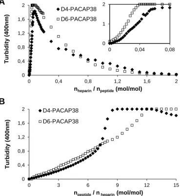
In fact, we could observed by visual inspection that the heparin solution become turbid upon PACAP addition. It has been previously shown that GAGs clustering plays a pivotal role in the endocytosis of cationic CPPs, such as WR9 [32] and penetratin [14]. Therefore, we explored if the lower GAGs-dependant cellular uptake of D4-PACAP and D6-PACAP could not be ascribed to a lower capacity to cluster sulfated GAGs. First, we attempted to analyze by dynamic light scattering the formation of molecular heparin-peptide complexes, but this approach was not appropriate as heparin-PACAP clusters aggregated too quickly and to large extent. Consequently, reliable data could not be obtained. Hence, we simply monitored the increase of solution turbidity at 400 nm upon the titration of the peptide into heparin and vice versa. When heparin (1.25 L; 100 M) was successively titrated into PACAP solution (1 mL; 50 M), we observed a rapid increase of turbidity after an initial baseline during the first few injections (Fig. 7A, inlet). Increasing the heparin/peptide ratio at a molar ratio of 0.8 and higher led to a less turbid solution. In the peptide-into-heparin titration experiment, PACAP solution showed a significant increase of turbidity at a molar ratio of npeptide/heparin = 2 and higher (Fig. 7B). Interestingly, D4- and D6-PACAP displayed a somewhat similar capacity to that of PACAP to form large particles in in the heparin-into-peptide titration experiment. In the peptide-into-heparin titration, the increase of turbidity at 400 nm was faster in the case of PACAP. Nonetheless, it is very unlikely that the slight variation in heparin clustering in vitro observed for D4-PACAP and D6-PACAP underlies the pronounced difference in the cellular uptake of these peptides by CHO K1 cells. Overall, these data indicate that the cellular uptake efficacy of PACAP does not necessary correlate to its in vitro binding to sulfated GAGs and/or GAGs clustering capacity. Instead, we showed that the conformational transition of a natively disordered cationic CPP is crucial for GAGs-mediated internalization.
4. Discussion
Cell surface GAGs were shown to interact with relatively high affinity with polycationic peptides [33-36] and to contribute significantly to the cellular uptake of CPPs [12-15]. It has been originally hypothesized that the contribution of GAGs in uptake is primarily related to the adsorption of CPPs to the outer leaflet of the plasma membrane through electrostatic interactions [37]. Over the last decade, several reports have instead proposed that the capacity of cationic CPPs to cluster GAGs can directly be ascribed to their cell penetrating efficacy [14, 15, 32]. For instance, it was shown that the internalization efficiency of penetratin derivatives correlates directly with their ability to cluster heparin in vitro [14]. On the other hand, it has been recently shown that the cellular uptake efficacy of CPPs derived from the human protein lactoferrin (hLF) did not correlate with the propensity of hLF peptides to cluster HS in
vitro [38]. Instead, the authors found a strong negative correlation between the extent of cellular uptake
and the stoichiometry of binding to HS of hLF peptide analogs [38]. Thus, although the contribution of cell surface GAGs in the uptake of cationic CPPs is well-recognized, the molecular mechanisms by which proteoglycans facilitate internalization remains unclear [11]. Moreover, even if some studies have reported that the binding of cationic peptides to sulfated GAGs induce the formation of a -helix [28, 29], the role(s) of this conformational shift in GAGs-mediated cellular uptake has not been investigated so far.
Herein, we focused on the mechanism of cellular uptake of PACAP in the context of cell surface GAGs. As previously reported for other cationic CPPs [11-15, 29], we showed here that GAGs play a pivotal role in the internalization of PACAP. It was originally assumed that the chirality and/or secondary structure of cationic CPPs did not play a pivotal role in their uptake efficacy [13]. Instead, the number of positively charged residues and their linear disposition were considered as the key factors for efficient internalization. However, it was recently reported that the uptake efficiency of a CPP derived from the
hLF protein is dependent on its conformation [39] and that L-nona-arginine peptide is internalized more efficiently than its all-D-counterpart [40]. As we observed that PACAP readily adopts a -helix in presence of heparin, we sought to investigate the role(s) of this heparin-induced conformational conversion in cellular uptake by destabilizing PACAP helical structure. D,D-analogs bound heparin with a relative affinity similar to that observed for PACAP and both peptides were efficient to induce GAGs clustering. Nonetheless, D, D-derivatives were poorly taken up by CHO K1 cells. In sharp contrast, in GAGs-deficient cells, the levels of uptake of D,D-analogs were around 80% of that PACAP. This indicates that this conformational conversion is essential in GAGs-dependent internalization whereas in GAGs-independent uptake, the formation of a helical secondary structure is not an absolute prerequisite for cellular uptake. To the best of our knowledge, this is the first report describing a key implication of conformational conversion of a cationic CPP in GAGs-mediated endocytosis. It has been recently shown that the stoichiometry of peptide-HS binding is a key determinant in the uptake efficiency of CPPs [38]. Unfortunately, because of the prompt aggregation in the initial phase of PACAP-into-heparin and heparin-into-PACAP titrations, we were unable to determine the binding stoichiometry by ITC. Nevertheless, it is hypothesized that the conformational shift of PACAP modified the stoichiometry of peptide-GAGs, resulting in a change in internalization level, an avenue that should be further investigated. Besides, formation of an -helix triggers the spatial gathering of Arg and Lys side chains and the formation of clusters of positive charges that might tighten GAGs-peptide interactions.
Overall, this study emphasizes the promoting roles of cell surface proteoglycans in the endocytosis of cationic CPPs and sheds light on the molecular basis of the high cell penetrating capacity of PACAP. Besides, we demonstrated that the uptake efficiency of a given cationic CPP does not necessarily correlate to its in vitro binding to sulfated GAGs and/or to its in vitro ability to cluster GAGs. Instead, this report describes that the adoption of an -helix is as a crucial step for GAGs-mediated endocytosis
of a cationic CPP. This observation will support the development of CPPs since it is not only GAGs binding/clusteringbut also peptide conformation that drives the extent of internalization.
Author contribution
A.T.N. and P.T.N designed, performed the experimental work and analyzed data. S.B. wrote the initial version of the paper and supervised the research. A.F. and D.C. participated in the design of the research and revised the paper
Acknowledgments
This work was supported by a grant from the Natural Sciences and Engineering Research Council of Canada (S.B.) and the Canadian Institutes of Health Research (A.F.). The authors thank Doan Ngoc Doan and Myriam Létourneau for their helpful discussions and Pr. Borhane Annabi for support with the flow cytometry analysis.
References
1. Jones, A. T. & Sayers, E. J. (2012) Cell entry of cell penetrating peptides: tales of tails wagging dogs, Journal of controlled release : official journal of the Controlled Release Society. 161, 582-91.
2. Milletti, F. (2012) Cell-penetrating peptides: classes, origin, and current landscape, Drug discovery today. 17, 850-60.
3. Futaki, S., Suzuki, T., Ohashi, W., Yagami, T., Tanaka, S., Ueda, K. & Sugiura, Y. (2001) Arginine-rich peptides. An abundant source of membrane-permeable peptides having potential as carriers for intracellular protein delivery, The Journal of biological chemistry. 276, 5836-40.
4. Tunnemann, G., Ter-Avetisyan, G., Martin, R. M., Stockl, M., Herrmann, A. & Cardoso, M. C. (2008) Live-cell analysis of cell penetration ability and toxicity of oligo-arginines, Journal of peptide science : an official publication of the European Peptide Society. 14, 469-76.
5. Doan, N. D., Chatenet, D., Letourneau, M., Vaudry, H., Vaudry, D. & Fournier, A. (2012) Receptor-independent cellular uptake of pituitary adenylate cyclase-activating polypeptide, Biochimica et biophysica acta. 1823, 940-9. 6. Doan, N. D., Letourneau, M., Vaudry, D., Doucet, N., Folch, B., Vaudry, H., Fournier, A. & Chatenet, D. (2012) Design and characterization of novel cell-penetrating peptides from pituitary adenylate cyclase-activating polypeptide, Journal of controlled release : official journal of the Controlled Release Society. 163, 256-65.
7. Vaudry, D., Falluel-Morel, A., Bourgault, S., Basille, M., Burel, D., Wurtz, O., Fournier, A., Chow, B. K., Hashimoto, H., Galas, L. & Vaudry, H. (2009) Pituitary adenylate cyclase-activating polypeptide and its receptors: 20 years after the discovery, Pharmacological reviews. 61, 283-357.
8. Hoare, S. R. (2005) Mechanisms of peptide and nonpeptide ligand binding to Class B G-protein-coupled receptors, Drug discovery today. 10, 417-27.
9. Bourgault, S., Chatenet, D., Wurtz, O., Doan, N. D., Leprince, J., Vaudry, H., Fournier, A. & Vaudry, D. (2011) Strategies to convert PACAP from a hypophysiotropic neurohormone into a neuroprotective drug, Current pharmaceutical design. 17, 1002-24.
10. Sze, K. H., Zhou, H., Yang, Y., He, M., Jiang, Y. & Wong, A. O. (2007) Pituitary adenylate cyclase-activating polypeptide (PACAP) as a growth hormone (GH)-releasing factor in grass carp: II. Solution structure of a brain-specific PACAP by nuclear magnetic resonance spectroscopy and functional studies on GH release and gene expression, Endocrinology. 148, 5042-59.
11. Favretto, M. E., Wallbrecher, R., Schmidt, S., van de Putte, R. & Brock, R. (2014) Glycosaminoglycans in the cellular uptake of drug delivery vectors - bystanders or active players?, Journal of controlled release : official journal of the Controlled Release Society. 180, 81-90.
12. Richard, J. P., Melikov, K., Brooks, H., Prevot, P., Lebleu, B. & Chernomordik, L. V. (2005) Cellular uptake of unconjugated TAT peptide involves clathrin-dependent endocytosis and heparan sulfate receptors, The Journal of biological chemistry. 280, 15300-6.
13. Jiao, C. Y., Delaroche, D., Burlina, F., Alves, I. D., Chassaing, G. & Sagan, S. (2009) Translocation and endocytosis for cell-penetrating peptide internalization, The Journal of biological chemistry. 284, 33957-65. 14. Amand, H. L., Rydberg, H. A., Fornander, L. H., Lincoln, P., Norden, B. & Esbjorner, E. K. (2012) Cell surface binding and uptake of arginine- and lysine-rich penetratin peptides in absence and presence of proteoglycans, Biochimica et biophysica acta. 1818, 2669-78.
15. Nakase, I., Tadokoro, A., Kawabata, N., Takeuchi, T., Katoh, H., Hiramoto, K., Negishi, M., Nomizu, M., Sugiura, Y. & Futaki, S. (2007) Interaction of arginine-rich peptides with membrane-associated proteoglycans is crucial for induction of actin organization and macropinocytosis, Biochemistry. 46, 492-501.
16. Bishop, J. R., Schuksz, M. & Esko, J. D. (2007) Heparan sulphate proteoglycans fine-tune mammalian physiology, Nature. 446, 1030-7.
17. Moremen, K. W., Tiemeyer, M. & Nairn, A. V. (2012) Vertebrate protein glycosylation: diversity, synthesis and function, Nature reviews Molecular cell biology. 13, 448-62.
18. Kaplan, I. M., Wadia, J. S. & Dowdy, S. F. (2005) Cationic TAT peptide transduction domain enters cells by macropinocytosis, Journal of controlled release : official journal of the Controlled Release Society. 102, 247-53.
19. Al-Taei, S., Penning, N. A., Simpson, J. C., Futaki, S., Takeuchi, T., Nakase, I. & Jones, A. T. (2006) Intracellular traffic and fate of protein transduction domains HIV-1 TAT peptide and octaarginine. Implications for their utilization as drug delivery vectors, Bioconjugate chemistry. 17, 90-100.
20. Gump, J. M., June, R. K. & Dowdy, S. F. (2010) Revised role of glycosaminoglycans in TAT protein transduction domain-mediated cellular transduction, The Journal of biological chemistry. 285, 1500-7.
21. Esko, J. D., Stewart, T. E. & Taylor, W. H. (1985) Animal cell mutants defective in glycosaminoglycan biosynthesis, Proceedings of the National Academy of Sciences of the United States of America. 82, 3197-201. 22. Bradshaw, A. C., Parker, A. L., Duffy, M. R., Coughlan, L., van Rooijen, N., Kahari, V. M., Nicklin, S. A. & Baker, A. H. (2010) Requirements for receptor engagement during infection by adenovirus complexed with blood coagulation factor X, PLoS pathogens. 6, e1001142.
23. Verdurmen, W. P., Wallbrecher, R., Schmidt, S., Eilander, J., Bovee-Geurts, P., Fanghanel, S., Burck, J., Wadhwani, P., Ulrich, A. S. & Brock, R. (2013) Cell surface clustering of heparan sulfate proteoglycans by amphipathic cell-penetrating peptides does not contribute to uptake, Journal of controlled release : official journal of the Controlled Release Society. 170, 83-91.
24. Esko, J. D., Weinke, J. L., Taylor, W. H., Ekborg, G., Roden, L., Anantharamaiah, G. & Gawish, A. (1987) Inhibition of chondroitin and heparan sulfate biosynthesis in Chinese hamster ovary cell mutants defective in galactosyltransferase I, The Journal of biological chemistry. 262, 12189-95.
25. Bourgault, S., Vaudry, D., Guilhaudis, L., Raoult, E., Couvineau, A., Laburthe, M., Segalas-Milazzo, I., Vaudry, H. & Fournier, A. (2008) Biological and structural analysis of truncated analogs of PACAP27, Journal of molecular neuroscience : MN. 36, 260-9.
26. Bourgault, S., Vaudry, D., Segalas-Milazzo, I., Guilhaudis, L., Couvineau, A., Laburthe, M., Vaudry, H. & Fournier, A. (2009) Molecular and conformational determinants of pituitary adenylate cyclase-activating polypeptide (PACAP) for activation of the PAC1 receptor, Journal of medicinal chemistry. 52, 3308-16.
27. Wray, V., Kakoschke, C., Nokihara, K. & Naruse, S. (1993) Solution structure of pituitary adenylate cyclase activating polypeptide by nuclear magnetic resonance spectroscopy, Biochemistry. 32, 5832-41.
28. Klocek, G. & Seelig, J. (2008) Melittin interaction with sulfated cell surface sugars, Biochemistry. 47, 2841-9. 29. Bechara, C., Pallerla, M., Zaltsman, Y., Burlina, F., Alves, I. D., Lequin, O. & Sagan, S. (2013) Tryptophan within basic peptide sequences triggers glycosaminoglycan-dependent endocytosis, FASEB journal : official publication of the Federation of American Societies for Experimental Biology. 27, 738-49.
30. Wieprecht, T., Dathe, M., Schumann, M., Krause, E., Beyermann, M. & Bienert, M. (1996) Conformational and functional study of magainin 2 in model membrane environments using the new approach of systematic double-D-amino acid replacement, Biochemistry. 35, 10844-53.
31. De Carufel, C. A., Nguyen, P. T., Sahnouni, S. & Bourgault, S. (2013) New insights into the roles of sulfated glycosaminoglycans in islet amyloid polypeptide amyloidogenesis and cytotoxicity, Biopolymers. 100, 645-55. 32. Ziegler, A. & Seelig, J. (2011) Contributions of glycosaminoglycan binding and clustering to the biological uptake of the nonamphipathic cell-penetrating peptide WR9, Biochemistry. 50, 4650-64.
33. Ziegler, A. & Seelig, J. (2004) Interaction of the protein transduction domain of HIV-1 TAT with heparan sulfate: binding mechanism and thermodynamic parameters, Biophysical journal. 86, 254-63.
34. Rullo, A., Qian, J. & Nitz, M. (2011) Peptide-glycosaminoglycan cluster formation involving cell penetrating peptides, Biopolymers. 95, 722-31.
35. Goncalves, E., Kitas, E. & Seelig, J. (2006) Structural and thermodynamic aspects of the interaction between heparan sulfate and analogues of melittin, Biochemistry. 45, 3086-94.
36. Ziegler, A. & Seelig, J. (2008) Binding and clustering of glycosaminoglycans: a common property of mono- and multivalent cell-penetrating compounds, Biophysical journal. 94, 2142-9.
37. Drin, G., Cottin, S., Blanc, E., Rees, A. R. & Temsamani, J. (2003) Studies on the internalization mechanism of cationic cell-penetrating peptides, The Journal of biological chemistry. 278, 31192-201.
38. Wallbrecher, R., Verdurmen, W. P., Schmidt, S., Bovee-Geurts, P. H., Broecker, F., Reinhardt, A., van Kuppevelt, T. H., Seeberger, P. H. & Brock, R. (2014) The stoichiometry of peptide-heparan sulfate binding as a determinant of uptake efficiency of cell-penetrating peptides, Cellular and molecular life sciences : CMLS. 71, 2717-29.
39. Duchardt, F., Ruttekolk, I. R., Verdurmen, W. P., Lortat-Jacob, H., Burck, J., Hufnagel, H., Fischer, R., van den Heuvel, M., Lowik, D. W., Vuister, G. W., Ulrich, A., de Waard, M. & Brock, R. (2009) A cell-penetrating peptide derived from human lactoferrin with conformation-dependent uptake efficiency, The Journal of biological chemistry. 284, 36099-108.
40. Verdurmen, W. P., Bovee-Geurts, P. H., Wadhwani, P., Ulrich, A. S., Hallbrink, M., van Kuppevelt, T. H. & Brock, R. (2011) Preferential uptake of L- versus D-amino acid cell-penetrating peptides in a cell type-dependent manner, Chemistry & biology. 18, 1000-10.
Figure Legends
Fig. 1. Sequence and structure of PACAP. (A) Primary structure of PACAP with basic residues indicated in italic bold letters. (B) Schematic ribbon representation of micelle-bound PACAP secondary structure (PDB code: 2D2P). (C) Helical wheel representation of the putative -helix segment of PACAP (Thr7-Lys38) with basic residues indicated in blue.
Fig. 2. Role of cell surface glycosaminoglycans in PACAP cellular uptake. (A) Representative flow cytometry histograms showing cellular uptake of 1 M PACAP in CHO K1 (red) and CHO pgs-A-745 (black) cells. (B) Cellular uptake of PACAP by CHO K1 and CHO pgs-A-745 cells. (C) Representative flow cytometry histograms showing cellular uptake of 1 M PACAP in CHO K1 cells treated with 8 UI/mL heparinase (black) and non-treated control cells (red). (D) Cellular uptake of PACAP by CHO K1 treated or not with 8 U/mL heparinase. (B,D) Data represent the relative mean fluorescence intensity ( S.E.M.) of gated cells from at least 3 individual experiments and results are expressed as a percentage of the median fluorescence for 5 M PACAP in CHO K1 cells. (A, B, C, D) Cells were incubated for 1h at 37C in 5% CO2 with fluorescein-PACAP, washed twice in HBSS, treated with 100 g/mL heparin for 5 min, detached by trypsin treatment, washed by centrifugation and resuspended in ice-cold sorting bufferbefore flow cytometry analysis.
Fig. 3. Role of cell surface glycosaminoglycans in PACAP binding to the cell surface. (A) Representative flow cytometry histograms showing membrane binding at 4 C of 1 M PACAP on CHO K1 (red) and CHO pgs-A-745 (black) cells. (B) Membrane binding at 4C of PACAP on CHO K1 and CHO pgs-A-745 cells. Data represent the relative mean fluorescence intensity ( S.E.M.) of gated cells from at least 3 individual experiments and results are expressed as a percentage of the median fluorescence for 5 M PACAP38 in CHO K1 cells. (A, B) Pre-chilled cells were incubated for 15 min at 4C with fluorescein-PACAP, washed 3 times with ice-cold HBSS, detached mechanically with a cell scraper, washed by centrifugation at 4C and resuspended in ice-cold sorting buffer before flow cytometry analysis.
Fig. 4. Conformational conversion of PACAP upon binding to heparin. Circular dichroism spectra of PACAP (50 M) in absence or in presence of increasing concentrations of heparin (3.125 to 50 M). Buffer in all experiments is 20 mM phosphate, 100 mM NaF, pH 7.4 and temperature was set at 25 C.
Fig. 5. Inhibition of heparin-induced conformational transition of PACAP with double-D susbtitutions. (A, B) Circular dichroism spectra of (A) [D-Ser11, D-Arg12, D-Leu23, D-Ala24]PACAP (D4-PACAP) and (B) [D-Ser11, D-Arg12, D-Leu23, D-Ala24, D-Gln33, D-Arg34]PACAP (D6-PACAP) in absence or in presence of increasing concentrations of heparin (3.125 to 50 M). Buffer in all experiments is 20 mM phosphate, 100 mM NaF, pH 7.4, temperature is 25 C and peptide concentration is 50 M. (C) Heparin affinity chromatography of PACAP and its derivatives. Superimposed chromatograms of PACAP (red), D4-PACAP (black) and D6-PACAP (green) elution with increasing NaCl concentration on a sepharose heparin column connected to a FPLC Aktapure system. Injection of 250 g of peptides in phosphate buffer (20 mM, pH 7.4) and the flow rate is 0.5 mL/min.
Fig. 6. Role of PACAP conformational conversion in GAGs-mediated cellular uptake. (A) Representative flow cytometry histograms showing cellular uptake of 1 M D4-PACAP38 (black), D6-PACAP (green) or D6-PACAP (red) in CHO K1 cells. (B) Cellular uptake of D6-PACAP38 and derivatives by CHO K1 cells. (C) Representative flow cytometry histograms showing cellular uptake of 1 M D4-PACAP (black), D6-D4-PACAP (green) or D4-PACAP (red) in CHO pgs-A-745 cells. (D) Cellular uptake of PACAP and derivatives by CHO pgs-A-745 cells. (B, D) Data represent the relative mean fluorescence intensity ( S.E.M.) of gated cells from at least 3 individual experiments and results are expressed as a percentage of the median fluorescence for PACAP used at the same concentration. (A,B,C,D) Cells were incubated for 1h at 37C in 5% CO2 with fluorescein-peptides, washed twice with HBSS, treated with 100 g/mL heparin for 5 min, detached by trypsin treatment, washed by centrifugation and resuspended in ice-cold sorting buffer before performing flow cytometry analysis.
Fig. 7. Light scattering showing heparin clustering by PACAP38 and its D,D-derivatives. (A) Titration of heparin into PACAP (), D4-PACAP () or D6-PACAP () monitored by turbidity at 400 nm. Each peak corresponds to the injection of 1.25 L of a 100 M heparin solution into a 1 mL 50 M peptide solution. Inset: emphasis on the initial phase of the titration assay. (B) Titration of PACAP (), D4-PACAP () and D6-PACAP () into heparin monitored by turbidity at 400 nm. Each peak corresponds to the injection of 12.5 L of a 200 M peptide solution into a 1 mL 10 M heparin solution. Data are expressed as a function of the heparin/peptide molar ratio (A) or peptide/heparin molar ratio (B). (A,B) All experiment were performed in 20 mM phosphate, 100 mM NaCl, pH 7.4.
Figure 1
HSDGIFTDS Y S R Y R KQM AV K K Y LAA VL G KR Y K QRV K N K -C O N H2 A B C N C0 40 80 120 1 10 100 1000 10000 E v e nt s FL1-H intensity (a.u.)
Figure 2
0 40 80 120 1 10 100 1000 10000 E v e nt s FL1-H intensity (a.u.) C D 0 20 40 60 80 5 1 0,5 0,1 R e la ti v e upta k e ( % Concentration (M) K1 pgs-A-745 0 20 40 60 80 100 5 1 0,5 R e la ti v e upta k e ( % ) Concentration (M) Control Treated K1 pgs-A-745 Control Treated0 40 80 120 1 10 100 1000 10000 E v e nt s FL1-H intensity (a.u.)
Figure 3
0 20 40 60 80 5 1 0,5 R e la ti v e upta k e ( % Concentration (M) K1 pgs-A-745 K1 pgs-A-745-25 -20 -15 -10 -5 0 5 10 15 20 25 190 200 210 220 230 240 250 260 M RE x 1 0 3 (de g. c m 2.d m ol -1) Wavelength (nm) + heparin 3.125µM + heparin 6.25µM + heparin 12.5µM + heparin 50µM
Figure 4
-25 -15 -5 5 15 25 190 210 230 250 M R E x 1 0 3 ( deg. c m 2.dm ol -1 Wavelength (nm) D4-PACAP38 + heparin 3.125 µM + heparin 6.25 µM + heparin 12.5 µM + heparin 50 µM -25 -15 -5 5 15 25 190 210 230 250 M R E x 1 0 3 ( deg. c m 2.dm ol -1 Wavelength (nm) D6-PACAP38 + heparin 3.125 µM + heparin 6.25µM + heparin 12.5 µM + heparin 50 µM 0 0,4 0,8 1,2 1,6 0 20 40 60 80 100 0 5 10 15 20 25 30 N a C l (M ) A bsorba nce a t 2 3 0 nm Elution volume (mL) D4-PACAP38 D6-PACAP38
Figure 5
0 40 80 120 1 10 100 1000 10000 E v e nt s FL1-H intensity (a.u.) 0 40 80 120 1 10 100 1000 10000 E v e nt s FL1-H intensity (a.u.) C D 0 10 20 5 1 0,5 R e la ti v e upta k e ( % ) Concentration (M) D6-PACAP38 0 20 40 60 80 100 120 5 1 0,5 R e la ti v e upta k e ( % ) Concentration (M) D4-PACAP38 D6-PACAP38
Figure 6
PACAP38 D4-PACAP38 D6-PACAP38 D4-PACAP38 D6-PACAP38Figure 7
B 0 0,4 0,8 1,2 1,6 0 0,4 0,8 1,2 1,6 2 T urbi di ty ( 4 0 0 nm )nheparin / npeptide (mol/mol)
D6-PACAP38 0 1 0 0,04 0,08 0 0,4 0,8 1,2 1,6 2 0 3 6 9 12 15 T u rbi d it y ( 400nm )
npeptide / n heparin (mol/mol)
D4-PACAP38 D6-PACAP38