Université de Montréal
A prevalence study of dental malocclusions in children with sleep
disorders
by
Jérémie Abikhzer
« Département de santé buccale – Section d’orthodontie » « Faculté de Médecine Dentaire »
Thesis presented to the « Faculté des études supérieures » to obtain a Master’s degree (M.Sc.)
in dental medicine orthodontic option
May, 2017
Résumé
Introduction : Les troubles respiratoires du sommeil (TRS) sont un continuum qui va du
ronflement à l’apnée du sommeil. Le ronflement est un bruit à l’inspiration, causé par la vibration des tissus mous des voies aériennes supérieures détendus par le sommeil. Le syndrome d’apnée du sommeil est caractérisé par l’arrêt partiel ou complet du flot respiratoire de façon répétitive et transitoire durant le sommeil. Alors que l’hypertrophie des adénoïdes/amygdales est le facteur primaire contribuant aux TRS pédiatriques, il pourrait y avoir d’autres origines à l’obstruction tel que les malformations craniofaciales. Le but de cette étude de prévalence est de faire le compte du nombre de patients qui bénéficieraient d’une évaluation dentaire et orthodontique parmi ceux qui ont des troubles respiratoires de sommeil vus au CHU Sainte-Justine. Notre hypothèse de recherche est que la prévalence de malocclusions et d’anomalies dento-squelettiques serait différente entre les enfants apnéiques et non-apnéiques. Méthodologie : Lors de cette étude prospective multicentrique, les patients qui vont compléter un enregistrement de sommeil pour diagnostiquer les troubles respiratoires du sommeil au laboratoire de sommeil du CHU Sainte Justine seront contactés pour participer à cette étude de prévalence (n=100). L’évaluation dentaire se fera durant le rendez-vous. Le questionnaire de dépistage de Gozal et les données polysomnographiques, orthodontiques et craniofaciales seront étudiées. Résultats : Un total de 100 patients a été recruté (58 M, 42F). L’âge moyen des patients était de 9.6 ± 4.05 (3-18 ans). Les patients étaient divisés en groupes (n=57) IAH < 2, (n=43) IAH ≥ 2. Le groupe IAH < 2 avait une moyenne 0.79 ± 0.53. Le groupe IAH ≥ 2 avait une moyenne de 7.79 ± 8.03. Aucune différence n’a été trouvé entre les groupes IAH et le IMC (p=0.303). Par contre, le score de Gozal était significatif pour dépister des IAH plus sévères (p=0.011) pour un score ≥ 2.72. Aucune différence significative n’a été trouvée entre les amygdales hypertrophiques (score ≥3) et l’IAH (p=0.426). De plus, aucune différence significative n'a été trouvée entre les groupes IAH pour les caractéristiques craniofaciales et dentaires. Les patients ayant des habitudes orales (morsures des ongles/joues/ lèvres, bruxisme, succion du pouce) avaient une tendance d’avoir un IAH <2 (p = 0.064). La régression logistique a conclu que les garçons sont plus à risque (OR=3.52, 95%CI 1.27-9.77), ceux avec des habitudes orales sont moins à risque (OR=0.33, 95%CI 0.13-0.89) et que le risque d’avoir l’apnée augmente de 1.09 pour chaque unité d’accroissement d’IMC.
Conclusions : La prévalence des malocclusions dentaires chez les enfants a été jugée non
significative entre les groupes de différentes sévérité d’IAH. Aucune corrélation significative n'a été trouvée entre la morphologie craniofaciale et dentaire et les données sur le sommeil. Néanmoins, il s'agit d'une analyse préliminaire. L'objectif de cette étude multicentrique est de recruter jusqu'à 400 enfants et une analyse plus approfondie sera effectuée. D'autres études sont recommandées pour tirer de meilleures conclusions et améliorer le pouvoir statistique dans le rôle de la morphologie craniofaciale et dentaire chez les enfants avec des troubles respiratoires du sommeil.
Abstract
Introduction: Sleep-disordered breathing (SDB) is a continuum that ranges from snoring to
sleep apnea. SDB occurs in children of all ages, from neonates to adolescents, and it is characterized by repeated events of snoring, and either partial (i.e. hypopnea) or complete (i.e. apnea) upper airway obstruction during sleep. While hypertrophy of the adenoids / tonsils is the primary factor contributing to pediatric SDB, there may be other origins to obstruction such as craniofacial malformations. The purpose of this prevalence study is to count the number of patients who would benefit from a dental and orthodontic assessment among those with sleeping breathing problems seen at the CHU Sainte-Justine. Our research hypothesis is that the prevalence of malocclusions and dento-skeletal abnormalities would be different between apneic and non-apneic children. Methods: In this prospective multicenter study, patients who will complete type 1 polysomnography to diagnose sleep disorders at the CHU Sainte Justine will be contacted to participate in this prevalence study (n=100). Dental and orthodontic evaluation will be done during the appointment. Gozal screening questionnaire, polysomnographic, orthodontic and craniofacial data will be studied. RESULTS: A total of 100 patients were recruited (58 M, 42 F). The mean age of the patients was 9.6 ± 4.05 (3-18 years). Patients were divided into groups (n = 57) AHI <2, (n = 43) AHI ≥ 2. The AHI <2 group had a mean AHI of 0.79 ± 0.53. The AHI ≥ 2 group had a mean AHI of 7.79 ± 8.03. No difference was found between AHI groups and BMI (p = 0.303). On the other hand, Gozal score was significant for detecting more severe AHI’s (p = 0.011) for a severity score ≥ 2.72. No significant difference was found between hypertrophic tonsils (score ≥3) and AHI (p = 0.426). In addition, no significant difference was found between AHI groups for craniofacial and dental characteristics. Patients with oral habits (nail/cheek/lip biting, bruxism, thumb sucking) tended to have an AHI <2 (p = 0.064). Logistic regression calculations concluded that boys are at higher risk (OR = 3.52, 95% CI 1.27-9.77), those with oral habits are less at risk (OR = 0.33, 95% CI 0.13-0.89) and that odds of having apnea increases by 1.09 for each unit of BMI increase. CONCLUSIONS: The prevalence of dental malocclusions in children was found to be insignificant among groups of different AHI severity. No significant correlation was found between craniofacial and dental morphology and sleep data. Nevertheless, this is a preliminary analysis. The objective of this multi-center study is to recruit up to 400 children and further analysis will be carried out. Further studies are recommended to draw better conclusions and improve statistical power in the role of craniofacial and dental morphology in children with sleep disorders.
Keywords: sleep-disordered breathing, obstructive sleep apnea, children, prevalence,
Table of Contents
Résumé ... 1 Abstract ... 2 Table of Contents ... 3 Table List ... 6 Figures List ... 7 Abreviations List ... 8 Acknowledgements ... 10 Chapter 1. Introduction ... 11 1.1 Sleep ... 11 1.2 History... 111.3 Obstructive sleep apnea syndrome (OSAS) ... 11
Chapter 2. Literature Review ... 13
2.1 Prevalence ... 13
2.1.1 Sex... 13
2.1.2 Age ... 14
2.1.3 Race... 14
2.2 Signs & Symptoms ... 15
2.3 Diagnosis... 16
2.3.1 Pediatric sleep questionnaires ... 18
2.4 Treatment ... 19
2.4.1 Adenotonsillectomy (T&A) ... 19
2.4.2 Continuous positive airway pressure (CPAP)... 20
2.4.3 Rapid maxillary expansion (RME) ... 21
2.4.4 Weight loss... 22
2.4.5 Intra-nasal steroids ... 23
2.4.6 Body position ... 23
2.5.1 Increased nocturnal respiratory effort ... 25
2.5.2 Intermittent hypoxemia ... 25
2.5.3 Sleep fragmentation ... 26
2.5.4 Learning & behavior ... 26
2.5.5 Alveolar hypoventilation ... 27
2.6 Untreated OSAS in children ... 27
2.7 Persistent OSAS ... 28
2.8 Craniofacial anatomy ... 31
2.9 Sleep bruxism (SB) ... 37
Chapter 3. Objectives and Hypothesis ... 38
3.1 Problematic ... 38
3.2 Type of study ... 38
3.3 Study purpose... 39
3.4 Hypothesis... 39
Chapter 4. Materials and Methods ... 40
4.1 Ethics committee ... 40 4.2 Patient selection ... 40 4.3 Data collected... 41 4.4 Initial examination ... 42 4.5 Statistical analysis ... 43 Chapter 5. Results ... 44 5.1 Patient description ... 44
5.2 Body mass index ... 47
5.3 Gozal score... 48
5.4 Descriptive polysomnographic data ... 50
5.5 AHI ... 52
5.6 Craniofacial morphology ... 54
5.6.1 Facial soft tissue characteristics ... 55
5.6.2 Intra-oral soft tissue characteristics ... 56
5.6.5 Correlations between malocclusions and polysomnographic data ... 59
5.6.6 Correlation between clinical data and numerical AHI ... 60
5.7 Prediction model ... 61
Chapter 6. Discussion ... 64
6.1 Subject description ... 64
6.2 BMI ... 65
6.3 Spruyt & Gozal questionnaire ... 65
6.4 Oral habits ... 66
6.5 Tonsils ... 67
6.6 Craniofacial characteristics ... 70
Chapter 7. Conclusion ... i
References ... ii
Annex 1 : Ethics committee ... xi
Annex 2 : Clinical exam ... xiii
Annex 3 : Adapted Gozal questionnaire ... xviii
Table List
Table 1: Sign & Symptoms of SDB in children ... 15
Table 2: AHI severity for children & adults ... 18
Table 3: Descriptive and polysomnographic data according to AHI ... 46
Table 4: Descriptive polysomnographic data ... 50
Table 5: Craniofacial data statistical significance ... 55
Table 6: Facial soft tissue data statistical significance ... 55
Table 7: Intra-oral data statistical significance ... 57
Table 8: Intra-oral data statistical significance ... 57
Table 9: Intra-oral data statistical significance ... 58
Table 10: Correlation between malocclusions and polysomnographic data ... 59
Table 11: Correlation between malocclusions and polysomnographic data ... 60
Table 12: Correlation between clinical data and numerical AHI ... 61
Table 13: Correlation between clinical data and numerical AHI ... 61
Table 14: Tests of model coefficients ... 62
Table 15 : Model summary ... 62
Table 16 : Classification table ... 63
Figures List
Figure 1: Polysomnography ... 16
Figure 2: Diagnosis & management of pediatric OSA ... 17
Figure 4: Adenotonsillectomy... 20
Figure 5: CPAP ... 21
Figure 6 : RME ... 22
Figure 7: UPPP ... 30
Figure 8: Genioglossus advancement ... 30
Figure 9: Tonsillar grading ... 32
Figure 11: Sex & AHI distribution ... 45
Figure 12: Age distribution ... 45
Figure 13: Age per AHI groups ... 46
Figure 14: BMI per AHI groups ... 47
Figure 15: Gozal score per AHI groups ... 48
Figure 16: AHI per Spruyt & Gozal severity score ... 49
Figure 17: AHI distribution per Gozal score ... 50
Figure 18: AHI per group ... 52
Figure 19 : AHI per tonsillar hypertrophy grade ... 53
Figure 20 : AHI distribution per tonsillar grade ... 53
Figure 21: Descriptive Craniofacial Data according to AHI ... 54
Abreviations List
OSAS: obstructive sleep apnea syndrome OSA: obstructive sleep apnea
SDB: sleep-disordered breathing CSA: central sleep apnea
AAP: American Academy of Pediatrics PSG: polysomnography
UARS: upper airway resistance syndrome PS: primary snoring
RERA: respiratory effort-related arousals AHI: apnea-hypopnea index
T&A: tonsils & adenoids
CPAP: continuous positive airway pressure NEPAP: nasal CPAP
RME: rapid maxillary expansion RDI: respiratory disturbance index EEG: electroencephalogram
ADHD: attention-deficit hyperactivity disorder DISE: drug-induced sleep endoscopy
UPPP: uvulopalatopharyngoplasty MRI: magnetic resonance imaging REM: rapid-eye movement
SB: sleep bruxism
To my wife, parents, siblings, Thank you for all your support &
encouragement, I love you all.
Acknowledgements
I would like to thank my research team for guiding and assisting me throughout my research project. Thank you, Dr. Nelly Huynh, for inspiring me in this field since the beginning of my undergraduate studies. I have learned so much throughout this whole journey and enjoyed every step of it. To my co-director, Dr. Andrée Montpetit, thank you for your advice in both the research and clinical aspects. Thanks to Dr. Sophia Laberge, Dr. Sheila Jacobs, and Sylvie Laporte for making this research possible. Finally, I would like to thank all my other team members for their valuable and appreciated help in this project: Dr. Julia Cohen-Levy and Dr. Mathieu Laramée.
I would also like to thank Mr. Pierre Rompré for his statistical expertise all along my research project. Thank you for your patience. Thank you, Dr. Gilles Lavigne, for accepting to preside my thesis; as well as Dr. Audrey Bellerive for taking the time to review my thesis and for all your valuable comments.
I would like to thank my classmates, Mélanie, Charles, Julien as well as all my other co-residents for making this whole journey so much more enjoyable.
Finally, I would like to show gratitude to our department chair, Dr. Claude Remise, as well as all the professors, clinicians, and staff of the department of Orthodontics for your expertise, teachings and your support throughout these three beautiful years. Your guidance and teachings will guide me throughout my career.
Chapter 1. Introduction
1.1 Sleep
Sleep is a universal biological procedure necessary in maintaining health. Sleep is defined by a physiological state of partial isolation from the environment. The average amount of sleep is between 6-9 hours for an adult and is more variable in children depending on age. Sleep is often described as recuperating when it is continuous and not disturbed. Sleep plays multiple functions: fatigue recuperation, biochemical functioning, immune function aid, memory and well-being. Its role is especially physiological in children. Development of a good night’s sleep in children is critical for proper growth mainly because growth hormone is secreted at its peak during nighttime.(1)
1.2 History
Sleep apnea research became much more regular in the 1950s. Around then, sleep apnea has been officially termed as a disorder. Common sleep apnea symptoms were called “Pickwickian syndrome” until the late 19th century originating from Charles Dickens literary contributions “The Pickwick Papers” description of “Fat Joe”.(2)
1.3 Obstructive sleep apnea syndrome (OSAS)
Sleep-disordered breathing (SDB) is described by an abnormal respiratory pattern during sleep. It comprises of snoring, mouth breathing, and pauses in breathing.(3) SDB is a continuum that ranges from snoring to sleep apnea. Breathing during sleep can be compromised by increased resistance in the upper airway or partial to complete collapse of the
relaxed by sleep. SDB occurs in children of all ages, from neonates to adolescents, and it is characterized by repeated events of snoring, and either partial (i.e. hypopnea) or complete (i.e. apnea) upper airway obstruction during sleep.
The Internal Classification of Sleep Disorders classifies SDB into five principal categories, two of which, are OSA and central sleep apnea (CSA). CSA is characterized by repeated episodes of absence or diminution of respiratory effort due to primary idiopathic reasons or secondary to a pathology.(1) The American Academy of Pediatrics (AAP) defines childhood OSA as a disorder in breathing during sleep with prolonged partial upper airway obstruction and/or intermittent complete obstruction with its associated signs and symptoms. However, the American Academy of Otolaryngology Head and Neck Surgery defines childhood OSAS when clinically SDB is supported by an abnormal polysomnography (PSG) with obstructive events before tonsillectomy.(3)
Chapter 2. Literature Review
2.1 Prevalence
The prevalence of habitual snoring in children, which is considered pathological, is currently estimated as high as 27% (4-6) and approximately 2% to 3% of children have clinical relevant sleep apnea.(7) Approximately 20% of snoring children who would undergo polysomnography would be diagnosed with OSA.(8) According to Marcus et al., prevalence of childhood OSAS can range between 1.2%-5.7% (9) and between 1%-10% according to Alexander et al.(3) Huynh et al. have reported primary snoring in children to be between 3.1%-12.1% and of OSAS to be between 0.7%-10.3%.(10) The peak incidence of pediatric OSAS is between 2 to 8 years old.(11)
2.1.1 Sex
Males have been shown to have a predominant ratio of 2:1 to females in adult sleep apnea. However, recent pediatric studies studying gender differences are limited and the results are inconclusive. In a review conducted by Lumeng et al. on gender differences in pediatric sleep apnea, fifteen studies showed a male predominance while 19 studies showed no sex difference. However, population samples were significantly higher in the studies who showed a higher prevalence in boys.(4) Only one study shows a higher girl predominance.(12) Gender differences become clearer when children enter puberty where hormonal differences play a role. Clearly, pubertal hormonal and physiologic changes potentiate the outcome of sex difference among many factors in SDB prevalence.(4)
2.1.2 Age
Numerous papers studied within their own population pool SDB variations with age. Most studies have shown no difference in age windows with parental-reported SDB symptoms. Only four studies have demonstrated an age difference in children with SDB.(4) One of those showed a significant decrease in parent-reported snoring between 4-12 years old.(13) Another study reported no statistically significant increase in snoring prevalence between 9-15 years old, but a marked age prevalence was seen after 15 years old.(14) Moreover, Ersu’s study reported a higher snoring prevalence amid 5-8 years old, then a prevalence decrease in 9-10 years old, and once again an increase with pubertal changes at around 11-13 years old.(15) However, data in children is insufficient to attest an SDB prevalence which differs analytically by age. Also, since parent-reported snoring alone has been used to screen children for PSG, additional underestimation of OSA prevalence is possible. Parent-reported snoring may be useful but not sufficient enough to differentiate pediatric primary snoring from OSA, and therefore further diagnostic tools such as PSG are recommended.(4)
2.1.3 Race
Race differences in pediatric SDB have also been reported controversial. African American’s have shown a higher potential association between race and prevalence of SDB amongst children in comparison to Caucasian’s.(16, 17) Also, subjectively, Hispanic parents have reported more SDB symptoms than Caucasian parents.(18) Yet, more extensive research is needed to come to better conclusions in regards to race differences in SDB prevalence.
2.2 Signs & Symptoms
Pediatric SDB has been associated with numerous daytime and nighttime symptoms which vary by age (Table 1). Daytime symptoms are generally seen in older children while nighttime symptoms are reported by parents instigating an initial consultation. Snoring is the most reported symptom in children.(3)
Table 1: Sign & Symptoms of SDB in children(10)
2.3 Diagnosis
SDB is primarily diagnosed by a clinical perspective. Presence of common relevant clinical observations such as chronic snoring, excessive fatigue and sleepiness, attention-deficit hyperactivity disorder, and learning difficulties may be helpful in guiding the clinician. SDB ranges from primary snoring (PS) to upper airway resistance syndrome (UARS) to obstructive sleep apnea (OSA). PS is defined by snoring without apneas, arousals on polysomnography (PSG) and gas exchange abnormalities. UARS is described by snoring with repetitive cycles of respiratory effort-related arousals (RERAs) without oxygen desaturation. UARS day-time symptoms can often resemble those of OSAS. UARS is diagnosed through an esophageal pressure monitor during overnight PSG (Figure 1).(3)
Figure 1: Polysomnography(19)
(Figure adapted from: https://www.thoracic.org/professionals/clinical-resources/sleep/sleep-fragments/images/slide3.jpg)
Per the 2012 AAP updated guidelines, PSG is the gold standard tool to diagnose childhood OSAS. However, due to different limitations, other accessory tools which come in aid to clinical evaluation are used today such as sleep videotaping, daytime nap PSG and nocturnal pulse oximetry. The limits of those tools include poorer sensitivity. PSG became the gold standard in diagnosing childhood OSAS due to poor sensitivity in differentiating OSAS and PS by clinical and physical evaluation alone (Figure 2).(3)
Figure 2: Diagnosis & management of pediatric OSA(3)
(Figure adapted from: Alexander et al. Pediatric obstructive sleep apnea syndrome. 2013)
Childhood OSAS diagnosis is different than adult OSAS. In adult OSAS, apnea is defined as a 10 second or more respiratory pause. In children, shorter respiratory pauses are clinically significant. Childhood apnea is defined by a complete air flow interruption of at least 2 breath periods, and hypopnea is defined by a 50% air flow reduction associated with awakening, arousal or desaturation of 3% or more for the same period.(3) Furthermore,
apnea-adults. The values for minimum oxygen saturation readings for children are >1 and <92 respectively. For adults, those values are >5 and <85 respectively.(20)
Pediatric OSA polysomnographic interpretations and performance have not been well defined. There exists controversy between different academies. The 2007 American Academy of Sleep Medicine guidelines defines as abnormal any of those signs; an AHI of 1 or more per hour, common arousals from sleep associated with increased respiratory effort, and arterial oxygen desaturation in association with apneic episodes. Abnormal AHI recordings also differ between adults and children (Table 2). Several studies consider an AHI of 1 or more as abnormal while some set the threshold at an AHI of 5 and more.(3)
Table 2: AHI severity for children & adults
2.3.1 Pediatric sleep questionnaires
Questionnaires have been used in most domains as a predictive and clinically useful tool for both the parent and clinician. Sleep questionnaires have been useful in sleep research since the late 1980s. Due to their increasing popularity, sleep questionnaires targeting pediatric
SDB have greatly increased and their heterogeneity has led to the need of better tools for more accurate diagnosis.(21) Therefore, Spruyt and Gozal have reviewed an extensive list of published and unpublished instruments and came up with a standardized 11 step instrument development tool based on proper psychometric norms.(22) In a recent publication, they arranged a set of six ordered questions from a wide-ranging list of questions that permits reasonable discernment along the SDB spectrum. Its high negative predictive value suggests that it will rarely misclassify a child with SDB as not presenting with SDB. It was also found that parent-reported snoring was found to be a relevant discriminant symptom factor in sleep questionnaires for screening apneic vs non-apneic snorers. The six questions used in our study are based on these subjective respiratory symptoms. Refer to annex 3 for full detail of sleep questionnaire.(23-26)
2.4 Treatment
2.4.1 Adenotonsillectomy (T&A)
The first-line and most common medical procedure for SDB in children is T&A (Figure 4). In the United States alone, 530,000 tonsillectomies are performed annually on children. The other frequent indication of T&A is for recurrent throat infections.(27) Anatomically, tonsils and adenoids occupy a large volume in the respiratory airways due to their frequent hypertrophy in children.(3) A meta-analysis conducted by Brietzke and Gallagher studied PSG data pre and post-T&A in children. The overall treatment success was found to be 82% with an average AHI reduction of 13 events/hr.(28) This data is more representative in a healthy non-obese children population. According to Friedman,
improvement in SDB after T&A is correlated to the degree of obesity.(29) A meta-analysis of 4 studies show an improvement of 10-25% in SDB after T&A in obese children.(30)
Figure 4: Adenotonsillectomy(31)
(Figure adapted from: Won et al. Surgical treatment of obstructive sleep apnea: upper airway and maxillomandibular surgery. 2008)
2.4.2 Continuous positive airway pressure (CPAP)
Although less successful, other adjunct medical therapies can be offered to children for SDB treatment. CPAP is considered the first-line treatment in adults. However, due to a higher success rate of surgery in pediatric OSA, CPAP is the second-line treatment in children (Figure 5). Home nasal CPAPs (NEPAP) are the most common CPAPs used in pediatrics.(32) A pilot study done by Kureshi et al. showed a 64% AHI improvement in children aged 8-16 years old. However, an improvement was not seen in 21% of children, and the AHI worsened in 14% of children. Better results were seen in older children and those with less hypercapnia. NEPAPs is therefore a potential alternative therapy in pediatric OSA when efficiency is confirmed with a polysomnographic study.(33) Family training with NEPAPs as well as
appropriate nasal cannula usage is important to prevent any craniofacial disturbance growth. Regular follow-ups can prevent complications such as local discomfort, skin ulceration, eye irritation as well as conjunctivitis.(34)
Figure 5: CPAP(35)
(Figure adapted from:
http://sleepapneadisorder.info/wp-content/uploads/2012/05/sleep-apnea-treatment-in-children.jpg)
2.4.3 Rapid maxillary expansion (RME)
Numerous studies have examined the effects of RME in pediatric OSA in children who present that indication (Figure 6).(10, 11, 36-40) The benefits of this treatment will be further developed below in the craniofacial anatomy section.
Figure 6 : RME(41)
(Figure adapted from: http://facialsurg.cc/images/expander.jpg?)
2.4.4 Weight loss
Other adjunct therapies can include weight loss in obese children. Only a couple studies show an improvement in pediatric OSA with weight loss but the degree of weight loss required in children has not been well studied. Weight loss and OSAS are more frequently studied in the adult population.(3) One of them, conducted by Verhulst et al. on 21 obese teenagers in a residential facility showed that with a median weight loss of 24 kg (11–48), the AHI decreased from 3.8 (2.2–58.3)/hour to 1.9 (0.6–27.7)/hour (p = 0.002). The authors calculated a significant decrease in incidence of moderate to severe OSAS from 33 to 9% (p = 0.05).(42) Furthermore, Kalra et al. studied the effect of bariatric surgery on 10 obese adolescents presenting with OSAS. Over a 5-month period and a mean weight loss of 58 kg, the mean AHI decreased from 9.2/hour pre-surgery to 0.65/hour post-surgery (p<0.01). However, to this date, the outcome of weight loss is unknown due to poor sample studies and loss of follow-ups.(43)
2.4.5 Intra-nasal steroids
Intra-nasal steroids have also been used for treating milder cases of pediatric OSAS. For example, a study done by Kheirandish-Gozal demonstrated that a budesonide protocol treatment in 48 children reduced their mean AHI from 3.7 ± 0.3 to 1.3 ± 0.2 (p < 0.001). Furthermore, the beneficial aspect of the medication persisted for at least 2 months post-treatment.(44) Others have shown that intra-nasal steroids such as prednisone or fluticasone could be helpful in pediatric OSA. In a case-control study, the treatment group showed a AHI value decrease from 10.7 ± 2.6/hour to 5.8 ± 2.2/hour (p = 0.03). Practically, intra-nasal steroids have been used in various clinical trials in children and are mostly indicated as second-resort treatment in children with mild OSAS.(11)
2.4.6 Body position
The role of body position on OSAS has not been well studied in children. Four conflicting studies have been analyzed based on retrospective sleep laboratories data correlating spontaneous body positioning with OSAS. The first study conducted by Pereira et al. showed an elevated respiratory disturbance index (RDI) in children younger than 3 years old sleeping in the supine position.(45) Another study conducted by the same research team showed no correlation between body position and OSAS in infants aged 8-12 months.(46) On the other hand, Fernandes do Prado et al. concluded that children aged 1-10 years old breathed better in a supine position.(47) Lastly, Dayyat et al. showed that although AHI was greater in children in the supine position, no significant AHI differences were found between body position and sleep. Further prospective studies are needed to evaluate appropriately body position with sleep in children.(48)
2.4.7 Myofunctional therapy
Current literature demonstrates the benefits of myofunctional therapy as an adjunct therapy in pediatric OSAS before and after T&A. It has been estimated to reduce AHI by 62% in children.(49) A study done by Villa et al. reported a 62% reduction in AHI between the experimental and control group in which 14 post-T&A children were randomly assigned to either a 2 month oropharyngeal exercise group or the control group.(50) Another study by Guilleminault et al. showed no recurrence of OSA in children 4 years post-T&A and maxillary expansion in combination with myofunctional therapy in comparison to a control group in which OSA relapsed.(51)
A recent meta-analysis by Huynh et al. reviewed the role of orthodontic treatment on obstructive sleep apnea management in children. Both RME and myofunctional mandibular advancement appliances were studied. Bearing in mind that only eight studies were deemed to fit all inclusion criteria, RME as well as myofunctional therapy may be effective in pediatric sleep apnea management. The authors conclude by stating that although orthodontic treatments may correct craniofacial morphology, a possible risk factor in pediatric SDB, one should be cautious in interpreting those results due to the lack in quantity and quality of studies.(38)
2.5 Morbidity
Although still not fully understood, the pathophysiology of OSA in children is multifactorial. Hypertrophic tonsils and adenoids are the principal cause of this condition.(8) Nevertheless, isolated adenotonsillar hypertrophy cannot be solely blamed as some children
develop OSA after adenotonsillectomy and some with enlarged tonsils and adenoids do not present any symptoms of OSA.(52)
The consequences of pediatric OSA can affect multiple physiological systems. An overview of the different systems will be expanded below. These consequences could have a direct and indirect impact on the quality of life and daily wellbeing of the patient as well as his entourage.
2.5.1 Increased nocturnal respiratory effort
Increased energy expenditure is a common side effect in children with OSA due to an increased respiratory effort. Other potential co-morbidities include dysphagia due to adenotonsillar hypertrophy and reduced tissue and systemic levels of insulin growth factor-1.(53) No evidence of irreversible somatic side effects has been proven once SDB is resolved in children.(8)
2.5.2 Intermittent hypoxemia
Persistent pulmonary hypertension, a major consequence of pediatric OSA, may arise from an increase in pulmonary artery pressure due to hypoxia-induced increases in pulmonary vasomotor contractility.(54) Intermittent hypoxia could also affect left ventricular function.(55) Although cardiovascular morbidity data is still limited in pediatric OSAS, studies have reported an increase of arterial blood pressure due to augmentation of sympathetic activity. This could induce permanent changes in the physical properties of blood vessels and therefore elevate blood pressure.(56)
2.5.3 Sleep fragmentation
Unlike adults, children with OSA experience sleep fragmentation instead of sleep deprivation. Sleep deprivation is defined as the occurrence of many arousals and awakenings at night in correlation with obstructive respiratory events. The consequences of sleep fragmentation are not known to this date.(8) Furthermore, unlike adults, assessment by parental questionnaires or a multiple sleep latency test show that children with OSA do not generally display excessive daytime sleepiness.(57) Therefore, we can generally assume that sleep fragmentation related-morbidity is of a lesser consequence in children since sleep architecture is slightly modified.(58) The main reason for this phenomenon is that children have less electroencephalogram (EEG) arousals than adults, and can therefore maintain and preserve sleep architecture better.(59) However, sleep pressure is increased and consequently leads to decreased arousability in children and therefore an increased frequency of nocturnal enuresis.(60)
2.5.4 Learning & behavior
A clear relationship between school performance and children with OSA has been well established.(61) Poor school performance in those children can translate into restlessness, hyperactivity, aggressive behavior and poor test performance. Poor memory and learning have also been shown to be directly correlated with children suffering of OSAS. However, considerable improvement in school performance and behavior has been documented in children post-treatment. This suggestive data demonstrates that neurocognitive side effects are partially reversible.(62) A cohort study conducted by Gozal et al. found that in the lowest 10th percentile performing 1st graders, a 6-9 fold increase in OSA incidence was shown.
those refusing treatment continued to perform poorly.(25) However, this needs to be taken into consideration since we cannot know for certain the actual learning potential of the child post-treatment and as a result cannot determine at which extent the reversibility of learning performance occurred (i.e. partial or complete).
There also exists a clear relationship between OSA and attention-deficit-hyperactivity disorder (ADHD) in children. Furthermore, up to 30% of children who display signs of frequent and loud snoring can show signs of hyperactivity and inattention.(63) PSG readings also show different sleep architecture in children with ADHD in comparison to healthy subjects.(64)
2.5.5 Alveolar hypoventilation
Alveolar hypoventilation is the result of continued upper airway resistance with diminished compensatory responses during sleep in children with OSA. It is developed by repeated periods of CO2 elevations and increased upper airway resistance during sleep.(3)
2.6 Untreated OSAS in children
The natural history of OSA in children can be variable mainly due to puberty and airway volume growth. A survey study conducted by Anuntaseree et al. in 1008 7-year-old children followed over a 3-year period concluded that 65% of habitual snorers did not continue snoring as they got older. However, 9% of children had developed OSAS with time. Deferment of treatment could have negative consequences, especially in children who have been diagnosed with mild OSAS.(65) Another study reported that no differences were found between healthy controls and children with primary snoring with a 3-year follow-up PSG. The
67) However, it must be noted that these 13 children were diagnosed with primary snoring, an early sign of OSA, and not OSAS.(66) Li et al. identified key factors associated with OSAS development in children if left untreated; boys, especially those presenting with tonsillar hypertrophy and obesity.(68) Lastly, the Childhood Adenotonsillectomy study suggests that children with mild to moderate OSA show a complete resolution with watchful waiting in patients with smaller waist circumferences.(69) When comparing watchful waiting to T&A in school-aged children, surgical treatment did not significantly improve attention in children measured with neuropsychological testing. However, it positively affected quality of life, polysomnographic findings, and reduced signs & symptoms. Polysomnographic data was normalized in 79% of the surgical treatment group, whereas the randomly assigned watchful waiting group showed 46% normalization in polysomnographic findings for reasons such as development of airway or regression of lymphoid tissue, regression to the mean and routine medical visits. Changes were more impressive in the more severe apneic children with surgical treatment. (70) In conclusion, observational waiting may be an acceptable management plan in children with mild OSA under few conditions: older age, normal airway volume and non-obese children.
2.7 Persistent OSAS
Numerous studies have shown a prevalence of up to 50% of children presenting with persistent OSAS after T&A confirmed by PSG.(71-73) Properly identifying persistent OSAS post-T&A is of outmost importance because of its high frequency. Tagaya et al. examined the risk factors in persistent OSAS 1.5 years post-T&A in 49 normal-weight children with up to three PSGs per child. 27% of children were still symptomatic at the 1.5-year follow-up. The
children commonly presented with allergic rhinitis and allergies (p<0.05). In both the symptomatic and asymptomatic groups, the following factors were both prevalent: atopic dermatitis, bronchial asthma food allergy, family history of OSAS, nocturnal enuresis, otitis media with effusion and sinusitis. Adenoid regrowth was found in 12% of children and all of them were still symptomatic post-T&A.(74)
When children present with persistent OSAS post-T&A, drug-induced sleep endoscopy (DISE) is a powerful diagnostic tool in analyzing remaining partial or complete sites of obstruction (nasal cavity, tongue base, velum/palate, oropharyngeal walls, epiglottis) in the airway system. DISE examines different levels of upper airway during spontaneous ventilation with the patient induced pharmacologically into an unconscious sedation simulating sleep. A retrospective study done by Truong et al. showed that DISE was an effective diagnostic tool for evaluating remaining sites of obstruction in children with persistent OSAS after T&A. Lingual tonsillar hypertrophy as well as laryngomalacia are possible remaining obstruction sites in persistent pediatric OSAS identified by DISE.(75)
Additional surgical therapies exist in children with persistent OSAS after T&A. Uvulopalatopharyngoplasty (UPPP) is a common secondary surgical option in children with persistent OSAS (Figure 7).
Figure 7: UPPP(31)
(Figure adapted from: Won et al. Surgical treatment of obstructive sleep apnea: upper airway and maxillomandibular surgery. 2008)
Furthermore, tongue base obstruction may be resolved with genioglossal advancement or lingual tonsillectomy, these with different varying degrees of success (Figure 8). (76) RME in pre-pubertal children has also shown great benefits in reducing AHI events. According to a study by Villa et al., 10 out of 14 children greatly benefited from a RME showing an AHI reduction, a decrease of daytime symptoms and snoring. The same results were also conclusive after a 24-month follow-up.(40)
(Figure adapted from: Won et al. Surgical treatment of obstructive sleep apnea: upper airway and maxillomandibular surgery. 2008)
2.8 Craniofacial anatomy
Craniofacial abnormalities are a frequent assessment in children with OSA due to its impingement on upper airway dimensions. Today, craniofacial morphology analysis is becoming significantly more important in the diagnosis and treatment planning in pediatric OSAS.(77)
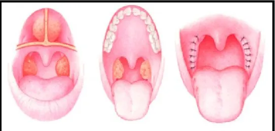
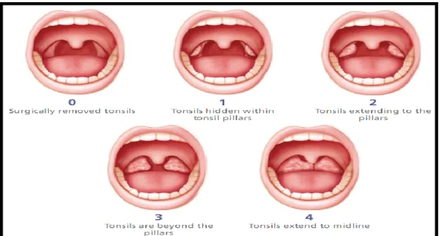
The principal risk factor in pediatric OSAS is adenotonsillar hypertrophy. Kang et al. examined the effect of adenotonsillar size and AHI in pediatric sleep apnea in 495 children. Brodsky’s scheme was used to evaluate tonsil size (Figure 9). A positive correlation was found between tonsil size and AHI in all different age groups (toddler, preschool, school and adolescents). However, adenoid size and AHI was positively associated in all groups except in the adolescent group. The following is consistent with normal adenoid growth pattern i.e. adenoid size decrease in adolescence.(78) At age 4, the adenoidal-nasopharyngeal space is the narrowest. Between ages 7 to 10, the face grows quickly and the space reaches its maximum volume. The space then continues to progressively decline until the age of 12 and decreases abruptly from 12 to 15 years old.(79) However, tonsil size is still prominent in both children and adolescents and is conclusive with Kang’s findings. Furthermore, the additive effects of both adenoid and tonsillar hypertrophy increase pediatric symptoms more than adenoidal hypertrophy or tonsillar hypertrophy only.(78)
Figure 9: Tonsillar grading(80)
(Figure adapted from: Brodsky et al. A comparison of tonsillar size and oropharyngeal dimensions in children with obstructive adenotonsillar hypertrophy. International journal of
pediatric otorhinolaryngology. 1987
Huynh et al. conducted a study on 604 children in a general orthodontic setting to assess associations between SDB with facial and dental morphology. They demonstrated that dolichofacial morphology and increased mandibular plane angle were significantly associated with several SDB symptoms. Also, SDB symptoms such as snoring, mouth breathing and daytime sleepiness were positively correlated with a narrow palate and decreased maxillary width. In the sagittal plane, retrognathia and overjet were not highly associated with SDB symptoms; however, they were statistically correlated with sleep bruxism and morning headaches. (10) Another study by Ameli et al. reported that in a suspected SDB pediatric population, 65% of subjects presented with dental malocclusions.(81) These findings support
A skeletal class II, increased overbite, maxillary constriction and a inferior hyoid bone position are all factors that may predispose children to apnea.(82) A dolichocephalic facial pattern and a narrow upper airway are common craniofacial characteristics in children with OSA.(40) A prospective study conducted by Schutz et al. showed a decrease in RERAs and RDIs in 16 children treated with an acrylic splint Herbst appliance combined with a maxillary expander (Figure 10). These children presented with a skeletal Class II pattern and a mild maxillary constriction. Furthermore, post-orthopedic functional treatment magnetic resonance imaging (MRI) showed a statistically significant increase in nasopharynx, oropharynx, hypopharynx total volume. A 6.1mm increase in effective mandible length as well as a 3.2mm maxillary expansion was calculated.(82)
Figure 10: Herbst appliance(83)
In general, a retrognathic mandible is concomitant with class II functional oral appliances which produces an anterior displacement of the mandible and the hyoid bone causing an anterior traction of the tongue.(84) As a result, in Schutz’s study, a 3.2mm posterior airway increase and reduced airway resistance was observed in children treated with a Herbst appliance. A proper swallowing pattern was also observed in those children due to anterior repositioning of the tongue. Proper swallowing also reduces tongue hypotonia, therefore helping the tongue to not fall back during the hypotonic stage of rapid-eye movement (REM) sleep.(82)
As discussed above, maxillary constriction may be seen in children with OSA. Rapid maxillary expansion is a potential adjunct treatment in pediatric sleep apnea, especially in post-adenotonsillectomy persistant OSA. Its main goal aside its sleep apnea benefit is to correct an existing maxillary posterior crossbite. A study by Villa et al. in 2007, demonstrated that an orthodontic treatment with RME significantly decreased OSA symptoms in 71.4% and AHI in 78.4% of children.(85) In a 36-month follow up study conducted by Villa et al. on the same 10 children, 80% of subjects showed a stable decrease in clinical and polysomnographic signs and symptoms of OSA.(40) Another study suggested that both RME and adenotonsillectomy may be essential to correct completely OSA and mouth breathing in children.(71) Guilleminault et al. showed that in 14.5% of children post-T&A, symptoms of OSA were still present three months post-surgery. He suggests that adjunct RME is necessary to resolve those signs and symptoms.(86) Villa et al. also demonstrated that mouth breathing was resolved in almost all children. RME therapy widens the buccal cavity and distorts the maxillary bone which enlarges space for adenoids and tonsils. These findings suggests that
adjunct orthodontic therapy should be proposed in children with OSA to help correct oral breathing as well provide benefits to airway obstruction.(40)
In healthy subjects, the nose is accountable for half of respiratory resistance. Therefore, any nasal obstruction due to craniofacial abnormalities could add to OSA factors. One of the principal goals of RME is to reduce nasal resistance. This is done by palatal expansion which increases the volume of the nasal and buccal cavities. Pharyngeal obstruction is therefore reduced by proper tongue repositioning in the buccal cavity. Also, in this study, 78.5% of children had hypertrophic tonsils and were chronic snorers. After RME, daytime and nighttime respiratory symptoms were reduced due to the enlargement of the buccal cavity.(87)
Children with deep/retrusive bites and crossbites had a superior improvement after RME in symptoms and polysomnographic variables with comparable amounts of intermolar distance gain than other children. This can be explained by additional benefits of orthodontic treatment in those cases in allowing proper tongue positioning and swallowing. Therefore, orthodontic treatment in children presenting with OSA and dental malocclusions should be commenced early to avoid developing its associated morbidities.(85) In that same study, the authors found no significant correlations between tonsillar hypertrophy and severity of SDB. AHI values were comparable no matter tonsillar grade. This outcome can perhaps explain how tonsillar hypertrophy is not always the mere risk factor in pediatric OSAS, particularly since RME improved the condition even in severe tonsillar hypertrophy. Relatively speaking, the new enlarged buccal cavity post-RME therapy makes tonsils appear relatively smaller.(85)
According to some authors, another craniofacial characteristic common in pediatric OSA is an ogival palate. During development, a posterior tongue position can contribute to the lateral palatine processes to expand vertically contouring the tongue before fusing at the midline causing that high-arched palate shape.(40) However, Smith et al. showed no difference in palatal height in children with OSA compared to controls using dental casts as measurement.(88)
According to Pirila-Parkkinen, cephalometric analysis is a valid method for measuring the dimension of the nasopharyngeal and retropalatal region.(89) A number of cephalometric studies have been conducted to better understand the craniofacial morphologic features of patients with SDB. Compromised breathing during sleep can arise from a combination of pathophysiological and anatomical features resulting in the narrowing of the upper airway. However, the precise location of the obstruction may differ from one person to another. These results reveal that individuals with SDB display few morphological dissimilarities in skeletal and soft tissue proportions, airway dimensions and hyoid positions. A shortened cranial base with reduced antero-posterior skeletal dimensions, variable outcomes in relation to hyoid bone position and an increase in both soft palate length and thickness are all examples of anatomical features which contribute to SDB.(90) Other factors such as a short mandibular body and mandibular retrusion are also associated with SDB.(91) Increased total and lower anterior face heights and larger craniofacial angles are also reported in SDB patients.(92)
Dental arch morphology is another risk factor in children with SDB. For example, narrower maxillae, deeper palatal height and shorter lower dental arch are associated with
SDB.(93) Increased overjet and reduced overbites are other significant examples of measures related to SDB.(94) Also, compared to controls, children with SDB present to have shorter maxillary arches and reduced intercanine widths.(88)
2.9 Sleep bruxism (SB)
Sleep bruxism is a sleep-related movement disorder also classified as a parafunction in dentistry.(95) Typically, SB teeth grinding is reported during childhood and adolescence with an overall prevalence ranging between 8% and 38% (96-98) and tends to decrease after adulthood from 8% to 3% in older adults.(99-101) SB is characterized by episodes of rhythmic masticatory muscle activity (RMMA) of the masseter and temporalis muscles. This activity can be observed and scored when electromyographic recordings are performed during sleep.(102) SB scoring relies on the recognition of RMMA, a succession of jaw muscle contractions, over the sleep period occurring mainly in light sleep stage N2. Grinding sounds, due to tooth contacts with jaw displacements, is the pathognomonic sign of SB that is usually reported by the patient’s sleep partner, siblings and/or parents. However, teeth grinding sounds do not occur during all RMMA/SB episodes. In children, SB can be associated with orofacial pain and headaches, and tooth damage.
Although the etiology of SB remains unknown, the multifactorial physiopathology is partly explained by re-activation of the cerebral cortex and autonomic nervous system during sleep, a process named sleep arousal, that occurs during periods of sleep instability.(103, 104) Increased respiratory amplitude is associated with RMMA,(105) which supports the hypothesis of an association between RMMA and breathing during sleep.
Studies have shown a higher incidence of sleep apnea or SB when they are comorbid. Moreover, SB teeth grinding decreased or disappeared in most children with sleep apnea who underwent adenotonsillectomy.(106, 107) Likewise, Bellerive et al. reported on a 32 patient sample study that although sleep and respiratory variables persisted, 65% of bruxers saw a reduction in RMMA after expansion.(108)
Chapter 3. Objectives and Hypothesis
3.1 Problematic
Our preliminary data suggest that from 604 patients (7-17 years) seen at the orthodontic clinic, up to 18% of respondents have compromised breathing during sleep and in whom comorbidities are also present.(10) Mandibular retrognathia, a narrow maxillary/mandibular ratio, a long and narrow face may be associated with these phenomena.(92, 94, 109, 110) Furthermore, preliminary studies on rapid palatal expansion or surgically assisted expansion, suggest an improvement in SDB.(36) Establishing a prevalence count in a multi-centric study across Canada and early detection of SDB and craniofacial development abnormalities may reduce the risk of developing the associated consequences.
3.2 Type of study
This study consists of a multi-centric prospective study in which the data will be collected in 5 clinical sites. Each clinical site will be responsible of recruiting 100 children completing a sleep study for a total of 500 subjects. One of the sites will recruit 100 children for the control group who do not present any sleep apnea symptoms confirmed by PSG.
3.3 Study purpose
The purpose of this prevalence study is to count the number of patients who would benefit from a dental and orthodontic evaluation among those presenting with respiratory sleep disorders seen at the CHU Sainte-Justine. Sleep questionnaire, polysomnographic, orthodontic and craniofacial data will be further analyzed.
3.4 Hypothesis
1. Research hypothesis:Our research hypothesis is that the prevalence of malocclusions and dento-skeletal abnormalities would be different between apneic and non-apneic children.
2. Null hypothesis:
The prevalence of malocclusions and dento-skeletal abnormalities would not be different between apneic and non-apneic children.
Chapter 4. Materials and Methods
4.1 Ethics committee
The project received the approval of the ethics committee of the CHU Sainte-Justine on March 31st 2014 and has been renewed yearly. Refer to annex 1 for ethics committee approval.
4.2 Patient selection
Patients referred for a polysomnographic sleep recording for proper diagnosis of SDB at the sleep laboratory of CHU Sainte-Justine were seen in this study. A total of 100 patients were examined. The patients seen at CHU Sainte-Justine take part in a national prospective cross-sectional study. Data is being collected at 4 other clinical sites, as well at a site that will provide control participants with no sleep apnea confirmed by polysomnography. Each of the other 4 clinical sites is recruiting patients with the same profile while the Dalhousie University site will recruit healthy participants. Gozal’s questionnaire as well as craniofacial and
orthodontic data collection took place during the scheduled appointment on the night of the polysomnographic sleep study. No additional visit or follow-up were required.
Inclusion Criteria
• Children aged 4-17 years willing to complete a polysomnographic sleep study at the sleep laboratory of the CHU Sainte-Justine for evaluation of snoring and apnea.
Exclusion Criteria
• Children with craniofacial anomalies linked to genetic syndromes. • Children currently under CPAP treatment.
4.3 Data collected
Facial and orthodontic data were collected. Refer to Annex 2 for full clinical exam. An adapted questionnaire from Spruyt & Gozal et al. was handed to one parent for further sleep information on the child presenting for the sleep study. Refer to Annex 3 for full sleep questionnaire.
The following polysomnographic data was collected. Sleep technicians trained in each site used a standardized evaluation method according to the parameters established by the American Academy of Sleep Medicine.
• Height • Weight
• Total sleep time • Sleep efficiency
• Respiratory Disturbance Index
• Mean and minimum oxygen saturation • Oxygen desaturation index
• Minimum and maximum respiratory rate • Minimum and maximum heart rate • TcCO2 range
• Total Hypopneas counts • Apnea Hypopnea Index (AHI)
4.4 Initial examination
Explanation of the procedure was given to the parent/guardian and the child. A customized consent formed was then given and read aloud before receiving proper consent. Refer to annex 4 for complete consent form. A standard orthodontic evaluation was completed on the night of the child’s polysomnographic sleep study at the CHU Sainte-Justine. A standard dental examination kit was used to record data as well as a Boley gauge to record distance. Following complete consent, data, and questionnaire collection, a 20$ gift certificate of their choice was given to the patient for accepting to participate in the study.
Children were then accompanied to their sleep study room. Complete type 1 polysomnographic measures were then set up by the on call registered inhalotherapist nurse. Sleep data was extracted for analysis in this study.
Dependent variables:
• Respiration during sleep • Questionnaires
• Sleep quality
Independent variables: • Craniofacial massif
• Dentition
4.5 Statistical analysis
An electronic data capture was used: Redcap ™ (Research Electronic Data Capture). The data was codified with an alphanumeric code to prevent patient identification. The Redcap ™ system uses a secure Web connection requiring authentication. Only members of the research project had access to the data.
Intraclass correlation was performed with an experienced orthodontist, Dr. Andrée Montpetit, for patient orthodontic evaluation. Fischer’s Exact Test, Mann-Whitney U-tests and two-sample t-test were used for prevalence calculation of dental malocclusions in children with SDB as well as Spearman correlation between dental malocclusions and type 1 polysomnographic data. A logistic regression was also done. Data was analyzed using SPSS 24 by an experienced statistician.
Chapter 5. Results
5.1 Patient description
In this prevalence study, 100 patients were recruited; 58 of them were male and 42 were female (Figure 11). The age ranged between 3-18 years old, the mean being 9.6 years old ± 4.05 (Figure 12). No patients were excluded from the study and all parents/guardians and patients consented to participate in the study. All patients included in this study responded to the inclusion criteria. No patients were secondarily excluded from the study after polysomnographic data analysis. Orthodontists were blinded to polysomnographic scores when doing clinical evaluation of patients on the night of sleep study. Kappa scores were rated excellent for ICC calculations. Subjects were separated in two different AHI groups (AHI < 2, AHI ≥ 2) for analytic purposes per the AAP guidelines.(111, 112)
The clinical data reported are based on the clinical evaluation done on the day of the polysomnography at the CHU Sainte-Justine. The polysomnographic data originate from the sleep study done the same night as the clinical evaluation compiled by sleep technicians from the site.
Figure 11: Sex & AHI distribution
The AHI <2 group had a mean age of 9.98 ± 3.83 and the AHI ≥ 2 group had a mean age of 9.16 ± 4.34 (Figure 13). No difference in age was found between AHI groups using t-test analysis (p=0.320). However, Fisher’s exact t-test showed that boys were more likely to present with sleep apnea (p=0.043).
Figure 13: Age per AHI groups
Table 3: Descriptive and polysomnographic data according to AHI
5.2 Body mass index
BMI per subject was calculated by dividing their weight (kg) by their height squared (m). No significant difference was found between AHI groups and BMI value in children (p=0.303) (Figure 14). The mean BMI value in the AHI < 2 group was 21.13 ± 7.78 and 23.21 ± 10.61 in the AHI ≥ 2 group.
5.3 Gozal score
The Gozal questionnaire is a helpful tool consisting of a set of six ordered questions along the SDB spectrum (frequency and intensity of snoring, breathing) given to the parent/legal tutor which allows screening of children at risk of SDB. No significant difference was found between Gozal score and AHI groups (p=0.220) (Figure 15). The mean Gozal score in the AHI < 2 group was 1.58 ± 0.92 and 1.82 ± 1.05 in the AHI ≥ 2 group.
Spruyt and Gozal severity score was then studied with a filter of ≥ 2.72. Spruyt and Gozal showed that pediatric patients with a score ≥ 2.72 have an increased risk of presenting an AHI ≥ 3.(23) Patients with a score < 2.72 had an AHI median of 1.4 (0-40.7) and patients with a score ≥ 2.72 had a median of 5.8 (0.3-23.4) (Figure 17). Mann-Whitney U test showed a statistical difference in that Gozal score was significant in predicting more severe AHI for a score ≥ 2.72 (p=0.011) (Figure 16).
Figure 17: AHI distribution per Gozal score
5.4 Descriptive polysomnographic data
The following data summarizes sleep recorded data for all subjects analysed by sleep technicians at the CHU Sainte-Justine (Table 4).
Table 4: Descriptive polysomnographic data
Group ox_desat_ind tcco2_r resp_dist_ind
ahi<2 N 48 34 45 Mean 5,1250 41,29 ,9556 Std. Deviation 19,69325 4,407 1,08283 Median ,9000 41,50 ,7000 Minimum ,00 33 ,00 Maximum 99,00 50 7,00 ahi>=2 N 36 27 34 Mean 18,6278 42,63 8,7647 Std. Deviation 30,59316 5,911 8,74164 Median 5,4000 43,00 5,0000 Minimum ,30 26 ,10 Maximum 100,00 52 40,70 Total N 84 61 79 Mean 10,9119 41,89 4,3165 Std. Deviation 25,68023 5,125 6,93781 Median 2,0000 42,00 1,4000 Minimum ,00 26 ,00
Group cap_ind ahi<2 N 55 Mean ,3581 Std. Deviation ,31427 Median ,3000 Minimum ,00 Maximum 1,30 ahi>=2 N 43 Mean 2,4319 Std. Deviation 5,23218 Median 1,3000 Minimum ,00 Maximum 33,50 Total N 98 Mean 1,2680 Std. Deviation 3,60253 Median ,5000 Minimum ,00 Maximum 33,50 oah_ind 26 ,2500 ,34205 ,2000 ,00 1,50 16 3,4563 3,98229 1,9000 ,00 12,50 42 1,4714 2,89080 ,3500 ,00 12,50 ma_ind 32 ,003 ,0177 ,000 ,0 ,1 24 ,150 ,2766 ,000 ,0 1,0 56 ,066 ,1938 ,000 ,0 1,0 ta_ind 55 ,3296 ,28383 ,3000 ,00 1,40 43 3,3672 5,03953 1,4000 ,00 22,90 98 1,6624 3,65198 ,5000 ,00 22,90 th_ind 31 ,5581 ,49178 ,4000 ,00 1,90 24 4,3250 4,23559 2,5500 ,60 18,50 55 2,2018 3,36603 ,9000 ,00 18,50 ah_ind 57 ,7940 ,53120 ,7000 ,00 1,90 43 7,7930 8,03331 4,6000 2,00 40,70 100 3,8036 6,29805 1,6000 ,00 40,70
5.5 AHI
Patients were divided into subgroups (AHI < 2, AHI ≥ 2) for analytic purposes. 57 patients had an AHI < 2. The mean AHI for this subgroup was 0.79 ± 0.53 with a median of 0.7. The AHI ≥ 2 subgroup was composed of 43 patients. The variability of this subgroup was greater with a mean of 7.79 ± 8.03 with a median of 4.6 (Figure 18).
AHI was then correlated with tonsillar hypertrophy grade per Brodsky’s tonsillar hypertrophy score. Mann-Whitney U test was used to establish statistical significance. The AHI median in the milder group was of 1.7 (0.1-14.5) and of 1.5 (0-40.7) in the more severe group (Figure 20). No statistical difference was found in AHI in terms of tonsillar hypertrophy grade (p=0.426) (Figure 19).
5.6 Craniofacial morphology
The following histogram summarizes the major craniofacial features in terms of prevalence in both AHI groups (Figure 21). Their statistical significance is listed below in their corresponding tables.
No significant difference was found between AHI groups for nominal craniofacial morphological characteristics using Fisher’s Exact Test (Table 5):
Table 5: Craniofacial data statistical significance
5.6.1 Facial soft tissue characteristics
No significant difference was found between AHI groups for nominal facial soft tissue characteristics using Fisher’s Exact Test (Table 6):
5.6.2 Intra-oral soft tissue characteristics
No significant difference was found between tongue size between AHI subgroups using Fisher’s exact test (p=1.0). A total of 5 patients presented with an observable clinical macroglossia in the AHI < 2 group and 4 patients in the AHI ≥ 2 group.
5.6.3 Intra-oral dental characteristics
The following histogram summarizes the major intra-oral dental characteristic in terms of prevalence in both AHI groups (Figure 22). Their statistical significance is listed below in their corresponding tables.
No significant difference was found between AHI groups for nominal intra-oral characteristics using Fisher’s Exact Test (Table 7):
Table 7: Intra-oral data statistical significance
No significant difference was found between AHI groups for numerical intra-oral characteristics using Mann-Whitney U Test (Table 8):
No significant difference was found between AHI groups for numerical intra-oral characteristics using two-sample t-test (Table 9):
Table 9: Intra-oral data statistical significance
5.6.4 Functional data
Functional data were also studied and their significance was analysed between AHI groups using Fisher’s exact test. Mouth breathers were found to be non-significant between AHI groups (p=0.473). The AHI <2 group had a total of 28 patients with reported mouth breathing and the AHI ≥ 2 had 21 patients with reported mouth breathing. Day or night time mouth breathing was not significant either (p=1.0).
A trend was found between patients with oral habits altogether (nail biting, cheek/lip biting, bruxism, thumb sucking) and an AHI <2 (p=0.064). However, each oral habit alone was found to be non-significant between AHI groups (nail biting p=0.140, cheek/lip biting p=1.0, bruxism p=0.650, thumb sucking p=0.632).
5.6.5 Correlations between malocclusions and polysomnographic data
Correlations between polysomnographic data and clinical variables were calculated by combing all normal values vs abnormal sleep apnea predictors. The clinical values were correlated with the following polysomnographic data: oxygen desaturation index, CO2 and TCO2 maximum range, total apnea index and apnea hypopnea index. The following table summarizes the lien between those correlations and their significance using Mann-Whitney U test (Table 10).
Spearman correlations test was used to calculate the following variables with the same polysomnographic data (Table 11).
Table 11: Correlation between malocclusions and polysomnographic data
5.6.6 Correlation between clinical data and numerical AHI
Correlations between clinical data and numerical AHI were done by combining all normal values vs abnormal known sleep apnea predictors and correlating them to the AHI. The following table summarizes their significance using Mann-Whitney U test (Table 12).
Table 12: Correlation between clinical data and numerical AHI
The following table summarizes other clinical data correlated to AHI using Spearman correlations (Table 13).
Table 13: Correlation between clinical data and numerical AHI
5.7 Prediction model
A logistic regression was calculated using age, sex, and all morphologic clinical variables with a p<0.20 in a univariate analysis. Polysomnographic variables were not included in the regression calculation since the sleep study would reveal if the patient has