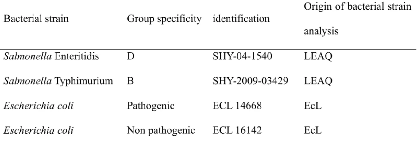
Université de Montréal
Production and immunogenicity of selected proteins of Salmonella Enteritidis
par
Y
UNC
UIDépartement de Sciences Cliniques
Faculté de médecine vétérinaire
Mémoire présenté à la Faculté de médecine vétérinaire en vue de
l’obtention du grade de maître ès sciences (M.Sc.) en sciences vétérinaires option
Hygiène vétérinaire et innocuité des aliments
November 2013
SUMMARY
Over the past years, Salmonella Enteritidis (SE) has become the most prevalent serovars isolated in Canadian patients. Most cases in humans are associated with consumption of chicken meat, raw egg and related products. For controlling Salmonella transmission and infection in poultry, available commercially killed vaccines poorly stimulate mucosal immunity, while the use of live vaccines remains controversial. Therefore an oral subunit vaccine may be a solution. Five bacterial proteins were chosen as potential candidates and identified as Glyceraldehyde-3-phosphate dehydrogenase, Enolase, Lipoamide dehydrogenase, DNA protection during starvation protein and Elongation factor-Tu. Our objectives were to produce and purify these proteins and study their immunogenicity. The proteins genes were amplified and cloned into pQE-30 vector, then transformed into Escherichia coli M15 for expression. Purification was performed using FPLC. SPF laying hens were separated into 6 groups and injected intramuscularly 3 times at 16, 20 and 28 weeks of age. Five groups were injected with a single protein respectively while the sixth group was injected with PBS as control. Eggs were collected during the duration of the experiment and blood was collected when hens were sacrificed at 36 weeks of age. IgY was extracted from egg yolk and serum and IgA from egg white. Immunodot, westernblot and ELISA were used to evaluate the immunogenicity of proteins and antibody levels they induced. We found that these five proteins could stimulate production of specific antibody in vivo. GAPDH, Enolase and DPS induced higher antibody titer than LpdA and Ef-Tu.
RÉSUMÉ
Au cours des dernières années, Salmonella Enteritidis est devenus les sérotypes les plus souvent isolés chez les patients canadiens, les cas étant liés à la consommation de viande de poulet et d’œufs crus. Les vaccins tués commercialement disponibles pour la volaille, stimulent mal l'immunité mucosale, tandis que l'utilisation de vaccins vivants reste controversée. Par conséquent, un vaccin sous-unitaire par voie orale peut être une solution. Cinq protéines bactériennes ont été choisies comme candidates potentielles et identifiées, soit Glyceraldehyde-3-phosphate dehydrogenase, Enolase, Lipoamide dehydrogenase, DNA protection during starvation protein et Elongation factor-Tu. Notre objectif a été de produire et de purifier ces protéines et de démontrer leur immunogénicité. Les gènes des protéines ont été amplifiés et clonés dans le vecteur pQE-30 pour expression dans Escherichia coli M15. La purification a été effectuée par FPLC. Des poules pondeuses SPF ont été séparées en 6 groupes et injectées par voie intramusculaire à different âges avec une des 5 protéines, ou le PBS chez le groupe témoin. Les œufs ont été ramassés pendant l'expérience et du sang a été prélevé à 36 semaines d'âge. Les anticorps IgY ont été extraits à partir du jaune d'oeuf et du sérum, et les IgA à partir du blanc d'oeuf. Des immunodots, westernblots et ELISA ont évalué l'immunogénicité des protéines et les niveaux d'anticorps induits . Nous avons constaté que ces cinq protéines pourraient stimuler la production d'anticorps spécifiques in vivo. GAPDH, Enolase et DPS ont induit des titres d'anticorps plus élevés que LpdA et EF-Tu.
TABLE OF CONTENTS SUMMARY………..…………i RÉSUMÉ……….……ii TABLE OF CONTENTS……….……..iii LIST OF TABLES……….…… vi LIST OF FIGURES………...vii ABBREVIATIONS……….……x ACKNOWLEGEMENT………..…..xii INTRODUCTION……….……..1 LITERATURE REVIEW……….……...4
1 General introduction of Salmonella……….…..……5
1.1 Classifications of Salmonella……….……….6
1.2 Prevalence of Salmonella……….……….……..7
1.2.1Prevalence in human………..……7
1.2.2Prevalence in chickens………..………..……...9
1.3 Source of Salmonella………...……….11
1.3.1 Sources of animal infection……….………...11
1.3.2 Sources of human infection………14
1.4 Transmission of Salmonella……….……….15
1.4.1 Horizontal transmission………...……...15
1.4.2 Vertical transmission………...…...16
1.5.1 Infections in human…………...……….….17
1.5.2 Infection in chickens………...18
2 Pathology and virulence of Salmonella………...20
2.1 Pathogenesis………..20
2.1.1 Getting to intestinal gut………...20
2.1.2 Adhesion……….21
2.1.3 Invasion………...22
2.1.4 Infection/dissemination………...24
2.1.5 Metabolic adaptation………...25
2.2 Virulence factors………...26
2.2.1 Salmonella Pathogenicity Island (SPI)………...26
2.2.2 LPS………..27
2.2.3 Flagellin……….……….28
2.2.4 OMP………29
3 Chicken immune responses………..……….……..29
3.1 Innate immune response………30
3.1.1 Constitutive response………..30
3.1.2 Heterophil………...32
3.1.3 Phagocytes………..33
3.2 Adaptive immune response………...34
3.2.1 Antigen presentation………...34
3.2.2 Humoral response……….………..35
3.3 Local immune reponse for Salmonella………..38
4 Control and prevention program for Salmonella……….39
4.1 Controlling programs..………..39
4.1.1 The Eurpean Union (EU)…………...……….40
4.1.2 The United States………41
4.1.3 Quebec………42
4.1.4 Other provinces in Canada………..42
4.2 Biosecurity methods………...……….…..44
4.3 Vaccines………47
4.3.1 Inactive vaccines………...…………..47
4.3.2 Attenuated live vaccines………..…………...48
4.3.3 Subunit vaccines………..…………...49
MATERIALS AND METHODS………...57
RESULTS………..67
GENERAL DISCUSSION………90
CONCLUSION………..…..102
LIST OF TABLE
LIST OF FIGUERS
Literature review
Figure 1. Incidence rate of Salmonella spp. and SE as reported to NESP from 2000 to 2010 (NESP Annual Summary, 2010)………..………...9
Results
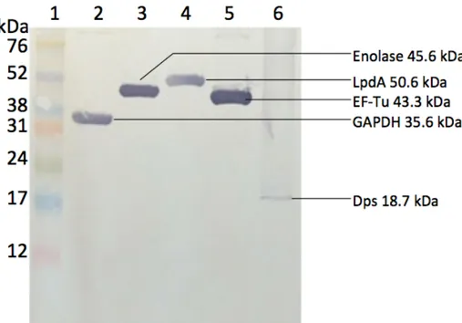
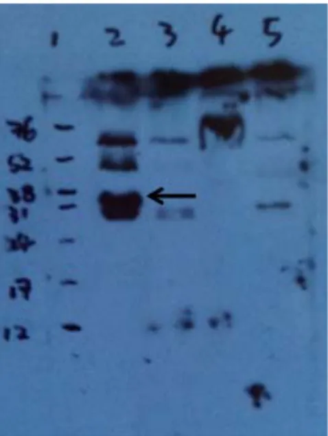
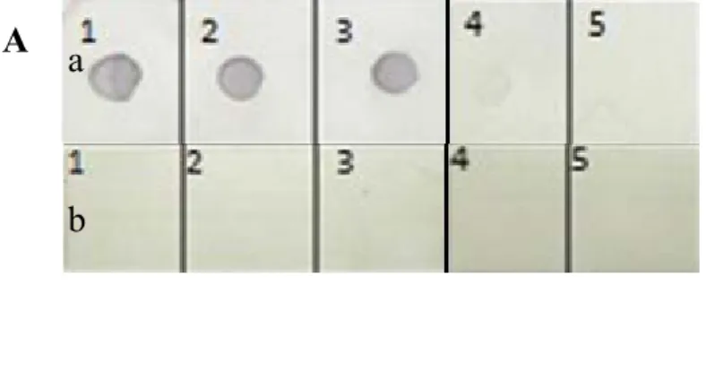
Figure 1. SDS-PAGE of recombinant GAPDH and EF-Tu purified by Ni-NTA matrix (Qiagen kit)………....73 Figure 2. SDS-PAGE of recombinant proteins purified by FPLC……….74 Figure 3. Anti-His Immunoblotting Analysis of Purified Recombinant Proteins …….75 Figure 4. Western blot analysis with antisera against SE from immune layers……….76 Figure 5. Immunoblotting analysis with specific IgY against recombinant GAPDH…77 Figure 6. Immunoblotting analysis with specific IgY against recombinant Enolase...78 Figure 7. Immunoblotting analysis with specific IgY against recombinant Dps...79 Figure 8. Immunoblotting analysis with specific IgY against recombinant LpdA……80 Figure 9. Immunoblotting analysis with specific IgY against recombinant EF-Tu…...81 Figure 10. Quantification of specific IgY against recombinant GAPDH in egg yolk of laying hens during post-immunization period analyzed using chicken IgG ELISA quantitation set………...82 Figure 11. Quantification of specific IgY against recombinant Enolase in egg yolk of laying hens during post-immunization period analyzed using chicken IgG ELISA quantitation set………...83
Figure 12. Quantification of specific IgY against recombinant Dps in egg yolk of laying hens during post-immunization period analyzed using chicken IgG ELISA quantitation set………...84 Figure 13. Quantification of specific IgY against recombinant LpdA in egg yolk of laying hens during post-immunization period analyzed using chicken IgG ELISA quantitation set………...85 Figure 14. Quantification of specific IgY against recombinant EF-Tu in egg yolk of laying hens during post-immunization period analyzed using chicken IgG ELISA quantitation set………...86 Figure 15. Comparision of specific IgY in egg yolk and serum………87 Figure 16. Quantification of specific IgA against recombinant GAPDH in egg white of laying hens during post-immunization period analyzed using chicken IgA ELISA quantitation set………...88 Figure 17. Quantification of specific IgA against recombinant Enolase in egg white of laying hens during post-immunization period analyzed using chicken IgA ELISA quantitation set………...89 Figure 18. Quantification of specific IgA against recombinant Dps in egg white of laying hens during post-immunization period analyzed using chicken IgA ELISA quantitation set………...90
ABBREVIATIONS
APC: Antigen presenting cell ATP: Adenosine triphosphate CD: Cluster of differentiation
CDC: Centers for Disease Control and Prevention CFU: Colony forming unit
CHEP: Canadian hatching egg producer CHEQ: Canadian hatching egg quality
CIPARS: the Canadian Integrated Program for Antimicrobial Resistance Surveillance CMI: Cell-mediated immunity
CTL: Cytotoxic T cell DC: Dendritic cell
DNA: Deoxyribonucleic acid
Dps: DNA protection during starvation prtein (DNA protecting protein) EFSA: the Europe Food Safety Authority
EF-Tu: Elongation factor thermo unstable ELISA: Enzyme-linked immunosorbent assay FAE: Follicle associated epithelium
FPLC: Fast protein liquidchromatography
GAPDH: Glyceraldehyde 3-phosphate dehydrogenase HACCP: Hazard analysis and critical control points HRP: Horseradish peroxidase
IEL: Intraepithelial lymphocyte IFN: Interferon
Ig: Immunoglobulin IL: Interleukin
LpdA: Lipoamide dehydrogenase LPS: Lipopolysaccharide
M cell: Microfold cell
MHC: Major histocompatibility complex MLN: Mesenteric lymph nodes
NAD: Nicotinamide adenine
NESP: National Enteric Surveillance Program NK: Natural killer cell
OD: Optical density
OFFSAP: On-farm food sfety assurance program OMP: Outer membrane protein
PBS: Phosphate buffered saline
PHAC: Public health agency of Canada PMN: Polymorphonuclear neutrophil PT: Phage type
RNA: Ribonucleic acid SCFA: Short-chain fatty acids SCV: Salmonella containing vacuole SPI: Salmonella Pathogenicity Island
ACKNOWLEGEMENTS
I want to thank my director, Dr. Martine Boulianne, for this precious chance to achieve my master study and have all these experiences of lab working, as well as for all the academic and technical guidance during the whole study period.
I want to thank my co-directors, Dr. Ann Letellier and Dr. Sylvette Laurent-Lewandowski, for their constant help and supports throughout my study and project.
I want to thank my colleagues and friends of CRSV and GRESA, Dr. Philippe Fravalo, Alexandre Thibodeau, Nicole Trottier,Bénédicte Bouchet, Rodolphe El Hajj Obeid, Andres Ramirez, Guillaume Larivière-Gauthier, Audrey Perron and Lila Maduro, for all the help and suggestions for my lab techniques.
I want to thank Canadian Poultry Research Council and Agriculture and Agri-Food Canada for the financial support for this project.
I want to thank members of thesis evaluation jury for all the advices and suggestions.
Last but not the least, I want to thank my parents for their constant encouragement and support during all these time.
Salmonella spp. is one of the major causes of foodborne illnesses in humans, which remains a
worldwide problem. Over the past years, the most prevalent serovars isolated in Canadian patients were Salmonella Enteritidis (SE) (CIPARS, 2007-2009). Human SE cases are most commonly associated with the consumption of contaminated eggs, egg products and more recently with poultry meat. For Salmonella infection in chickens, we have observed an increase in the prevalence of SE in samples from abattoir, retail meat and animal clinical isolates (CIPARS, 2007-2009). The Canadian egg industry has over the past years developed several programs to detect and limit SE contamination. However, even with good biosecurity protocols and various controlling programs, breeder and layer flocks still become infected with SE and can transmit it horizontally and vertically. In order to prevent SE contamination, vaccination has been suggested and various killed vaccines are commercially available. Although they were able to induce an immune response, these vaccines did not protect SE challenged hens from excreting the bacteria and even laying positive SE eggs at 55 and 65 weeks of age (Tran et al., 2010). In fact, the cell-mediated response appears to play an important role in the resolution of Salmonella infection, presumably because Salmonella is an intracellular facultative pathogen. Unfortunately, commercially available killed vaccines poorly stimulate mucosal immunity, and live vaccine use remains controversial due to risk of virulence recovery and food safety concerns. Another vaccine strategy is the development of sub-unit vaccine orally administered using protein antigens. Thus subunit vaccine may then induce efficient stimulation of mucosal immune system to protect birds from Salmonella.
For this project, five candidate proteins have been chosen in our lab from previous work, based on their immune reactions to SE and ST whole-cell antigens as well as well
conservation in both Salmonella serovars. Their cell-surface expression and immunogenicity were also reported in other different species in different precious researches. These 5 proteins were identified as Glyceraldehyde-3-phosphate dehydrogenase (GAPDH), Enolase, Lipoamide dehydrogenase (LpdA), DNA protection during starvation protein (Dps) and Elongation factor-Tu (EF-Tu). In this project, based on biological and immunological characters of these selected proteins, we have the hypothesis: these proteins are immunogenic and able to induce immune responses in laying hens and antibody production in both sera and eggs (yolk and white). Our main objectives are to produce and demonstrate the immunogenicity of these immunoreactive proteins, with the ultimate objective of eventually developing a new subunit vaccine against Salmonella Enteritidis and Salmonella Typhimurium in laying and breeder hens. For the protein production, we need to use an optimal method to purify these proteins effectively once they are overexpressed in E.coli M15 cells, in order to ensure the satisfactory quality and purity of produced proteins. After production of these selected proteins, we will test if each protein is immunogenic i.e., able to induce specific antibody in vivo (as measured in eggs and sera) after immunization laying hens with each of them. If these proteins are demonstrated to be immunogenic to be capable of inducing specific antibody production, we will continue to have a closer look at specific antibody titer levels and changes post vaccination and maintenance of blood, egg yolk and egg white antibody titers.
1 GENERAL INTRODUCTION OF SALMONELLA
Salmonellosis is an important zoonotic infection, and human salmonellosis causes widespread morbidity and economic loss. In recent years, some countries have observed a marked increase in the number of human cases (Hendriksen et al., 2011). Much of this increase has been associated with poultry meat and table eggs consumption (Foley et al., 2011). The predominance of Salmonella Enteritidis as a human pathogen has overshadowed other
Salmonella serovars, many of which are capable of causing serious illness such as Salmonella
Typhimurium DT 104 (Bohaychuk et al., 2006; Foley et al., 2008; Foley et al., 2011).
The economic losses associated with human salmonellosis are not only associated with the cost of investigations, treatment, and prevention of illness but also may affect the whole chain of food production. Estimated annual costs for salmonellosis might have reached billions of dollars in the United States and Canada in the 1990s only (Sockett, 1991; Clark et al., 2001).
Although primarily an intestinal bacteria, Salmonella is widespread in the environment and is commonly found in farm effluents, human sewage, and in any material subject to fecal contamination. Salmonellosis has been recognized in all countries, especially in the areas of intensive animal husbandry, such as poultry and swine production (Berends et al., 1996; Akkina et al., 1999). Although disease can affect all species of domestic animals, young and gestation animals are the most susceptible. However, most infected animals are asymptomatic carriers i.e. are infected without showing any sign of illness. The challenge in the food animal industry resides not only in the detection of these asymptomatic carriers to avoid
contamination of the food chain, but also mostly in preventing animals to becoming infected. Vaccination could therefore be an important tool in a prevention and control program.
1.1 Classification of Salmonella
Salmonella is a genus of rod-shaped, Gram-negative, non-spore-forming, predominantly
motile enterobacteria with diameters around 0.7 to 1.5 µm, lengths from 2 to 5 µm, and flagella which grade in all directions (i.e., peritrichous). They are chemoorganotrophs, obtaining their energy from oxidation and reduction reactions using organic sources, and are facultative anaerobes. Most Salmonella produce hydrogen sulfide that is important for bacteriological isolation (Clark and Barrett, 1987). They are found worldwide in cold- and warm-blooded animals (including humans), and in the environment (Yue, 2012).
Genus Salmonella includes two species: Salmonella bongori and Salmonella enterica.
Salmonella enterica is subdivided into 6 subspecies (enterica, salamae, arizonae, diarizonae, houtenae and indica). Salmonella enterica subspecies enterica has 2610 different serotypes
and Salmonella Enteritidis belongs to this subspecies, The serotypes are characterized by three surface antigens: the flagella “H” antigen, the oligosaccharide “O” antigen and the polysaccharide “Vi” antigen (found in Typhi and Paratyphi serotypes) (Bronze and Greenfield, 2005).
Salmonella serovars can be divided into two groups, those that are host adapted and others that
and the paratyphoid Salmonellae (S. paratyphi A, S. paratyphi B, and S. paratyphi C) for human, and S. Choleraesuis (swine), S. Dublin (cattle), S. Abortusovis (sheep), S. Pullorum (poultry) and S. Gallinarum (poultry) for animals (Barrow, 1992; Selander et al., 1992; Bolton
et al., 1999; Uzzau et al., 2001). On the other hand, non-host-specific Salmonella serovars
cause salmonellosis in humans and a wide variety of animal hosts as well. Salmonella enterica serovar Enteritidis (SE) belongs to this group, and have presented serious continuous challenge in food safety and poultry industry over the past years being responsible for significant health problems in human (Mølbak et al., 2006).
1.2 Prevalence of Salmonella
1.2.1 Prevalence in human
Salmonella is considered as one of the most common foodborne illness etiologic agent in
human. In a recent CDC (Centers for Disease Control and Prevention) summary of USA,
Salmonella Enteritidis (SE) was described as the most prevalent serotype responsible for
17.5% of all human salmonellosis (CDC, 2009). In the EU, SE is the serovar most frequently associated with human illness. In 2008, a total of 131,468 confirmed cases of human salmonellosis (notification rate 26.4 per 100,000 populations) were reported from 27 European countries. The total number of reported human salmonellosis cases in the EU has decreased steadily by several thousand cases annually since 2004, from 195,947 cases in 2004 to 133,258 cases in 2008 (EFSA, 2008). In 2009, the number of salmonellosis cases in humans decreased by 17.4 %, compared to 2008, and the statistically significant decreasing trend in
the European Union continued for the fifth consecutive year. In total 108,614 confirmed human cases were reported in 2009 and in particular, human cases caused by SE decreased markedly. It is assumed that the observed reduction of salmonellosis cases is mainly attributed to successful implementation of national Salmonella control programs in poultry (EFSA, 2009).
In Canada, from 2005 to 2010, Salmonella accounted for the most frequent cases in human when comparing with other foodborne pathogens, which include Campylobacter, E. coli,
Shigella, Vibrio, etc. Based on the latest NESP (National Enteric Surveillance Program)
annual report (2010), in all provinces of Canada, including Quebec, Salmonella caused much more human cases than other select major organism groups as (NESP Annual Summary, 2010). Among all the serovars of Salmonella, SE was one of the most important public health concerns. From 2005 to 2010, the ranking among the top three serovars remained unchanged with SE being the most frequently reported, followed by ST and Salmonella Heidelberg, (NESP Annual Summary, 2010). PT8 and PT13 were the predominant phage types found in human SE (CIPARS Annual Report, 2008).
In 2010 a record high of 2827 SE isolations were reported to NESP. SE was the most prevalent cause of human salmonellosis in Canada representing approximately 39% of all human Salmonella isolates reported in 2010. The proportion of salmonellosis cases attributed to SE has been steadily increasing over time, from 14% in 2000 to 39% in 2010 (Figure 1).
Figure 1. Incidence rate of Salmonella spp. and SE as reported to NESP from 2000 to 2010 (NESP Annual Summary, 2010).
1.2.2 Prevalence in chickens
In United States, the presence of SE was identified in 35% of layer flocks (end of laying period) from cecal sample collected in Northern US slaughterhouses between 1991 and 1995 (Ebel and Schlosser, 2000). The prevalence of environmental contamination in SE was 7.1% for 200 laying hens’ farms in 15 states (Garber et al. 2003). Meanwhile, the high percentage of samples positive for SE was detected in ground chickens (0.46%, 8/1722 samples) and broilers (0.26%, 124/47090 samples) (White et al. 2007). In 2010, CDC investigated a multistate outbreak of SE infections in the United States, and a total of 3,578 cases in humans were identified from May 1 to November 30, 2010. According to the data from the investigations conducted by public health official in 11 states, the shell eggs were a likely source of this huge outbreak. Certain egg suppliers and farms conducted a nationwide egg recall after the outbreak was reported (CDC, 2010). This was the largest egg recall in American history. Throughout
the whole country, more than 500 million eggs were involved in the nationwide recall (FDA, 2010). Total costs to American shell-egg producers is not clear yet, however, the negative media attention produced a drop in prices that cost the shell-egg industry over $100 million in September 2010 only (Capturing Recall Costs Measuring and Recovering the Losses. 2011.).
In Europe, SE was one of the most predominant serotypes detected in laying hens and their eggs in 2008. Over the past 20 to 25 years, SE has been frequently isolated from environmental samples of egg-laying flocks when compared to other isolates of other serotypes (EFSA, 2007). The prevalence of SE and ST in laying flocks has increased from 0.5% (2005) to 2.3% (2006) and 3.2% (2007) (EFSA. 2009), even with the nations control programs in place. The EU has passed legislation that requires member states to actively work to reduce the presence of Salmonella in poultry flocks at all levels of production by setting up national control programs that must target specific Salmonella serovars (most regulations currently only cover SE and ST). This has resulted in a significant decrease in the prevalence of SE in broiler, layer and breeding flocks since 2008 (Keery, 2010).
In Canada, SE has become the third most important Salmonella serovar from chicken sources including samples from abattoir and retail meat, while the isolation rate, which was less than 1% in 2002, increasing to 7% in 2006 (CIPARS annual report, 2006; PHAC, 2007). SE isolated from chicken fecal samples has also increased sharply over the past five years, going from just less than 1% in 2002 to 13% in 2006 in the samples collected at slaughter (PHAC, 2007). In parallel, the increase has also been observed in broilers with successive increases observed over the last 3 years (2006: 20% 2007: 30% 2008: 40% percentage cultures positive)
(Middleton, 2009). In Canada, in 2008, in an abattoir surveillance program, Salmonella isolates were recovered from 27% (234/851) from chicken cecal samples, in which the SE was the second most common serovar (19%, 45/234) following Salmonella Kentucky (40%, 93/234). In Canadian retail meat surveillance, Salmonella isolates were recovered from 40% (382/960) of retail chicken samples, in which Kentucky (31%, 120/382), Heidelberg (20%, 78/382) and Enteritidis (16%, 62/382) were most frequent serovars. Also in 2008, in the province of Quebec, Salmonella was present in42% (120/287) of retail chicken samples, and
Salmonella Enteritidis was the third most prevalent (16%, 62/382) compared to all other
serovars. Regarding surveillance of animal clinical isolates, including layer hens, broiler chickens, and their environment, the most common Salmonella serovar was Enteritidis, accounting 47% of all the 209 Salmonella isolates (CIPARS, 2008).
1.3 Source of Salmonella
1.3.1 Sources of animal infection
Poultry may acquire Salmonella infection from various sources, including parent birds, feedstuffs, rodents, wild birds, and other vehicles.
Wild animals provide a Salmonella reservoir, and are consequently a potential source for transmission of infection to domestic animals. Birds of all species, rodents, foxes, badgers, and other animals have been shown to be sources of Salmonella (Edel et al., 1976; Euden, 1990; Evans and Davies, 1996). Insects, such as litter beetles and flies, have been observed to be risk
factors for the re-introduction of Salmonella into poultry houses after depopulating, cleaning and restocking the premises (Davies and Wray, 1995).
Henzler and Opitz (1991) demonstrated that the most important vectors of Salmonella transmission are rodents, especially mice. In 1992, they tested 2103 environmental samples and 715 mice and rats from five of the farms were rated as clean of SE and five as contaminated based on culture results of environmental samples for SE. On contaminated farms, SE was isolated from 24.0% of the mice and 7.5% of the environmental samples, which represented 75.3% of all Salmonella isolations from mice but only 18.0% of Salmonella isolations from environmental samples on these farms. SE was not detected in mice on clean farms. Rodents can be long-term sources of Salmonella infection: it was found that 3-week-old chicks can acquire infection via mice artificially infected with SE 2 and 5 months previously. Artificially and naturally infected rodents were found to excrete 104–106 cfu/g in some individual droppings; while their droppings can be contaminated for up to 3 months post infection (Meerburg and Kijlstra, 2007). So it suggested that rodents should be included in all epizootiological studies of poultry production facilities as a risk factor.
Salmonella can contaminate feed, which is a potential source of Salmonella in animals
entering the food chain (Guard-Petter 2001). When the feed is contaminated with the bacteria, the birds will get infected and potentially introduce Salmonella to the whole flock or their offspring. When infected birds defecate in open water drinkers, they will contaminate the drinking water. The open water suppliers are also easy to be contaminated by dust, rodents and other wild animals when they contact the water source.
Humans (such as farm staff, veterinarians, and visitors) and domesticated animals (such as cats and dogs) can also serve as vectors of Salmonella introduction into food animal flocks (Kinde et al., 2005; Hoelzer et al., 2011). Moreover, Salmonella may also be transmitted by airborne spread of aerosols from, for instance, manure, human waste dumps and contaminated water (Hardman et al., 1991; Zongo et al., 2010; Henrigues et al., 2013). Moreover, contaminated working cloths and boots, cages and vehicles will cause potential cross contaminations between flocks, hatchery, and even chicken farms.
In addition, the presence of large amount of dust in poultry houses may also be a risk. Indeed dust has been recognized as a vehicle of transmission of Salmonella when large numbers of organisms are present and it could cause infection in flocks (Harbaugh et al., 2006).
Once chickens get infected with the organisms in the surrounding environment, in addition to contaminate other birds, the bacteria will be excreted with feces and the infection will exist in reproductive organs and tracts as well. Egg might be infected either via its formation process in reproductive tracts or via penetration of bacteria in the feces through the egg shell, and the infection in the egg will be transmitted to offspring at last (Gantois et al., 2009). Environmental contamination in hatcheries also can be a key factor of egg contamination (Skov et al., 1999). Nest boxes, hatchers or hatchery trucks can lead to outer shell contamination (Schoeni et al, 1995). Contaminated eggshells have long been thought to lead to the spread of Salmonella in the hatchery (Cox et al., 2000). Cox et al. (1990, 1991) found that breeder and broiler hatcheries were highly contaminated with Salmonella. In the broiler
hatchery, this contamination was detected on 71% of eggshell fragments, 80% of chick conveyor belt swab samples, and 74% of samples of pads placed under newly hatched chicks to gather feces (Cox et al., 2000). The presence of chicken manure and other moist organic materials facilitate the survival and growth of Salmonella by providing the required nutrients and physical protection (Gantois 2009).
1.3.2 Sources of human infection
Human most commonly gets Salmonella via ingestion of contaminated food. Food products derived from raw or undercooked eggs and poultry meat are the most important sources for human infection with Salmonella. Chicken carcasses may contain Salmonella either because animals are infected or because they were in contact with contaminated feces as well as possible cross-contamination from the slaughter or transport equipment during processing (Salmonella Surveillance: Annual Summary, 2006; Rasschaert et al., 2008).
Unpasteurized milk and beef can be the common vehicles of food-borne infection too, and other foods cross-contaminated during preparation, storage or serving may be involved as well. An increase numbers of infections have also occurred following ingestion of contaminated uncooked vegetables, fruits, etc (Barak et al, 2005). Pork and pork products are also increasingly recognized as an important source of human salmonellosis (Jansen et al., 2007). Moreover, direct or indirect contact with animals colonized with Salmonella is considered to be another source of infection, including contact during visits to petting zoos and farms (Friedman et al., 1998). Recently, turtles were reported as an important source of
Salmonella in humans, especially in children, and have caused several outbreaks in USA in
2012 (CDC, 2013). This has led to stricter regulations regarding the sales of these reptiles in pet shops. In the EU, Human SE cases are reported most commonly associated with the consumption of contaminated eggs and poultry meat (EFSA, 2008).
1.4 Transmission of Salmonella
Salmonella can be spread by horizontal transmission to other hosts including humans as well
as vertical transmission, via an egg-associated (trans-ovarian) transmission to progeny.
1.4.1 Horizontal transmission
Chickens can get infected by Salmonella via various vectors in their surrounding environment and then horizontally transmit it to others. These vehicles include feed and water, as well as wild animals. In one flock, Salmonella can be transmitted via contaminated feces to individuals. Contaminated chickens become intestinal carriers, shedding the microorganism through their feces for long periods of time. In a study by Nakamura et al. (1993), shedding of SE persisted for more than 28 weeks after infection of newly hatched group-housed chicks in a seeder bird model. In the study of Van Immerseel et al. in 2004, both high dose (109 cfu) and
very low dose (102 cfu) of SE resulted in persistent excretion for at least 18 weeks in chickens. Contamination will also occur between flocks as contaminated transport vehicle, introduction of the contaminated birds or flocks, and so on, may infect birds. Moreover, wild birds, mammals, rodents, insects etc. are generally regarded as the main reservoir for Salmonella in
the environment (Meerburg and Kijlstra, 2007).
1.4.2 Vertical transmission
The vertical transmission occurs when reproductive organs are infected with Salmonella by direct contamination of the yolk, albumen, eggshell membranes or eggshells before oviposition. This route was believed to be important in the large number of egg-associated outbreaks (Okamura et al., 2001a, b; Gantois, 2009).
Salmonella is introduced to the egg from infected ovaries or oviduct tissue before the hen lays
the egg (Keller et al. 1995). Salmonella can gain access to the peritoneal cavity, ovary and oviduct areas of an adult hen with resulting contamination of the structures of the egg, such as the yolk, membranes and shell, during ovulation and egg formation within the oviduct (Keller
et al, 1995). Adult laying hens infected may carry the organism in their large intestines and
shed it in their feces, which may lead to contamination of the eggshell surface then the contents of eggs. The eggshell structure provides several readily accessible sites, including shell surface, shell pore and the outer and inner shell membranes, for Salmonella to reside, often as a result of environmental contamination. This will lead to the laying of contaminated eggs, and these infected chicks will grow up to become pullets and subsequently lay contaminated eggs (Sanchez et al, 2002).
However, research has shown that 0 to 0.6% of the eggs with infected contents laying by hens having contaminated reproductive tract, which indicated that there might be factors within the
eggs that control the pathogen before the eggs are laid (Barrow and lovell, 1991; Keller et al., 1995; Gast, 1994; Gantois, 2009). While it the proportion of infected eggs laid by infected hens could be greatly different when layers got experimental infected, which implied that oral-exposure doses of Salmonella Enteritidis for laying hens can significantly affect both the frequency and location of deposition of this pathogen inside eggs (Gast et al., 2013). Once in the egg, the albumin is not ideal to bacterial survival. The reproductive tract produces and incorporates into the albumen antimicrobial components that are growth restricting for
Salmonella. The most well known are lysozyme and ovotransferrin. Lysozyme may affect
integrality of the cell wall of Gram-negative bacteria by forming pores, and ovotransferrin may create an iron-deficient environment for bacteria to inhibit their growth and interact with the membrane and interfere with biological functions of the bacterial cytoplasmic membrane (Gantois et al., 2009).
1.5 Infections of Salmonella
1.5.1 Infections in human
Most people are probably exposed to Salmonella from time to time, either from contaminated foods or from environmental sources. The incidence of Salmonella is especially high in infants and young children, elderly, and patients suffering chemotherapy and immunodeficiency, and this is because of either an immature immune protection or suppressed immune function (Sirinavin and Garner, 2000; Kendall et al., 2003). Under certain conditions, this exposure leads to clinical infection, subclinical infection or asymptomatic carriage.
When human are infected with non-typhoid Salmonella, they will have diarrhea (sometimes bloody). Stomachache, fever, nausea, and vomiting are the most classic symptoms of gastroenteritis. In most of cases, salmonellosis symptoms will develop within 24-48 hours after exposure. In some other situations, patients may be asymptomatic and symptoms develop as late as 10 days after exposure (Mølbak and Neimann, 2002; Onwuezobe et al., 2012).
1.5.2 Infection in chickens
Several factors can affect the susceptibility of poultry to Salmonella colonization. Young chicks are more susceptible to Salmonella infection and gut colonization from hatching to 96 hours of age because of immature immune system (Bohez et al., 2007). The stress from environment, transport and other diseases also may result in weak resistance to Salmonella infection. In other cases, some feed additives, such as antimicrobials and anticoccidials, may interfere with the inner-balance by killing gut microflora without purpose, which intern to destroy the integrity of intestinal mucosal protection. Salmonella would then take place of these microflora and get chance to colonize on the intestinal wall to facilitate the infection (Bailey, 1988; Foley, 2011).
Mortality rates in poultry infected with SE PT4 were 2% in broilers during the first 48 hours of life, with a cumulative mortality and morbidity rate of 6% and 20% respectively, at 5 days of age (McIlroy et al., 1989). Affected young chicks may exhibit symptoms including anorexia, adypsia, depression, ruffled feathers, huddling together in groups, reluctance to
move, drowsiness, somnolence, dehydration, white diarrhea and stained or pasted vents (Baskerville et al., 1992). Laying flocks are often clinically normal, despite the isolation of SE from fecal droppings, dust and litter (Hinton et al., 1989; McIlroy et al., 1989). However, clinical signs are sometimes found in laying hens. Salmonellosis in broilers due to ST infection has been characterized by growth retardation, blindness, twisted necks, lameness and mortality and cull rates that varied between 1.7% and 10.6% in flocks during the first 2 weeks of age (Padron, 1990).
Post-mortem lesions seen in chicks affected with salmonellosis may consist of dehydration, emaciation, an unresorbed or poorly resorbed yolk sac, and infection of the yolk sac with sometimes presence of necrotic debris. Lesions related to septicemia such as splenomegaly, hepatomegaly, necrotic foci and petechiation in the liver and spleen, various serositis such as airsacculitis, perihepatitis, pericarditis and peritonitis have been described in all type birds as well as lesions to the ovary and oviduct in mature birds. Watery intestinal contents and reddened areas of the mucosal surface of the duodenum, ileum and colon, typhlitis, with or without bloodstained or inspissated cecal cores have been reported, (Wray and Wray, 2000).
In newly hatched chicken, SE can cause diarrhea and septicemia with invasion and infection of a variety of internal organs including liver, spleen, peritoneum, ovary and oviduct (Lutful Kabir, 2010). When the animals become infected with SE, extensive interstitial edema of the lamina propria and the submucosa of the intestines can be observed within one day of infection, followed by a rapid influx of granulocytes and macrophages (Desmidt et al, 1996).
In this regard, the course of SE infection in young chicken resembles that in susceptible humans.
2 PATHOLOGY AND VIRULENCE OF SALMONELLA
Salmonella is orally taken up by the hen and enters the intestinal tract. Bacteria possess
different strategies to attach and colonize the intestinal lumen are able to invade the intestinal epithelial cells. As a consequence, immune cells, more specifically macrophages, are attracted to the site of invasion and enclose the Salmonella bacteria. This allows the bacteria to survive and multiply in the intracellular environment of the macrophage. These infected macrophages migrate to the internal organs such as the reproductive organs.
2.1 Pathogenesis
2.1.1 Getting to the intestinal gut
Salmonella is usually orally taken by hosts. Salmonella first need to pass the acidic
environment of the proventriculus so that it can move into the small intestine. Thus bacterial resistance against these acidic conditions plays an important role in infection (Kwon and Ricke, 1998; Marcus et al, 2000). Two types of acid-tolerance systems have been described: one is activated on exposure to an acidic environment during log phase growth, which is regulated by the ferric uptake regulator (Fur) (Foster and Hall, 1996) and is activated by exposure to pH 5 with short life about 20 to 40 minutes (Rychlik and Barrow, 2005), and the
other one develops during stationary phase, which depends on the alternate sigma factor RpoS (Fang et al, 1992; Seshadri and Samuel, 2001). The log phase rpoS-dependent acid resistance also can be observed after at least 60 minutes of adaptation period (Rychlik and Barrow, 2005). Besides, PhoPQ and OmpR are also pH-response regulators (Bang et al, 2002). PhoPQ is a two-component signal transduction system present in Salmonella. The phoPQ-dependent acid tolerance response mainly protects Salmonella in inorganic acid environment. OmpR is central to the stationary phase-inducible acid tolerance, and its activity is induced to protect
Salmonella survival in organic acid environment (Rychlik and Barrow, 2005).
2.1.2 Adhesion
After its successful passage through the stomach and upon entering the gut, Salmonella has to counterbalance the intestinal peristalsis and to ensure its adhesion for gut colonization. Adhesion to host tissues is a crucial step during pathogenesis: the first tight contact between host and microbe is a prerequisite for triggering distinct processes like biofilm formation or protein translocation that may then be followed by entry into the host cell and later systemic dissemination.
The various adhesion systems present in Salmonella have been organized into different categories. Fimbrial and non-fimbrial adhesins are the two major groups of adhesive structures (Soto and Hultgren, 1999), with the latter group including two adhesins (SiiE and BapA) from the type I secretion system (T1SS) and autotransported adhesins from the type V secretion system (T5SS). Additionally, several surface structures of Salmonella, whose main functions
are primarily not involved in adhesion, also contribute in part to the attachment and colonization of host tissues. These structures include flagellum, the Type III Secretion System (TTSS) and LPS. These factors will be described in the next section.
2.1.3 Invasion
Salmonella must be able to adhere to and invade the epithelial cell layer lining the intestine in
order to cause enteritis and/or systemic disease.
The primary sites of invasion are the Peyer’s patches, which contain specialized membranous epithelial cells (M cells) in the follicle-associated epithelium (FAE) that overlay aggregation of lymphoid cells. Experiments in mice suggest that bacterial entry and destruction of M cells play a major role in the invasion process in order to reach the Peyer’s patches (Jones et al, 1994; Monack et al, 1996; Jensen et al, 1998). The M cells facilitate host colonization and are able to sample the bacteria from the lumen content and present it to the immune system. The FAE facilitating uptake of bacteria is helped by four features: low quantities of mucus which is associated with the absence of goblet cell, low concentration of secretary IgA (sIgA) since lacking of polymeric immunoglobulin receptors, and an inner glycocalyx as well as an irregular brush border (Jepson and Clark, 2001). Salmonella is capable of invading enterocytes as well as M cells, and dendritic cells (DC) and microphages have also been implicated in the transfer of Salmonella across the intestinal epithelium.
Invasion of the intestinal mucosa results in an extrusion of the infected epithelial cells into the intestinal lumen and a destruction of microvilli, which leads to a loss of absorptive surface. The invasion of epithelium causes structural damage to intestinal wall cells (Burkholder and Bhunia, 2009). Bacterial invasion of the M cells or enterocytes also elicits an acute inflammatory response in the host intestinal epithelium, characterized by the production of the proinflammatory cytokines, which stimulate the influx of polymorphonuclear leukocytes into the infected mucosa (Eckmann et al, 1993; Saarinen et al, 2002.). In addition, invasion of the M cells and enterocytes and the following inflammation may bring adverse consequences. Cell death and sloughing of the FAE provide new opportunities for bacteria to invade the submucosal tissues (Jepson and Clark. 2001).
The molecular mechanisms by which Salmonella modulates the intracellular trafficking remain largely to be defined, but it has been demonstrated that Salmonella can invade non-phagocytic cells through its type III secretion system (T3SS-1), which induces a Trigger entry process (Jantsch et al, 2011), which is characterized by dramatic cytoskeletal rearrangements and the apparition of large membrane ruffles at the bacterial entry site (Velge et al., 2012). However, it was demonstrate that SPI-1 facilitates systemic infection but is not essential for invasion and systemic spread of the organism in chickens (Desin et al., 2009), since Salmonella is able to induce Zipper entry system via outer membrain protein Rck to invade cells (Rosselin et al., 2010). The Rck invasin expressed on Salmonella outer membrane interacts with its receptor on the host cell membrane, leading the invading bacteria are tightly bound to the host cell membrane, and only minor cytoskeletal protein rearrangements are initiated by specific contact between bacterial ligands (invasin) and host cell surface receptors
(Velge et al., 2012). Zipper entry system is a T3SS-independent invasion mechanism, which implys that SPI-1 is not unique factor for Salmonella cell invasion any more (Rosselin et al., 2010).
2.1.4 Infection/dissemination
The bacteria can be taken up by macrophages, survive and then be carried as engulfed bacteria to systemic sites through the lymphatic system (Richter-Dahlfors et al, 1997). Survival of
Salmonella within macrophages is generally considered to be essential for the translocation of
bacteria from the gut-associated lymphoid tissue to the liver and spleen (Gantois et al, 2009).
Once Salmonella has breached the epithelial barrier, it comes into contact with cells of the reticuloendothelial system, in particular resident macrophages that are intimately associated with M cells. Salmonella can proliferate in epithelial cells and non-activated macrophages. The bacteria are demonstrated to primarily replicate in macrophages, as it is found in the lymphatic tissues and organs during systemic infection (Jantsch et al, 2011).
It has reported that SPI-2 is essential for the intracellular survival and replication of the bacteria. SPI-2 carries genes that encode for a second Type III Secretion System (TTSS-2) that is structurally and functionally distinct from the TTSS that is encoded by SPI-1 mediating invasion (Jantsch et al, 2011). Via TTSS-2, Salmonella may deliver proteins into Salmonella-containing-vacuoles or through the vacuolar membrane into the host cytosol. This process
influences intracellular trafficking and contributes to the intracellular survival of Salmonella in macrophages (Uchiya et al, 1999).
After host cells invasion, Salmonella can survive and replicate within a modified phagosome known as the Salmonella-containing vacuole (SCV) with an active modification (Steele-Mortimer, 2008; Jantsch et al, 2011). It is documented that the avoidance of phagolysosomal fusion is unlikely to be a major pathogenic strategy of Salmonella. Studies in various cell types also demonstrated that the vacuole acidifies; however, depending on the mechanism of host cell entry, vacuolar acidification may be delayed in both macrophages and epithelial cells (Jantsch et al, 2011). In addition, the ability of Salmonella to survive exposure to lysosomal contents is mediated by its resistance to antimicrobial peptides, nitric oxide, and oxidative killing, and these features are important for its survival within macrophages and to virulence (Jantsch et al, 2011).
2.1.5 Metabolic adaptation
During the various stages of an infection, Salmonella encounters a variety of environmental challenges, such as nutrient starvation, oxidative stress and digestive enzymes. Salmonella is equipped with a series of adaptive mechanisms that enable it to survive these challenges. Apart from the various tightly controlled Salmonella pathogenicity islands (SPI) that function at various stages of the infection, a sophisticated regulation of bacterial metabolism appears to exist. One group of genes that play a role in metabolic adaptation is the starvation stress response genes, called “starvation-stress response genes” (Spector, 1998). These genes encode
for metabolic functions that are required for Salmonella to survive in the starving host environment during infection and thus can be considered as determinants of virulence. The identification of genes that are required for bacterial survival at a certain stage of infection deserves more attention as they may provide opportunities to develop attenuated strains with vaccine potential.
2.2 Virulence factors
2.2.1 Salmonella Pathogenicity Island (SPI)
Many virulences of S. enterica are encoded by genes on Salmonella Pathogenicity Island
(SPI). At present, 12 different SPI have been described (Hensel, 2004). The major SPIs of SE
include SPI-1, SPI-2, SPI-3, SPI-4 and SPI-5. SPI-1 and SPI-2 have been studied most frequently. Salmonella pathogenicity islands 1 and 2 (SPI-1 and SPI-2) each encode a specialized type III secretion system (T3SS) that enables Salmonella to manipulate host cells at various stages of the invasion/infection process (Winer et al., 2010). The SPI-1 encoded T3SS can induce cytoskeletal rearrangements resulting in the uptake of S. enterica even by host cells so that it is required for the transport of S. enterica proteins across the cytoplasmic membrane of a host cell into its cytosol (Kaniga et al., 1995). The effector proteins encoded within SPI-1 are translocated into the host cell cytoplasm through the secretion apparatus. In particular, AvrA, SipABCD, SopE, SopE2, SopB, and SopD are translocated into the host enterocyte by the secretion machinery encoded by SPI-1. These effector proteins orchestrate the cytosol changes that result in uptake of Salmonella. It is believe that SPI-1 is essential for
Salmonella invasion host cells until recent that some studies demonstrate that SPI-1 is not necessary for the cell invasion anymore but can facilitate the rapid host cell invasion (Desin et al., 2009). SPI-2 encoded T3SS is required for the transport of S. enterica proteins across the phagosomal membrane (Cirillo et al., 1998; Hensel et al., 1998) and increase intracellular survival (Hensel et al, 1995; Vazquez-Torres and Fang, 2001). Hensel et al. (1998) and Cirillo
et al. (1998) have demonstrated that SPI-2 is required for survival in host phagocytes.
Subsequent research by Vazquez-Torres et al. (2000) suggests that SPI-2 may interfere with trafficking of the NADPH oxidase to Salmonella-containing vacuoles thereby preventing phagocyte-dependent oxidative killing. It has been also recently suggested that SE SPI-2 T3SS facilitates invasion and systemic spread in chickens, although alternative mechanisms for these processes appear to exist for the systemic spread levels of SPI-2 mutants could match that of the wild-type strain (Winer et al., 2010). SPI-3 genes are involved both in gut colonization due to MisL-dependent fibronectin binding and intracellular survival due to high-affinity magnesium transport encoded by mgtABC (Smith et al., 1998; Dorsey et al., 2005). SPI-4 genes are required for the intestinal phase of disease by coding for non-fimbrial adhesin (Morgan et al., 2004), and the genes localized in SPI-5 are co-regulated with either SPI-1 or SPI-2 genes and therefore code for effector proteins transported by either of these T3SS (Knodler et al., 2002). However, the vast majority of this information has been obtained in a mouse model and ST, and much less data are available for SE or poultry although poultry in particular represent major reservoirs of SE.
LPS of Salmonella, which is made of lipid A, the core oligosaccharide chains and O polysaccharides, is a major component of the outer membrane and an important toxin that interacts with the host immune system to induce inflammation and produce septic shock, fever, and death.
Salmonella can modulate the structure of the O-antigen as a means of dampening host innate
immune responses, and an action that presumably enhances the microorganism’s ability to persist and survive in the host (Ernst et al, 2001). The lipid A has a potential biological activity that is able to cause pathophysiological effects, tells the endotoxic shock, pyrogenicity, complement activation, coagulation changes and hemodynamic changes. Lipid A contributes to the pathogen or toxic activity of Salmonella. LPS is considered a component that has the capacity to stimulate cytokine synthesis (Henderson et al, 1996).
2.2.3 Flagellin
Flagellin composes protein subunits of flagella. Flagellin is typically diphasic in Salmonella. The availability of two genetic systems (genes distantly located on the chromosome) expressing different flagellins could help the microorganism to survive the host’s defenses.
Flagella exist in two forms termed antigenic phase 1 and phase 2 and H antigens are then two-phase (Popoff, 2001). SE antigen H is called a single-two-phase (two-phase 1: g, m), while ST has both type of antigen H (phase 1: i and phase 2: 1,2). Research has shown that flagellin g, m is highly antigenic. This protein is extracted in relatively pure form from the surface of
Salmonella. Flagellin (g, m) is used commercially for the serological analysis of serogroup
identification because the production of this antigen is relatively easy. This distinguishes the SE infection and those of other strains of Salmonella (McDonough et al, 1998).
2.2.4 OMP
OMPs interface the cell with the environment, thus representing important virulence factors with a significant role in the pathobiology of gram-negative bacteria and bacterial adaptation (Hamid and Jain, 2008). The OMPs of gram-negative bacteria are immunologically important because of their accessibility to the host defense system. OMPs (82.3 and 75.6 kDa) were shown to be involved in attachment of Salmonella Enteritidis to intestinal epithelial cell lines (Fadl et al., 2002). SE membrane usually contains three major proteins of the outer membrane: OmpC (36 kDa), OmpF (35 kDa) and OmpA (33 kDa). A Canadian study (Poppe et al, 1993) with 318 SE isolates primarily from poultry and their environment showed 35 of 36 strains had the same profile of OMPs (42, 40 and 37 kDa). The expression of OMPs in SE can be significantly influenced by conditions of growth of the bacterium (Chart et al, 1993). Chart et al. (1993) found that the expression of iron regulated OMPs: 74, 78 and 81 kDa was induces when SE is growing in trypticase soy broth containing ovotransferin. Recently, outer membrane protein of SE Rck was reported can induce a Zipper enter system to help the bacterial invasion to host cells (Rosselin et al., 2010).
Chicken immune system is divided into two types of immunity – innate and adaptive. Innate immunity such as physical barriers and chemical barrier prevents the entry of pathogens. Meanwhile, adaptive immunity takes over when innate immunity fails to stop an invading pathogen. Adaptive immunity involves targeted recognition of specific molecular features on the surface of a pathogen, resulting in a series of events intended to eliminate that pathogen and establish protection to subsequent challenges. Although innate immunity is effective, this response is normally unable to fight against pathogens and prevent disease completely. On the other hand, acquired immunity not only can protect birds against pathogens, but also will provide more rapid and effective protections when the host gets infected with the same pathogens again. Adaptive immunity can be further divided to humoral and cell-mediated immunity.
3.1 Innate immune responses
Innate responses are considered important in the earliest phases of microbial invasion, to rapidly limit the spread of the pathogen very rapidly until adaptive responses become mobilized to clear the infection. Also, the innate and adaptive responses are highly integrated. The earliest pathogen recognition events that occur in the body lead to recruitment and enhancement of innate responses, as well as activation of the adaptive immune system (Davison et al., 2008).
Innate immune responses are important in controlling the early phases of infection with
Salmonella. When Salmonella is infected orally, it will enter gastrointestinal tract to try to
attach and colonize the epithelial cells and in turn to cause further infection. In the gastrointestinal tract mucosa, there are several physical and chemical defenses to help to stop the bacteria.
There is also mucus lining on the intestinal epithelium. It is a mixture of glycoproteins produced by goblet cells whose viscous slimy consistency can trap bacteria and prevents them from reaching the surface of the epithelial cells. In addition, mucosal cells are constantly being replaced and old cells are ejected into the lumen, which is also helpful to stop bacteria reaching and colonizing the intestinal epithelia (Salyers et al., 2011). Mucus also possesses proteins that have a certain antibacterial activity. One example is the lysozyme, which can digest the Gram-negative cell wall if breaches in the outer membrane are made by membrane-disrupting substances, such as the bile salts found in the intestine. Another example is the lactoferrin, an iron-binding protein that sequesters iron and deprives bacteria of this essential nutrient.
The epithelium lining the intestinal tract consists of tightly packed cells which are attached to each other by protein structures called tight junctions. The tight binding of epithelial cells to one another prevents bacteria from transiting through the epithelial layer. To get through the epithelium, bacteria must either take advantage of breaches caused by wounds or be capable of invading epithelial cells, passing between them or passing through them to get to underlying tissue.
Antimicrobial peptides (AMP) are important components of the natural defenses and have been isolated from most living organisms. They will form pores in the membrane of bacteria and fungi leading to cell death to eliminate the further infection. Defensins are toxic peptides isolated from most living organisms, and only β-defensins exist in chickens (Xiao et al., 2004). They kill bacteria by forming pores in their membranes and collapsing the proton motive force that is essential for bacteria survival. In the crypts of the intestinal mucosa, defensins offer presumably protection to the intestinal stem cells, which divide constantly to replenish the intestinal mucosa to eliminate bacterial adhesion (Salyers et al., 2011). It was also reported that β -defensin antimicrobial peptides might play a role in intestinal epithelium and vagina immune responses against Salmonella Enteritidis (Derache et al., 2009; Anastasiadou et al., 2013).
3.1.2 Heterophils
Heterophils are the avian Polymorphonuclear leukocytes (PMN) that are an essential component of the innate immune system. Heterophils actually contribute to host resistance against Salmonella infection (Swaggerty et al., 2005). Heterophils in poultry are equivalent to the neutrophils in mammals and are important mediators of natural resistance during bacterial infections (Davison et al., 2008). Because of their early response and their ability to kill pathogens, heterophils are considered a biomarker for assessing the competence of innate immunity in poultry (Swaggerty, 2003). In chickens, heterophils accumulate in the propria mucosae of the caeca within 18 hours after an experimental infection with a SE field strain
(Van Immerseel, 2002). During infection, Salmonella can be rapidly detected and killed by the various functions of heterophils (Kogut et al., 1994; Kogut, 2001). Detection of bacterial Toll-like receptors stimulates heterophil phagocytosis and oxidative burst (Kogut et al. 2001; Farnell et al. 2003) and induces expression of pro-inflammatory cytokines (Kogut et al. 2005). Antimicrobial substances contained in heterophil granules can be released through degranulation to kill phagocytized bacteria (He et al. 2005). It has also been implied that heterophils play a central role in protecting the host against colonization and invasion of intestine mucosa (Kogut et al., 1994; Van Immerseel, 2002).
3.1.3 Phagocytes
Phagocytes include immature dendritic cells, monocytes and heterophils that ingest and kill bacteria. They are able to defend the blood and tissue once the bacteria breach the epithelial surface successfully. When bacteria encounter the phagocyte, they are first engulfed by endocytosis into a phagosome. Fusion of phagosomes and lysosomes to form the phagolysosome releases toxic lysosomal enzymes and proteins that kill most bacteria. Debris from dead bacteria is then released by exocytosis (Salyers et al., 2011). The oxidative burst of phagocytosis activate products are reactive oxygen (ROI) and reactive derivatives of nitrogen (RNI) such as chloramines, hydrogen radicals, and the hydrogen peroxide. ROI are necessary to protect against Salmonella. These compounds kill bacteria very efficiently within macrophages (Raupach and Kaufmann, 2001).
Assessing the ability of heterophils and monocytes in phagocytosis revealed that heterophils phagocytose more SE than monocytes do (Stabler et al., 1994). In addition, heterophils can kill intracellular Salmonella, while the majority of non-opsonized Salmonella survive within monocytes. Therefore, heterophils are capable of killing bacteria more effectively than monocytes (Stabler et al., 1994; Swaggerty et al., 2005).
3.2 Adaptive immune responses
3.2.1 Antigen presentation
B lymphocytes express surface immunoglobulin (Ig) molecules with great specificities for antigens, and T lymphocytes recognize processed antigens on antigen presenting cells (APCs). Upon binding of an antigen to B cells expressing surface immunoglobulin (Ig), cell division and clonal expansion ensue and Igs with identical antigen specificity are secreted from the differentiated B cells. In contrast, T cells only recognize small fragments of antigens in association with MHC molecules that have been processed by APCs (Lillehoj and Trout, 1996).
Immunization with antigens through the gut induces the production of local antibody and cellular responses. The nature of the antigen influences the mode of antigen uptaking, processing, presenting, and the type of APC. Dendritic cells, macrophages, and epithelial cells are representative of APCs in the gut. In addition, Peyer’s patches are critical to initiate antigen-specific immune response to pathogens capable of penetrating M cells, which may
pinocytose and phagocytose both soluble and particulate (e.g., viruses and bacteria) antigens in the lumen of the gut (Cerutti and Rescigno, 2008). The M cells can present antigen to underlying lymphoid cells, leading to the sensitization of lymphoid cells present in distinct T and B-cell zones in the PP (Lillehoj and Trout, 1996).
3.2.2 Humoral immune response
The humoral immunity mediated by antibodies produced by B cells. There are three classes of antibodies that are produced in the chicken after exposure to a pathogenic organism: IgM, IgY (IgG), and IgA (Lipman et al., 2005). IgY is detected after 5 days following exposure, peaks at 3 to 3 1/2 weeks, and then slowly decreases (the method of antibody detection was not mentioned, while ELISA is widely used for antibody analysis) (Esatu et al., 2012). IgYs are the most important protective sera antibody in the chicken and is measured by most serological test systems. Tran et al. (2010) found that high titers of serum IgY could not be associated with reduction of intestinal SE burden after an experimental challenge. This suggested that during Salmonella infection, IgY protection might not as important as IgA
protection. IgA and IgM are the predominant Igs in the local intestinal mucosa. IgM appears
after 4-5 days following exposure to a disease organism and then disappears by 10-12 days. It is effective in elimination of microbes (Lillehoj and Trout, 1996). IgA appears after 5 days following exposure. Secretary IgA can prevent environmental antigen influxing into internal body compartments, neutralize viruses and microbial toxins, and prevent microbial pathogens adhering and colonizing mucosal surfaces, as well as facilitate antigen catch by binding to M cells (Lillehoj and Trout, 1996; Cerutti and Rescigno, 2008).
The B cells respond by producing antibodies after day 5 following bacteria exposure via macrophages. The lag period occurs because the B-cells must be programmed and undergo clonal expansion to increase their numbers. If the chicken is exposed a second time to the same disease, the response is quicker and a much higher level of antibody production occurs (memory immune responses) (Chalghoumi et al., 2009) which is the concept for vaccination. Antibodies do not have the capability to kill bacteria directly. To respond to bacterial infection, antibodies in serum need to activate the complement to mediate their antibacterial effect, which are opsonophagocytosis and direct bacterial lysis. In the mucosal surface, on the other hand, since IgA cannot activate the complement system, its major role is neutralization of antigens by binding to bacterial surface antigens and preventing the cells from attaching to their targets on epithelial cells.
Because Salmonella is intracellular bacteria, antibodies are unlikely to protect the host against the intracellular stage of infection (Lillehoj et al., 1996). Cell-mediated response is therefore necessary to lyse infected cells. Only when the bacteria are released into the extracellular environment, the antibodies can then they participate in the elimination of bacteria (Erf, 2004). So, to fight Salmonella infection, cell-mediated immune response is more important.
For endogenous antigens or intracellular pathogens, the cell-mediated immunity is the functional aspect of the avian immune system that works to destroy the infected cell to expose pathogens then to kill them.
Examples of actions by cell-mediated responses include activation of macrophages, cell lysis by cytotoxic T lymphocytes and natural killer (NK) cells, all mediated by cytokines released by T helper cells or other cells (Lillehoj and Trout, 1996). Cytokines are chemical messengers that coordinate the interactions between immune cells as one of their extensive functions. Cytokines are crucial stimulators of the initiation and maintenance of the immune response and play a role as effector molecules themselves to impact the duration and strength of the response (Kogut, 2000).
T lymphocytes are the antigen specific cells in the cell-mediated immunity(CMI) response, capable of recognizing a wide range of pathogens. T lymphocytes are subclassified by surface markers and receptors. All T cells express a CD3 complex on their cell surface, independent of the T cell receptor presenting. T helper cells are typically identified by CD4 surface markers, serving primarily a regulatory role in adaptative immunity, both cell-mediated and humoral. T helper cells function to activate macrophages by secretion of cytokines and stimulate B cell growth and differentiation. Cytotoxic T lymphocytes (CTLs) can be identified by typically having CD8 on their surface and are important in lysis of intracellular pathogen infected cells and tumor cells (Moser and Leo, 2010). CTLs recognize foreign antigens in the context of MHC class I molecules and initiate the cyctotoxic effects, whereas helper T cells recognize antigens in association with MHC class II molecules and differentiate to Th1 and Th2 with the
effects of different cytokines, then in turn to stimulate the cytotoxic activation of macrophage and CTLs, and secrection of antibodies, respectively (Lillehoj and Trout, 1996; Tizard, 2009).
NK cells have been postulated to play an important role as a primary host defense mechanism against tumors, bacteria, and viruses, as well as in the homeostasis of normal tissues (Herberman et al., 1978). The observation that chicken intestinal intraepithelial lymphocyte (IEL) contain NK cells that mediate spontaneous cytotoxicity (Chai and Lillehoj, 1988) suggests that NK cells may play an important role in local defense (Lillehoj and Trout, 1996).
3.3 Local immune response for Salmonella
The local immune system comprises T cells, a large number of B and plasma cells. The mucosa-associated lymphoid tissue (MALT) has special structures responsible for the first line of defense to the mucosal surface. With MALT pathogens are limited to adherence to the epithelium and intestinal colonization. Peyer's patches (PP) are more inductive site for IgA responses to pathogens and antigens ingested in the gastrointestinal tract (Lillehoj et al., 1996).
The initiation of Salmonella infection appears originally in the mucosal surface where a humoral immune response usually occurs after infection. The immunoglobulin (Ig) is the predominant local secretory IgA, although the responses of IgY and IgM can also be observed. IgA is synthesized locally by plasma cells and is secreted through the mucous membranes and are present in secretions (Tizard, 2009). IgA acts like inhibiting the adhesion of