Revue de micropal´eontologie 50 (2007) 27–57
Original article
Early to Middle Devonian miospores from northern Saudi Arabia
Miospores du D´evonien Inf´erieur `a Moyen au nord de l’Arabie Saoudite
Pierre Breuer
a,∗, Abdullah Al-Ghazi
b,c, Mansour Al-Ruwaili
b, Kenneth T. Higgs
d,
Philippe Steemans
a, Charles H. Wellman
caLaboratoire de pal´eobotanique, pal´eopalynologie et micropal´eontologie, universit´e de Li`ege, all´ee du 6-aoˆut, B18, Sart-Tilman, 4000 Li`ege, Belgium bSaudi Aramco, P.O. Box 10781, 31311 Dhahran, Saudi Arabia
cDepartment of Animal and Plant Science, University of Sheffield, Alfred Denny Building, Western Bank, Sheffield S10 2 TN, United Kingdom dDepartment of Geology, University College Cork, Cork, Ireland
Abstract
Well-preserved palynomorph assemblages are recovered from the Devonian Jauf and Jubah formations in five shallow boreholes in the northern part of Saudi Arabia. These fully cored boreholes overlap stratigraphically to form a 1640 ft composite sequence. Miospores dominate the palynological assemblages of most samples. The assemblages are mainly composed of trilete spores but also contain cryptospores and monolete spores. One new genus, sixteen new species and two new varieties of miospores are described from the studied assemblages: Artemopyra inconspicua nov. sp., Artemopyra recticosta nov. sp., Camarozonotriletes filatoffii nov. sp., Camarozonotriletes rugulosus nov. sp., Cymbohilates baqaensis nov. sp., Cymbohilates comptulus nov. sp., Cymbohilates heteroverrucosus nov. sp., Cymbosporites asymmetricus nov. sp., Dibolisporites pilatus nov. sp., Dictyotriletes biornatus nov. sp., Gneudnaspora divellomedia (Chibrikova) Balme, 1988 var. divellomedia, Gneudnaspora divellomedia (Chibrikova) Balme, 1988 var. minor nov. var., Latosporites ovalis nov. sp., Scylaspora costulosa nov. sp., Squamispora arabica nov. gen. and sp., Stellatispora multicostata nov. sp., Zonotriletes armillatus nov. sp. and Zonotriletes simplicissimus nov. sp. Their stratigraphic distribution is compared to the well-established Devonian West European zonation of Streel et al. (1987) (Streel, M., Higgs, K.T., Loboziak, S., Riegel, W., Steemans, P., 1987. Spore stratigraphy and correlation with faunas and floras in the type marine Devonian of the Ardenne-Rhenish region. Review of Palaeobotany and Palynology 50, 211–219). A late Pragian-Givetian age is suggested for this sequence. No characteristic Eifelian taxa are recorded, but this could be explained by a gap in palyniferous samples.
© 2007 Elsevier Masson SAS. All rights reserved. R´esum´e
Des assemblages palynologiques bien conserv´es ont ´et´e retrouv´es dans les formations de Jauf et de Jubah travers´ees par cinq forages situ´es dans le nord de l’Arabie Saoudite. Ces forages se chevauchent stratigraphiquement et forment une s´equence composite de 1640 pieds. Les miospores dominent les assemblages palynologiques de la plupart des ´echantillons. Les assemblages sont principalement compos´es de spores tril`etes mais ils contiennent ´egalement des cryptospores et des spores monol`etes. Un nouveau genre, seize nouvelles esp`eces, deux nouvelles vari´et´es de miospores sont d´ecrits `a partir des assemblages ´etudi´es : Artemopyra inconspicua nov. sp., Artemopyra recticosta nov. sp., Camarozonotriletes filatoffii nov. sp.,
Camarozonotriletes rugulosus nov. sp., Cymbohilates baqaensis nov. sp., Cymbohilates comptulus nov. sp., Cymbohilates heteroverrucosus nov. sp., Cymbosporites asymmetricus nov. sp., Dibolisporites pilatus nov. sp., Dictyotriletes biornatus nov. sp., Gneudnaspora divellomedia (Chibrikova)
Balme, 1988 var. divellomedia, Gneudnaspora divellomedia (Chibrikova) Balme, 1988 var. minor nov. var., Latosporites ovalis nov. sp., Scylaspora
costulosa nov. sp., Squamispora arabica nov. gen. et sp., Stellatispora multicostata nov. sp., Zonotriletes armillatus nov. sp. et Zonotriletes simpli-cissimus nov. sp. Leur distribution stratigraphique est compar´ee `a la zonation d´evonienne de Streel et al. (1987) (Streel, M., Higgs, K.T., Loboziak,
S., Riegel, W., Steemans, P., 1987. Spore stratigraphy and correlation with faunas and floras in the type marine Devonian of the Ardenne-Rhenish region. Review of Palaeobotany and Palynology 50, 211–219) ´etablie sur le continent Europ´een. Un ˆage Praguien sup´erieur `a Givetien est sugg´er´e pour cette s´equence. Aucun taxon caract´eristique de l’Eifelien n’a ´et´e trouv´e, ceci pourrait ˆetre expliqu´e par une lacune d’´echantillons productifs. © 2007 Elsevier Masson SAS. All rights reserved.
Keywords: Devonian; Miospores; Biostratigraphy; Saudi Arabia Mots cl´es : D´evonien ; Miospores ; Biostratigraphie ; Arabie Saoudite
∗Corresponding author.
E-mail address:piet79@yahoo.fr(P. Breuer).
0035-1598/$ – see front matter © 2007 Elsevier Masson SAS. All rights reserved. doi:10.1016/j.revmic.2007.01.009
28 P. Breuer et al. / Revue de micropal´eontologie 50 (2007) 27–57
1. Introduction
During the Devonian, Arabia formed part of the northern passive margin of Gondwana, and lay just in southern tropi-cal latitudes (Beydoun, 1991). Much of Arabia was subaerially exposed, with shallow seas extending over the remaining area. Substantial Devonian deposits accumulated, essentially as broad transgressive-regressive cycles: a retrogradational cycle (fining-upward) encompassing the Tawil and Jauf formations, and a progradational cycle (coarsening-upward) through much of the Jubah Formation (Sharland et al., 2001). Most of these deposits occur in subsurface, although limited surface outcrops abut against the Precambrian Arabian Shield in the northwest and southwest of Saudi Arabia. Palynology (particularly dispersed plant spores) is the primary tool used in biostratigraphical dating and correlation of the Devonian deposits of Arabia supple-menting marine faunas that are confined to the Jauf Formation (Boucot, 1984; Boucot et al., 1989). To date, however, only relatively few publications document the taxonomy and distribu-tion of dispersed spores in the Devonian of Arabia (Hemer and Nygreen, 1967; Loboziak and Streel, 1995; Steemans, 1995; Al-Hajri et al., 1999; Loboziak, 2000). Recently, Saudi Aramco drilled a number of fully cored shallow boreholes in northern Saudi Arabia with the intention of shedding light on the nature of the Devonian deposits of this area. This paper reports on a palynological investigation of these boreholes. The aims of this paper are twofold:
• to describe in detail the taxonomy of Early and Middle Devo-nian spores from these boreholes;
• to document the stratigraphical distribution of the spores in order elucidate the age of the deposits, and possibly to facili-tate establishing a local biostratigraphy.
2. Geological setting
The Devonian strata of Saudi Arabia occur within a conformable package of Late Silurian-earliest Carboniferous deposits, that are subdivided into the Tawil, Jauf and Jubah formations (Steineke et al., 1958; Powers et al., 1966; Powers, 1968; Meissner et al., 1988). A regional disconformity separates this package from older (middle Silurian: Wenlock) deposits of the underlying Qalibah Formation. Regional unconformities also separate this package from younger strata above: either the pre-Unayzah Unconformity (from the Permo-Carboniferous Unayzah Formation) or the pre-Khuff Unconformity (from the Upper Permian Khuff Formation). Current understanding of the ages of these deposits suggests that the oldest deposits of the Tawil Formation, occurring directly above the disconformity, are of Late Silurian (Ludlow-Pridoli) age, and the youngest parts are of the formation of Early Devonian (Pragian) age (Stump et al., 1995; Al-Hajri and Paris, 1998). The Jauf and Jubah formations are usually Emsian to Frasnian in age, although it has recently been discovered that in places the Jubah Formation deposits extend up into the latest Famennian to earliest Tournaisian ( Al-Hajri et al., 1999; Clayton et al., 2000).
Lithologies are dominated by siliciclastics (Tawil Forma-tion), mixed siliciclastics and carbonates (Jauf Formation) and a return to siliciclastics (Jubah Formation). The alternating silici-clastics and carbonates of the Jauf Formation have been used to subdivide this formation into five members: the Sha’iba (oldest), Qasr, Subbat, Hammamiyat and Murayr (youngest) members. The deposits of the Late Silurian-earliest Carboniferous package are generally continental to nearshore shallow marine. There are regional-scale facies changes. For example, the Jauf Formation changes from marine in northwestern Saudi Arabia to marginal marine/continental in central and southern regions (Al-Hajri et al., 1999; Al-Hajri and Owens, 2000).
Dispersed spores are the chief biostratigraphical tool utilized to date and correlate the deposits of the Tawil, Jauf and Jubah formations (Al-Hajri et al., 1999). It should be noted, however, that the occurrence of rich spore assemblages is sporadic. In many of the extensive sandstone and limestone sequences, rich spore assemblages are only recovered from thin siltstone/shale intercalations. Furthermore, as one moves east, burial depths of the Devonian deposits increase, and high thermal maturation lev-els negatively affect preservation of palynomorphs. Additional biostratigraphical evidence is provided by other fossil groups collected at the surface exposures (e.g., Boucot et al., 1989; Forey et al., 1992). For example, trilobites and conodonts indi-cate that the uppermost Sha’iba and Qasr members (lower Jauf Formation) are Pragian-early Emsian in age and brachiopods suggest that the Hammamiyat Member (upper Jauf Formation) is late Emsian in age.
3. Previous Devonian miospore studies in Saudi Arabia
Hemer and Nygreen (1967)were the first to report on spore assemblages from the Devonian of Saudi Arabia. These were from core and cuttings samples of a 1341 ft Devonian sequence in borehole S-462 in northern Saudi Arabia. They assumed that this interval represented a non-marine extension of the Jauf Formation that outcropped some 70 km away (but now con-sidered as part of the Jubah Formation, see below). Based on spore assemblages they subdivided the strata into four zones: Zone I (1465–1670 ft, Upper Devonian assemblage); Zone II (1670–1940 ft, Upper Devonian); Zone III (1940–2300 ft, Mid-dle Devonian); Zone IV (2700–2750 ft, probably Givetian, lower diversity assemblage in sandy facies).
Loboziak and Streel (1995) examined Devonian cuttings from borehole TRBH-1 also in northern Saudi Arabia, with addi-tional data from boreholes DMMM-45 and SDGM-211 from eastern Saudi Arabia. Based on taxa common to the Euramer-ican region, they applied the Devonian spore zonation scheme developed in the Ardenne-Rhenish region byStreel et al. (1987). In all three wells, they assigned the spore assemblages from the uppermost Tawil and lower Jauf formations to the AB-FD Oppel Zone range (Emsian age). Higher in the sequence, in the Sub-bat Member of the Jauf Formation in boreholes TRBH-1 and DMMM-45, they recognized the FD (Min) and AP Oppel Zones (late Emsian age). Few characteristic spore taxa occurred above this latter zone in the Jubah Formation andLoboziak and Streel (1995)considered these strata to belong to an undifferentiated
P. Breuer et al. / Revue de micropal´eontologie 50 (2007) 27–57 29 AP-AD Oppel Zone range (latest Emsian to early Givetian).
Stratigraphically above this, in borehole TRBH-1, occur assem-blages they assigned to the AD (Lem) and TA Oppel Zones (Givetian).
Steemans (1995)published reports of Devonian spore assem-blages from cuttings in boreholes DMMM-45 and UDYN-1. In DMMM-45, the miospores were recovered from the lower part of the Tawil Formation and assigned to the MN, BZ and PoW (W and Pa) Oppel Zones (Lochkovian to Pragian). Those recovered from the Jauf Formation were assigned to the AB Oppel Zone (Emsian). In UDYN-1,Steemans (1995)reported Lochkovian spore assemblages from the Tawil Formation (MN-R Interval Zone) and Givetian-Frasnian spore assemblages from the upper Tawil to lower Jauf formations.
Al-Hajri et al. (1999)provided a detailed description of the ‘operational palynological zonation’ developed by the oil indus-try for the Devonian strata of Saudi Arabia. Because it was developed by oil industry geologists working largely with bore-hole cuttings, this zonation is based primarily on first down-bore-hole occurrence of taxa (i.e., extinctions), although it also considers first common down-hole occurrences/co-occurrences, and acme zones. The scheme consists of six zones and four subzones, from top to base: D0, D1, D2, D3A, D3B, D3/D4, D4A, D4B. These were age-calibrated based on comparisons with the estab-lished spore zonation schemes of Richardson and McGregor (1986)andStreel et al. (1987), that were both established in the Euramerican ‘Old Red Sandstone’ continent and the Ardenne-Rhenish region, respectively.
From the same sequence/borehole studied by Hemer and Nygreen (1967)(see above),Loboziak (2000)described spore assemblages from Jubah Formation cuttings of borehole S-462 from northern Saudi Arabia over the interval 1465–2806 ft. The oldest assemblages were interpreted to be of late early Eifelian age and the youngest of late early Frasnian age, again based on comparisons with the spore biostratigraphy scheme developed byStreel et al. (1987).
Clayton et al. (2000) described latest Devonian-earliest Carboniferous spore assemblages from the uppermost Jubah Formation in wells of eastern Saudi Arabia. In HRML-51, Strunian assemblages characterized by Retispora lepidophyta were recovered (D0 Palynozone of Al-Hajri et al., 1999). Latest Famennian and earliest Tournaisian assemblages in ABSF-29 were referred to the ‘Verruciretusispora famenensis Assemblage’ and the ‘Indotriradites explanatus Assemblage’, respectively. This work clearly demonstrates that in places the Jubah Formation extends into the Carboniferous.
4. Materials and methods
This study concerns cored boreholes drilled as part of the Saudi Aramco shallow core investigation of the Devonian deposits of northwestern Saudi Arabia. The studied boreholes are located in two distinct areas separated by ca. 350 km (Fig. 1). Two boreholes (BAQA-1 and BAQA-2) are near Baq’a in the Widyan Basin, while the others (JNDL-1, JNDL-3 and JNDL-4) are located in the vicinity of Domat Al-Jandal in the Tabuk Basin. Lithostratigraphical logs of the borehole
cores and their stratigraphical interpretation and correlation are illustrated inFig. 2. At both locations, the cored boreholes overlap, and correlation is easily achieved using evidence from lithology and wireline logs (and confirmed by palynological evidence reported herein). BAQA-1 and BAQA-2 cover an interval from the uppermost Tawil Formation, through the Sha’iba, Qasr, Subbat and lowermost Hammamiyat members of the Jauf Formation. JNDL-1, JNDL-3 and JNDL-4 cover an interval from the upper part of the Subbat Member of the Jauf Formation, through the Hammamiyat and Murayr members of the Jauf Formation, and into the lower part of the Jubah Formation. In total, a 1640 ft composite sequence is covered by the five cored boreholes (Fig. 2). Distribution of the samples collected and palynologically analyzed in this study is illustrated inFig. 2with details provided inAppendix A.
Samples from BAQA-1, BAQA-2, JNDL-3 and JNDL-4 were prepared in the Palynological Research Facility of the University of Sheffield whereas these from JNDL-1 were pro-cessed in the laboratory of ‘Pal´eobotanique, Palynologie et Micropal´eontologie’ of the University of Li`ege. All the 188 sam-ples were prepared using standard palynological acid maceration techniques. The vast majority of samples were productive, yield-ing palynomorphs that are well preserved and of low thermal maturity (T.A.I. ca. 2). Because of the low thermal maturity, oxidation of the organic material was not required. Samples were equally subdivided between KTH/PS/CHW and logged, with PB examining the entire sequence and collating the data. All figured material is housed in the Centre for Palynology of the University, Department of Animal and Plant Sciences, Uni-versity of Sheffield and in the collections of the laboratory of ‘Pal´eobotanique, Palynologie et Micropal´eontologie’, Univer-sity of Li`ege. Individual specimens are located by providing borehole, slide details and England Finder Co-ordinates (EFC).
5. Systematic palaeontology
5.1. List of species
The identified miospore taxa are reported in the following list (in alphabetical order). Figured taxa are indicated (Plates 1–12):
Acinosporites acanthomammillatusRichardson, 1965
Acinosporites apiculatus (Streel) Streel, 1967 (Plate 4, Fig. 1)
Acinosporites lindlarensisRiegel, 1968(Plate 4, Fig. 2) Acinosporites spp.
Ambitisporites avitusHoffmeister, 1959(Plate 4, Fig. 3) Ambitisporites eslae (Cramer and D´ıez) Richardson et al., 2001(Plate 4, Fig. 4)
Amicosporites streeliiSteemans, 1989(Plate 4Figs. 5 and 6) Anapiculatisporites petilus Richardson, 1965 emend. McGregor and Camfield, 1982
Aneurospora goensisLele and Streel, 1969
Aneurospora cf. tojoidesCramer, 1966a
Aneurospora spp.
Apiculiretusispora arenorugosaMcGregor, 1973
Apiculiretusispora brandtii Streel, 1964 (Plate 4, Figs. 7 and 8)
30 P. Breuer et al. / Revue de micropal´eontologie 50 (2007) 27–57
Fig. 1. Geological map of the Arabian Peninsula illustrating the location of boreholes investigated in this study. Fig. 1. Carte g´eologique de la P´eninsule arabique indiquant l’emplacement des forages analys´es au cours de cette ´etude.
Apiculiretusispora plicata (Allen) Streel, 1967 (Plate 4, Fig. 9)
Apiculiretusispora spp.
Archaeozonotriletes chulus (Cramer)Richardson and Lister, 1969(Plate 4, Figs. 10–12)
Artemopyra inconspicua nov. sp. (Plate 1, Figs. 1–5) Artemopyra recticosta nov. sp. (Plate 1, Figs. 6–12) Biornatispora spp.
Brochotriletes foveolatusNaumova, 1953(Plate 4, Fig. 13) Camarozonotriletes filatoffii nov. sp. (Plate 4, Figs. 14–23; Plate 5, Fig. 1)
Camarozonotriletes rugulosus nov. sp. (Plate 5, Figs. 2–9) Camarozonotriletes sextantiiMcGregor and Camfield, 1976 (Plate 5, Fig. 10)
Chelinospora cantabricaRichardson et al., 2001
Chelinospora cf. hemiesfericaRichardson et al., 2001
Chelinospora spp.
Cirratriradites diaphanusSteemans, 1989(Plate 5, Fig. 11) Clivosispora verrucata McGregor, 1973 (Plate 5, Figs. 12–14)
Cymbohilates baqaensis nov. sp. (Plate 1, Figs. 13–19) Cymbohilates comptulus nov. sp. (Plate 2, Figs. 1–7) Cymbohilates cymosusRichardson, 1996(Plate 2, Fig. 8) Cymbohilates heteroverrucosus nov. sp. (Plate 2, Figs. 9–12; Plate 3, Figs. 1 and 2)
Cymbosporites asymmetricus nov. sp. (Plate 5, Figs. 15–19; Plate 6, Figs. 1 and 2)
Cymbosporites dammamensisSteemans, 1995(Plate 6, Figs. 3–5)
Cymbosporites? senexMcGregor and Camfield, 1976(Plate 6, Figs. 6 and 7)
Dibolisporites echinaceus (Eisenack) Richardson, 1965 (Plate 6, Fig. 8)
Dibolisporites eifeliensis (Lanninger)McGregor, 1973(Plate 6, Fig. 9)
Dibolisporites pilatus nov. sp. (Plate 6, Figs. 10–13) Dibolisporites spp.
Dictyotriletes biornatus nov. sp. (Plate 7, Figs. 1–9) Dictyotriletes emsiensis (Allen)McGregor, 1973 (Plate 7, Figs. 10 and 11)
P. Breuer et al. / Revue de micropal´eontologie 50 (2007) 27–57 31
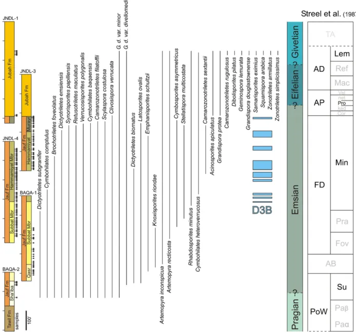
Fig. 2. Stratigraphic ranges of selected spore taxa encountered in this study compared with the west European zonation ofStreel et al. (1987). The biozones that are not recognized in the study are in grey. D3B is a recurrent sphaeromorph-dominated palynosubzone of significance for regional correlations (see text for details). Fig. 2. Distribution stratigraphique d’une s´election des spores observ´ees au cours de l’´etude et comparaisons avec la zonation ouest-europ´eenne deStreel et al. (1987). Les biozones non identifi´ees au cours de l’´etude sont en gris. D3B est une sous-palynozone r´ecurrente `a sphaeromorphes dominants, significative pour les corr´elations r´egionales (voir le texte pour les d´etails).
Dictyotriletes favosusMcGregor and Camfield, 1976
Dictyotriletes subgranifer McGregor, 1973 (Plate 7, Figs. 12 and 13)
Dictyotriletes?gorgoneus inMcGregor, 1973(Plate 7, Figs. 14 and 15)
Dyadaspora murusattenuataStrother and Traverse, 1979
Emphanisporites annulatusMcGregor, 1961(Plate 8, Fig. 1) Emphanisporites decoratusAllen, 1965
Emphanisporites mcgregorii Cramer, 1967 (Plate 8, Figs. 2 and 3)
Emphanisporites rotatus McGregor emend.McGregor, 1973 (Plate 8, Fig. 4)
Emphanisporites schultzii McGregor, 1973 (Plate 8, Figs. 5 and 6)
Geminospora lemurata (Balme) Playford, 1983 (Plate 8, Figs. 7–9)
Gneudnaspora divellomedia (Chibrikova)Balme, 1988var. divellomedia (Plate 3, Figs. 3–9)
Gneudnaspora divellomedia (Chibrikova)Balme, 1988var. minor nov. var. (Plate 3, Figs. 10–16)
Grandispora douglastownenseMcGregor, 1973(Plate 8, Fig. 10)
Grandispora protea (Naumova)Moreau-Benoˆıt, 1976(Plate 9, Fig. 1)
?Knoxisporites riondaeCramer and D´ıez, 1975(Plate 9, Figs. 2 and 3)
Latosporites ovalis nov. sp. (Plate 12, Figs. 8–12) Raistrickia spp.
32 P. Breuer et al. / Revue de micropal´eontologie 50 (2007) 27–57
P. Breuer et al. / Revue de micropal´eontologie 50 (2007) 27–57 33
34 P. Breuer et al. / Revue de micropal´eontologie 50 (2007) 27–57
P. Breuer et al. / Revue de micropal´eontologie 50 (2007) 27–57 35
36 P. Breuer et al. / Revue de micropal´eontologie 50 (2007) 27–57
P. Breuer et al. / Revue de micropal´eontologie 50 (2007) 27–57 37
38 P. Breuer et al. / Revue de micropal´eontologie 50 (2007) 27–57
P. Breuer et al. / Revue de micropal´eontologie 50 (2007) 27–57 39
40 P. Breuer et al. / Revue de micropal´eontologie 50 (2007) 27–57
P. Breuer et al. / Revue de micropal´eontologie 50 (2007) 27–57 41 Plate 1. Each miospore is identified by borehole, sample, slide numbers and England Finder Co-ordinate. Magnification× 1000. It is applicable to all plates. Fig.
1. Artemopyra inconspicua nov. sp. JNDL-1, 495.0’, 60855, E38/3. Fig. 2. Artemopyra inconspicua nov. sp. BAQA-1, 227.1’, 03CW110, G24/1. Holotype. Hilum
exhibits a pseudo-trilete mark. Fig. 3. Artemopyra inconspicua nov. sp. JNDL-3, 368.8’, 03CW160, W45. Fig. 4. Artemopyra inconspicua nov. sp. BAQA-1, 223.5’, 03CW109, F35/2. Paratype. Fig. 5. Artemopyra inconspicua nov. sp. BAQA-1, 219.2’, 03CW107, F43-44. Paratype. Hilum exhibits a pseudo-trilete mark. Fig.
6. Artemopyra recticosta nov. sp. BAQA-1, 219.2’, 03CW107, X31/2. Fig. 7. Artemopyra recticosta nov. sp. JNDL-1, 495.0’, 60854, U37/4. Holotype. Fig. 8.
Artemopyra recticosta nov. sp. JNDL-3, 368.8’, 03CW160, N43. Paratype. Fig. 9. Artemopyra recticosta nov. sp. JNDL-1, 156.0’, 60839, U44/4. Hilum exhibits a
pseudo-trilete mark that does not reach curvatura. Fig. 10. Artemopyra recticosta nov. sp. JNDL-1, 155.6’, 60837, P28/2. Fig. 11. Artemopyra recticosta nov. sp. JNDL-1, 155.6’, 60837, A29/4. Fig. 12. Artemopyra recticosta nov. sp. JNDL-1, 155.6’, 60837, L44. Fig. 13. Cymbohilates baqaensis nov. sp. BAQA-2, 54.8’, 03CW129, S44/1. Holotype. Fig. 14. Cymbohilates baqaensis nov. sp. BAQA-2, 54.8’, 03CW129, Q23/4. Paratype. Fig. 15. Cymbohilates baqaensis nov. sp. BAQA-2, 54.8’, 03CW129, S38/1. Fig. 16. Cymbohilates baqaensis nov. sp. BAQA-2, 50.2’, 03CW126, V33/2. Fig. 17. Cymbohilates baqaensis nov. sp. BAQA-2, 50.2’, 03CW126, S47/1. Paratype. Fig. 18. Cymbohilates baqaensis nov. sp. BAQA-2, 50.8’, 03CW127, M28. Fig. 19. Cymbohilates baqaensis nov. sp. BAQA-2, 52.0’, 03CW128, J33/1.
Planche 1. Chaque miospore est identifi´ee par le puits, l’´echantillon, le num´ero de lame et ses coordonn´ees England-Finder. Grossissement×1000 pour toutes les planches. Figs. 2 et 5. Le hilum pr´esente une pseudo-marque tril`ete. Fig. 9. Le hilum pr´esente une pseudo-marque tril`ete qui n’atteint pas les curvatures.
Plate 2. Fig. 1. Cymbohilates comptulus nov. sp. BAQA-1, 222.5’, 03CW108, Y35. Fig. 2. Cymbohilates comptulus nov. sp. BAQA-1, 205.8’, 03CW106, N50/2. Fig.
3. Cymbohilates comptulus nov. sp. BAQA-1, 205.8’, 03CW106, W25. Holotype. Fig. 4. Cymbohilates comptulus nov. sp. BAQA-1, 285.5’, 03CW111, V30. Fig. 5. Cymbohilates comptulus nov. sp. BAQA-1, 285.5’, 03CW111, W31. Fig. 6. Cymbohilates comptulus nov. sp. BAQA-1, 222.5’, 03CW108, P48. Paratype. Fig. 7. Cymbohilates comptulus nov. sp. BAQA-1, 205.8’, 03CW106, O23/1. Paratype. Fig. 8. Cymbohilates cymosusRichardson, 1996. BAQA-2, 134.4’, 03CW137, M34/2. Permanent tetrad. Fig. 9. Cymbohilates heteroverrucosus nov. sp. JNDL-1, 172.7’, 60845, L28. Holotype. Fig. 10. Cymbohilates heteroverrucosus nov. sp. JNDL-1, 167.8’, 60843, D27/3. Fig. 11. Cymbohilates heteroverrucosus nov. sp. JNDL-1, 174.6’, 60847, E33/1. Fig. 12. Cymbohilates heteroverrucosus nov. sp. JNDL-1, 177.0’, 60850, P48/1.
Plate 3. Fig. 1. Cymbohilates heteroverrucosus nov. sp. JNDL-1, 177.0’, PPM009, G33. Fig. 2. Cymbohilates heteroverrucosus nov. sp. JNDL-1, 177.0’, 60849, H44. Paratype. Fig. 3. Gneudnaspora divellomedia (Chibrikova)Balme, 1988var. divellomedia. JNDL-1, 167.8’, PPM006, Q36/3. Fig. 4. Gneudnaspora divellomedia (Chibrikova)Balme, 1988var. divellomedia. JNDL-1, 156.0’, 60839, U32/1. Hilum exhibits a pseudo-trilete mark. Fig. 5. Gneudnaspora divellomedia (Chibrikova)
Balme, 1988var. divellomedia. JNDL-1, 156.0’, PPM004, C45. Fig. 6. Gneudnaspora divellomedia (Chibrikova)Balme, 1988var. divellomedia. JNDL-1, 155.6’, 60837, M27. Fig. 7. Gneudnaspora divellomedia (Chibrikova)Balme, 1988var. divellomedia. JNDL-1, 174.6’, PPM008, U31. Fig. 8. Gneudnaspora divellomedia (Chibrikova)Balme, 1988var. divellomedia. BAQA-1, 219.2’, 03CW107, G49. Fig. 9. Gneudnaspora divellomedia (Chibrikova)Balme, 1988var. divellomedia. JNDL-4, 52.3’, 03CW188, V26. Fig. 10. Gneudnaspora divellomedia (Chibrikova)Balme, 1988var. minor nov. var. BAQA-2, 57.2’, 03CW131, E25/4. Fig. 11.
Gneudnaspora divellomedia (Chibrikova)Balme, 1988var. minor nov. var. BAQA-2, 50.2’, 03CW126, V30. Fig. 12. Gneudnaspora divellomedia (Chibrikova)
Balme, 1988var. minor nov. var. BAQA-2, 54.8’, 03CW129, G49. Paratype. Fig. 13. Gneudnaspora divellomedia (Chibrikova)Balme, 1988var. minor nov. var. JNDL-1, 172.7’, PPM007, M49/3. Fig. 14. Gneudnaspora divellomedia (Chibrikova)Balme, 1988var. minor nov. var. BAQA-2, 52.0’, 03CW128, T32/3. Fig. 15.
Gneudnaspora divellomedia (Chibrikova)Balme, 1988var. minor nov. var. BAQA-2, 50.2’, 03CW126, B49/2. Holotype. Fig. 16. Gneudnaspora divellomedia (Chibrikova)Balme, 1988var. minor nov. var. BAQA-2, 50.8’, 03CW127, L38/1. Fig. 17. Tetrahedraletes medinensisStrother and Traverse, 1979. JNDL-1, 156.0’, 60840, F43/4.
Plate 4. Fig. 1. Acinosporites apiculatus (Streel)Streel, 1967. JNDL-1, 495.0’, PPM014, O29. Fig. 2. Acinosporites lindlarensisRiegel, 1968. JNDL-1, 162.3’, PPM005, P37/4. Fig. 3. Ambitisporites avitusHoffmeister, 1959. BAQA-2, 52.0’, 03CW128, O47/1. Fig. 4. Ambitisporites eslae (Cramer and D´ıez)Richardson et al., 2001. BAQA-1, 371.1’, 03CW118, V36/3. Fig. 5. Amicosporites streeliiSteemans, 1989. BAQA-1, 395.2’, 03CW121, G26/3. Fig. 6. Amicosporites streelii
Steemans, 1989. BAQA-1, 285.5’, 03CW111, V44. Fig. 7. Apiculiretusispora brandtiiStreel, 1964. BAQA-1, 219.2’, 03CW107, U26/2. Fig. 8.
Apiculiretusis-pora brandtiiStreel, 1964. BAQA-1, 223.5’, 03CW109, V30. Fig. 9. Apiculiretusispora plicata (Allen)Streel, 1967. BAQA-1, 222.5’, 03CW108, U26. Fig. 10.
Archaeozonotriletes chulus (Cramer)Richardson and Lister, 1969. JNDL-1, 162.3’, 60841, N32/2. Fig. 11. Archaeozonotriletes chulus (Cramer)Richardson and Lister, 1969. JNDL-1, 162.3’, 60841, H45/3. Fig. 12. Archaeozonotriletes chulus (Cramer)Richardson and Lister, 1969. BAQA-2, 133.0’, 03CW136, Q25/1. Fig.
13. Brochotriletes foveolatusNaumova, 1953. BAQA-1, 223.5’, 03CW109, T23/2. Fig. 14. Camarozonotriletes filatoffii nov. sp. BAQA-1, 223.5’, 03CW109, S48/4.
Fig. 15. Camarozonotriletes filatoffii nov. sp. BAQA-1, 285.5’, 03CW111, N51/2. Fig. 16. Camarozonotriletes filatoffii nov. sp. BAQA-1, 223.5’, 03CW109, N45/2. Fig. 17. Camarozonotriletes filatoffii nov. sp. BAQA-1,.223.5’, 03CW109, N24. Fig. 18. Camarozonotriletes filatoffii nov. sp. BAQA-1, 219.2’, 03CW107, V39/1. Fig. 19. Camarozonotriletes filatoffii nov. sp. BAQA-1, 222.5’, 03CW108, W30. Paratype. Fig. 20. Camarozonotriletes filatoffii nov. sp. BAQA-1, 222.5’, 03CW108,
V44/4. Fig. 21. Camarozonotriletes filatoffii nov. sp. BAQA-1, 223.5’, 03CW109, M37. Paratype. Fig. 22. Camarozonotriletes filatoffii nov. sp. BAQA-2, 133.0’, 03CW136, H25. Holotype. Fig. 23. Camarozonotriletes filatoffii nov. sp. BAQA-1, 227.1’, 03CW110, Q47.
Plate 5. Fig. 1. Camarozonotriletes filatoffii nov. sp. BAQA-1, 222.5’, 03CW108, J-K36. Tetrad. Fig. 2. Camarozonotriletes rugulosus nov. sp. JNDL-1, 156.0’, 60840, T48. Holotype. Fig. 3. Camarozonotriletes rugulosus nov. sp. JNDL-1, 167.8’, 60842, G51/4. Fig. 4. Camarozonotriletes rugulosus nov. sp. JNDL-1, 156.0’, 60839, M49/3. Paratype. Fig. 5. Camarozonotriletes rugulosus nov. sp. JNDL-1, 156.0’, PPM004, J41/4. Fig. 6. Camarozonotriletes rugulosus nov. sp. JNDL-1, 172.7’, 60846, P46/2. Paratype. Fig. 7. Camarozonotriletes rugulosus nov. sp. JNDL-1, 155.6’, 60838, J42/2. Fig. 8. Camarozonotriletes rugulosus nov. sp. JNDL-1, 174.6’, PPM008, L-M44. Fig. 9. Camarozonotriletes rugulosus nov. sp. JNDL-1, 172.7’, PPM007, C47/4. Fig. 10. Camarozonotriletes sextantiiMcGregor and Camfield, 1976. JNDL-4, 75.0’, 03CW192, E32/1. Fig. 11. Cirratriradites diaphanusSteemans, 1989. BAQA-2, 50.2’, 03CW126, B45. Fig. 12. Clivosispora
verrucataMcGregor, 1973. BAQA-1, 222.5’, 03CW108, M-N26. Fig. 13. Clivosispora verrucataMcGregor, 1973. BAQA-1, 395.2’, 03CW121, F47/1. Fig. 14.
Clivosispora verrucataMcGregor, 1973. BAQA-1, 285.5’, 03CW111, U35. Fig. 15. Cymbosporites asymmetricus nov. sp. JNDL-3, 413.2’, 03CW166, C40. Fig.
16. Cymbosporites asymmetricus nov. sp. JNDL-4, 160.7’, 03CW211, V42. Paratype. Fig. 17. Cymbosporites asymmetricus nov. sp. JNDL-4, 214.3’, 03CW226,
X52. This specimen displays a local slight detachment of the exospore. Fig. 18. Cymbosporites asymmetricus nov. sp. JNDL-4, 214.3’, 03CW111, V51. Fig. 19.
Cymbosporites asymmetricus nov. sp. JNDL-4, 42.0’, 03CW186, R48.
Plate 6. Fig. 1. Cymbosporites asymmetricus nov. sp. JNDL-3, 413.2’, 03CW166, T42/1. Holotype. Fig. 2. Cymbosporites asymmetricus nov. sp. JNDL-3, 368.8’, 03CW160, L49. Fig. 3. Cymbosporites dammamensisSteemans, 1995. BAQA-2, 133.0’, 03CW136, R26/2. Fig. 4. Cymbosporites dammamensisSteemans, 1995. BAQA-2, 134.4’, 03CW137, N42. Fig. 5. Cymbosporites dammamensisSteemans, 1995. BAQA-1, 371.1’, 03CW118, T54. Fig. 6. Cymbosporites? senexMcGregor and Camfield, 1976. BAQA-1, 285.5’, 03CW111, W44/1. Fig. 7. Cymbosporites? senexMcGregor and Camfield, 1976. JNDL-1, 495.0’, 60855, O30. Fig. 8.
Dibolisporites echinaceus (Eisenack)Richardson, 1965. BAQA-2, 50.8’, 03CW127, P25/3. Fig. 9. Dibolisporites eifeliensis (Lanninger)McGregor, 1973. BAQA-2, 134.4’, 03CW137, J30/1. Fig. 10. Dibolisporites pilatus nov. sp. JNDL-1, 177.0’, PPM009, N39. Fig. 11. Dibolisporites pilatus nov. sp. JNDL-1, 167.8’, PPM006, V48. Holotype. Fig. 12. Dibolisporites pilatus nov. sp. JNDL-1, 174.6’, 60847, M29/3. Fig. 13. Dibolisporites pilatus nov. sp. JNDL-1, 167.8’, 60842, R42/3. Paratype.
42 P. Breuer et al. / Revue de micropal´eontologie 50 (2007) 27–57
Plate 7. Fig. 1. Dictyotriletes biornatus nov. sp. BAQA-1, 308.3’, 03CW112, E48/2. Fig. 2. Dictyotriletes biornatus nov. sp. JNDL-4, 448.6’, 03CW267, U33/2.
Fig. 3. Dictyotriletes biornatus nov. sp. BAQA-1, 366.9’, 03CW117, L27/1. Paratype. Fig. 4. Dictyotriletes biornatus nov. sp. JNDL-4, 448.6’, 03CW267, H34/3.
A branch of the trilete mark is visible. Fig. 5. Dictyotriletes biornatus nov. sp. BAQA-1, 345.5’, 03CW114, O53. Holotype. Fig. 6. Dictyotriletes biornatus nov. sp. JNDL-4, 495.2’, 03CW275, L41/4. Fig. 7. Dictyotriletes biornatus nov. sp. JNDL-4, 464.1’, 03CW269, M37/1. Fig. 8. Dictyotriletes biornatus nov. sp. BAQA-1, 219.2’, 03CW107, L40/4. Paratype. Fig 9. Dictyotriletes biornatus nov. sp. BAQA-1, 169.1’, 03CW103, K26. Fig. 10. Dictyotriletes emsiensis (Allen)McGregor, 1973. BAQA-2, 56.0’, 03CW128, X46. Fig. 11. Dictyotriletes emsiensis (Allen)McGregor, 1973. BAQA-2, 134.4’, 03CW137, Q29/3. Fig. 12. Dictyotriletes
subgraniferMcGregor, 1973. BAQA-1, 366.9’, 03CW117, O31. Fig. 13. Dictyotriletes subgraniferMcGregor, 1973. BAQA-2, 134.4’, 03CW137, T39/2. Fig. 14.
Dictyotriletes?gorgoneus inMcGregor, 1973. BAQA-2, 50.8’, 03CW127, G54. Fig. 15. Dictyotriletes?gorgoneus inMcGregor, 1973. BAQA-2, 50.8’, 03CW267, D24.
Plate 8. Fig. 1. Emphanisporites annulatusMcGregor, 1961. JNDL-1, 172.7’, 60846, S30/2. Fig. 2. Emphanisporites mcgregoriiCramer, 1967. JNDL-1, 177.0’, PPM009, K47/3. Fig. 3. Emphanisporites mcgregoriiCramer, 1967. BAQA-2, 134.4’, 03CW137, E52. Fig. 4. Emphanisporites rotatus McGregor emend.McGregor, 1973. BAQA-1, 345.5’, 03CW114, H50/1. Fig. 5. Emphanisporites schultziiMcGregor, 1973. BAQA-1, 395.2’, 03CW121, G50. Fig. 6. Emphanisporites schultzii
McGregor, 1973. JNDL-4, 182.5’, 03CW220, R38. Incomplete tetrad. Fig. 7. Geminospora lemurata (Balme)Playford, 1983. JNDL-1, 172.7’, PPM007, O49/3. Fig.
8. Geminospora lemurata (Balme)Playford, 1983. JNDL-1, 167.8’, 60842, E34. Fig. 9. Geminospora lemurata (Balme)Playford, 1983. JNDL-1, 162.3’, 60841, G30/2. Fig. 10. Grandispora douglastownenseMcGregor, 1973. JNDL-1, 177.0’, 60850, P45/4.
Plate 9. Fig. 1. Grandispora protea (Naumova) Moreau-Benoˆıt, 1976. JNDL-1, 156.0’, PPM004, H44/4. Fig. 2. ?Knoxisporites riondaeCramer and D´ıez, 1975. BAQA-1, 285.5’, 03CW111, R48/3. Fig. 3. ?Knoxisporites riondaeCramer and D´ıez, 1975. BAQA-1, 366.9’, 03CW117, M32/2. Fig. 4. Retusotriletes maculatus
McGregor and Camfield, 1976. BAQA-1, 219.2’, 03CW107, F35. Fig. 5. Retusotriletes maculatusMcGregor and Camfield, 1976. BAQA-1, 285.5’, 03CW111, Q34.
Fig. 6. Retusotriletes rotundus (Streel)Lele and Streel, 1969. BAQA-1, 371.1’, 03CW118, P33. Fig. 7. Retusotriletes triangulatus (Streel)Streel, 1967. BAQA-1, 345.5’, 03CW114, M35/1. Fig. 8. Rhabdosporites minutusTiwari and Schaarschmidt, 1975. JNDL-3, 368.8’, 03CW160, H45/1. Fig. 9. Rhabdosporites minutus
Tiwari and Schaarschmidt, 1975. JNDL-1, 177.0’, PPM009, U32. Fig. 10. Rhabdosporites minutusTiwari and Schaarschmidt, 1975. JNDL-1, 156.0’, 60839, S36.
Fig. 11. Samarisporites eximius (Allen)Loboziak and Streel, 1989. JNDL-1, 162.3’, PPM005, S45/3.
Retusotriletes maculatus McGregor and Camfield, 1976 (Plate 9, Figs. 4 and 5)
Retusotriletes rotundus (Streel)Lele and Streel, 1969(Plate 9, Fig. 6)
Retusotriletes triangulatus (Streel) Streel, 1967 (Plate 9, Fig. 7)
Retusotriletes spp.
Rhabdosporites minutus Tiwari and Schaarschmidt, 1975 (Plate 9, Figs. 8–10)
Samarisporites eximius (Allen) Loboziak and Streel, 1989 (Plate 9, Fig. 11)
Scylaspora costulosa nov. sp. (Plate 10, Figs. 1–5)
Squamispora arabica nov. gen. et sp. (Plate 10, Figs. 6–10)
Stellatispora multicostata nov. sp. (Plate 11, Figs. 1–4) Synorisporites papillensisMcGregor, 1973(Plate 11, Figs. 5 and 6)
Tetrahedraletes medinensis Strother and Traverse, 1979 (Plate 3, Fig. 17)
Verruciretusispora dubia (Eisenack)Richardson and Rasul, 1978
Verrucosisporites flexibilisTurnau, 1986
Verrucosisporites polygonalis Lanninger, 1968 (Plate 11, Fig. 7)
Verrucosisporites spp.
Zonotriletes armillatus nov. sp. (Plate 11, Figs. 8–12;Plate 12, Figs. 1 and 2)
Zonotriletes simplicissimus nov. sp. (Plate 12, Figs. 3–7) Zonotriletes spp.
5.2. Systematic descriptions
New taxa of cryptospores, trilete spores and monolete spores are described below. Within each group, taxonomic descriptions are arranged alphabetically by genus and species.
5.2.1. Hilate cryptospores
Genus Artemopyra Burgess and Richardson emend. Richardson, 1996
Type species: Artemopyra brevicosta Burgess and Richardson, 1991.
Artemopyra inconspicua nov. sp.
Plate 1, Figs. 1–5
Derivation of name: Refers to the characteristics of the muri;
inconspicuus = not well visible (Latin).
Holotype:Plate 1, Fig. 2, borehole BAQA-1, sample 227.1’, slide 03CW110, EFC G24-1.
Paratypes:Plate 1, Fig. 4, borehole BAQA-1, sample 223.5’, slide 03CW109, EFC F35-2.Plate 1, Fig. 5, borehole BAQA-1, sample 219.2’, slide 03CW107, EFC F43-44.
Diagnosis: A distally laevigate Artemopyra. Hilum
sculp-tured with weakly defined, radially disposed, straight muri, extending from the curvatura for between half to all of the way to the proximal pole.
Description: Amb circular to sub-circular. A diffuse to clear
curvatura delimits a more or less circular hilum. Proximal sur-face is characterized by scars which often resemble a trilete mark. The hilum radius commonly equals to 3/4 to 4/5 of the amb radius. Hilum sculptured with closely spaced, radially dis-posed muri which are straight, 0.5–1m wide and 0.5–1 m apart at the equator, becoming thinner towards the proximal pole. Typically the muri are weakly defined and extend from the curvatura for half to all of the way to the proximal pole. There are numerous muri, usually between 60 and 90. Distal surface laevigate.
Dimensions: 36 (48) 57m, 20 specimens measured. Comparison: Artemopyra brevicosta Burgess and Richardson, 1991 has more robust, radial muri which always are confined to the equatorial region of the contact area. Artemopyra recticosta nov. sp. possesses thicker muri which
P. Breuer et al. / Revue de micropal´eontologie 50 (2007) 27–57 43 are less numerous (18–46). Artemopyra inconspicua nov. sp.
sometimes can be mistaken for Gneudnaspora divellomedia (Chibrikova) Balme, 1988 var. divellomedia because of the inconspicuous nature of the muri.
Occurrence: Boreholes BAQA-1 and JNDL-1, 3, 4. Jauf
and Jubah formations. Emsian-Givetian. Artemopyra recticosta nov. sp.
Plate 1, Figs. 6–12
?1986. Emphanisporites orbicularis – Turnau, p. 345, Plate 4, Figs. 4 and 9.
2007. Artemopyra? spp. – Wellman Plate 20, Fig. i.
Derivation of name: Refers to the straight muri;
rec-tus = straight, costa = rib (Latin).
Holotype:Plate 1, Fig. 7, borehole JNDL-1, sample 495.0’, slide 60854, EFC U37/4.
Paratype:Plate 1, Fig. 8, borehole JNDL-3, sample 368.8’, slide 03CW160, EFC N43.
Diagnosis: A distally laevigate Artemopyra. Hilum
sculp-tured with radially disposed, straight muri extending from the curvatura for between half to all of the way to the proximal pole.
Description: Amb circular to sub-circular. A diffuse to clear
curvatura 0.6–2m wide delimits a more or less circular hilum. Proximal surface is characterized by different types of scars which sometimes show a shape similar to a trilete mark. The hilum radius equals or is more or less 4/5 of the amb radius. Hilum sculptured by radially disposed muri which are straight and up 0.7–2.6m wide at the equator, becoming thinner towards the proximal pole. They extend from the curvatura for half to all of the way to the proximal pole. There are usually 18–46 muri. Distal surface laevigate.
Dimensions: 34 (51) 70m, 36 specimens measured. Comparison: Artemopyra laevigata Wellman and Richardson, 1996 is generally smaller (33–48m) and possesses proximal radial muri which are highly variable in appearance and sometimes bifurcate. Artemopyra brevicosta Burgess and Richardson, 1991 is smaller (22–49m) and shows proximal radial muri which can be slightly sinuous and are short, less than 1/2 of the hilum radius. Emphanisporites orbicularisTurnau, 1986represents probably the same taxon, although this is described as a trilete spore? Artemopyra spp. inWellman (2007)represents the same species described here. The trilete spore Emphanisporites rotatus McGregor emend. McGregor, 1973 has the same kind of muri as Artemopyra recticosta nov. sp.
Remark: Some specimens of Artemopyra recticosta nov. sp.
in our material possess a darkened area which is located at or near the proximal pole.
Occurrence: Boreholes BAQA-1 and JNDL-1, 3, 4. Jauf
(Subbat-Murayr members) and Jubah formations. Emsian-Givetian.
Genus CymbohilatesRichardson, 1996emend.
Type species: Cymbohilates horridusRichardson, 1996.
Emended diagnosis: Proximally hilate cryptospore monads.
Exospore, sculptured subequatorially and distally with grana,
coni, spinae, bacula, verrucae or biform elements, sometimes fused in groups. Contact area (hilum) sculptured or smooth, with random or concentric folds, and/or radial muri. Hilum, more or less circular, curvatural ridge distinct or barely perceptible.
Remark: The diagnosis is enlarged to include either
subequatorially and distally or wholly sculptured forms. The discrete positive elements can be of all types.
Cymbohilates baqaensis nov. sp.
Plate 1, Figs. 13–19
Derivation of name: Refers to the locality where this species
was found; baqaensis = from the locality of Baq’a in Saudi Ara-bia (Latin).
Holotype:Plate 1, Fig. 13, borehole BAQA-2, sample 54.8’, slide 03CW129, EFC S44/1.
Paratypes:Plate 1, Fig. 14, borehole BAQA-2, sample and slide 54.8’, slide 03CW129, EFC Q23/4.Plate 1, Fig. 17, bore-hole BAQA-2, sample 50.2’, slide 03CW126, EFC S47/1.
Diagnosis: A Cymbohilates with small bacula. The proximal
surface possesses different types of scars which sometimes have a shape similar to a trilete mark.
Description: Amb circular to sub-circular. An
undulat-ing and irregular curvatura delimits a more or less circular, smooth hilum. The smooth proximal surface shows different types of scars such as multiple radial branches, cross frac-tures, pseudo-trilete mark, or simple slit. Exospore is sculptured subequatorially and distally with evenly distributed bacula 0.75–2m high, 0.75–1.5 m wide at base and 1.5–3 m apart. The tops of elements are flat or slightly concave, with generally bifurcate shape.
Dimensions: 30 (36) 40m, 16 specimens measured. Comparison: Cymbosporites baculatusTurnau et al., 2005 also possesses bacula but without bifurcate shape. According to Turnau (pers. comm.), this taxon is different but is probably related. Cymbosporites dammamensisSteemans, 1995appears very similar but is a trilete miospore, although its trilete mark is not always clearly perceptible. Some specimens have probably been misinterpreted inSteemans (1995)and have to be revised.
Remark: Some specimens can be preserved as dyads. Occurrence: Boreholes BAQA-1, 2 and JNDL-3, 4. Jauf
Formation (Sha’iba-Murayr members). Pragian-Emsian. Cymbohilates comptulus nov. sp.
Plate 2, Figs. 1–7
?1988. Gneudnaspora sp. (Chibrikova) – Balme, p. 17, Plate 3, Fig. 15.
Derivation of name: Refers to the fine ornamentation of the
distal surface; comptulus = finely decorated (Latin).
Holotype:Plate 2, Fig. 3, borehole BAQA-1, sample 205.8’, slide 03CW106, EFC W25.
Paratypes:Plate 2, Fig. 6, borehole BAQA-1, sample 222.5’, slide 03CW108, EFC P48. Plate 2, Fig. 7, borehole BAQA-1, sample 205.8’, slide 03CW106, EFC O23/1.
Diagnosis: A Cymbohilates with small bacula, spinae and
coni sparsely distributed. The proximal surface often possesses narrow radial scars in the exospore.
44 P. Breuer et al. / Revue de micropal´eontologie 50 (2007) 27–57
P. Breuer et al. / Revue de micropal´eontologie 50 (2007) 27–57 45
46 P. Breuer et al. / Revue de micropal´eontologie 50 (2007) 27–57
P. Breuer et al. / Revue de micropal´eontologie 50 (2007) 27–57 47 Plate 10. Fig. 1. Scylaspora costulosa nov. sp. JNDL-3, 353.8’, 03CW159, L33/3. Fig. 2. Scylaspora costulosa nov. sp. BAQA-1, 227.1’, 03CW110, L24. Holotype.
Fig. 3. Scylaspora costulosa nov. sp. BAQA-2, 50.8’, 03CW127, O49/1. Paratype. Fig. 4. Scylaspora costulosa nov. sp. BAQA-2, 133.0’, 03CW136, M45/1. Fig. 5. Scylaspora costulosa nov. sp. BAQA-2, 50.8’, 03CW127, J40. Fig. 6. Squamispora arabica nov. gen. and sp. JNDL-1, 172.7’, 60846, H29. Paratype. Fig. 7.
Squamispora arabica nov. gen. and sp. JNDL-1, 177.0’, PPM009, H45. Fig. 8. Squamispora arabica nov. gen. and sp. JNDL-1, 174.6’, PPM008, C43. Fig. 9. Squamispora arabica nov. gen. and sp. JNDL-1, 174.6’, 60848, L40/2. Paratype. Fig. 10. Squamispora arabica nov. gen. and sp. JNDL-1, 177.0’, 60849, U31.
Holotype.
Plate 11. Fig. 1. Stellatispora multicostata nov. sp. JNDL-3, 462.3’, 03CW175, L30-31. Fig. 2. Stellatispora multicostata nov. sp. JNDL-4, 182.5’, 03CW220, U41/4. Holotype. Fig. 3. Stellatispora multicostata nov. sp. JNDL-3, 462.3’, 03CW175, H26. Paratype. Fig. 4. Stellatispora multicostata nov. sp. JNDL-4, 197.8’, 03CW224, H39/3. Fig. 5. Synorisporites papillensisMcGregor, 1973. BAQA-1, 376.4’, 03CW119, C26/3. Fig. 6. Synorisporites papillensisMcGregor, 1973. BAQA-1, 308.3’, 03CW112, H40/4. Fig. 7. Verrucosisporites polygonalisLanninger, 1968. BAQA-1, 406.0’, 03CW123, M48/3. Fig. 8. Zonotriletes armillatus nov. sp. JNDL-1, 156.0’, PPM004, P43. Holotype. Fig. 9. Zonotriletes armillatus nov. sp. JNDL-1, 156.0’, 60840, X32/3. Fig. 10. Zonotriletes armillatus nov. sp. JNDL-1, 155.6’, 60837, O34. Fig. 11. Zonotriletes armillatus nov. sp. JNDL-1, 155.6’, PPM003, P30/4. Fig. 12. Zonotriletes armillatus nov. sp. JNDL-1, 155.6’, 60838, J43/1. Plate 12. Fig. 1. Zonotriletes armillatus nov. sp. JNDL-1, 162.3’, 60841, F29/1. Paratype. Fig. 2. Zonotriletes armillatus nov. sp. JNDL-1, 167.8’, 60843, L41. Paratype. Fig. 3. Zonotriletes simplicissimus nov. sp. JNDL-1, 177.0’, PPM009, R47/1. Fig. 4. Zonotriletes simplicissimus nov. sp. JNDL-1, 172.7’, 60845, F28/1. Paratype. The transverse attachment lines of the flange on the central body are distinguishable. Fig. 5. Zonotriletes simplicissimus nov. sp. JNDL-1, 177.0’, PPM009, P32/4. Holotype. Fig. 6. Zonotriletes simplicissimus nov. sp. JNDL-1, 177.0’, 60850, K44/2. The transverse attachment lines of the flange on the central body are distinguishable. Fig. 7. Zonotriletes simplicissimus nov. sp. JNDL-1, 177.0’, 60849, K43/3. Paratype. Fig. 8. Latosporites ovalis nov. sp. BAQA-1, 222.5’, 03CW108, F40. Paratype. Fig. 9. Latosporites ovalis nov. sp. BAQA-1, 345.5’, 03CW114, K23. Holotype. Fig. 10. Latosporites ovalis nov. sp. BAQA-2, 54.8’, 03CW129, Q36.
Fig. 11. Latosporites ovalis nov. sp. BAQA-1, 371.1’, 03CW118, R45. Fig. 12. Latosporites ovalis nov. sp. BAQA-2, 52.0’, 03CW128, Q51/3.
Description: Amb circular to sub-circular. The hilum is well
defined by a curvatura and its slightly granulate character is barely perceptible. Narrow, radial, irregular scars commonly characterize the proximal surface. The hilum radius equals about 3/4 of the amb radius. Exospore is sculptured sube-quatorially and distally with evenly distributed and spaced elements 0.5-1.5m high and 1–3 m apart. The ornamenta-tion varies from bacula to spinae and coni. Exospore 1–3m thick.
Dimensions: 48 (58) 70m, 28 specimens measured. Comparison: Cymbohilates amplus Wellman and Richardson, 1996 possesses a very similar distal orna-mentation but it is significantly larger (73–104m) than Cymbohilates comptulus nov. sp. Moreover, Cymbohilates amplus does not possess narrow radial tears in the exospore on the proximal surface. Gneudnaspora sp. in Balme (1988) also appears very similar to Cymbohilates comptulus nov. sp., however it is only represented by a single specimen that is ornamented with evenly disposed cones about 1m high and 1m in basal diameter.
Occurrence: Boreholes BAQA-1, 2 and JNDL-4. Jauf
Formation (Sha’iba-Hammamiyat members). Pragian-Emsian. Cymbohilates heteroverrucosus nov. sp.
Plate 2, Figs. 9–12;Plate 3, Figs. 1 and 2
Derivation of name: Refers to the ornament variability on a
same specimen; hetero- = different, verrucosus = bearing verru-cae (Latin).
Holotype:Plate 2, Fig. 10, borehole JNDL-1, sample 167.8’, slide 60843, EFC D27/3.
Paratype:Plate 3, Fig. 2, borehole JNDL-1, sample 177.0’, slide 60849, EFC H44.
Diagnosis: A Cymbohilates bearing verrucae and bacula of
different type and size, which are distributed unevenly on the whole surface of the cryptospore.
Description: Amb circular to sub-circular. Hilum often is
barely perceptible and commonly shows narrow scars that may
resemble a pseudo-trilete mark. Distal and proximal surfaces bear a varied sculpture of verrucae and bacula, 0.5–7m wide at base and up to 4m high. The elements can have a flared base or more parallel sides. The ornament size and type can be very variable on the same specimen. The small and largest elements are distributed unevenly, however the contact area normally pos-sesses the smallest verrucae.
Dimensions: 45 (53) 62m, 34 specimens measured. Occurrence: Boreholes JNDL-1, 3, 4. Jauf
(Hammamiyat-Murayr members) and Jubah formations. Emsian-Givetian. Genus GneudnasporaBalme, 1988emend.
1991. Laevolancis – Burgess and Richardson, p. 606.
Type species: Gneudnaspora kernickiiBalme, 1988.
Emended diagnosis: Proximally hilate cryptospore monads.
Amb circular to oval. Exospore alveolate or laevigate, proxi-mally attenuated. Contact face bordered by a weakly to strongly developed circular to oval curvatural ridge. Different types of scars can be present on the contact face, from an irregular, simple, pseudo-trilete or pseudo-tetralete scar to an irregular sub-stellate opening.
Remark: The material in the present study is very similar
to that figured and described byBalme (1988)and the authors consider that the proximal structure of Gneudnaspora has been misinterpreted by Balme. It corresponds to scars, sometimes looking like a trilete or monolete mark, but commonly hav-ing no particular shape. In addition, some specimens do not show these scars and are therefore similar to genus Laevolan-cisBurgess and Richardson, 1991described from the Silurian. Finally, we consider that the latter form is a junior synonym of the genus GneudnasporaBalme, 1988, following the diagnosis as emended here.
The diagnosis of the genus Gneudnaspora is restricted to include only proximally hilate cryptospore monads that are entirely laevigate. Thus, Gneudnaspora sp. in Balme (1988), which bears small cones, has to be allocated to the genus Cym-bohilatesRichardson, 1996 emended here. As Gneudnaspora
48 P. Breuer et al. / Revue de micropal´eontologie 50 (2007) 27–57 divellomedia (Chibrikova) Balme, 1988is here considered to
be the valid combination, the type species for Laevolancis, L. divellomedia (Chibrikova) Burgess and Richardson, 1991, becomes invalid. The existing species of Laevolancis are herein transferred to Gneudnaspora.
Gneudnaspora divellomedia (Chibrikova)Balme, 1988var. divellomedia
Plate 3, Figs. 3–9
1988. Gneudnaspora divellomedium (Chibrikova) – Balme, p. 125, Plate 3, Figs. 1–7.
Diagnosis: An entirely laevigate Gneudnaspora showing
dif-ferent types of scars on its contact face.
Description: Amb circular to sub-circular. A diffuse to clear
curvatura delimits a more or less circular hilum. The hilum radius equals 3/4 to 6/7 amb radius. Contact face shows different types of scars, from an irregular, simple, trilete or pseudo-tetralete, to an irregular sub-stellate shape. Proximal and distal surfaces are entirely laevigate.
Dimensions: 32 (54) 82m, 17 specimens measured. Remark and comparison: Specimens assigned to
Laevolan-cis divellomedia (Chibrikova) Burgess and Richardson, 1991 are not considered here to be conspecific with Gneudnas-pora divellomedia (Chibrikova)Balme, 1988var. divellomedia. Laevolancis divellomedia is generally smaller than Gneudnas-pora divellomedia var. divellomedia. Furthermore, the former species very often shows an intact contact face whilst the latter usually possesses different types of scars.
Occurrence: Boreholes BAQA-1, 2 and JNDL-1, 3, 4.
Jauf (upper Sha’iba-Murayr members) and Jubah formations. Emsian-Givetian.
Gneudnaspora divellomedia (Chibrikova)Balme, 1988var. minor nov. var.
Plate 3, Figs. 10–16
1959. Archaeozonotriletes divellomedium –Chibrikovap. 65, Plate 9, Fig. 4.
1966b. Hispanaediscus bernesgae –Cramerp. 82, Plate 1, Figs. 2 and 11, Text-fig. 2.
1968. Spore no. 2651 –Magloire, 1968, Plate 1, Fig. 6. 1969.?Archaeozonotriletes cf. divellomedium Chibrikova – Richardson and Lister, p. 238, Plate 43, Fig. 12.
1974. Archaeozonotriletes (?) divellomedium (Chibrikova) – ArkhangelskayaPlate 1, Fig. 51.
1974. Zonaletes (?) divellomedium (Chibrikova) – Arkhangelskaya, Plate 6, Figs. 3 and 4.
1974.?Archaeozonotriletes divellomedium Chibrikova – McGregorPlate 1, Figs. 35 and 40.
1978. Hispanaediscus sp. –McGregor and Narbonne, 1978, p. 1296, Plate 1, Figs. 20–22.
1980. Zonaletes (?) divellomedium (Chibrikova) – ArkhangelskayaPlate 5, Fig. 34.
1984.?Stenozonotriletes irregularis (Schultz) –McGregorp. 37, Plate 1, Fig. 26.
1986. Archaeozonotriletes cf. divellomedium Chibrikova – Buret and Moreau-Benoˆıt, 1986, Plate 1, Fig. 1.
1986. Tholisporites divellomedium (Chibrikova) – Turnau, p. 349, Plate 2, Fig. 12, Plate 4, Fig. 14.
1991. Laevolancis divellomedia (Chibrikova) – Burgess and Richardson, p. 607, Plate 2, Figs. 4 and 6.
Derivation of name: Refers to the small size of the amb;
minor = smaller (Latin).
Holotype:Plate 3, Fig. 15, borehole BAQA-2, sample 50.2’, slide 03CW126, EFC B49/2.
Paratype:Plate 3, Fig. 12, borehole BAQA-2, sample 54.8’, slide 03CW129, EFC G49.
Diagnosis: A Gneudnaspora of small size, very simple
form.
Description: Amb circular to sub-circular. A diffuse to clear
curvatura delimits a more or less circular hilum. Hilum radius is commonly 3/4 to 4/5 spore radius. Contact face sometimes shows a small simple scar. Proximal and distal surfaces are entirely laevigate.
Dimensions: 28 (31) 34m, 21 specimens measured. Remark: As the genus LaevolancisBurgess and Richardson, 1991is here considered to be a junior synonym of genus Gneud-nasporaBalme, 1988, Laevolancis divellomedia (Chibrikova) Burgess and Richardson, 1991requires reassignment. Since it is suggested above that Laevolancis divellomedia differs from Gneudnaspora divellomedia (Chibrikova)Balme, 1988var. div-ellomedia, the reassignment necessitates introduction of a new variety, Gneudnaspora divellomedia (Chibrikova)Balme, 1988 var. minor nov. var.
Comparison: Gneudnaspora divellomedia (Chibrikova)
Balme, 1988var. divellomedia has a similar structure but is larger (32–82m). The smaller new variety shows a little size variabil-ity. Furthermore, the larger variety always possesses different types of scars, unlike Gneudnaspora divellomedia (Chibrikova) Balme, 1988var. minor nov. var., which often possesses an intact contact face. Gneudnaspora plicata (Burgess and Richardson) nov. comb. appears very similar and is thus difficult to distin-guish, however it is only known from Wenlock strata.Steemans et al. (1996)define a morphon which includes the two taxa and record intermediate forms with a range of thin to thick wall thicknesses.
Occurrence: Boreholes BAQA-1, 2 and JNDL-1, 3, 4. Jauf
(Sha’iba-Murayr members) and Jubah formations. Pragian-Givetian.
Gneudnaspora plicata (Burgess and Richardson) nov. comb. 1991. Gneudnaspora plicata – Burgess and Richardson, p. 607, Plate 2, Fig. 8.
Remark: As the genus LaevolancisBurgess and Richardson, 1991is here considered to be a junior synonym of genus Gneud-nasporaBalme, 1988, other species that have been described under Laevolancis become invalid and have to be transferred to the valid genus. Gneudnaspora plicata (Burgess and Richard-son) nov. comb. is not recorded in the present assemblages.
Gneudnaspora chibrikovae (Steemans et al.) nov. comb. 2000. Gneudnaspora chibrikovae – Steemans et al. p. 99, Plate 2, Figs. n–o; Plate 3, Fig. a.
Remark: Gneudnaspora chibrikovae (Steemans et al.) nov.
P. Breuer et al. / Revue de micropal´eontologie 50 (2007) 27–57 49
5.2.2. Trilete spores
Genus Camarozonotriletes Naumova exIshchenko, 1952
Type species: Camarozonotriletes devonicus Naumova, 1953.
Camarozonotriletes filatoffii nov. sp.
Plate 4, Figs. 14–23;Plate 5, Fig. 1
?1972.?Reticulatisporites sp. – Kemp, p. 115, Plate 55, Fig. 9.
Derivation of name: After the name of the Australian
paly-nologist employed by Saudi Aramco, John Filatoff.
Holotype:Plate 4, Fig. 22, borehole BAQA-2, sample 133.0’, slide 03CW136, H25.
Paratypes: Plate 4, Fig. 19, borehole BAQA-1, sample 222.5’, slide 03CW108, W30.Plate 4, Fig. 21, borehole BAQA-1, sample 223.5’, slide 03CW109, EFC M37.
Diagnosis: A small Camarozonotriletes with an indistinctly
defined spine-bearing reticulum.
Description: Amb sub-circular to sub-triangular. Contact
area sub-triangular to triangular with rounded corners and laevigate. Laesurae simple or accompanied by narrow labra, straight, 3/5 to 4/5 of the radius in length. Exospore darker adjacent to rays, forming a more or less triangular darkened and presumably thickened zone around the proximal pole and extending to the end of the rays. Cingulum 1–2m thick equa-torially opposite the trilete rays and up to 6m interradially. Distal and equatorial sculpture composed of spines generally 1–2m high, 0.5–1.5 m wide at their base, and 1.5–2.5 apart. Elements are commonly situated on indistinctly defined muri, about 1–2.5m wide, that form a reticulum. Lumina are generally 3–7m in their longest diameter. Cingulum slightly darker than central area.
Dimensions: 24 (29) 35m, 35 specimens measured. Comparison: The distal sculpture distinguishes
Camaro-zonotriletes filatoffii nov. sp. from all other species of Camarozonotriletes. Camarozonotriletes sextantii McGregor and Camfield, 1976 is larger (37–60m). Its distal and equatorial sculpture does not possess a reticulum and the ornaments are evenly distributed.?Reticulatisporites sp. in Kemp (1972) is similar but does not exhibit spines on the reticulum.
Occurrence: Boreholes BAQA-1, 2 and JNDL-3, 4. Jauf
Formation (Sha’iba-Murayr members). Pragian-Emsian.
Camarozonotriletes rugulosus nov. sp.
Plate 5, Figs. 2–9
Derivation of name: Refers to the wrinkled appearance of
the distal face; rugulosus = slightly wrinkled (Latin).
Holotype:Plate 5, Fig. 2, borehole JNDL-1, sample 156.0’, slide 60840, EFC T48.
Paratypes: Plate 5, Fig. 4, borehole JNDL-1, sample and slide 156.0’, slide 60839, EFC M49/3.Plate 5, Fig. 6, borehole JNDL-1, sample 172.7’, slide 60846, EFC P46/2.
Diagnosis: A triangular Camarozonotriletes with thin
rugu-lae.
Description: Amb triangular. The corners are slightly
rounded, while the margins are slightly convex, straight, or sometimes slightly concave. Exospore thin. Laesurae distinct, simple and straight. The trilete mark radius is at least longer than 4/5 of the amb radius. Curvaturae are not easily distinguish-able. Cingulum generally 1–2m thick equatorially opposite the trilete rays and 3–5m interradially. Contact faces laevi-gate. Distal face sculptured with very thin, 0.5m in maximum width.
Dimensions: 37 (46) 59m, 30 specimens measured. Comparison: Leiotriletes horridulus Ishchenko, 1958, an Upper Mississippian species of Russia, resembles Camaro-zonotriletes rugulosus nov. sp. according to the original description, but it is smaller (30–35m). Furthermore, this taxon is not interradially thickened.
Remark: As the rugulae are very thin, they may be barely
visible or undistinguishable because of bad preservation. Its thin exospore seems delicate.
Occurrence: Borehole JNDL-1. Jubah Formation. Givetian.
Genus CymbosporitesAllen, 1965
Type species: Cymbosporites cyathusAllen, 1965. Cymbosporites asymmetricus nov. sp.
Plate 5, Figs. 15–19;Plate 6, Figs. 1 and 2
Derivation of name: Refers to the asymmetry of the trilete
mark; asymmetricus = asymmetrical (Latin).
Holotype:Plate 6, Fig. 1, borehole JNDL-3, sample 413.2’, slide 03CW166, EFC T42/1.
Paratype:Plate 5, Fig. 16, borehole JNDL-4, sample 160.7’, slide 03CW211, EFC V42.
Diagnosis: A Cymbosporites with small spines densely
dis-tributed. One interradial area of the contact surface is smaller than the other two.
Description: Amb oval to sub-triangular with a distinctly
elongate axis aligned with one of the laesurae. Contact faces laevigate with the one, transected by the long axis, being smaller than its neighbours. The smallest interradial angle, formed by the two shorter trilete mark branches, is commonly between 70◦and 100◦. Laesurae distinct, straight to slightly sinuous and accompanied by prominent labra commonly 1–2.5m in over-all width. Radius of contact surface generover-ally equals to 3/4 of the amb radius. Proximo-equatorial and distal regions patinate and sculptured with spines (ca. 1m high), which are densely distributed. Some specimens can exhibit slight separations of the exospore. Exospore 2–4m thick.
Dimensions: 36 (51) 67m, 15 specimens measured. Comparison: Apiculiretusispora plicata (Allen) Streel, 1967, has a similar size and ornamentation, and sometimes also an asymmetrically placed laesurae, but differs in not being patinate.
Occurrence: Boreholes JNDL-3, 4. Jauf Formation
(Subbat-Murayr members). Emsian.
Genus DibolisporitesRichardson, 1965
Type species: Dibolisporites echinaceus (Eisenack) Richardson, 1965.
50 P. Breuer et al. / Revue de micropal´eontologie 50 (2007) 27–57 Dibolisporites pilatus nov. sp.
Plate 6, Figs. 10–13
Derivation of name: Refers to the pilate distal surface;
pila-tus = made up of pila (Latin).
Holotype:Plate 6, Fig. 11, borehole JNDL-1, sample 167.8’, slide PPM006, EFC V48.
Paratype:Plate 6, Fig. 13, borehole JNDL-1, sample 167.8’, slide 60842, EFC R42/3.
Diagnosis: A thick-walled Dibolisporites with high pilate
elements irregularly distributed.
Description: Amb sub-triangular to sub-circular. Exospore
3–6.5m thick equatorially. Contact faces laevigate. Laesurae distinct, straight to slightly sinuous and accompanied by labra up to 4m in overall width. Distal surface sculptured with pila or other constricted ornaments with parallel sides, which always possess a rounded top. The elements are commonly 2–4m wide at base and 4–7m high, rarely 10 m high. The ornament size is very variable on the same specimen; short and tall elements are irregularly distributed.
Dimensions: 54 (68) 79m, 27 specimens measured. Remark: This taxon cannot be attributed to the genus
RaistrickiaSchopf et al., 1944emend.Potoni´e and Kremp, 1954 because this latter includes forms sculptured with rather abruptly blunted elements.
Occurrence: Borehole JNDL-1. Jubah Formation. Givetian.
Genus Dictyotriletes (Naumova)Potoni´e and Kremp, 1954
Type species: Dyctyotriletes mediareticulatus (Ibrahim)
Potoni´e and Kremp, 1954.
Dictyotriletes biornatus nov. sp.
Plate 7, Figs. 1–9
Derivation of name: Refers to the muri formed by rows of
grana; biornatus = two ornament types (Latin).
Holotype:Plate 7, Fig. 5, borehole BAQA-1, sample 345.5’, slide 03CW114, EFC O53.
Paratypes:Plate 7, Fig. 3, borehole BAQA-1, sample 366.9’, slide 03CW117, EFC L27/1.Plate 7, Fig. 8, borehole BAQA-1, sample 219.2’, slide 03CW107, EFC L40/4.
Diagnosis: A Dictyotriletes with muri formed by grana. Description: Amb triangular to sub-circular. Trilete mark
rarely visible. Exospore thin proximally. Distal and proximo-equatorial areas are thicker and reticulate. Muri of reticulum formed by orientated rows of grana (1–2m wide) that are commonly merged at the base. Polygonal lumina of retic-ulum 4–9m in greatest diameter is about 30 to 40 in number.
Dimensions: 41 (55) 71m, 29 specimens measured. Remark: Some specimens show a further merging of
sev-eral grana so as to form solid muri. Other specimens, which are not attributed to Dictyotriletes biornatus nov. sp., have less well organized lumina with some grana occurring inside the lumina. All these specimens are probably related and need further inves-tigation.
Occurrence: Boreholes BAQA-1, 2 and JNDL-3. Jauf
For-mation (upper Sha’iba-Hammamiyat members). Emsian.
Genus ScylasporaBurgess and Richardson, 1995
Type species: Scylaspora scriptaBurgess and Richardson, 1995.
Scylaspora costulosa nov. sp.
Plate 10, Figs. 1–5
Derivation of name: Refers to the nature of the contact faces,
costulosus = possessing weakly defined ribs (Latin).
Holotype:Plate 10, Fig. 2, borehole BAQA-1, sample 227.1’, slide 03CW110, EFC L24.
Paratype:Plate 10, Fig. 3, borehole BAQA-2, sample 50.8’, slide 03CW127, EFC O49/1.
Diagnosis: A Scylaspora with anastomosing sinuous muri or
rugulae on the contact faces. Muri are more perceptible towards the curvaturae.
Description: Amb circular to sub-circular. Distal surface
strongly convex. Proximal surface flattened pyramidal with weakly concave contact faces that are delimited equatorially by curvaturae perfectae. Trilete mark often gaping at the proximal pole. Laesurae distinct, simple and straight. There is marked encroachment of the distal surface due to invagination of adja-cent curvaturae where they conjoin at the radial extremities. The muri on contact faces (ca. 1m wide) are generally radially aligned but are slightly sinuous and rugulate in places. They are obvious close to the curvaturae perfectae and commonly become faint towards the proximal pole. Exospore outside the contact faces entirely laevigate, and 1–2.5m thick equatorially.
Dimensions: 46 (65) 90m, 14 specimens measured. Comparison: Scylaspora rugulata (Riegel) nov. comb. has
a darker subtriangular area at the proximal pole and the muri are more prominent, more angular and visible on the whole con-tact surface. Scylaspora chartulata (McGregor and Narbonne, 1978)Rubinstein and Steemans, 2002appears very similar but may possess minute, closely spaced grana on the contact areas. Scylaspora fayersiiWellman, 2007possesses very similar muri but also a subtriangular-subcircular area of thinner exospore centered on the proximal pole that extends 1/3 to 1/2 of the way to the equator. This area is surrounded by a discrete dark band of thickened exospore. Moreover, this species is smaller (33–50m) than Scylaspora costulosa nov. sp.
Occurrence: Boreholes BAQA-1, 2 and JNDL-3, 4. Jauf
Formation (Sha’iba-Murayr members). Pragian-Emsian. Scylaspora rugulata (Riegel) nov. comb.
1965. Retusotriletes dubius (Eisenack) – Richardson, p. 564, Plate 88, Fig. 6.
1973. Retusotriletes rugulatus – Riegel, p. 82, Plate 10, Figs. 2–5.
Remark: Retusotriletes rugulatus Riegel, 1973 is not recorded in the present assemblages but is here reassigned to the genus Scylaspora Burgess and Richardson, 1995because it possesses the typical proximal sculpture of this genus. As Rubinstein and Steemans (2002)consider the genus Rugosis-porites Dufka exJansonius et al., 1998to be a junior synonym of Scylaspora, its type species is reassigned here to the valid genus.