Université de Montréal
Molecular Determinants of Congenital
Hypothyroidism due to Thyroid Dysgenesis
par
Rasha Abu-khudir
Département de Biochimie et médecine moléculaire Faculté de Médecine
Thèse présentée à la Faculté des Études Supérieures en vue de l’obtention du grade de Philosophiæ Doctor (Ph.D.)
En Biochimie
Avril, 2014
ii
Université de Montréal Faculté des études supérieures
Cette thèse intitulée:
Molecular Determinants of Congenital
Hypothyroidism due to Thyroid Dysgenesis
Présentée par Rasha Abu-khudir
a été évaluée par un jury composé des personnes suivantes :
Dre Muriel Aubry, présidente-rapporteuse Dr Johnny Deladoëy, directeur de recherche Dre Cheri L. Deal, co-directrice de recherche
Dr Stéphane Roy, membre du jury Dr Gabor Szinnai, examinateur externe Dr Janos G. Filep, représentant du doyen de la FES
iii
Résumé
L’hypothyroïdie congénitale par dysgénésie thyroïdienne (HCDT) est la condition endocrinienne néonatale la plus fréquemment rencontrée, avec une incidence d’un cas sur 4000 naissances vivantes. L’HCDT comprend toutes les anomalies du développement de la thyroïde. Parmi ces anomalies, le diagnostic le plus fréquent est l’ectopie thyroïdienne (~ 50% des cas). L’HCDT est fréquemment associée à un déficit sévère en hormones thyroïdiennes (hypothyroïdisme) pouvant conduire à un retard mental sévère si non traitée. Le programme de dépistage néonatal assure un diagnostic et un traitement précoce par hormones thyroïdiennes. Cependant, même avec un traitement précoce (en moyenne à 9 jours de vie), un retard de développement est toujours observé, surtout dans les cas les plus sévères (c.-à-d., perte de 10 points de QI).
Bien que des cas familiaux soient rapportés (2% des cas), l’HCTD est essentiellement considérée comme une entité sporadique. De plus, plus de 92% des jumeaux monozygotiques sont discordants pour les dysgénésies thyroïdiennes et une prédominance féminine est rapportée (spécialement dans le cas d’ectopies thyroïdiennes), ces deux observations étant clairement incompatible avec un mode de transmission héréditaire mendélien. Il est donc cohérent de constater que des mutations germinales dans les facteurs de transcription thyroïdiens connus (NKX2.1, PAX8, FOXE1, and NKX2.5) ont été identifiées dans seulement 3% des cas sporadiques testés et furent, de plus, exclues lors d’analyse d’association dans certaines familles multiplex. Collectivement, ces données suggèrent que des mécanismes non mendéliens sont à l’origine de la majorité des cas de dysgénésie thyroïdienne. Parmi ces mécanismes, nous devons considérer des modifications épigénétiques, des mutations somatiques précoces (au stade du bourgeon thyroïdien lors des premiers stades de l’embryogenèse) ou des défauts développementaux stochastiques (c.-à-d., accumulation aléatoire de mutations germinales ou somatiques). Voilà pourquoi nous proposons un modèle «2 hits » combinant des mutations (épi)génétiques germinales et somatiques; ce modèle étant compatible avec le manque de transmission familial observé dans la majorité des cas d’HCDT.
Dans cette thèse, nous avons déterminé si des variations somatiques (épi)génétiques sont associées à l’HCTD via une approche génomique et une approche gène candidat. Notre approche génomique a révélé que les thyroïdes ectopiques ont un profil d’expression différent
iv
des thyroïdes eutopiques (contrôles) et que ce profil d’expression est enrichi en gènes de la voie de signalisation Wnt. La voie des Wnt est cruciale pour la migration cellulaire et pour le développement de plusieurs organes dérivés de l’endoderme (p.ex. le pancréas). De plus, le rôle de la voie des Wnt dans la morphogénèse thyroïdienne est supporté par de récentes études sur le poisson-zèbre qui montrent des anomalies du développement thyroïdien lors de la perturbation de la voie des Wnt durant différentes étapes de l’organogénèse. Par conséquent, l’implication de la voie des Wnt dans l’étiologie de la dysgénésie thyroïdienne est biologiquement plausible.
Une trouvaille inattendue de notre approche génomique fut de constater que la calcitonine était exprimée autant dans les thyroïdes ectopiques que dans les thyroïdes eutopiques (contrôles). Cette trouvaille remet en doute un dogme de l’embryologie de la thyroïde voulant que les cellules sécrétant la calcitonine (cellules C) proviennent exclusivement d’une structure extrathyroïdienne (les corps ultimobranchiaux) fusionnant seulement avec la thyroïde en fin de développement, lorsque la thyroïde a atteint son emplacement anatomique définitif.
Notre approche gène candidat ne démontra aucune différence épigénétique (c.-à-d. de profil de méthylation) entre thyroïdes ectopiques et eutopiques, mais elle révéla la présence d’une région différentiellement méthylée (RDM) entre thyroïdes et leucocytes dans le promoteur de FOXE1. Le rôle crucial de FOXE1 dans la migration thyroïdienne lors du développement est connu et démontré dans le modèle murin. Nous avons démontré in vivo et in vitro que le statut de méthylation de cette RDM est corrélé avec l’expression de FOXE1 dans les tissus non tumoraux (c.-à-d., thyroïdes et leucocytes). Fort de ces résultats et sachant que les RDMs sont de potentiels points chauds de variations (épi)génétiques, nous avons lancé une étude cas-contrôles afin de déterminer si des variants génétiques rares localisés dans cette RDM sont associés à la dysgénésie thyroïdienne.
Tous ces résultats générés lors de mes études doctorales ont dévoilé de nouveaux mécanismes pouvant expliquer la pathogenèse de la dysgénésie thyroïdienne, condition dont l’étiologie reste toujours une énigme. Ces résultats ouvrent aussi plusieurs champs de recherche prometteurs et vont aider à mieux comprendre tant les causes des dysgénésies thyroïdiennes que le développement embryonnaire normal de la thyroïde chez l’homme.
v
Mots-clés: L’hypothyroïdie congénitale, Dysgénésie thyroïdienne, L’ectopie thyroïdienne,
Variations somatiques, La voie de signalisation Wnt, La variabilité du nombre de copies, FOXE1, Rrégulation épigénétique, Région différentiellement méthylée (RDM).
vi
Abstract
Congenital hypothyroidism from thyroid dysgenesis (CHTD) is the most common congenital endocrine disorder with an incidence of 1 in 4,000 live births. CHTD includes multiple abnormalities in thyroid gland development. Among them, the most common diagnostic category is thyroid ectopy (~ 50 % of cases). CHTD is frequently associated with a severe deficiency in thyroid hormones (hypothyroidism), which can lead to severe mental retardation if left untreated. The newborn biochemical screening program insures the rapid institution of thyroid hormone replacement therapy. Even with early treatment (on average at 9 d), subtle developmental delay is still be observed in severe cases (i.e., IQ loss of 10 points).
Although there have been some reports of familial occurrence (in 2% of the cases), CHTD is mainly considered as a sporadic entity. Furthermore, monozygotic (MZ) twins show a high discordance rate (92%) for thyroid dysgenesis and female predominance is observed in thyroid dysgenesis (especially thyroid ectopy), these two observations being incompatible with simple Mendelian inheritance. In addition, germline mutations in the thyroid related transcription factors NKX2.1, PAX8, FOXE1, and NKX2.5 have been identified in only 3% of sporadic cases and linkage analysis has excluded these genes in some multiplex families with CHTD. Collectively, these data point to the involvement of non-Mendelian mechanisms in the etiology of the majority of cases of thyroid dysgenesis. Among the plausible mechanisms are epigenetic modifications, somatic mutations occurring in the thyroid bud early during embryogenesis, or stochastic developmental events. Hence, we proposed a two-hit model combining germline and somatic (epi)genetic variations that can explain the lack of clear familial transmission of CTHD.
In this present thesis, we assessed the role of somatic (epi)genetic variations in the pathogenesis of thyroid dysgenesis via a genome-wide as well as a candidate gene approach. Our genome wide approach revealed that ectopic thyroids show a differential gene expression compared to that of normal thyroids, with enrichment for the Wnt signalling pathway. The Wnt signalling pathway is crucial for cell migration and for the development of several endoderm-derived organs (e.g., pancreas). Moreover, a role of Wnt signalling in thyroid organogenesis was further supported by recent zebrafish studies which showed thyroid abnormalities resulting from the disruption of the Wnt pathway during different steps of organogenesis. Thus, Wnt pathway involvement in the etiology of thyroid ectopy is biologically plausible.
vii
An unexpected finding of our genome-wide gene expression analysis of ectopic thyroids was that they express calcitonin similar to normally located (orthotopic) thyroids. Such a finding, although in contradiction with our current knowledge of the embryological development of the thyroid attributes C cell origins to extrathyroidal structures (ultimobrachial bodies) upon fusion with a fully-formed, normally situated gland.
Using a candidate gene approach, we were unable to demonstrate any differences in the methylation profile between ectopic and eutopic thyroids, but nevertheless we documented the presence of a differentially methylated region (DMR) between thyroids and leukocytes in the promoter of FOXE1, a gene encoding the only thyroid related transcription factor known to play a crucial role in regulating the migration of the thyroid precursors during development as shown by animal studies. We demonstrated by in vivo and in vitro studies that the methylation status of this DMR is correlated with differential expression of FOXE1 in non-tumoral tissues (thyroids and leukocytes). Knowing that DMRs are hotspots for epi(genetic) variations, its screening among CTHD patients is justifiable in our search for a molecular basis of thyroid dysgenesis, currently underway in a case-control study.
The results generated during my graduate studies represent unique and novel mechanisms underlying the pathogenesis of CHTD, the etiology of which is still an enigma. They also paved the way for many future studies that will aid in better understanding both the normal and pathogenic development of the thyroid gland.
Keywords: Congenital hypothyroidism, Thyroid dysgenesis, Ectopic thyroid, Somatic
variations, Wnt signalling pathway, Copy number variants (CNVs), Calcitonin-producing C cells, FOXE1, Epigenetic regulation, Differentially methylated region (DMR).
viii
Table of Contents
Résumé ... iii
Abstract ... vi
List of tables... xi
List of figures ... xii
Abbreviation List ... xiii
Acknowledgements ...xix
CHAPTER 1: INTRODUCTION ...1
1. The thyroid gland ... 1
1.1. Embryology and development ...1
1.1.1. Normal thyroid development ...1
1.1.2. Abnormal thyroid development ...5
1.2. Anatomy and Histology ... 7
1.2.1. Anatomy ...7
1.2.2. Histology ...9
1.3. Physiology ... 10
1.3.1. Synthesis and secretion of THs ... 10
1.3.2. Transport of THs... 13
1.3.3. Peripheral metabolism of THs ... 14
1.3.4. Control of THs synthesis and secretion ... 16
1.3.5. Actions of THs ... 17
1.3.5.1. Genomic actions of THs ... 18
1.3.5.2. Nongenomic (extranuclear) actions of THs... 19
2. Congenital Hypothyroidism (CH) ... 21
2.1. Definition and classification ... 21
2.2. Epidemiology ... 24
ix
2.4. Diagnosis... 26
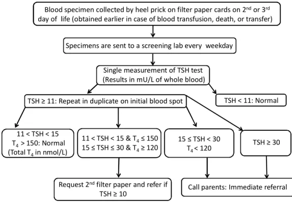
2.4.1. Newborn screening (NBS) ... 26
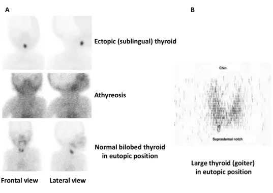
2.4.2. Other diagnostic studies ... 29
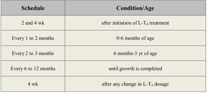
2.5. Treatment ... 32
3. Permanent primary CH due to thyroid dyshormonogenesis (TDHG) ... 33
4. Permanent primary CH due to thyroid dysgenesis (CHTD) ... 37
4.1. Molecular mechanisms of CHTD ... 38 4.1.1. Monogenic mechanisms ... 38 4.1.1.1. TSHR ... 43 4.1.1.2. NKX2.1 ... 47 4.1.1.3. PAX8 ... 49 4.1.1.4. FOXE1 ... 54 4.1.1.5. NKX2.5/CSX ... 58 4.1.1.6. HHEX/PRH ... 60
4.1.1.7. Inductive signals and other genes ... 62
4.1.1.7.1. Inductive signals ... 62
4.1.1.7.2. Other genes ... 67
4.1.2. Alternative mechanisms ... 73
4.1.2.1. Multigenic model of TD ... 74
4.1.2.2. Copy number variations (CNVs) ... 75
4.1.2.3. Early somatic mutations ... 78
4.1.2.4. Epigenetic modifications ... 79
4.1.2.4.1. DNA methylation ... 81
4.1.2.4.2. Histone modifications ... 85
4.1.2.4.3. Interplay between DNA methylation and histone modifications ... 88
5. Hypothesis and project objectives ... 91
CHAPTER 2: RESULTS ... 93
1. Transcriptome, Methylome and Genomic Variations Analysis of Ectopic Thyroid Glands. ... 93
x
2. Evidence for Calcitonin-Producing Cells in Human Lingual Thyroids. ... 120
3. Role for tissue-dependent methylation differences in the expression of FOXE1 in non-tumoral thyroid glands ... 135
CHAPTER 3: GENERAL DISCUSSION... 181
1. Genome-wide approach ... 182
1.1. Wnt signalling and thyroid ectopy ... 182
1.2. Expression of calcitonin in ectopic lingual thyroids ... 188
2. Candidate gene approach ... 189
CHAPTER 4: CONCLUSIONS AND PERSPECTIVES ... 195
xi
List of tables
Table I: Timing of morphogenetic events during thyroid embryonic development in different
species. ...8
Table II: Physiologic effects of thyroid hormones. ... 18
Table III: Monitoring schedule for serum total or free T4 and TSH. ... 33
xii
List of figures
Figure 1: Roles and functional interactions of intrinsic genetic factors involved in thyroid
morphogenesis. ...4
Figure 2: Developmental stages of the thyroid and the list of transcription factors and thyroid-specific genes involved in cell fate determination and differentiation. ...4
Figure 3: Anatomy of the thyroid gland and surrounding structures. ...9
Figure 4: Histology of thyroid gland; (1) Thyroid follicle, (2) Follicular cells, (3) Parafollicular (C cells)... 10
Figure 5: Thyroid hormone synthesis in thyrocytes. ... 11
Figure 6: Deiodinase-mediated activation or inactivation of T4 and T3... 15
Figure 7: Screening algorithm for CH in Quebec. ... 28
Figure 8: Thyroid scintigraphy. ... 30
Figure 9: Canonical function of DNA methylation and tissue-specific gene regulation. ... 83
Figure 10: DNA methylation-mediated transcriptional regulation at genomic regions other than promoter elements ... 85
Figure 11: Examples of the recruitment of proteins to modified histones. ... 86
Figure 12: Cross-talk between histone modifications. ... 88
Figure 13: The Wnt signalling pathways. ... 185
Figure 14: A single base pair deletion in the DMR of FOXE1 promoter. ... 197
xiii
Abbreviation List
5caC 5-carboxylcytosine 5fC 5-formylcytosine 5hmC 5-hydoxymethylcytosine 5mC 5-methylcytosineAAP American Academy of Pediatrics
AC Adenylate cyclase
aCGH Array comparative genomic hybridization AIS Androgen insensitivity syndrome
APC Adenomatous polyposis coli
AR Androgen receptor
AVE Anterior visceral endoderm
BAT Brown adipose tissue
BHC Benign hereditary chorea
bHLH Basic helix-loop-helix
BLTS Triad of brain, lung, thyroid syndrome
BM Bone maturation
BMPs Bone morphogenic proteins
BMR Basal metabolic rate
C cells Calcitonin-producing cells
Ca2+/NADPH Calcium- and reduced nicotineamide adenine dinucleotide phosphate cAMP Cyclic adenosine monophosphate
CCH Central congenital hypothyroidism CDU Color Doppler ultrasonography
CGIs CpG islands
CH Congenital hypothyroidism
CHD Congenital heart disease
CHI Congenital hyperinsulinism
CHTD Congenital hypothyroidism due to thyroid dysgenesis
cKO Conditional Knockout
CNPs Copy number polymorphisms CNS Central nervous system CNVs Copy number variations
CTCF CCCTC-binding factor d Developmental day D1 Type 1 deiodinase D2 Type 2 deiodinase D3 Type 3 deiodinase DAG 1,2-Diacylglycerol
xiv DEHAL1 Iodotyrosine dehalogenase 1
DIT Di-iodotyrosine
DMRs Differentially methylated regions
DNA Deoxyribonucleic Acid
DNMTs DNA methyltransferases
DQ Developmental quotient
Ds Selenoprotein iodothyronine deiodinases DUOX2 Dual oxidase type 2
DVL Disheveled
E Embryonic day
ECD Extracellular domain
ECM Extracellular matrix
ELC Ether link cleavage
EMSA Electrophoretic mobility shift assay
ER Endoplasmic reticulum
ESCs Embryonic stem cells EZH2 Enhancer of zeste 2
FGFR Fibroblast growth factor receptor
FNAH Familial non-autoimmune hyperthyroidism
fT3 Free T3
fT4 Free T4
Fzd Frizzled
GDFs Growth/differentiation factors
GIT Gastrointestinal tract
GnRH Gonadotrophin-releasing hormone
GO Gene ontology
GPCRs G protein-coupled receptors GpHR Glycoprotein hormone receptor GSK3β Glycogen synthase kinase 3β
GW Gestational weeks
H2O2 Hydrogen Peroxide
HAT Heterodimeric amino acid transporter HAT activity Histone acetyl transferase activity HCP High CpG-density promoters HDAC Histone deacetylase
HMT Histone methyltransferase hpf Hours post-fertilization
HPT axis Hypothalamic-Pituitary-Thyroid axis ICP Intermediate CpG-density promoters IGF-1 Insulin-like growth factor-1
xv
IHD Iodohexadecanal
IP3 Inositol 1,4,5 trisphosphate
IQ Intelligence quotient
IRD Inner ring deiodination
IRDS Infant respiratory distress syndrome ITD Iodide transport defects
JNK Jun N-terminal Kinase
KO Knockout
LATs L-type amino acid transporters
LBW Low birth weight
LCP Low CpG-density promoters
LCRs Low-copy repeats
LIM Lin11, Isl-1, and Mec-3
LOF Loss-of-function
LOH Loss of heterozygosity L-T4 Levothyroxine
MAPK Mitogen activated protein kinase
MBDs Methyl-CpG-binding domain (MBD)-containing proteins MCT Monocarboxylate transporter
MeCPs Methyl-CpG-binding proteins
MeDIP Methylated DNA immunoprecipitation
miRNA MicroRNA
MIT Mono-iodotyrosine
MMEJ Microhomology-mediated end-joining
MNG Multinodular goitrous
MO Morpholino antisense oligonucleotide Na+/K+ ATPase Na+/K+-adenosine triphosphatase NAHR Non-allelic homologous recombination NASH Nonautoimmune subclinical hypothyroidism
NBS Newborn screening
ncRNAs Non-coding RNAs
NGS Next-generation sequencing NHEJ Nonhomologous end joining
NIS Na+/I‾ symporter
NTCP Na+/ taurocholate cotransporting polypeptide NTH Neonatal transient hypothyroidism
OATP Organic anion-transporting polypeptide ORD Outer ring deiodination
PcG Polycomb group
xvi
PD Paired-box DNA binding domain
PI3K Phosphatidyl Inositol 3 Kinase PIOD Partial iodide organification defect
PKA Protein Kinase A
PLC Phospholipase C
PRC2 Polycomb repressive complex 2 pTFCs Precursors of thyroid follicular cells PTMs Post-translational modifications
PU Phenylthiourea
RA Retinoic acid
rhTSH Recombinant human thyroid-stimulating hormone RTHs Resistance to thyroid hormones
RXRs Retinoid X receptors
SDs Segmental duplications
SET Su(var), Enhancer of zest, and Trithorax SFRPs Secreted frizzled-related proteins SNP Single nucleotide polymorphisms
T3 Tri-iodothyronine
T3S Sulfated T3
T4 Thyroxine
TAMs Thyronamines
TBG Thyroxine-binding globulin
TBII Thyrotropin-binding inhibitor immunoglobulin
TD Thyroid dysgenesis
TDA Thyroid developmental anomalies
TDHG Thyroid dyshormonogenesis
T-DMR Tissue-specific differentially methylated region
TEs Transposable elements
TET Ten-eleven translocation
TFCs Thyroid follicular cells
TFs Transcription factors
Tg Thyroglobulin
TGFβ Transforming growth factor beta
THCMTD Thyroid hormone cell membrane transport defects THMD Thyroid hormone metabolism defects
THOX2 Thyroid oxidase 2
THs Thyroid hormones
TIOD Total iodide organification defects
TMD Transmembrane domain
xvii
TRAPs Thyroid receptor auxiliary proteins TRB-Ab Thyrotropin receptor blocking antibody
TRE TH response elements
TRH Thyrotropin-releasing hormone
TRs Thyroid receptors
TRα Thyroid receptor alpha
TRβ Thyroid receptor beta
TSH Thyroid-stimulating hormone
TSHR Thyroid-stimulating hormone receptor UBB Ultimobranchial bodies
UPD Uniparental isodisomy
US Ultrasound
WES Whole-exome sequencing
Wnt wingless-int-1
xviii
“Your assumptions are your windows on the world.Scrub them off every once in a while, or the light won't come in” Isaac Asimov To my family
xix
Acknowledgements
First of all, I would like to express my sincere gratitude and deep appreciation towards my supervisor, Dr. Deladoëy, who took a chance and gave me one of the best opportunities to join his lab. Thank you for the opportunity, for the continuous encouragement and guidance, and most of all for believing in me.
Next, I would like to express my deep thanks to my co-supervisor Dr. Deal. Many thanks for being always there, for your continuous academic and personal help and support, and for pushing me to my excel. Dr. Deal, I will miss your warm feelings and your pretty smile.
I sincerely thank all the Jury members for kindly and promptly accepting to evaluate my doctoral thesis. It is a great honor for me to receive your comments and suggestions that will eventually improve this work.
I would like to acknowledge the incredible support I received from Dr. Guy Van-Vliet during my doctoral studies, especially during writing this thesis. I cannot thank you enough for your insightful suggestions that helped me to improve my thesis.
Also, I wish to extend my sincere thanks to all past and present members of the lab, Jean paquette, Isabelle Vandernoot, Fabien Magne, and Stéphanie Larrivée-Vanier. Thank you for your invaluable support, kindness, and over all for our active discussions.
A special thanks to Dr. Mounib Elchebly and Dr. André Tremblay who their assistance and support are greatly appreciated.
It is a pleasure to thank Mme Sylvie Beauchemin from the department of Biochemistry and Molecular Medicine, for her invaluable help in secretarial work. She is such a nice and helpful person. My deep thanks is also extended to Mme Sandy Lalonde from the research center at Sainte-Justine Hospital, for her sympathy and cooperation.
Certainly, this thesis would not have been realizable and accomplished without the joint financial support of my Egyptian government (Egyptian Ministry of Higher Education) and University of Montreal (Faculty of Graduate Studies). Furthermore, the financial support I have received as grants from the Foundation of stars, the Sainte-Justine University Hospital Foundation, and the Biochemistry department, is gratefully acknowledged.
xx
To my dearest friend, Suzanne, thank you for being always there, for helping me through the challenges, for lending an ear, rejoicing through the good times, and for the life lessons.
Finally, I cannotignore the unwavering support of my family. Mum, thank you for your
endless support, your advice, your sacrifices, and your continuous prayers. Really, your advice
and education that you gave me is a huge bunch of keys that will allow me to open the closed
doors of this world. Many thanks for being always there. To my dear brother, many thanks for
your endless support through my whole life.
To my father, although I don’t get to have you in my life as much as I would want to, the
confidence and pride that you have placed in me, stimulate me always to move further. I miss
you.
I am mightily grateful to my beloved husband, Dr. Reda Gaafar, for being my pillar of strength. Reda, thanks for your patience, your sacrifices, for encouraging and supporting me throughout my graduate studies, and beyond all for your endless love. I hope I did not let you down. To my greatest accomplishment of all: Youmna and Haneen. My two little angels, I really cannot tell you how much I appreciate your understanding of how important was this stage in my life. Many thanks for being always beside me, encouraging me, and above all for your patience. I hope that you are proud of me.
CHAPTER 1: INTRODUCTION
1. The thyroid gland
In humans, the thyroid gland is considered to be one of the largest endocrine glands in the body, weighing 15-20 grams in normal adults. It synthesizes, stores, and secretes thyroid hormones (THs), thyroxine (T4) and tri-iodothyronine (T3). The production and secretion of THs
is under the regulation of the hypothalamic-pituitary-thyroid (HPT) axis. Mainly through the nuclear thyroid hormone receptors (TRs), TRα and TRβ, THs exert various physiological responses: they are responsible for the maintenance of the basal metabolic rate as well as controlling pre- and post-natal growth and differentiation of numerous tissues, notably the brain, through their effects on protein, lipid and carbohydrate metabolism. In addition to THs, the thyroid gland secretes the serum calcium-lowering hormone, calcitonin (Kirsten, 2000).
1.1. Embryology and development
1.1.1. Normal thyroid development
In vertebrates, similar to other endodermal-derived organs, the morphogenesis of the thyroid gland begins in the floor of the primitive pharynx with a specification event during which a monolayer of endodermal cells, precursors of thyroid follicular cells or pTFCs, are committed to attaining the thyroid fate. Subsequently, thickening of the monolayer of committed endodermal cells representing the thyroid anlage results in the formation of a multilayered structure, the thyroid placode, by embryonic day (E) 9-9.5 in mice and E22 in humans (De Felice and Di Lauro, 2011). The formation of the thyroid placode is followed by its expansion into the underlining mesenchyme resulting in the formation of the thyroid bud (median thyroid primordium). By E10.5 in mice, the thyroid bud is recognized as a narrow necked flask-like structure that promptly becomes a diverticulum (De Felice and Di Lauro, 2004). The thyroid bud begins to migrate caudally along the anterior neck region while it is connected by a narrow channel (thyroglossal duct) to a small hole (foramen cecum) at its site of origin in the floor of the pharynx. Consequent to the atrophy of the thyroglossal duct (E11.5 in mice and E30-40 in humans), the median primordium detaches from the pharyngeal floor and starts its lateral expansion by E12 in mice (De Felice and Di Lauro, 2004). At E13.5 in mice and E45-50 in
2
humans, the median thyroid primordium reaches its final destination (Van Vliet, 2003; De Felice and Di Lauro, 2004). The mechanisms underlying the translocation of the thyroid primordium have not yet been clarified and different ones have been proposed among which are active transport (De Felice et al., 1998), involvment of cardiac mesoderm and major blood vessels (Alt et al., 2006b; Fagman et al., 2006; Opitz et al., 2012), or relocalization secondary to the differential growth of the whole embryo (Gasser, 2006).
Upon reaching its final location in front of the trachea, fusion between both the median thyroid primordium and the ultimobranchial bodies (UBB) or the lateral primordia occurs by E14 in mice and E60 in humans (De Felice and Di Lauro, 2004). The UBB are a pair of transient embryonic structures derived from the fourth and fifth (caudal) pharyngeal pouches in mouse and human, respectively to which the precursors of the parafollicular or C cells, cells devoted to the production of calcitonin, have migrated from the neural crest and become localized (Pearse and Carvalheira, 1967; Fontaine, 1979; Fagman and Nilsson, 2010). In contrast to the established neuronal origin of the progenitors of the ultimobranchial C cells in avian species (Kameda, 1995), that of mammals has been recently challenged (Kameda et al., 2007b). In this regard, Kameda et al. have shown that the murine ultimobranchial cells, that ultimately differentiate into thyroid C cells, are endodermally derived. Moreover, they have also indicated that the UBB were not occupied by neural crest-derived cells at any developmental stage as well as that the thyroid C cells exhibited no expression of the neural crest markers (Kameda et al., 2007b). Recently, calcitonin-producing C cells have been reported in human ectopic lingual thyroids, thus suggesting that UBB are not the sole source of C cells in humans and that the interactions between TFCs and calcitonin-producing cells occur early during embryonic development than previously known (Vandernoot et al., 2012).
At E15 the bilobed shape of the gland is attained and by E15.5 (around E70 in humans), the final steps of thyroid morphogenesis are detected by the appearance of follicular organization. Subsequently, activation of the terminal or functional differentiation takes place whereby the thyroid follicular cells (TFCs) ultimately exhibit the expression of the set of proteins known to be involved in the biosynthesis of THs, including thyroglobulin (Tg), thyroid peroxidase (TPO), Na+/I‾ symporter (NIS), and the thyroid-stimulating hormone (TSH) receptor (TSHR) (De Felice and Di Lauro, 2004; 2011).
3
Currently, knowledge concerning the signals inducing the process of commitment or specification of a group of multipotent cells to the thyroid fate is still limited. However, studies carried out in animal models have pointed to the plausible role of extrinsic (acting outside the pTFCs) genetic factors such as Nodal-related signals as well as a number of mesenchymal inductive signals in the specification of the thyroid (Fagman and Nilsson, 2010; De Felice and Di Lauro, 2011; Fagman and Nilsson, 2011).
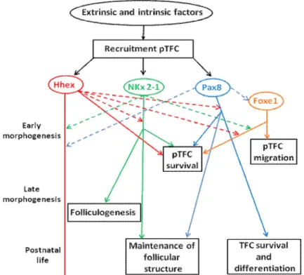
On the other hand, intrinsic genetic factors (acting inside the pTFCs) involved in subsequent morphogenetic steps and their functional interactions have been identified (Figure 1) (De Felice and Di Lauro, 2011). The pTFCs are differentiated from other cells in the primitive pharynx by the coexpression of four transcription factors: thyroid transcription factor 1 (TTF-1 or NKX2.1), paired homeobox-8 (PAX8), thyroid transcription factor 2 (TTF-2 or FOXE1), and hematopoietically expressed homeobox (HHEX) (De Felice and Di Lauro, 2004; 2011). Based on their early expression in the thyroid anlage, it has been suggested that these transcription factors collaborate to drive the budding, migration, survival, and growth of the pTFCs (De Felice and Di Lauro, 2004; Santisteban and Bernal, 2005; De Felice and Di Lauro, 2011). In addition, they are also involved in controlling the terminal differentiation of the TFCs via the regulation of thyroid-specific gene expression as well as the maintenance of the differentiated phenotype of the mature TFCs (Figure 2) (Damante et al., 2001; De Felice and Di Lauro, 2004; Santisteban and Bernal, 2005; De Felice and Di Lauro, 2011). Animal models deficient in thyroid transcription factors pointed to their crucial role in thyroid development: In the absence of either Nkx2-1, Pax8, Foxe1, or Hhex, a proper specification of the thyroid anlage occurs but subsequent morphogenesis of the thyroid is severely impaired (Wendl et al., 2002; Elsalini et al., 2003; Parlato et al., 2004).
4
Figure 1: Roles and functional interactions of intrinsic genetic factors involved in thyroid
morphogenesis (adapted From De Felice and Di Lauro, 2011). Dashed arrows: involvement in the initiation or maintenance of other factors. Solid arrows: controlling crucial steps of thyroid morphogenesis and physiology.
Figure 2: Developmental stages of the thyroid and the list of transcription factors and
thyroid-specific genes involved in cell fate determination and differentiation (modified from Santisteban and Bernal, 2005).
5
In addition to the above mentioned thyroid-enriched transcription factors, several recent studies in animal models have shed light on novel regulators of thyroid development and differentiation. Among the new genetic factors are those involved in early stages of thyroid morphogenesis including those encoding the basic helix-loop-helix (bHLH) transcription factor Mash1 (Kameda et al., 2007a), the LIM homeodomain transcription factor Isl1 (Westerlund et al., 2008), and the bHLH transcriptional repressor Hes1, a known target of the Notch signalling pathway (Carré et al., 2011). In addition, genes implicated in late morphogenetic stages were also identified among which are the Hox genes, Hoxa-3 and its paralogs Hoxb-3 and Hoxd-3 (Manley and Capecchi, 1995; 1998), TSHR, the gene encoding for the TSH receptor (Postiglione et al., 2002), and the Eyes absent (Eya) gene Eya1 (Xu et al., 2002). Recently, it has been shown that mouse models deficient in the microRNA-processing enzyme Dicer exhibit impaired postnatal thyroid function, thus indicating the role of an intact microRNA processing machinery in maintaining thyroid homeostasis (Frezzetti et al., 2011; Rodriguez et al., 2012). It has been shown that genes expressed in the foregut endoderm known to play crucial roles in pharyngeal development are involved in the developmental process of the thyroid as well, among which are genes involved in fibroblast growth factor receptor (FGFR) signalling (Celli et al., 1998; Ohuchi et al., 2000; Revest et al., 2001; De Felice and Di Lauro, 2004; Lania et al., 2009), the Nkx.2 class homeobox genes Nkx2.5 and Nkx2.6 (Tanaka et al., 2001), the gene encoding Hoxa5 (Meunier et al., 2003), the candidate gene of the 22q11 deletion syndrome, Tbx1 (Fagman et al., 2007; Lania et al., 2009), as well as the gene encoding the novel regulator of thyroid development, the tyrosine kinase receptor EphA4 (Andersson et al., 2011). The role of the above mentioned genes in the development of the thyroid gland will be discussed in detail later in this chapter.
1.1.2. Abnormal thyroid development
Abnormalities in thyroid gland development, designated thyroid dysgenesis (TD), include a thyroid gland that is either ectopically located (thyroid ectopy), completely absent (athyreosis), or severely reduced in size (thyroid hypoplasia) (Van Vliet, 2003; De Felice and Di Lauro, 2004). The ectopic thyroid tissue occurs consequent to the aberrant migration of all or part of the thyroid precursor cells. Although it is commonly located at the base of the tongue (lingual position), the ectopic gland can be detected in other locations along the normal migratory route
6
followed by the thyroid precursors from the foramen cecum to the neck (Van Vliet, 2003; De Felice and Di Lauro, 2004). Rarely, ectopic thyroid tissue has been detected in several other sites either within the head and neck region (e.g. submandibular, trachea, iris, and pituitary) or distantly (e.g. heart, lung, duodenum, gall bladder, adrenal gland, ovary and uterus). The presence of ectopic thyroid tissue in or near the heart could be attributed to developmental disturbances of organs sharing a common embryological origin. In subdiaphragmatic positions (e.g. duodenum, gall bladder, and adrenal gland), heterotropic differentiation of unspecified endodermal cells might be the underlying etiology. Moreover, it has been reported that thyroid tissue located in the ovary can arise within a teratoma, an encapsulated tumor containing more than one differentiated tissue (Ibrahim and Fadeyibi, 2011; Noussios et al., 2011).
Thyroid ectopy is the most common cause of permanent primary congenital hypothyroidism due to thyroid dysgenesis (CHTD), where it represents ~ 50 % of the cases (Van Vliet and Deladoëy, 2012). In the majority of cases, the ectopic thyroid is the only detectable thyroid tissue (Batsakis et al., 1996). Ectopic thyroids exhibit both normal histological organization and normal capacity to trap and organify iodine and hence to produce THs and Tg (Leger et al., 1988; Gallo et al., 2001; Toso et al., 2009). Consequently, the congenital hypothyroidism observed in most of the patients with thyroid ectopy is likely attributed to the smaller amount of tissue (due to absence of lateral lobes) and from a limited TSH-dependent compensatory growth (Stoppa-Vaucher et al., 2010). Of note, some euthyroid individuals (with normal thyroid function) have been found to have thyroid ectopy (Castanet et al., 2010a; Stoppa-Vaucher et al., 2011). In spite of being generally limited, the thyroid hormone producing capability of ectopic thyroids remains stable over time, thus reflecting a normal postnatal survival of the ectopic cells (Grant et al., 1989; Léger and Czernichow, 1990).
In addition to thyroid ectopy, absence of TFCs or athyreosis is the second common variant of TD (Van Vliet, 2003; De Felice and Di Lauro, 2004; Castanet et al., 2010a). Although the specification of the thyroid bud occurs, subsequent abnormalities leading to defects in either survival and/or proliferation of the precursors of TFCs (pTFCs) is the underlying cause resulting in absence of TFCs. Lack of differentiation of TFCs or shifting to another fate has been suggested as alternative mechanisms leading to the disappearance of TFCs (Van Vliet, 2003; De Felice and Di Lauro, 2004). A rare variant of TD is hypoplasia of an orthotopic (normally located) bilobed thyroid gland. Finally, absence of one of the two lobes of the thyroid, mainly the
7
left one, is a rare variant of TD referred to as hemiagenesis (Van Vliet, 2003; De Felice and Di Lauro, 2004).
Thyroid dysgenesis (TD) is the most common cause of congenital hypothyroidism (CH). The characteristics of congenital hypothyroidism from thyroid dysgenesis (CHTD) and its underlying pathogenesis will be discussed in later sections.
1.2. Anatomy and Histology
1.2.1. Anatomy
The thyroid is a butterfly-like shaped gland located in the anterior portion of the neck, just below the larynx on opposite sides of, and anterior to, the trachea. In mammals and in some reptiles, it consists of two lobes joined by the isthmus in the midline (Figure 3) (Stathatos, 2006). Occasionally, a pyramidal lobe exists ascending from the isthmus towards the hyoid bone. The thyroid is surrounded by a fibrous capsule, the extension of which into the body of the gland leads to the formation of septae, thus producing an irregular and incomplete lobulation (Stathatos, 2006).
Major differences in the anatomy of the thyroid are observed among classes of vertebrates: In birds and amphibians, the thyroid consists of two unconnected lobes. Moreover, in cartilaginous fish, the thyroid follicles are arranged in the form of a compact gland surrounded by a capsule (Gorbman, 1986). On the other hand, absence of glandular organization is observed in the majority of bony fish (teleosts) where the thyroid follicles are non-encapsulated and loosely distributed (Gorbman, 1986), as is observed in zebrafish where the thyroid follicles are loosely dispersed along the ventral aorta in the lower jaw region and hence no bilateral lobes are formed (Wendl et al., 2002). In contrast to what is observed in mouse and humans concerning the composite structure of the thyroid gland resulting from the fusion of the median and lateral primordia (Fagman et al., 2006), the two embryonic structures remain separated in chicken (Kameda, 1995) and zebrafish (Alt et al., 2006a).
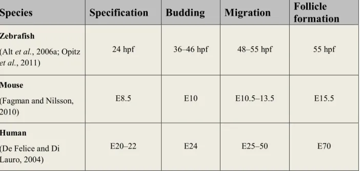
In addition to differences in thyroid anatomy, species-specific timing of crucial embryonic developmental steps do exist as well (Table I) (Deladoëy, 2012).
8
Table I: Timing of morphogenetic events during thyroid embryonic development in different
species (modified from Deladoëy, 2012).
Species
Specification
Budding
Migration
Follicle
formation
Zebrafish
(Alt et al., 2006a; Opitz
et al., 2011)
24 hpf 36–46 hpf 48–55 hpf 55 hpf
Mouse
(Fagman and Nilsson, 2010)
E8.5 E10 E10.5–13.5 E15.5
Human
(De Felice and Di Lauro, 2004)
E20–22 E24 E25–50 E70
hpf: hours post-fertilization; E: embryonic day
The thyroid gland is highly vascularized (Figure 3) (Stathatos, 2006). Its arterial supply is provided by two superior and two inferior arteries that originate from the external carotid arteries and thyrocervical trunks, respectively. The thyroid ima artery, a branch of either the brachiocephalic or the aorta, contributes infrequently to the blood supply of the thyroid. Three pairs of veins, superior, middle and inferior, are responsible for the venous drainage of the thyroid gland. Both the superior and middle veins drain into the internal jugular vein while the inferior ones drain into the brachiocephalic and subclavian veins (Stathatos, 2006).
9
Figure 3: Anatomy of the thyroid gland and surrounding structures (From Stathatos, 2006).
1.2.2.
Histology
The thyroid follicle is the main histological and functional unit of the thyroid gland (Figure 4) (Stathatos, 2012). Each follicle consists of a single layer of secretory epithelial cells known as the thyroid follicular cells (TFCs) or thyrocytes surrounded by a basement membrane. The lumen of each thyroid follicle is filled with a homogenous colloid, mostly formed of thyroglobulin (Tg), a macromolecular glycoprotein that functions as a scaffold for the synthesis of THs. A significant variation in the size of follicles within the same thyroid gland is attributed to the variability of the colloid content within the different follicles. In addition to TFCs, the parafollicular or C cells (calcitonin-producing cells which participate in calcium regulation) are found in between the follicles (Stathatos, 2006; 2012).
10
Figure 4: Histology of thyroid gland; (1) Thyroid follicle, (2) Follicular cells, (3) Parafollicular
(C cells) (From Stathatos, 2012).
1.3. Physiology
1.3.1. Synthesis and secretion of THs
Synthesis of thyroid hormones (THs) takes place in the thyroid follicles. It requires a normally developed thyroid gland, sufficient dietary iodide intake, as well as a set of successive biochemical steps (Figure 5) (Bizhanova and Kopp, 2009). To be able to synthesize THs, thyrocytes have to trap iodide from the blood, at their basolateral membrane, in an active process against a concentration gradient. Such an energy consuming process is mediated by the Na+/I‾ symporter (NIS), which transports two Na+ and oneI‾ down the Na+ ion gradient generated from the activity of Na+/K+-adenosine triphosphatase (Na+/K+ ATPase). The NIS-mediated iodide transport increases its concentration in thyrocytes 20- to 40-fold compared to that in serum (Stathatos, 2012). The release of the trapped iodide in the lumen across the apical membrane is mediated by an iodide channel, the candidate of which is the anion transporter pendrin (Song et al., 2007). However, the elaborated function of pendrin and its role as an apical iodide
11
transporter is still a matter of debate and the reported data are controversial (Bizhanova and Kopp, 2011; Twyffels et al., 2011).
Figure 5: Thyroid hormone synthesis in thyrocytes (From Bizhanova and Kopp, 2009).
At the cell-lumen boundary and in the presence of hydrogen peroxide (H2O2), thyroid
peroxidase (TPO) catalyzes the organification of iodide via its coupling to selective tyrosyl residues in thyroglobulin (Tg) leading to the formation of mono- and diiodotyrosines (MIT and DIT). Coupled to TPO at the apical membrane is the dual oxidase type 2 (DUOX2), originally known as thyroid oxidase 2 (THOX2), a calcium- and reduced nicotineamide adenine dinucleotide phosphate (Ca2+/NADPH)-dependent oxidase, required for the production of H2O2.
12
DUOXA2 is a specific maturation factor required by DUOX2 for the proper translocation of the DUOX2/DUOX2A complex from the endoplasmic reticulum (ER) to the apical plasma membrane of thyrocytes where H2O2 is produced (Ohye and Sugawara, 2010). In humans and
many other species, generation of H2O2 as well as binding of oxidized iodide to tyrosine residues
of Tg and formation of THs are activated by the TSH-dependent phospholipase C-Ca2+ -diacylglycerol (DAG) pathway and inhibited by cyclic adenosine monophosphate (cAMP) signalling cascades (Song et al., 2007). In addition, the generation of H2O2 is inhibited at high
concentrations of iodide (the Wolff-Chaikoff effect) (Wolff and Chaikoff, 1948) via the iodinated lipid, 2-iodohexadecanal (IHDA) (Corvilain et al., 1988; Panneels et al., 1994). In many vertebrate systems, H2O2 activates signal transduction pathways downstream of insulin and
growth factors (Rhee et al., 2005) and it enhances proliferation in various mammalian cells at physiological levels (Stone, 2004). However, a signalling role of H2O2 in thyrocytes has not been
yet elucidated, but it can be deduced from studies carried on other cells (Song et al., 2007). In comparison with the amounts of iodide incorporated into proteins, H2O2 is largely produced in
excess within the thyroid cells (Song et al., 2007). Similar to what is observed in other cell types, H2O2 exerts toxic effects on thyroid cells in vitro that are potentially mutagenic if not properly
repaired: at concentrations lower and higher than 0.1 mm, H2O2 induces DNA single- and
double-strand breaks, respectively (Mondello et al., 2002; Chico Galdo et al., 2006). Moreover, apoptosis and necrosis are among the effects of high H2O2 levels on thyrocytes (Riou et al.,
1999; Demelash et al., 2004). In vivo, an H2O2-induced mutagenesis in human thyroid cells is
substantially supported (Maier et al., 2006; Song et al., 2007). Consequently, the thyroid cells exhibit a number of defense mechanisms against the generated H2O2 among which is the
stringent separation of both the iodination system, located at the apical membrane of the cell, and the interior of the cell (Song et al., 2007).
In addition to the formation of MIT and DIT, TPO catalyzes the coupling of two residues of DIT or one DIT and MIT to form either T4 or T3, respectively. Iodinated Tg is stored in the
follicular lumen until needed (Bizhanova and Kopp, 2009). For THs to be released, iodinated Tg is reabsorbed into follicular cells by pinocytosis. Subsequent lysosomal digestion leads to the release of T4 and T3 via the basolateral membrane into the bloodstream through an unknown
13
1 (DEHAL1) and the released iodide is recycled for further synthesis of THs (Bizhanova and Kopp, 2009).
1.3.2.
Transport of THs
The majority (99 %) of the T4 and T3 released into the plasma remains inactive by being
bound to carrier proteins, mainly thyroxine-binding globulin (TBG; 70-80 %) and what is left to thyroid-binding prealbumin (transthyretin) and albumin. Hence, less than 1 % of the total plasma THs are in a free state. The effects of THs on peripheral tissues are exerted by these biologically active free T4 (fT4) and T3 (fT3) hormones. Several other serum proteins, in particular high
density lipoproteins, bind T4 and T3 (Stathatos, 2012).
In order to be peripherally metabolized and to exert their intracellular genomic actions, THs have to be firstly transported across the plasma membrane (Visser et al., 2011). Earlier, it had been presumed that THs are transferred across the phospholipid bilayer by simple diffusion owing to their lipophilic structure. However, several experimental lines of evidence have pointed out the role of specific transporters in the process of THs' uptake by target cells (Krenning et al., 1981; Hennemann et al., 1986; Krenning et al., 1988), among which are those studies showing that, in certain cells, the cellular uptake of THs is energy- and Na+-dependent (Hennemann et al., 2001). Such observations have been reinforced by the identification of the foremost THs transporters, the organic anion transporter subtypes oatp2 and oatp3 (Abe et al., 1998). Later, various transporters of iodothyronine derivatives have been identified, including the Na+/ taurocholate cotransporting polypeptide (NTCP) (Friesema et al., 1999; Visser et al., 2010), members of the organic anion-transporting polypeptide (OATP) family including the specific THs transporter OATP1C1 (Friesema et al., 1999; Hagenbuch and Meier, 2003; Hagenbuch, 2007; van der Deure et al., 2010), the L-type amino acid transporters (LATs) 1 (LAT1) and 2 (LAT2) which belong to the heterodimeric amino acid transporter (HAT) family (Friesema et al., 2001; Jansen et al., 2005), as well as various members of the monocarboxylate transporter (MCT) family among which is the specific THs transporter MCT8 and the aromatic amino acid transporter MCT10 (Friesema et al., 2003; Visser et al., 2007; van der Deure et al., 2010). While a number of plasma membrane transporters involved in the intracellular uptake of THs have been identified, those incorporated in the translocation of THs across the membranes surrounding
14
either the mitochondria or the nucleus (if required), where the biologically active TH (T3)
mediates its effects on transcription, are yet unidentified (Visser et al., 2011).
1.3.3.
Peripheral metabolism of THs
The major TH secreted by the thyroid gland is the prohormone thyroxine (T4), with only
a minor amount of T3 (less than 10% of the total blood TH) being secreted under normal
conditions. The majority of T4 is converted to the biologically active 3,5,3′-triiodothyronine (T3).
Hence, the majority of circulating T3 is derived from the secreted T4 instead of from the thyroid
itself (Kirsten, 2000).
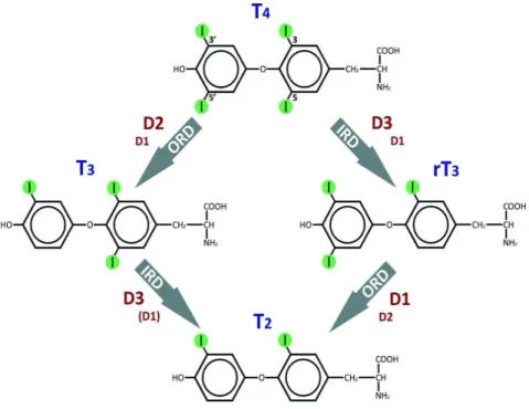
The conversion of the long-lived less active T4 to the short-lived more active T3 occurs
via the outer (phenolic) ring 5′-deiodination (ORD) of T4, a process catalyzed by two
selenoprotein iodothyronine deiodinases (Ds) types 1 (D1) and 2 (D2) (Darras and Van Herck, 2012) (Figure 6). The deiodinase D1 is expressed in the thyroid, liver, and kidney and is known to be responsible for the conversion of the majority of T4 to T3 in circulation (Gereben et al.,
2008a; Mebis and van den Berghe, 2009). D2 is expressed in brain, pituitary, brown adipose tissue (BAT), skeletal muscle, and thyroid and is responsible for the local conversion of T4 to T3
(Gereben et al., 2008a; Mebis and van den Berghe, 2009).
On the other hand, type 3 deiodinase (D3), expressed mainly in placenta, brain, skin, and several fetal tissues, is considered to be the main T3 and T4 inactivating deiodinase (Gereben et al., 2008a; Mebis and van den Berghe, 2009). Through inner (tyrosyl) ring 5-deiodination (IRD), it catalyzes the transition of both T3 and T4 to their inactive metabolites 3,3′-diiodothyronine (T2)
and reverse T3 (rT3), respectively (Figure 6) (Darras and Van Herck, 2012). Owing to its broader
substrate specificity, D1 is also capable of terminating the action of T3 and prohibiting the
activation of T4 byconverting them into their inactive metabolites via IRD (Darras and Van
15
Figure 6: Deiodinase-mediated activation or inactivation of T4 and T3 (From Darras and Van
Herck, 2012).
Integration of the activities of the iodothyronine deiodinases plays an important role in maintaining concentrations of serum T3: Bianco et al. have pointed out to the role of the mutual
changes in the activity of the main activating (D2) and inactivating (D3) deiodinases in maintaining thyroid gland homeostasis in response to variations in plasma T4 and T3
concentrations (Bianco et al., 2002). Altered thyroid hormone metabolism related to abnormalities in iodothyronine deiodinases has been observed in a number of clinical conditions including critical illness, referred to as non-thyroidal illness or low T3 syndrome. It is associated
with lower levels of circulating T3 and even T4 in severe cases, as well as increased levels of rT3
andthyrotropin (TSH) levels that are within the normal range (Mebis and van den Berghe, 2009). In addition to deiodination, other alternative metabolic pathways of THs include conjugation of the phenolic hydroxyl group of the iododthyronines with sulfate (sulfation) or glucuronic acid (glucuronidation) (Wu et al., 2005): Sulfation of T4 completely blocks its ORD,
while it robustly enhances the IRD of both T3 and T4. On the other hand, sulfated T3 (T3S) can
act as a reservoir for the biologically active T3 that is recovered via the action of tissue sulfatase
(Wu et al., 2005). The glucuronidated iodothyronines are excreted in bile and then eliminated through fecal excretion or recycled in the enterohepatic cycle (Wu et al., 2005). Oxidative
16
deamination of the alanine side-chain of T3 and T4 leads to the formation of the acetic acid
derivatives, 3,3′,5-triiodothyroacetic acid (triac) and tetraiodothyroacetic acid (tetrac), respectively (Wu et al., 2005). The acetic acid derivatives of THs are mainly metabolized via deiodination as well as by conjugation (sulfation and glucuronidation) that might be followed by monodeiodination (Wu et al., 2005). In addition, the endogenous biologically active thyronamines (TAMs), namely 3-iodothyronamine (3-T1AM) and thyronamine (T0AM), are
derived from the iodothyronine precursors via decarboxylation of their alanine side-chain. Recently, it has been shown that, in addition to decarboxylation, the biosynthesis of TAMs involves both phenolic and tyrosyl rings deiodination mediated by D1 or D2 (phenolic) as well as D1 or D3 (tyrosyl) deiodinases (Gereben et al., 2008b; Piehl et al., 2011). While the physiological effects exerted by TAMs were found to be opposite to those of THs, it has been suggested that TAMs can either refine or act as antagonists of THs (Gereben et al., 2008b; Piehl et al., 2011). Finally, ether link cleavage (ELC) is considered to be a minor metabolic pathway of THs, the importance of which is illustrated during infections as it exhibits bacterial killing activity (Wu et al., 2005).
1.3.4.
Control of THs synthesis and secretion
The plasma levels of THs are under the regulation of the hypothalamic-pituitary-thyroid (HPT) axis (Stathatos, 2012). At low serum levels of T4 and T3, the heterodimeric glycoprotein
thyrotropin or thyroid-stimulating hormone (TSH) is released from the anterior pituitary thyrotropes. TSH is considered to be the key regulatory factor controlling the synthesis and secretion of THs. The release of TSH itself is under the regulation of the thyrotropin-releasing hormone (TRH) secreted from the hypothalamus. The actions of TSH are mediated via the TSH-receptor (TSHR), a seven-transmembrane G protein-coupled TSH-receptor located at the basolateral membrane of the TFCs (Stathatos, 2012). Binding of TSH to its receptor leads to the activation of the enzyme adenylate cyclase (AC) via an activated Gs protein. The consequent increase in cAMP intracellular levels together with the activation of cAMP-dependent protein kinase A (PKA) mediate the TSH-dependent synthesis of THs as well as the development of TFCs (Yen, 2001; Rivas and Santisteban, 2003; Bursuk, 2012; Stathatos, 2012). Nearly every step along the process of THs' synthesis and secretion is stimulated by TSH. TSH stimulates the synthesis of Na+/I‾ symporter, thyroid peroxidase, and thyroglobulin, which are involved in iodide uptake,
17
organification, and formation of iodinated Tg, respectively. As well, TSH stimulates the generation of H2O2 via the activation of PLC-Ca2+ / DAG cascade, a key control stage in the
synthesis process. Moreover, TSH stimulates the internalization of Tg by TFCs, its degradation, and the subsequent release of THs into the blood circulation. Hence, TSH assures the sufficient uptake of iodine by the TFCs and its efficient release in the circulation as THs (Dunn and Dunn, 2001). On the other hand, high plasma levels of THs exert a negative feedback action on the HPT axis leading to the inhibition of TSH secretion and the concomitant decrease in synthesis and secretion of THs (Stathatos, 2012). The mechanism leading to the inhibition of TSH secretion involves the type 2 deiodinase (D2)-mediated conversion of T4 to T3 in the pituitary
(Yen, 2001). Moreover, TSH production is negatively regulated, either directly or indirectly, via THs. Direct negative regulation of TSH occurs by TH-mediated decrease in transcription of the glycoprotein hormone α- and TSH β-subunit genes (Yen, 2001). Similarly, THs negatively regulate the transcription of TRH which leads at last to the decrease in transcription of TSH mRNA (Hulbert, 2000; Yen, 2001).
The other major regulator of THs' synthesis and secretion, in addition to TSH, is iodine availability. An inverse relationship between the amounts of iodine available within the TFCs and the synthesis of THs is observed: inadequate amounts of iodine lead to increased TSH stimulation and uptake of iodine, faster iodine turnover, and increased production of T3 relative
to T4. However, excess amounts of iodine hinder TPO activity by inhibiting the production of
H2O2 (the Wolff-Chaikoff effect) (Wolff and Chaikoff, 1948), thus blocking Tg iodination and
finally inhibiting the synthesis of THs (Dunn and Dunn, 2001; Stathatos, 2012). In addition, tissue-specific as well as hormone-dependent regulation of expression of the THs' metabolic enzymes, the three iodothyronine deiodinases, represent an additional layer of regulation exerted on the synthesis and secretion of THs (Santisteban and Bernal, 2005).
1.3.5.
Actions of THs
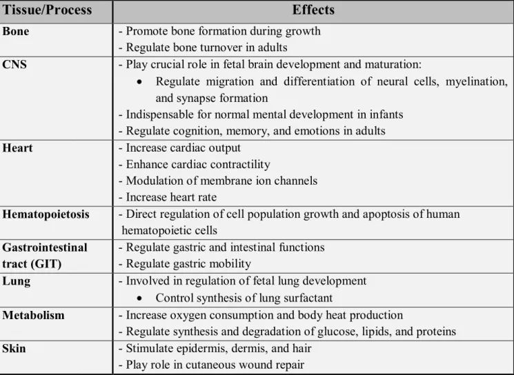
Thyroid hormones play critical roles in regulating a large number of body functions including growth, differentiation, and metabolism. The physiological effects of THs affects nearly all tissues (Table II) (Kirsten, 2000).
18
Table II: Physiologic effects of thyroid hormones (modified from Kristen, 2000).
Tissue/Process
Effects
Bone - Promote bone formation during growth
- Regulate bone turnover in adults
CNS - Play crucial role in fetal brain development and maturation:
Regulate migration and differentiation of neural cells, myelination, and synapse formation
- Indispensable for normal mental development in infants - Regulate cognition, memory, and emotions in adults
Heart - Increase cardiac output
- Enhance cardiac contractility
- Modulation of membrane ion channels - Increase heart rate
Hematopoietosis - Direct regulation of cell population growth and apoptosis of human
hematopoietic cells
Gastrointestinal tract (GIT)
- Regulate gastric and intestinal functions - Regulate gastric mobility
Lung - Involved in regulation of fetal lung development
Control synthesis of lung surfactant
Metabolism - Increase oxygen consumption and body heat production
- Regulate synthesis and degradation of glucose, lipids, and proteins
Skin - Stimulate epidermis, dermis, and hair
- Play role in cutaneous wound repair
1.3.5.1. Genomic actions of THs
In target tissues, the intracellular genomic effects of the THs are elicited by their specific binding, with high affinity, to the nuclear thyroid hormone receptors (TRs), the two major isoforms of which are TRα and TRβ. TRα and TRβ are members of a large superfamily of nuclear hormone receptors (steriod/thyroid hormone receptor superfamily) that mediate ligand-dependent transcriptional regulation subsequent to recognition and binding to specific DNA sequences, the TH response elements or TREs, located at the regulatory regions of THs' responsive genes (Yen, 2001; Huang et al., 2008). Through alternative splicing, the TRα gene encodes several proteins, among which TRα-1 is the only authentic TR while the others may act as inhibitors. On the other hand, the two isoforms of TRβ, TRβ-1 and TRβ-2, are generated as a
19
result of alternate promoter choice. Both TRβ-1 and TRβ-2 are authentic TRs that can mediate TH-mediated transcriptional regulation (Yen, 2001; Huang et al., 2008).
TRs are capable of binding to TREs in the form of monomers or homodimers, the role of which in the transcriptional regulation is not clearly understood (Yen, 2001; Viguerie and Langin, 2003; Huang et al., 2008). However, heterodimerization with TR auxiliary proteins (TRAPs), mainly retinoid X receptors (RXRs), leads to enhancement of TR binding to TREs (Yen, 2001). In addition, interactions with other nuclear proteins, such as coactivators or corepressors, are involved in either transcriptional activation or repression of basal transcription, respectively (Yen, 2001; Viguerie and Langin, 2003; Huang et al., 2008). Upon binding of the natural ligand of TR, T3, the TR/RXR heterodimer undergoes conformational changes that
ultimately lead to the substitution of a corepressor complex by a coactivator one. The transcriptionally active coactivator complex bears a histone acetyl transferase (HAT) activity that eventually leads to an accessible chromatin structure and hence a marked increase of gene transcription above its basal level (Viguerie and Langin, 2003). In the absence of ligand, the TR/RXR heterodimer interacts with a corepressor complex having either a histone deacetylase activity (HDAC) or able to interact directly with the basal transcriptional machinery thus leading to repression of transcription (Viguerie and Langin, 2003). In addition to the ligand-dependent modulation of TRs' transcriptional activity, it has been shown that phosphorylation of TRs regulates their transcriptional activity by altering DNA binding ability as well as tissue-specific stability (Yen, 2001; Chen et al., 2003; Huang et al., 2008).
1.3.5.2. Nongenomic (extranuclear) actions of THs
Although the majority of the actions exerted by the THs involve the nuclear TRs (genomic actions), a number of non-genomic actions, not directly influencing nuclear gene expression, have been described (Bassett et al., 2003; Cheng et al., 2010; Davis et al., 2011). Such nongenomic actions are initiated at the plasma membrane, in the cytoplasm, or in intracellular organelles, such as the mitochondria (Cheng et al., 2010; Davis et al., 2011). Moreover, the nongenomic actions of THs are mediated via either extranuclear TRs or cell surface receptors (certain integrins) and they are associated with the release of intracellular secondary messengers and the activation of a number of protein kinase signalling pathways (Bassett et al., 2003; Davis et al., 2011). In fact, certain alterations in gene transcription might
20
occur due to effects of THs initiated at the plasma membrane integrin receptor (Davis et al., 2011). Moreover, other nongenomic actions of T3, involving extranuclear TR isoforms, can start in the cytoplasm and ends with gene transcription (Moeller et al., 2006; Lei et al., 2008). Hence, it is the site of initiation that differentiates between genomic and nongenomic actions of THs rather than whether or not altered gene expression occurs (Davis et al., 2011). In contrast to the genomic actions of THs, the nongenomic ones are characterized by being rapid (occur within seconds to minutes), unaffected by inhibitors of both transcription and translation, and exhibit agonist/antagonist affinity different from those of classical TRs (Bassett et al., 2003).
Among the nongenomic actions of THs occurring at the plasma membrane is the regulation of the basal activity of a number of plasma membrane ion pumps. Such an action is mediated via a TH cell surface receptor found on integrin αvβ3 and leads to the activation of the mitogen activated protein kinase (MAPK) signal transduction cascade (Davis et al., 2005; Davis et al., 2010). Furthermore, other non-genomic actions induced mainly by T4 and to a lesser
extent by T3 include nuclear translocation of TRβ1 and those resulting in the activation of gene
transcription leading to the modulation of angiogenesis and tumor cell proliferation (Davis et al., 2009; Cheng et al., 2010; Davis et al., 2011). Moreover, trafficking of intracellular proteins (shuttling of TRα1 as well as several other proteins from cytoplasm to nucleus) and the transcription of genes involved in glucose metabolism are T3-induced nongenomic actions
mediated via the plasma membrane receptor on integrin αvβ3T3 and involves the activation of the signal transducing protein phosphatidyl Iinositol 3-Kinase (PI3K) (Cheng et al., 2010). At the TH receptor on integrin αvβ3, both the binding and actions of THs is blocked by the deaminated derivative of T4, tetraiodothyroacetic acid (tetrac), at both the T4/T3 site and the T3-specific site
(Cheng et al., 2010; Lin et al., 2011).
In the cytoplasm, interactions between T3 and the extranuclear TRs isoforms, TRβ1 and
TRα1, involve the activation of the signal transducing protein PI3K as is observed in the plasma membrane. Consequently, transcription of genes involved in glucose metabolism as well as plasma membrane insertion and activation of Na+,K+-ATPase. In addition, it was reported that a truncated form of TRα1 (TR∆α1) mediated the T4 and rT3-induced regulation of actin