Université de Montréal
Understanding the Transcriptional Control of EIF4E and its
Dysregulation in Acute Myeloid Leukemia: Role of NF-κB
par
Fadi Mounir Hariri
Département de Pathologie et Biologie Cellulaire
Faculté de Médecine
Thèse présentée à la Faculté de Médecine
en vue de l’obtention du grade de Philosophiæ Doctor (Ph.D.) en Biologie Moléculaire
option Biologie des Systèmes
Aout, 2014 © Hariri, 2014
Résumé
EIF4E, le facteur d’initiation de la traduction chez les eucaryotes est un oncogène puissant et
qui se trouve induit dans plusieurs types de cancers, parmi lesquels les sous-types M4 et M5 de la leucémie aiguë myéloblastique (LAM). EIF4E est régulé à plusieurs niveaux cependant, la régulation transcriptionnelle de ce gène est peu connue. Mes résultats montrent que EIF4E est une cible transcriptionnelle directe du facteur nucléaire « kappa-light- chain-enhancer of activated B cells » (NF-κB).Dans les cellules hématopoïétiques primaires et les lignées cellulaires, les niveaux de EIF4E sont induits par des inducteurs de NF-κB. En effet, l’inactivation pharmaceutique ou génétique de NF-κB réprime l’activation de EIF4E. En effet, suite à l’activation de NF-κB chez l’humain, le promoteur endogène de EIF4E recrute p65 (RelA) et c-Rel aux sites évolutionnaires conservés κB in vitro et in vivo en même temps que p300 ainsi que la forme phosphorylée de Pol II. De plus, p65 est sélectivement associé au promoteur de EIF4E dans les sous-types LAM M4/M5 mais non pas dans les autres sous-types LAM ou dans les cellules hématopoïétiques primaires normales. Ceci indique que ce processus représente un facteur essentiel qui détermine l’expression différentielle de EIF4E dans la LAM. Les analyses de données d’expressions par séquençage de l’ARN provenant du « Cancer Genome Atlas » (TCGA) suggèrent que les niveaux d’ARNm de EIF4E et RELA se trouvent augmentés dans les cas LAM à pronostic intermédiaire ou faible mais non pas dans les groupes cytogénétiquement favorables. De plus, des niveaux élevés d’ARNm de EIF4E et
RELA sont significativement associés avec un taux de survie relativement bas chez les
patients. En effet, les sites uniques κB se trouvant dans le promoteur de EIF4E recrutent le régulateur de transcription NF-κB p65 dans 47 nouvelles cibles prévues. Finalement, 6 nouveaux facteurs de transcription potentiellement impliqués dans la régulation du gène
EIF4E ont été prédits par des analyses de données ChIP-Seq provenant de l’encyclopédie des
éléments d’ADN (ENCODE). Collectivement, ces résultats fournissent de nouveaux aperçus sur le control transcriptionnel de EIF4E et offrent une nouvelle base moléculaire pour sa dérégulation dans au moins un sous-groupe de spécimens de LAM. L’étude et la compréhension de ce niveau de régulation dans le contexte de spécimens de patients s’avère important pour le développement de nouvelles stratégies thérapeutiques ciblant l’expression du gène EIF4E moyennant des inhibiteurs de NF-κB en combinaison avec la ribavirine.
Mots-clés : EIF4E, NF-κB, La Régulation Transcriptionnelle, La Leucémie Aiguë Myéloblastique.
Abstract
The eukaryotic translation initiation factor EIF4E is a powerful oncogene that is overexpressed in cancers, including the M4 and M5 subtypes of acute myeloid leukemia (AML). EIF4E is regulated at multiple levels; however not much is known about the transcriptional regulation of this gene. My findings show that the nuclear factor kappa-light- chain-enhancer of activated B cells (NF-κB) is a direct transcriptional regulator of EIF4E. EIF4E levels are induced in primary hematopoietic cells and in cell lines in response to NF-κB activating stimuli. Pharmacological and genetic inhibition of NF-κB suppresses EIF4E levels. NF-κB factors RelA (p65) and c-Rel are recruited to evolutionarily conserved κB sites in the
EIF4E promoter in vitro and in vivo following NF-κB activation concurrent with the
recruitment of p300 and phosphorylated Pol II. Furthermore, p65 is selectively associated with the EIF4E promoter in M4/M5 AML subtypes but not in other AML subtypes or normal primary hematopoietic cells and thus represents an underlying factor in determining the differential expression of EIF4E in AML. Analysis of gene expression RNA-Seq data from The Cancer Genome Atlas (TCGA) suggests that EIF4E and RELA mRNA levels are upregulated in intermediate and poor prognosis AML but not in the cytogenetically favorable group. Additionally, elevated EIF4E and RELA mRNA levels are significantly associated with worst patient survival outcome. Furthermore, 8 new putative NF-κB target genes that may be regulated with a pattern similar to EIF4E in poor prognosis AML were in silico predicted from Chip-Seq data. Finally, 6 new transcription factors that may be implicated in EIF4E gene regulation were predicted from the analysis of ChIP-Seq data from the encyclopedia of DNA elements (ENCODE). Collectively, these findings could offer novel insights into the transcriptional regulation of EIF4E and a novel molecular basis for its dysregulation in AML. Understanding this level of regulation within the context of patient specimens is important for the development of novel therapeutic strategies to target EIF4E gene expression with specific NF-κB inhibitors combined with ribavirin.
Table of Contents
Chapter 1: Introduction 1
1.1 Cap- Dependent Eukaryotic Translation Initiation 2
1.1.1 A 7-methyl guanosine cap structure is required for cap-dependent translation initiation 2 1.1.2 Molecular mechanism of cap-dependent translation initiation 4
1.2 Alternative Mechanisms for Translation Initiation 6
1.3 EIF4E Functions in mRNA Export and Translation 6
1.3.1 Structure of the EIF4E gene, alternative splicing and homology 6 1.3.2 The EIF4E structure reveals a distinct mode for cap-recognition required in mRNA export
and translation 7
1.4 EIF4E regulation is multifaceted with distinct levels of control 12
1.4.1 A c-Myc centric view for EIF4E transcriptional control 12
1.4.2 The stability of EIF4E mRNA is regulated by HuR and AUF1 14
1.4.3 EIF4E activity is modulated by protein interactions with a multitude of regulators 14 1.4.4 EIF4E activity is modulated through post-translational modifications 16
1.5 EIF4E is Overexpressed in 30% of Human Cancers and is a Plausible Candidate for Clinical
Targeting 17
1.6 Acute Myeloid Leukemia: A Hematopoietic Malignancy with Aberrant EIF4E Expression
and Activity 18
1.6.1 Cytogenetic and Molecular classification of AML to predict patient prognosis 19
1.6.2 EIF4E is overexpressed in poor prognosis AML 21
1.7 Disruption of NF-κB activity in AML alters EIF4E expression and localization 21
1.8 An Overview of the NF-κB Pathway and its Dysregulation in AML 22
1.8.1 NF-κB transcription factors 22
1.8.2 NF-κB signaling pathways 23
1.8.3 NF-κB factors exert a bimodal transcriptional activity 26
1.8.4 NF-κB is constitutively activated in cancer 28
1.8.5 Strategies to target NF-κB activity 29
1.9 Hypothesis and Main Objectives 31
Chapter 2: The eukaryotic translation initiation factor EIF4E is a direct transcriptional target of NF-κB and is aberrantly regulated in acute myeloid leukemia 42
2.1 Introduction 45
2.2 Materials and Methods 46
2.2.1 Primary cell isolation and treatments 46
2.2.2 Cell culture 47
2.2.3 Antibodies and Primers 48
2.2.4 Promoter Analysis and validation of NF-κB sites 48
2.2.5 Expression Analysis 48
2.3 Results 49
2.3.1 NF-κB activation stimulates EIF4E expression in hematopoietic cell lines 49 2.3.2 The NF-κB subunits cRel and p65 directly alter EIF4E promoter activity. 49 2.3.3 NF-κB recruits p300 and Pol II to the EIF4E promoter in vivo 51 2.3.4 NF-κB activation induces EIF4E transcription in primary human cells 53 2.3.5 EIF4E transcription is elevated in cells with constitutively active NF-κB 56 2.3.6 Elevated NF-κB activity in M4 and M5 AML specimens underlies, at least in part, EIF4E
dysregulation 56
2.4 Discussion 61
2. 5 Conclusion 63
Bibliography 64
Supplementary Material 67
Chapter 3: Analysis of public gene expression and transcription factor binding data reveals a correlation between NF-κB and EIF4E mRNA expression levels in AML and unravels an
intricate control mechanism for EIF4E 77
Abstract 78
3.1 Introduction 79
3.1.1 Public Databases and Limitations 81
3.2 Methods for acquisition and processing of public gene expression and transcription factor
binding data 83
3.2.1 Choice and mining of datasets 83
3.2.2 Microarray data analysis in R Bioconductor 83
3.2.4 ChIP-Seq quality control and data analysis with IGV, MEME, TFSEARCH, R
Bioconductor and Panther 84
3.3 Results 86
3.3.1 EIF4E and NF-κB RELA mRNA expression levels follow a positive Pearson correlation in
AML 86
3.3.2 EIF4E and RELA mRNA expression levels are upregulated in intermediate and poor
prognosis AML and predict poor overall survival outcomes 86
3.3.3 The RelA and p50 consensus binding sites are significantly enriched in the ENCODE NFkB
ChIP-Seq datasets from B-lymphoblast cells 89
3.3.4 RelA (p65) is enriched in the EIF4E promoter and intron and can be recruited indirectly to
the EIF4E locus 91
3.3.5 In silico prediction of 8 new putative NF-κB target genes that may be regulated with a
pattern similar to EIF4E in poor prognosis AML 94
3.3.6 In silico prediction of 6 new putative EIF4E transcriptional regulators 96
3.4 Discussion 99
3.5 Conclusion 101
Bibliography 102
Supplementary Material 106
Chapter 4: Discussion 117
4.1 EIF4E is a Direct NF-κB Transcriptional Target and Functions as an Amplifier of NF-κB
Activity 118
4.2 NF-κB as a Complex Regulator of EIF4E expression: Evidence for Post-Transcriptional and
Post-Translational Control 121
4.3 EIF4E gene regulation is multifaceted and involves several transcription factors 126
4.4 Selective Recruitment of NF-κB Proteins to the EIF4E Promoter Underlies its Differential
Regulation in AML 127
4.5 Elevated NF-κB (RELA) and EIF4E levels in AML Serve as Poor Risk Markers and Suggest a
New approach of Targeting to Inhibit EIF4E Expression 128
Bibliography 131
List of Tables
Chapter 1Table 1. Classification of AML into five clinical prognosis groups based on the
underlying cytogenetic and molecular aberrations. 20
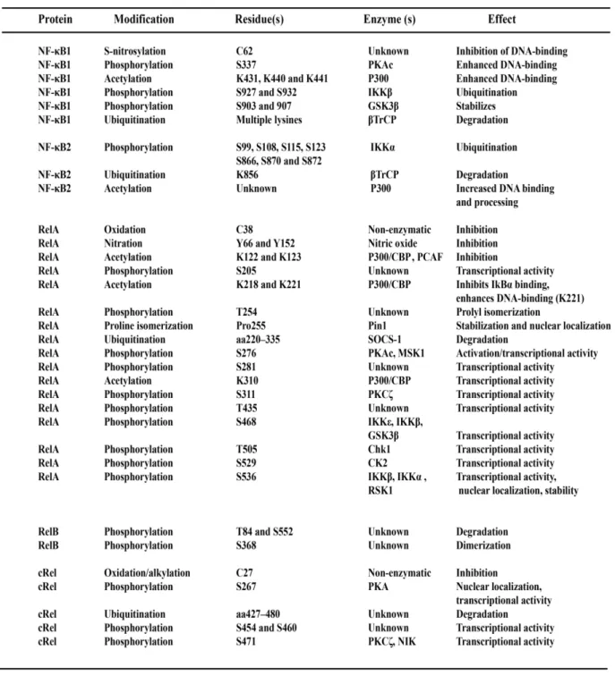
Table 2. Post-translational modifications of NF-κB transcription factors have diverse
impacts on functionality. 30
Chapter 2
Supplementary Table 1. List of the AML patient specimens and cell lines used in
this study. 75
Supplementary Table 2. List of oligonucleotide sequences used in this study. 76 Chapter 3
Supplementary Table1. An overview of the 10 B-lymphoblast cell lines presented
in this study. 111
Supplementary Table2. List of the cell lines and the ChIP-Seq datasets used in this
study. 111
Supplementary Table3. NF-κB RelA and NF-κB1 (p50) motif enrichment in the
investigated samples are statistically significant. 112
Supplementary Table4. List of 47 predicted NF-κB target genes containing EIF4E
κB sites in their promoter and/ or intron. 113
Supplementary Table5. List of the functional annotation groups and their underlying
List of Figures
Chapter 1Figure 1. The mechanism of eukaryotic translation initiation. 5 Figure 2. The initiation factor EIF4E gene comprises 8 exons that codes for a
cap-binding protein with a unique structure. 8
Figure 3: EIF4E functions at two levels: mRNA export and translation initiation. 11 Figure 4. The Myc-centric view depicting EIF4E transcriptional control. 13 Figure 5. EIF4E is regulated at multiple levels through transcript stability, protein
interactions and post-translational modifications. 15
Figure 6. The NF-κB signaling pathways involve two main transduction
modules: canonical and non-canonical. 24
Chapter 2
Figure 1. Stimulation of BJAB cells with PMA leads to NF-κB dependent
EIF4E transcriptional upregulation. 50
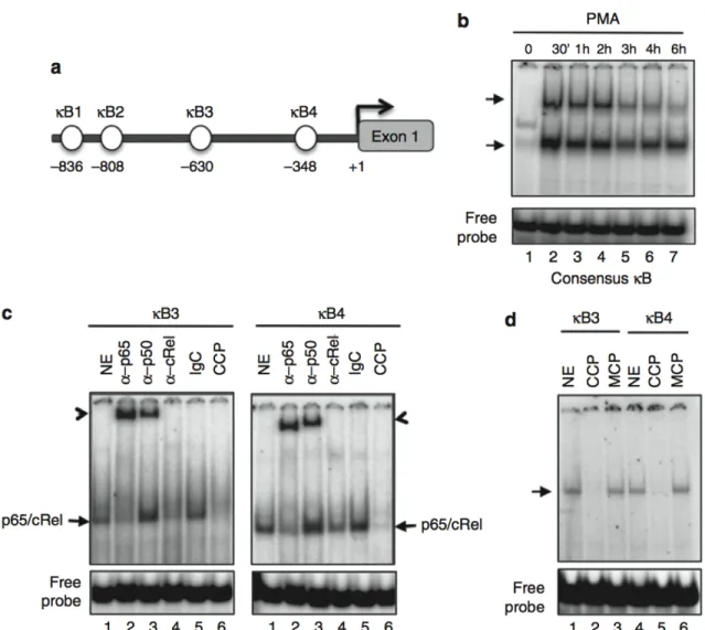
Figure 2. The EIF4E promoter contains four κB sites preferentially bound by
cRel-p65 NF-κB complexes. 52
Figure 3. NF-κB complexes are recruited to the EIF4E promoter and promote
transactivation. 54
Figure 4. PMA Stimulation of primary human PBMCs increases EIF4E expression
in an NF-κB dependent manner. 55
Figure 5. Constitutively active NF-κB regulates EIF4E expression in KM-H2 cells. 57 Figure 6. NF-κB recognition of the EIF4E promoter elements in AML cell lines. 58 Figure 7. Selective NF-κB recognition of the EIF4E promoter elements in
M4/M5 AML. 60
Supplemental Figure 1. 71
Supplemental Figure 2. The EIF4E promoter harbors 4 NF-κB elements. 72 Supplemental Figure 3. Electrophoretic mobility shift assay of BJAB nuclear extracts
Supplemental Figure 4. EIF4E is elevated with nuclear localization in the
M0 AML cell line KG1a. 74
Chapter 3
Figure 1. EIF4E and NF-κB RELA mRNA expression levels are positively
correlated in an AML microarray gene expression study. 87
Figure 2. Elevated EIF4E and NF-κB RELA mRNAs predict worse survival
outcomes in AML. 88
Figure 3. The RelA and p50 consensus binding sites are significantly enriched in the ENCODE NF-κB ChIP-Seq datasets suggesting the formation of RelA and
p50 complexes in the regulatory regions of target genes. 90
Figure 4. NF-κB (RelA) is enriched in the EIF4E promoter and first intron. 92 Figure 5. In silico prediction of new putative NF-κB target genes that are upregulated
in poor prognosis AML specimens. 95
Figure 6. In silico prediction of 6 new putative EIF4E transcriptional regulators. 97 Supplementary Figure 1. Intermediate and poor prognosis AML groups predict
inferior survival outcomes. 108
Supplementary Figure 2. Overview of the various stages in an NF-κB ChIP-Seq
experiment. 109
Supplementary Figure 3. NF-κB (RelA) is enriched in the promoter and intron
of two validated NF-κB target genes: NFκBIA and BCL2. 110
Chapter 4
Figure 1. Two intronic κB elements are bound by NF-κB complexes. 123 Figure 2. EIF4E is a downstream NF-κB target that acts as an amplifier of NF-κB
activity to drive proliferative gene expression. 124
Figure 3. Regulation of EIF4E gene expression through NF-κB may involve
post-transcriptional or post-translational events. 125
Figure 4. The NF-κB inhibitor Bay11-7082 results in a downregulation of EIF4E
Abbreviations
4E-BE 4E Basal Element
4E-BP 4E Binding Protein
4E-HP 4E Homologue Protein
4E-SE 4E Sensitivity Element
4E-T 4E Transporter
4GI 4E/4G Inhibitor
AEG Astrocyte Elevated Gene
AhR Aryl Hydrocarbon Receptor
AIDS Acquired Immunedefficiency Syndrome
AKIP PKA Interacting Protein
AKT Aktinic Kinase
AML Acute Myeloid Leukemia
AML/ETO2 AML/Eight Twenty One Fusion Protein
ANOVA Analysis of Variance
AP1 Activating Protein1
APL Acute Pro-myelocytic Leukemia
ARE AU Rich Element
AUF1 ARE Binding Factor
BAFFR B-Cell-Activating Factor Receptor
Bcl B-Cell Lymphoma
BCR B-Cell Receptor
BCR/Abl Breakage Cluster Region/Abelson Kinase Fusion Protein
Bp Base Pairs
C/EBP CAAT/Enhancer Binding Protein
CARMA1 CARD-Containing MAGUK Protein
CBP CREB Binding Protein
CCND1 CyclinD1
CCP Consensus Cold Probe
CHD9 Chromodomain Helicase DNA Binding Protein 9
CHFR Checkpoint with Forkhead and RING domain Protein
ChIP Chromatin Immunoprecipitation
ChIP-Seq Chromatin Immunoprecipitation coupled to Sequencing
cIAP Cellular Inhibitor of Apoptic Pathways
CML Chronic Myelogenous Leukemia
COMMD1 Copper Metabolism with MURR Domain 1
COX Cyclooxygenase
CREB cAMP Response Element Binding Protein
CRISPR Clustered regularly interspaced short palindromic repeats
CRM1 Chromosome Region Maintenance 1
CYLD Cylindromatosis Deubiquitinase
CYTH4 Cytohesin4
DNA Deoxyribonucleic Acid
DR5 Death Receptor 5
DSIF DRB Sensitivity Inducing Factor
EGF Epidermal Growth Factor
eIF Eukaryotic Initiation Factor (e.g. EIF4E, EIF4G, EIF4A)
ELAM Endothelial Leukocyte Adhesion Molecule
EMSA Electrophoretic Mobility Shift Assay
ENCODE Encylcopedia of DNA Elements
FAB French American British
FBS Fetal Bovine Serum
FDR False Discovery Rate
GAPDH Glyeraldehyde 3-Phosphate Dehydrogenase
GnRH Gonadotropin Releasing Hormone
GTP Guanosine Triphosphate
HAT Histone Acetyl Transferase
HDAC Histone Deacetylase
HOIP/HOIL Heme-Oxidized IRP2 Ubiquitin Ligase with Interacting Protein
HoxA9 Homeodomain Box Protein A9
ICAM Intercellular Adhesion Molecule
IgG Immunoglobulin G
IgM Immunoglobulin G
IGV Integrative Genomics Viewer
IKK IκB Kinase
IL Interleukin
ING4 Inhibitor of Growth 4
IRE Internal Ribosomal Entry
IRES Internal Ribosomal Entry Site
ITAF IRES Trans Acting Factors
IκB Inhibitor of NF-κB
IκB-SR IκB Super Repressor
Kbp Kilo Base Pairs
KDa Kilo Dalton
LRA Luciferase Reporter Assay
LRP/PRC Leucine-Rich Pentatricopeptide Repeat Containing
LTβ Lymphotoxin beta
M7GDP 7-Methyl Guanosine Diphosphate
MALT1 Mucosa-Associated Lymphoid Tissue Lymphoma 1
MAPK Mitogen Activated Protein Kinase
Mcl1 Myeloid Cell Leukemia Protein 1
MCP Mutant Cold Probe
MEME Multiple EM for Motif Elicitation
MLL Mixed-Lineage Leukemia Protein
MMP Matrix Metalloproteinase
Mnk MAP Kinase Interacting Serine/Threonine Kinase
mTOR Mammalian Target of Rapamycin
MyD88 Myeloid Differentiation Primary Response 88 NCBI National Center for Biotechnology Information
NELF Negative Regulator of Transcriptional Elongation Factor
NEMO NF-κB Essential Modulator
NEXT-GEN Next Generation
NF-κB Nuclear Factor Kappa-light-chain-enhancer of activated B-cells
NFAT Nuclear Factor of Activated T-cells
NFκBIA NF-κB Inhibitor alpha
NIK NF-κB Inducing Kinase
NURR NR4A Nuclear Receptor
OCT Octamer Factor
ODC Ornithine Decarboxylase
PABP PolyA Binding Protein
PAX5 Paired Box Protein
PBMC Peripheral Blood Mononuclear Cells
PCR Polymerase Chain Reaction
PDLIM2 PDZ And LIM Domain Protein 2
PKC Protein Kinase C
PLXNA1 Plexin A1
PM Perfect Match
PMA Phorbol-12-myristate-13-acetate
PML Promyelocytic Leukemia
PML/RARA PML Protein/Retinoic Acid Receptor alpha Fusion Protein PPP1R15B Protein Phosphatase 1, Regulatory (Inhibitor) Subunit 15B
PRH Proline-Rich Protein HaeIII Subfamily
pTEFb Positive Regulator Transcription Elongation Factor b
PTL Parthenolide
PU.1 Purine Rich (PU) box Binding Protein
RANBP2 Ran-Binding Protein 2
REC Research Ethics Committee
RHD Rel Homology Domain
RING Really Interesting New Gene Protein
RIP Receptor Interacting Protein
RMA Robust Multi-Array
RNA Ribonucleic Acid
RNA-Seq RNA Sequencing
RNAP RNA Polymerase
RNGTT RNA Guanylyl transferase triphosphatase
RNMT RNA Methyl Transferases
RNP Ribonuclear Protein
RPKM Reads Per Kilobase of exon model per Kilobase mapped reads
RPS Ribosomal Protein S
SAH S-Adenosylhomocysteine
SAHH S-Adenosylhomocysteine Hydrolase
SCF-βTrCP SKP-Cullin-F-box/Beta-Transducin Repeat Containing
Shh Sonic Hedgehog
siRNA Small interfering RNA
SIRT Sirtuin
SNX32 Syntaxin 32
SOD Superoxide Distmutase
SP1 Specificity Protein 1
STAT Signal Transducer and Activator of Transcription
SUMO Small Ubiquitin-Like Modifier 1
TAB TAK Binding Protein
TAD Transactivation Domain
TAK Transforming Growth Factor β Activated Protein Kinase TAP/NXF1 Tip Associating Protein/Nuclear Export Factor1
TBP TATA Binding Protein
TCR T-cell Receptor
TFIIB Transcription Factor II B
TK Thymidine Kinase
TNF-α Tumor Necrosis Factor alpha
TRADD TNF Receptor Associated Death Domain
TRAF1 TNF Receptor Associated Factor1
TRED Transcription Regulatory Element Database
TSS Transcription Start Site
Ubc/Uev Ubiquitin C/Ubiquitin Conjugating Enzyme Variant
UCSC University of California, Santa Cruz
USER Untranslated Sequence Elements for Regulation
UTR Untranslated Region
VCAM Vascular Cell Adhesion Molecule
VEGF Vascular Endothelial Growth Factor
WDR33 WD Repeat Domain 33
To my family, My wife, And all of my friends,
Acknowledgements
I would like to express my earnest gratitude to my supervisor Dr. Katherine Borden for her guidance throughout the years spent at her laboratory. Her meticulous nature has been integral for the success of the work presented in this thesis.
I genuinely acknowledge all the funding agencies that had supported my research: the Fonds de recherche en santé du Québec (FRSQ), the Cole Foundation and the Faculté des études supérieures et postdoctorales (FESP).
I would also like to thank Dr. Meztli Arguello for her help and support as well as the Borden lab members, past and current, for all the discussions and memorable experiences.
In addition, I would like to express my sincere thankfulness to Dr. Alain Verreault as well as my committee members: Dr. Martine Raymond and Dr. Muriel Aubrey for their helpful comments and constructive criticisms; their recommendations for my third chapter have propelled me into the realm of bioinformatics, which I profoundly appreciate. Furthermore, I would like to recognize the support of Dr. Koren Mann and Dr. John Hiscott for their fruitful collaborations.
My wholehearted recognition goes to my parents for constantly believing in me and for their warm support that transcended the vast continental distance separating us. I would also like to convey my love and appreciation to my wife, Rola, for her incessant care and affection. A heartfelt thanks goes to Dr. Georges Nemer for his constant advice and encouragement. Finally, I would like to thank my friends Iman, Aline, Gloria, Lama, Joe, Jacob, Alex, Georges, Moutih as well as the IRIC community for all the unforgettable moments and for being part my PhD journey.
Chapter 1: Introduction
It is imperative that global rates of gene expression are strictly regulated to achieve optimal spatiotemporal RNA concentrations required to elicit a biological response1. Eukaryotic cells regulate gene expression through transcription, mRNA stability and post-transcriptional modifications, as well as protein synthesis (translation) and post-translational modifications. The RNA regulon model offers a blueprint in which cells regulate gene expression and protein synthesis. Cis-acting elements are positioned within the untranslated regions of transcripts known as untranslated sequence elements for regulation (USER) that recruit RNA binding proteins (RNPs) to modulate mRNA stability, export and translation1,2.
Translational control is rapid and represents an important hallmark of cell development to modulate cell growth, proliferation and differentiation. Dysregulated expression and activity of components of the translation apparatus have been linked to cell transformation and carcinogenesis3. One key rate-limiting component, the eukaryotic initiation factor EIF4E, is upregulated in a plethora of cancers4,5. The transcriptional regulation of EIF4E and its dysregulation in acute myeloid leukemia (AML) is the focus of this thesis.
1.1 Cap- Dependent Eukaryotic Translation Initiation
Protein synthesis or mRNA translation is comprised of three distinct stages: initiation, elongation and termination3. The rate-limiting step in protein synthesis is translation initiation when the target mRNA is recognized by the translation machinery which catalyzes ribosomal assembly to commence protein synthesis3,6. Three mechanisms have been described so far for eukaryotic mRNA translational initiation7. These are (1) cap-dependent scanning8, (2) scanning-independent ribosomal shunting7 and (3) cap-independent scanning-independent internal ribosomal entry (IRE)9. In this thesis, I present my research on the initiation factor EIF4E, an integral component in the cap-dependent mode of translation initiation.
1.1.1 A 7-methyl guanosine cap structure is required for cap-dependent translation initiation
Following DNA transcription, the nascent pre-mRNA transcripts undergo a series of modifications including the amendment of a 7-methyl guanosine cap structure to the 5’ end of transcripts6. The process of mRNA cap methylation constitutes a key step that is essential for
gene expression. It involves two enzymatic reactions catalyzed by the RNA guanylyl transferase triphosphatase (RNGTT), that forms the 5’ guanylylated end on transcripts, and RNA methyl transferase (RNMT), which methylates the added 5’ guanosine. It is has been demonstrated that elevating c-Myc expression promotes an increase in the proportion of capped transcripts, thus leading to increased rates of protein synthesis. c-Myc enhances capping by promoting RNA pol II phosphorylation as well as the upregulation of s-Adenosyl Homocysteine Hydrolase (SAHH) which neutralizes s-Adenosyl Homocysteine (SAH), an inhibitory bi-product of methylation reactions10.
The cap structure is essential for mRNA stability11, splicing12, nucleo-cytoplasmic export13 and acts as a marker that interacts with the translation initiation machinery14. Evidence for cap requirement in translation initiation came from studies using protein synthesis-competent wheat germ extracts. In these studies, only capped reovirus RNAs formed an interaction with the 40S ribosomal subunit and were efficiently translated11,14. Addition of m7GDP cap analogs to the in vitro translation reaction or removal of the m7 cap structure from the viral transcripts diminished the translation of these RNAs15, corroborating the importance of the cap structure in mRNA translation.
In an attempt to decipher the mechanism through which the cap structure triggers mRNA translation, cap-binding complexes were identified and isolated using ribosomal washes from
Artemia salina16 as well as from capped viral RNA studies. These complexes were originally
referred to as cap binding proteins (CBP) I and II and are now known as eukaryotic translation initiation factors 4E (EIF4E) and 4F (EIF4F) respectively17,18. The EIF4E-cap interaction was displaced by cap analogs suggesting specificity. Furthermore, purified preparations of the 25KDa EIF4E protein revealed an integral role in stimulating mRNA translation of only capped viral transcripts19. The EIF4F molecule was later shown to comprise EIF4E in complex with a 46KDa RNA helicase (EIF4A) and a 220KDa mRNA-ribosome bridging factor (EIF4G)20.
1.1.2 Molecular mechanism of cap-dependent translation initiation
As illustrated in figure 1, the dissociation of the 80S ribosome marks the start of mRNA translation facilitated by EIF6, which binds the 60S ribosomal subunit as well as EIF3 and EIF1A that bind the 40S ribosome. The 40S subunit associated with EIF3 and EIF1A is loaded with EIF2 together with GTP and the initiator tRNA, Met-tRNA, forming the 43S pre-initiation complex3,21. This complex is then recruited to the 5’ end of transcripts through its interaction with the EIF4F cap-binding complex.
The EIF4F cap-binding complex is formed in three stages, first, EIF4E binds the 5’ capped end of transcripts; next, the scaffolding protein EIF4G binds EIF4E and recruits the 43S pre-initiation complex to mRNA through its ability to interact with EIF3. The RNA helicase EIF4A and a “scanning” protein EIF4B are then recruited through EIF4G and are required to unwind the complex 5’ untranslated regions (5’UTR) and scan for the start codon positioned in the consensus “Kozak” initiator sequence3,8,21. The circularization of the complex is attained through the bridging properties of EIF4G that interacts with the polyA binding protein (PABP), which in turn binds the polyadenylated 3’ end of the transcripts. This subsequent association of the 43S pre-initiation complex and EIF4F results in the 48S complex3,21.
The scanning properties of EIF4F allows the 48S complex to traverse the bound transcripts in a 5’ to 3’ fashion until the start codon (AUG) is located. Subsequently, the initiation factors are released in a process catalyzed by EIF5, a GTPase activating protein. The dissociation of these initiation factors allows the association of the 60S subunit and the commencement of translation elongation3. The association of 43S complex with mRNA through EIF4E constitutes the rate-limiting step in translation initiation3,6. The abundance and activity of the EIF4E protein is highly regulated in a multifactorial manner and will be discussed thoroughly in this chapter.
Figure 1. The mechanism of eukaryotic translation initiation. (A) Translation initiation commences with the dissociation of the 80S ribosomal subunit catalyzed by EIF1A, EIF3 and EIF6. The 40S subunit forms the 43S pre-initiation complex with EIF1A and EIF6 together with EIF2 and the Met initiator tRNA. (B) The 43S complex is recruited to the 5’ end of transcripts through its interaction with the EIF4F complex. The EIF4F complex is formed from three main proteins: the cap binding protein EIF4E, a scaffolding protein EIF4G and an RNA helicase EIF4A (coupled to the scanning factor EIF4B). The EIF4F complex can interact with the 43S complex through EIF3 and and recruits it to the bound transcripts forming the 48S complex. Circularization is achieved through the interaction of EIF4G with the poly A binding protein (PABP). The 48S complex traverses the transcript in a 5’ to 3’ manner to locate the start codon. Translation initiation ends with the release of the initiation factors and the joining of the 60S ribosomal subunit.
1.2 Alternative Mechanisms for Translation Initiation
Alternative mechanisms for translation initiation have been previously described and involve cap-independent strategies7. Some viral and cellular mRNAs are innately uncapped and are thus translated by a process involving internal initiation. These transcripts harbor an internal ribosome entry site (IRES), a structural element in the 5’UTR. This alternative mode of initiation involves direct recruitment of the initiation factors and ribosome complex to the IRES element independent of EIF4E through IRES trans acting factors (ITAFs)7,9,21. Under patho-physiological and stress conditions, cap-dependent translation is impaired; however, mRNA translation of a subset of transcripts is maintained with a translation initiation re-programming in favor of IRES-mediated translation21. Translational profiling experiments performed under physiological and stress conditions such as, mitosis, differentiation and apoptosis as well as following heat shock and hypoxia, revealed that 10-15% of all mRNAs are translated potentially through an IRES-mediated process when cap-dependent translation is compromised21. This mechanism allows cells to adapt in response to various physiological and stress stimuli. Interestingly, since this process still requires most of the translational machinery, several picornaviruses have evolved strategies to hijack the host’s translational machinery for viral protein synthesis through site-specific cleavage of EIF4G’s amino-terminus compromising EIF4G-EIF4E interaction without altering its binding properties to other factors. This process diverts the host’s EIF4G protein from cap-dependent translation in favor of viral IRES-mediated protein synthesis22.
Finally, a role for the cap binding complex Cbc1 in mRNA translation has been demonstrated. In yeast, global translation is suppressed in response to osmotic stress; however, mRNAs encoding stress protective proteins remain selectively translated to allow survival. This process was shown to be dependent on Cbc123.
1.3 EIF4E Functions in mRNA Export and Translation
1.3.1 Structure of the EIF4E gene, alternative splicing and homology
The human eukaryotic initiation factor EIF4E gene spans more than 50 kilobase pairs (kbp) and is situated on chromosome 4. It is a functionally conserved gene encoded by 8 exons with three possible transcript variants illustrated in figure 2A. The most common transcript variant
1 (4749 bp) produces a 217 amino acid protein. Through alternative splicing, the longest transcript variant 2 (4842 bp) uses an alternative exon in the 3’ coding region producing a 248 amino acid protein with an extended C-terminus. Transcript variant 3 (3406 bp) has a longer N-terminus producing a 237 amino acid protein by using an alternative exon in the 5’UTR and 5’ coding region with a distinct initiation codon AUG. The alternatively spliced transcripts 2 and 3 have been predicted from cDNA libraries; however, their expression and biological functions are yet to be investigated24.
Interestingly, two EIF4E mammalian paralogues have been described and are referred to as EIF4E-2, also known as 4E-HP, and EIF4E-324,25. These proteins are distinct in their structure, function and expression pattern from the main ubiquitously expressed EIF4E-1 protein. EIF4E-2 is highly expressed in the testis whereas EIF4E-3 is mostly expressed in muscles, lung and spleen24. These paralogues have been shown to bind the 7-methyl cap26,27; however are, unlike EIF4E-1, incapable of functionally rescuing the growth-arrested phenotype in S.
cerevisiae with a deletion in the cdc33 gene, an EIF4E orthologue, suggesting that EIF4E-2
and EIF4E-3 fulfill distinct tissue-dependent functions24. In fact, studies have shown that EIF4E-2 and EIF4E-3 are not associated with EIF4G and are thus not part of the translation competent EIF4F complex. Furthermore, these proteins are incapable of binding known EIF4E-1 regulators such as the EIF4E binding protein 1(4E-BP1)26,27. These studies suggest that EIF4E paralogues may function as negative regulators of EIF4E-1 activity by competing for the same pool of capped transcripts and thus impede EIF4E-1 functions. The structure and function of the main EIF4E-1 protein will be further discussed and will be referred to as EIF4E.
1.3.2 The EIF4E structure reveals a distinct mode for cap-recognition required in mRNA export and translation
The EIF4E protein is of relatively small size at 25 KDa and is present as part of the EIF4F complex as well as in free form. The human and mouse cap-bound EIF4E structures have been elucidated with X-ray crystallography revealing a unique structure underlying EIF4E’s cap binding properties24.
Figure 2. The initiation factor EIF4E gene comprises 8 exons that codes for a cap-binding protein with a unique structure. (A) Alternative splicing of the EIF4E gene produces three transcript variants, the most common being transcript variant 1 which produces a 217 amino acid protein. Transcript variant 2 uses an alternative exon in the 3’ coding region resulting in a 248 amino acid protein with a longer C-terminus. Transcript variant 3 has a longer N-terminus producing a 237 amino acid protein by using an alternative exon in the 5’ coding region. (B) Crystal structure of the mouse EIF4E bound the 7-methyl cap (blue) reveals a unique “cupped hand” structure composed of 8 anti-parallel beta strands supported by 3 alpha helices. Cap binding involves aromatic pi-pi stacking and requires W56 (red) and W102 (orange). (C) The 7-methyl cap (yellow) is nestled in a binding groove involving 7 amino acids: W56, W102, E103, W166, R157, K159 and K162 (see text for details). (D) The EIF4E dorsal surface binds protein partners that serve a regulatory purpose. Several key residues include H37, P38, L131, E132 and L135 (shown in red). Disruption of V69 (orange) and W73 (beige) hinders EIF4E’s interaction with EIF4G and 4E-BPs. All cartoons were generated from the 1L8B structure, obtained from pdb.org, using pyMol.
The EIF4E protein consists of eight anti-parallel beta-sheets supported by three alpha-helices to form the palm and back of a cupped hand respectively28-30 as illustrated in figure 2B. Studies have shown that EIF4E recognizes the 7-methyl cap through intercalation between two aromatic residues W56 and W102, also known as π-π stacking31,32.
Additional residues involved in cap recognition are highlighted in figure 2C and include a polar E103 that interacts with the nitrogen moiety of the cap, a W166 residue forming a hydrophobic interaction with the methyl group of the cap, and three positively charged residues R157, K159, K162 that interact with the phosphate backbone of the cap. Nuclear magnetic resonance (NMR) solution structure of the cap-free EIF4E form, apo-EIF4E, revealed that this factor remains structured; however, key structural variations in the cap-binding pocket and the dorsal surface were noticed compared to the cap-bound EIF4E. Structural alterations in the S4-H4 loop distal to the cap binding pocket appears to be essential in regulating conformational changes in EIF4E following cap binding33.
Structural studies with mouse EIF4E bound to EIF4G and 4E-BP1, an EIF4E regulator, revealed a requirement for the dorsal region in the EIF4E protein. Several amino acids (H37, P38, V69, W73, L131, E132 and L135) situated in the dorsal surface mediate EIF4E’s interaction with its binding partners and regulators34 (figure 2D). Disruption of two key phylogenetically conserved residues V69 and W73 hinders EIF4E’s interaction with EIF4G and 4E-BPs35,36. Interestingly, phylogenetic alignment studies of EIF4E revealed that only around 170 amino acids representing the EIF4E core are conserved in all eukaryotes. This region includes the amino acids involved in cap recognition as well as binding to EIF4E partners and regulators35,37. Accordingly, EIF4E is functionally conserved whereby mammalian EIF4E is capable of rescuing the growth-arrested phenotype in S. cerevisiae with a deletion in the cdc33 gene, an EIF4E orthologue, although it harbors only 30% sequence identity with its yeast counterpart38,39.
Subcellular localization experiments revealed a dynamic nuclear and cytoplasmic localization for EIF4E. EIF4E functions at two levels: mRNA export and mRNA translation, both of which require cap recognition24 (figure 3). Transcripts that harbor a complex GC rich highly
structured 5’UTR have been shown to be translated more efficiently, in an EIF4F dependent fashion, than transcripts with short unstructured 5’UTRs40-42. These transcripts have been dubbed as EIF4E translationally sensitive targets and include genes involved in cell proliferation and survival such as PIM1, VEGF, MYC, ODC and many more41,43-45.
The nuclear localization of EIF4E (up to 68%)46 suggests EIF4E nuclear functions. In fact, EIF4E has been shown to promote the export of transcripts containing a 50 nucleotide 4E-sensitivity element (4E-SE) in their 3’UTR1,44,47,48. Transcripts containing a 4E-SE element are bound by the export factor LRPPRC that interacts with EIF4E resulting in a CRM1-dependant export; unlike bulk mRNA export which is TAP/NXF1 dependent48,49. EIF4E export targets are also of the pro-proliferative nature and include MYC, CCND1 (CyclinD1), ODC and many more44,47. Interestingly, EIF4E was also shown to reprogram the nuclear pore complex to enhance the export of its target genes. Specifically, the RanBP2 cytoplasmic fibrils reduce the release and recycling of export factors to the nucleus, thereby impeding EIF4E mediated export; EIF4E circumvents this inhibitory mechanism by indirectly reducing the levels of RanBP250.
Eukaryotic cells organize the regulation of mRNA pools involved in the same biological process at the post-transcriptional level by altering the activities of RNPs interacting with these transcripts through USER sequences2. EIF4E functions to promote the nuclear export of its target genes and subsequently enhances the translation of the cytoplasmic EIF4E sensitive transcripts; these functions are independent of ongoing transcription and protein synthesis48. EIF4E export and translation targets are involved in cell proliferation and survival, accordingly, EIF4E is a central node of an RNA regulon that directs cell survival48. Importantly, not all EIF4E transcripts are sensitive at both export and translation levels24.
Figure 3: EIF4E functions at two levels: mRNA export and translation initiation. In the nucleus, EIF4E promotes the export of transcripts containing a unique 50 nucleotide element in their 3’ UTR referred to as the 4E sensitivity element (4E-SE). EIF4E export is CRM1 dependent. In the cytoplasm, EIF4E enhances the translation of transcripts containing a complex highly structured 5’UTR. EIF4E export or translationally sensitive transcripts include genes involved in cell proliferation and survival (e.g. ODC, MYC, CCND1, VEGF, PIM1). EIF4E overexpression has been associated with cell transformation and tumor promotion owing to its proliferative and anti-apoptic program.
Finally, EIF4E’s pro-survival program endows oncogenic properties for this initiation factor; in fact, EIF4E is overexpressed in an estimated 30% of human cancers4,5.
1.4 EIF4E regulation is multifaceted with distinct levels of control
EIF4E expression and activity are regulated at multiple levels through transcription, mRNA stability, protein interactions as well as post-translational modifications. These distinct modes of regulation are illustrated in figures 4 and 5. Dysregulation in EIF4E control modules have been linked to malignant transformation6.
1.4.1 A c-Myc centric view for EIF4E transcriptional control
The identification and cloning of the EIF4E promoter was achieved through screening human genomic DNA libraries using 5’c-DNA probes corresponding to EIF4E exons51. The transcription start site (TSS) of EIF4E was mapped with RNase protection assays51,52. The
EIF4E promoter lacks a TATA box but harbors a polypyrimidine tract at position -25 distal to
the TSS known as the EIF4E basal element (4EBE)53. This element binds the heteronuclear ribonuclear protein K (hnRNPK) that recruits the TATA binding protein (TBP) and consequently the basal transcription machinery to the EIF4E promoter53,54. Studies of the
EIF4E promoter revealed multiple E-box elements that were shown to bind c-Myc and
transactivate the EIF4E gene51 as seen in figure 4. The tumor suppressor p53 was shown to repress EIF4E through luciferase reporter assays (LRA) possibly through sequestering c-Myc and hindering its interaction with the promoter55. In addition, the sonic hedgehog pathway was also shown to upregulate EIF4E in neural cells through Myc56. Accordingly, for the past 16 years EIF4E transcriptional regulation has been solely the purview of Myc; however, EIF4E transcript is still inducible in Myc null fibroblasts following serum stimulation57 suggesting that other mechanisms are involved in EIF4E transcription. Consistent with this idea, a recent report suggests that EIF4E is also a C/EBP target58. In fact, the EIF4E promoter is enriched with binding sites for a plethora of transcription regulators including NF-κB, STAT, PU.1, PAX, NFAT, GATA, SP1 and many more59. This doctoral thesis focuses on the transcriptional regulation of EIF4E through NF-κB and its dysregulation in AML.
Figure 4. The Myc-centric view depicting EIF4E transcriptional control. The EIF4E promoter harbors multiple E-box elements that recruit c-Myc and upregulate EIF4E expression. The tumor suppressor p53 represses EIF4E expression by sequestering Myc and hindering its interaction with the promoter. The sonic hedgehog (SHH) pathway induces a Myc-dependent EIF4E expression in neuronal cells. These pathways illustrate Myc-centric mechanisms for the transcriptional regulation of EIF4E.
1.4.2 The stability of EIF4E mRNA is regulated by HuR and AUF1
Three conserved AU rich elements (AREs) have been identified in the 3’UTR of EIF4E. These elements direct HuR binding to stabilize the EIF4E transcript. On the other hand, a competing destabilizing protein, p42 AUF1, was shown to reduce EIF4E transcript stability. Furthermore, HuR is upregulated in cancer cells with elevated EIF4E expression and depletion of HuR in cancer cells results in EIF4E downregulation60. Thus EIF4E expression is also modulated through post-transcriptional events independent of EIF4E transcript levels (figure 5A).
1.4.3 EIF4E activity is modulated by protein interactions with a multitude of regulators
Another mode of EIF4E regulation involves its interaction with binding proteins that confer an activating or inhibitory effect6 (figure 5B). Experiments conducted with Far-Western hybridization led to the isolation of two small proteins that interact with EIF4E and were referred to as EIF4E binding proteins 4E-BP1 and 4E-BP261. Many proteins that bind EIF4E share the following small conserved amino acid motif YXXXXLϕ, where X is any residue and ϕ is a hydrophobic amino acid62. Accordingly, 4E-BPs compete with EIF4G to bind EIF4E and prohibit its access to the translational apparatus. In fact, 4E-BPs enhance the cap binding affinity in EIF4E thereby sequestering the bound EIF4E-mRNA complex to inhibit translation61,63. The dual cytoplasmic and nuclear localization of 4E-BPs suggests that these binding proteins can alter both EIF4E export and translation activities64. Importantly, 4E-BPs are regulated by phosphorylation through the mammalian target of rapamycin (mTOR); this reduces the interaction with EIF4E and increases translational activity65.
Interestingly, mice lacking 4E-BPs were not more prone to developing cancers than controls 66-68 suggesting a redundancy in EIF4E regulators. A multitude of proteins have been identified that contain the consensus EIF4E binding motif including more than 200 homeodomain proteins; these proteins can act as positive and/or negative regulators of EIF4E activity24. The PRH/Hex homeodomain protein is a negative regulator of EIF4E’s nuclear export functions69,70 whereas HoxA9 was shown to promote both EIF4E export and translation functions71.
Figure 5. EIF4E is regulated at multiple levels through transcript stability, protein interactions and post-translational modifications. (A) The EIF4E mRNA harbors 3 AU rich elements (AREs) in the 3’UTR that bind HuR and AUF1. EIF4E transcript stability is enhanced by HuR and reduced with AUF1. (B) EIF4E activity is modulated by multiple protein-protein interactions. HoxA9 promotes the export and translation functions of EIF4E, whereas PRH and PML inhibit EIF4E-mediated export. On the other hand, 4E-BPs can inhibit both EIF4E export and translation activities. (C) EIF4E activity is also regulated through post-translational modifications. EIF4E sumoylation and phosphorylation enhance its translation functions. Ubiquitination of the EIF4E protein promotes its proteasomal-mediated degradation.
Additional homeodomain proteins shown to alter EIF4E activity include Otx, Engrailed 2, Emx2, Bicoid and Hox1172.
Furthermore, two additional distinct protein families have been shown to interact with EIF4E and regulate its activity; they lack the YXXXXLϕ motif and include the RING domain containing proteins as well as the virus protein linked to the genome (VPg). Promyelocytic leukemia (PML) and arenaviral Z proteins are RING domain containing proteins that impede EIF4E activity by reducing its affinity to the 7-methyl cap (~100 fold)24. Nuclear PML was shown to impede EIF4E’s mRNA export function73. The potyviral VPg was shown to form a complex with EIF4E and reduces its affinity for the mRNA cap74.
The regulation of EIF4E through binding partners is thus multifaceted24 involving an abundance of proteins expressed at various stages of the cell cycle and development to ensure optimal levels of EIF4E activity.
1.4.4 EIF4E activity is modulated through post-translational modifications
A fourth level of EIF4E regulation involves post-translational modifications of the EIF4E protein6 including phosphorylation, ubiquitylation and sumoylation (figure 5C). The phosphorylation status of EIF4E reflects the translation rate and growth state of the cell6. The stress and cytokine activated p38 mitogen activated protein kinase (p38 MAPK) pathway converges at two EIF4E kinases, Mnk1 and Mnk2, to phosphorylate EIF4E at residue S209 75-77. EIF4E phosphorylation enhances its export function and cell transformation capacity78. Furthermore, EIF4E could also be modified by ubiquitylation and SUMO1 conjugation79-81.
This described multifactorial regulation of EIF4E ensures that ideal levels of EIF4E protein and activity are maintained to achieve an appropriate biological effect in response to physiological stimuli. Aberrant regulation of EIF4E expression and/or activity has been linked to malignancies as well as cell transformation.
1.5 EIF4E is Overexpressed in approximately 30% of Human Cancers and is a Plausible Candidate for Clinical Targeting
EIF4E target genes at the export and translation levels impart a proliferative effect48. In fact, microinjection of EIF4E in quiescent fibroblasts promotes DNA synthesis82. In addition, EIF4E overexpression decreases cell cycle transit time in HeLa cells, whereas downregulation of EIF4E using anti-sense methods increases transit time in a dose-dependent fashion83. These studies demonstrated a role of EIF4E in supporting cell cycle progression and cell transformation6. Furthermore, anti-apoptic functions have also been described for EIF4E whereby overexpression in NIH3T3 cells blocks apoptosis following serum deprivation84. Additionally, EIF4E overexpression impedes Myc-driven apoptosis84. Taken together, these studies highlight EIF4E’s pro-survival properties. EIF4E is overexpressed in a multitude of cancers including hematopoietic malignancies and solid tumors4,5.
EIF4E overexpression is underlined by several factors and is correlated with poor prognosis. EIF4E overexpression at the RNA and protein levels has been described in epithelial cancers including breast, colon, prostate, lung, cervix and squamous head and neck carcinoma. EIF4E gene amplification has also been described in head and neck as well as breast carcinomas. Hematopoietic cancers with elevated EIF4E levels include AML and Hodgkin and non-Hodgkin lymphomas4. In this thesis, I present a new mechanism underlying the dysregulation of EIF4E in AML. A brief overview on AML and its classification systems will be covered in this chapter.
Increased levels of EIF4E drive cell transformation and oncogenesis, accordingly, EIF4E represented a plausible candidate for clinical targeting85. Several preclinical and clinical methods have been described including synthetic peptides, anti-sense oligos, suicide gene therapy as well as a cap mimetic. Through a high throughput fluorescence polarization binding screen, the 4EGI-1 synthetic peptide was identified. This peptide inhibits the association of EIF4E with the EIF4G and blocks the formation of an active EIF4F complex. 4EGI-1 was shown to posses preferential activity in transformed cells and reduces the expression of MYC and BCL2L1 (BclXL), both of which are EIF4E targets86. Small molecule analogs for 4EGI-1 have been described to target EIF4E in T-cell leukemia and non-small-cell lung cancer cells85.
Anti-sense oligonucleotides targeting EIF4E have been described in breast, prostate and head and neck carcinoma xenograft models87-89. In addition, a suicide gene therapy strategy has effectively targeted EIF4E in a head and neck carcinoma mouse xenograft model by fusing a complex structured 5’UTR upstream of the toxic gene encoding thymidine kinase TK, thereby promoting its expression in cells with upregulated EIF4E90. Furthermore, a novel strategy in epithelial ovarian cancer with upregulated gonadotropin releasing hormone (GnRH) receptor has also been described by fusing the EIF4E negative regulator 4EBP1 to agonists of the GnRH receptor to inhibit EIF4E activity in those cells91.
Finally, an effective EIF4E targeting strategy using a 7-methyl cap mimetic, ribavirin, has been demonstrated in AML92,93, breast cancer94 as well as head and neck carcinoma mouse xenograft model93. Ribavirin, an antiviral drug established in hepatitis C treatment, is structurally similar to the 7-methyl cap and was shown to physically interact with EIF4E93,95,96 to impede its oncogenic functions in phase II clinical trial of poor prognosis AML, leading to clinical response with no adverse drug-related side effects92. This provided a novel clinical approach to target mRNA translation in cancers with elevated EIF4E. Importantly, ribavirin is the only EIF4E inhibitor to date that has provided promising clinical outcomes. Furthermore, similar exciting findings have been observed in AML patients treated with Ribavirin plus low dose cytarabine (Ara-C) with remission up to two years (Assouline et al. In press). Finally, Ribavirin resistance has been observed in AML patients and has been attributed to a GLI1 driven UGT1A-dependent glucuronidation of ribavirin. In this same study, ribavirin resistance was overcome by genetic or pharmacological inhibition of GLI1, suggesting a novel strategy to overcome ribavirin resistance in the treated patients97.
1.6 Acute Myeloid Leukemia: A Hematopoietic Malignancy with Aberrant EIF4E Expression and Activity
Acute myeloid leukemia is a hematological malignancy of the myeloid lineage of blood cells; this malignancy affects the immature myeloid population of cells (myeloblasts) that expand at the expense of normal cells98. AML is a disease of older adults (> 60 years of age) with a median age at diagnosis of 67 years. The yearly incidence of new AML diagnoses in the USA
is 17.6/100,000 for individuals > 65 years of age, compared to 1.8/100,000 for individuals < 65 years (2008 statistics)99. The average 5-year overall survival (OS) rates, in patients receiving therapy, range from 5–15% in older adults and approach 30% in younger adults with AML99. The French-American-British classification (FAB) has classified AML into 9 distinct groups based on of the type of cell from which the leukemia has developed as well as the degree of maturity. The groups are: M0 (minimally differentiated), M1 (no maturation), M2 (granulocytic maturation), M3 (promyelocytic leukemia), M4 (myelomonocytic), M5a (monoblastic), M5b (monocytic), M6 (erythrocytic) and M7 (megakaryocytic)98,100.
Several epigenetic and genetic anomalies underscore AML blasts progression. Epigenetic and/ or cytogenetic aberrations may result in the overexpression or the formation of fusion genes resulting in a blockade of myeloid differentiation and the formation of immature myeloblasts. Additionally, aberrant constitutive activation of cell receptors (e.g. Flt3, c-kit) confers a proliferative advantage to blast cells98. Notably, many AML cases are characterized by a normal karyotype.
1.6.1 Cytogenetic and Molecular classification of AML to predict patient prognosis
The four decades old FAB AML classification system has been subject to criticism, revisions and re-evaluations since the techniques required in classifying AML samples into FAB groups are very descriptive and rely on cell morphological features as well as simple cytochemical assays101. Accordingly, the World Health Organization (WHO) classifies AML into three clinical prognosis groups: favorable, intermediate and poor prognosis. The criteria required in this diagnostic classification are based on clinical data (patient history), cytogenetic analysis, immunophenotyping and biological features. This system aimed at offering a clinically relevant approach to determine prognostic parameters and plan more effective treatment regimen102,103.
The advent of sequencing technologies has improved the WHO cytogenetic prognostic model through establishing molecular markers in all cytogenetic AML groups. This approach allowed for the classification of 5 prognostic subgroups with significant differences in OS thus
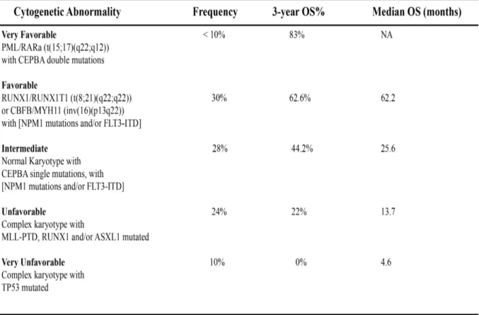
Table 1. Classification of AML into five clinical prognosis groups based on the underlying cytogenetic and molecular aberrations. The five distinct AML prognosis groups are shown with the underlying anomalies and frequency of occurrence. The 3-year overall survival (%) as well as the median survival rates for patients harboring the outlined anomalies are presented when applicable. NA, Not Applicable. Adapted from Grossmann et al.104
leading to a model based on molecular markers that is more comprehensive than standalone cytogenetics104. A summary of this prognostic classification system with the underlying cytogenetic and molecular aberrations and the associated OS rates is summarized in Table 1.
1.6.2 EIF4E is overexpressed in poor prognosis AML
A striking trend for EIF4E overexpression was observed in M4/M5 poor prognosis primary AML specimens but not in most M1/M2 specimens, with more than 100 samples tested70. The EIF4E export function is also augmented in M4/M5 AML; these samples show a predominant EIF4E nuclear accumulation. The nuclear function of EIF4E was shown to contribute to leukemogenesis by enhancing the export of target genes imparting a pro-proliferative and anti-differentiation program70. Molecular targeting of EIF4E with ribavirin (1µM) in M4/M5 primary AML specimens resulted in an EIF4E relocalization from the nucleus to the cytoplasm and inhibited EIF4E export functions leading to growth suppression. On the other hand, M1/M2 AML specimens with normal EIF4E levels were inhibited at a much higher concentration; additionally, M1 AML specimens with high EIF4E were also affected93. Accordingly, targeting EIF4E with ribavirin in poor prognosis AML patients led to clinical response92. These findings suggest that AML cells overexpressing EIF4E evolved an EIF4E dependency for proliferation and survival and thus have an oncogene addiction to EIF4E92,93.
The molecular underpinnings for EIF4E’s differential regulation in AML will be discussed in chapter 2. Furthermore, the expression pattern of this oncogene has not been investigated in clinical prognosis AML groups and accordingly, this notion will be discussed in chapter 3.
1.7 Disruption of NF-κB activity in AML alters EIF4E expression and localization
The nuclear factor kappa-light-chain-enhancer of activated B-cells (NF-κB) pathway is constitutively activated in primary leukemia specimens105. The link between NF-κB and
EIF4E came from experiments performed in M5 primary AML and bc-CML (blast crisis
Chronic Myelogenous Leukemia) specimens. In these cells, genetic NF-κB disruption with a super repressor (IκB-SR) resulted in a downregulation of EIF4E transcript and protein70.
Furthermore, NF-κB suppression led to the re-organization of EIF4E nuclear bodies and its co-localization with PRH70, a negative regulator of EIF4E activity69.
These findings suggest that EIF4E expression and activity are regulated through the NF-κB pathway; however, the molecular mechanism underlying this control was not further investigated. The transcriptional regulation of EIF4E through NF-κB and its dysregulation in AML is the focus of this thesis.
1.8 An Overview of the NF-κB Pathway and its Dysregulation in AML
The NF-κB factors belong to a family of ubiquitous and inducible regulators first discovered by Sen and Baltimore in 1986 in the nuclei of activated B-cells106. They are evolutionary conserved from Cnidarians to humans but are absent in yeast and C. elegans, suggesting that they might have been lost during evolution107. They have been implicated in development as well as host defense and immune functions. Aberrations in this pathway have been linked to a variety of human diseases including arthritis, asthma, atherosclerosis, AIDS, inflammation as well as malignant transformation and oncogenesis108.
1.8.1 NF-κB transcription factors
The mammalian NF-κB pathway is comprised of five distinct transcription regulators classified into two groups. The first group includes NF-κB1 (p105/p50) and NF-κB2 (p100/p52), whereas the second group includes RelA (p65), RelB and c-Rel. These proteins share a conserved 300 amino acid Rel homology N-terminus domain required for dimerization and DNA binding. NF-κB transcription factors exist as homo- or heterodimers and bind 10 bp cognate DNA sequences known as κB sites following the consensus motif 5’-GGGRNYYYCC-3’ where R is a purine, Y is a pyrimidine and N is any nucleotide. Members of the first group are synthesized as precursor proteins (p105 and p100) containing ankyrin repeats that shield the nuclear localization signal; accordingly, these proteins must undergo limited proteolysis to yield the active subunits (p50 and p52). Members of the second NF-κB group share a C-terminus transactivation domain to regulate target gene expression108.
In resting cells, NF-κB transcription factors reside in the cytoplasm and are kept inactive by the IκB family of inhibitors. Signals that elicit an NF-κB response trigger a cascade leading to the phosphorylation and degradation of the IκB-α inhibitor thus liberating NF-κB dimers that translocate to the nucleus and modulate gene transcription107.
NF-κB gene deletion studies suggested a role for these transcription regulators in development and immune response108. NFκB1 (p50) is critical for the survival of non-activated B-cells109.
NFκB2 (p52) is required for antigen presentation in dendritic cells and macrophages as well as
the maintenance of lymph node and splenic architecture110. RELA (p65) knockouts are embryonic lethal due to defective fetal liver development111. Accordingly, p65 NF-κB has been implicated in cell survival and has been shown to promote induced lymphocyte proliferation and isotype switching112. The c-Rel NF-κB factor is important for B-cell proliferation in response to immunogens as well as cytokine production in T-cells and macrophages113. RelB has been shown to cross talk with the aryl hydrocarbon receptor (AhR) pathway and mediate an inflammatory response114.
1.8.2 NF-κB signaling pathways
The NF-κB axis is activated by diverse stimuli and physiological conditions including cytokines and growth factors, viruses and bacteria and their products, carcinogens, tumor promoters, reactive oxygen species, stress as well as apoptosis inducers108. Two major NF-κB signal transduction pathways have been described: canonical and non-canonical, these are illustrated in figure 6.
The canonical pathway is the classic NF-κB activation pathway (figure 6A) that is triggered by ligand binding to cell surface receptors such as the tumor necrosis factor receptor, cytokine receptor, toll-like receptor as well as B- and T-cell receptors107. Receptor activation recruits scaffolding proteins (e.g. RIP, TRAF, TRADD, MyD88) and converges on an NF-κB activating module known as the IκB Kinase complex (IKK)115. The IKK complex is composed of three subunits: two catalytic subunits (IKKα and IKKβ) as well as a regulatory subunit IKKγ, also known as the NF-κB essential modulator (NEMO)108. The recruitment and