Direction des bibliothèques
AVIS
Ce document a été numérisé par la Division de la gestion des documents et des archives de l’Université de Montréal.
L’auteur a autorisé l’Université de Montréal à reproduire et diffuser, en totalité ou en partie, par quelque moyen que ce soit et sur quelque support que ce soit, et exclusivement à des fins non lucratives d’enseignement et de recherche, des copies de ce mémoire ou de cette thèse.
L’auteur et les coauteurs le cas échéant conservent la propriété du droit d’auteur et des droits moraux qui protègent ce document. Ni la thèse ou le mémoire, ni des extraits substantiels de ce document, ne doivent être imprimés ou autrement reproduits sans l’autorisation de l’auteur.
Afin de se conformer à la Loi canadienne sur la protection des renseignements personnels, quelques formulaires secondaires, coordonnées ou signatures intégrées au texte ont pu être enlevés de ce document. Bien que cela ait pu affecter la pagination, il n’y a aucun contenu manquant.
NOTICE
This document was digitized by the Records Management & Archives Division of Université de Montréal.
The author of this thesis or dissertation has granted a nonexclusive license allowing Université de Montréal to reproduce and publish the document, in part or in whole, and in any format, solely for noncommercial educational and research purposes.
The author and co-authors if applicable retain copyright ownership and moral rights in this document. Neither the whole thesis or dissertation, nor substantial extracts from it, may be printed or otherwise reproduced without the author’s permission.
In compliance with the Canadian Privacy Act some supporting forms, contact information or signatures may have been removed from the document. While this may affect the document page count, it does not represent any loss of content from the document.
Réponses des neurones du noyau sensoriel principal du trijumeau à la stimulation de leurs afférences primaires
par
Alexandre Pastor Bernier
Département de Physiologie Faculté de Médecine
Mémoire présenté à la Faculté des études supérieures en vue de l'obtention du grade de maitrise (l\1.Sc)
en Sciences Neurologiques 2-530-1-0
Août, 2007
©Alexandre Pastor Bernier, 2007
Faculté des études supérieures
Ce mémoire intitulé :
Réponses des neurones du noyau sensoriel principal du trijumeau à la stimulation de leurs afférences primaires
présenté par: Alexandre Pastor Bernier
Sera évalué par un jury composé des personnes suivantes Réjean Dubuc
Présiùent-Illl'porteur Arlette Kolta Directeur ùe recherche
James P. Lu~d Cc Hlirecteur Richard Robitaille
Résumé
La mastication est une activité générée par un réseau de neurones communément nommé générateur de patron central ou GPC, qui se trouve dans le tronc cérébral et qui peut être activé par des influx corticaux ou sensoriels. De plus en plus d'évidences suggèrent que la partie dorsale du noyau sensoriel principal du trijumeau, (NVsnpr) pourrait former le cœur du GPC de la mastication. Le but de cette étude était de déterminer si l'activation tonique des inputs sensoriels à ce noyau génère une activité rythmique dans ses neurones. Pour tester cette hypothèse, nous avons effectué des enregistrements extracellulaires de neurones du NVsnpr dans une préparation de tranche in vitro et examiné les effets de la stimulation répétitive du tractus du trijumeau et de l'application locale de NMDA et d'APV en présence de différentes concentrations de calcium extracellulaire, ([C~le). Les effets de la stimulation répétitive sur les patrons de décharge des neurones du NVsnpr sont divers (excitateurs ou inhibiteurs) mais dans quelques cas la stimulation peut entraîner un changement du patron de décharge des cellules du NVsnpr (tonique à rythmique en bouffées). Dans ces cas, un index de rythmicité (RI) calculé démontre que la décharge devient rythmique après la stimulation (RI~O.OI), alors qu'elle ne l'était pas avant. L'effet
rhythmogénique de la stimulation du tractus peut être mimé par l'application locale de NMDA et peut être bloqué par l'application locale d'APV. Dans plusieurs cas d'enregistrements multiples, les neurones qui changent leur patron de décharge deviennent synchrones. Ces résultats suggèrent que la stimulation répétitive des afférences sensorielles in vitro dans des [Ca2+]e physiologiques peut initier dans les neurones du NVsnpr des activités rythmiques en bouffées qui ressemblent à la mastication et cet effet dépend de l'activation des récepteurs NMDA.
Mots clés: Génération de patron central, mastication, noyau sensoriel principal du trijumeau, inputs sensoriels, tractus du trijumeau, calcium, récepteurs NMDA.
Abstract
Mastication is an activity generated by a network of neurons that is ca lIed a central pattern generator or CPG. The masticatory CPG is located in the brainstem and can be activated either by cortical or sensory inputs. Increasing evidence suggests that neurons in dorsal part of the trigeminal main sensory nucleus (NVsnpr) have the intrinsic properties and synaptic connections that cou Id allow it to form the core of the masticatory CPG. The purpose of this study was to detennine whether the activation of peripheral inputs contributes to generation of rhythmic activity in NVsnpr. To test this hypothesis we recorded extracellularly from neurons in NVsnpr in a brainstem slice in vitro preparation and studied the effect of sustained stimulation of the trigeminal tract and local application of NMDA and APV under different extracellular concentrations of calcium, ([Ca2+]e). The effects of tonic stimulation on the frring pattern ofNVsnpr neurons were diverse (either excitatory or inhibitory) but in sorne cases tonic stimulation switched the neurons firing pattern from tonic firing to bursting. A rhythm index (RI) calculated in these cases shows that cell firing becomes rhythmic after stimulation (RI~O.Ol). The rhythmogenic effect oftrigeminal
tract stimulation could be reproduced by application ofNMDA and blocked by local application of APV. In several cases of multiple unit recodings the neurons that change their firing pattern became synchronous. These results suggest that stimulation of sensory afferents in vitro and in physiological [Ca2+]e can elicit masticatory-like rhythmic bursting activities in NVsnpr neurons and that this effect relies on the activation ofNMDA receptors.
Key words: Central pattern generation, mastication, trigeminal main sensory nucleus, peripheral inputs, trigeminal tract, calcium, NMDA receptors.
v
TABLE OF CONTENTS
RÉSUMÉ (Français) ... 111 ABSTRACT (English) ... .IV INDEX ... V SECTION [- INTRODUCTION ... V1 SECTION II - ARTICLE ... VIII SECTION III - DISCUSSiON ... .IX LIST OF FIGURES ... X-XI LIST OF ABBREVIATIONS ... XII ACKNOWLEDGEMENTS ... XlV
SECTION 1
INTRODUCTION
1. PREAMBLE ... 2
1.1 THE MASTICATION PROCESS ... 3
2.1 THE NEURAL CORRELATES OF MASTICATION ... 5
2.1.1 The trigeminal nerve ... 5
2.1.2 The motor branches of the trigeminal nerve ... 5
2.1.3 The sensory branches of the trigeminal nerve ... 7
2.2 THE TRIGEMINAL SySTEM ... 10
2.3 THE MASTICATORY CENTRAL PATTERN GENERATOR. ... 13
2.3.1 Definition ... 13
2.3.2 Localization ... 15
2.3.3 Early models ... 16
3. THE MAIN SENSORY NUCLEUS, NVsnpr ... 21
3.1 NVsnpr morphology and localization ... 21
3.2 NVsnpr connectivity ... 22
3.2.1. Cortical inputs ... 22
3.2.2. Peripheral inputs: Somatotopy of NV snpr ... 23
3.2.3. Neurochemical studies ... 26
.
'3. THE MAIN SENSORY NUCLEUS, NVsnpr (Continuation) ... .
3.3 Outputs from NVsnpr ... 28
3.3.1 Projections to higher centers: Thalamus ... 28
3.3.2 Projections to the reticular formation and NVmot.. ... 30
3.4 Evidence that NVsnpr may form the core for the CPG for mastication ... 32
4. THE GENERAL HYPOTHESIS ... 37
5. HYPOTHESIS OF THIS STUDY ... 38
SECTION II
ARTICLE
ABSTRACT ... 41
INTRODUCTION ... 42
MA TERIALS AND METHODS ... 44
Preparation of slices ... 44
Electrophysiological recordings ... .45
Drug applications ... 45
Analysis ... 46
RESULTS ... 47
Age dependency of firing pattern ... .47
[Ca2+]e dependency of firing pattern ... ~ ... .47
Effects of stimulation of the trigeminal tract on cells firing pattern ... .48
Distribution of neurons and effects ofrepetitive stimulation ... ..49
Analysis of conversion from tonic to burst firing ... .49
Optimal parameters of stimulation are required for the initiation of rhythmic bursting activity ... 51
Role of NMDA receptors in rhythm generation ... 51
DISCUSSION ... 53 ACKNOWLEDGEMENTS ... 59 ABBREVIA TIONS ... 60 REFERENCES ... 61 FIGURE LEGENDS ... 65 FIGlTRES ... 68 .
SECTION III
DISCUSSION
1. The pattern of aetivity of NVsnpr neurons depends on the age
of the animaIs ... 81
2. The pattern of aetivity of NV snpr neurons de pends on [Ca2+]e ... 82
3. Rhythm analysis ... 83
4. Synehronization of NVsnpr neurons ... 84
5. Funetional implications of the location ofrhythmie neurons in NVsnpr..85
6. Effeetiveness of peripheral stimulation ... 85
7. Role of tonie versus phasic sensory afferent stimulation ... 87
8. CPG eonverts tonie inputs into rhythmic bursts ... 88
9. Basic properties of the mastieatory CPG ... 89
10. Putative meehanisms for burst generation ... 90
CONCLUSION ... 95
x
LIST OF FIGURES
SECTION 1 and III
FIG. 1 Branches of the trigeminal nerve and their fields ... 6 FIG. 2 The trigeminal nuclear complex ... 8 FIG. 3 Trigeminal sensory and motor fi bers and their associated nuclei ... 11 FIG. 4 NVsnpr and surrounding structures in transverse and horizontal
sections of the rat brainstem ... 12 FIG. 5 Early CPG model.. ... ~ ... 17
FIG. 6 Somatotopic organization of primary afferents within the trigeminal ganglion and NV snpr. ... 24 FIG. 7 Intrinsic properties of neurons in NVsnpr ... 33 FIG. 8 Intrinsic properties of neurons in NVsnpr ... 36 FIG. 9 Mechanism underlying the initiation
LIST OF FIGURES
SECTION II
«Effect of the stimulation of sensory inputs on the firing of neurons of the trigeminal main sens ory nucleus»
FIG. 1 Patterns of spontaneous activity ... 68
FIG. 2 Percentages of tonic and bursting neurons at different [Ca2+]e (mM) ... 69
FIG. 3 Effects of repetitive stimulation of the trigeminal tract on sUent and tonically firing neurons ... 70
FIG. 4 Effects of stimulation on spontaneously bursting neurons ... 71
FIG. 5 Localisation of the effects of repetitive stimulation of the trigeminal tract...72
FIG. 6 Analysis of burst firing units ... 73
FIG. 7 Rhythm index distribution ... 74
FIG. 8 Frequency of the bursts elicited by repetitive stimulation calculated from the Fourier transform analysis (FFf) ... 75
FIG. 9 Synchronization of two units after repetitive stimulation ... 76
FIG. 10 Effects of stimulation parameters on probability of bursting ... 77
FIG. 11 Effect of NMDA application ... 78
LIST OF ABBREVIATIONS
ACSF: Artificial cerebrospinal fluid ADP: After-depolarization
AHP: After-hyperpolarization
AMPA: a-amino-3-hydroxyl-5-methyl-4-isoxazolepropionic acid APV: D,L-2-amino-5-phosphonovaleric acid,
BK: Big Ca2+ activated K+ conductances
[Ca2+]e : Extracellular concentration of Ca2+ CMA: Cortical Masticatory Area
CPG :Central Pattern Generator DNQX: 6,7-dinitro-quinoxaline
dPGC :Paragigantocellular reticular formation EMG :Electro-myogram
EPSP: Excitatory postsynaptic potential GABA: y-aminobutyric acid
GC :Gigantocellular reticular nucleus
GCo :Gigantocellular reticular nucleus pars oralis HRP: Horse-radish peroxidase
INaP: Persistent sodium conductance IPSP: Inhibitory postsynaptic potential JC: Jaw-closing
JO: Jaw-opening
MRF : Medial bulbar reticular formation NintV :Intertrigeminal region
NMDA: N-methyl-D-aspartic acid, NMDA nPontC:Nucleus Pontis pars caudalis nPontO : Nucleus Pontis pars oralis
NVd : dorsal portion of NVsnpr equivalent in the cat NVmes: Trigeminal mesencephalic nucleus
NVmot: Trigeminal motor nucleus
NV snpr: Trigeminal main sensory nucleus NVsp: Trigeminal spinal nucleus
NVspc: Trigeminal spinal nucleus pars caudalis NVspi: Trigeminal spinal nucleus pars interpolaris NVspo: Trigeminal spinal nucleus pars oralis
NVv: ventral portion of NVsnpr equivalent in the cat PCRt Parvocellular reticular formation
PeriV: Peri-trigeminal reticular area PKC: Prote in Kinase C
PSPs: Postsynaptic potentials
Regio h : PeriV equivalent in the rabbit SK: Small Ca2+ activated K+ conductances SupV : Supratrigeminal region
TNSC: Trigeminal nuclear sensory complex TTX: Tetrodotoxin
ACKNOWLEDGEMENTS
This work was carried out at the department ofPhysiology, GRSNC, Groupe de Recherche du Système Nerveux Central, Faculty of Medicine, University of Montreal, under the supervision of Dr. Arlette Kolta and co-supervision of Dr James P.Lund. 1 am very grateful to my supervisor Arlette Kolta and co-supervisor James P .Lund for their attention and engagement in research. 1 would also like to thank my lab colleagues for their friendly and positive attitude during my study, in particular Drs Dorly Verdier and FuXing Zhang. 1 am also very grateful to Louise Grondin for her sincere friendship and technical support. Finally, 1 would like to dedicate this work to my beloved parents Francisco Javier Pastor and Isabel Bernier, to whom 1 owe so much, to my little brother Juan Carlos Pastor Bernier, for his healthy sense of humour, and to Gisela Zamudio Capitanachi, for her advice, constant encouragement, and for always being my source of inspiration.
Montréal, August 2007. Alex Pastor Bernier.
SECTION 1
INTRODUCTION
1. PREAl\ffiLE
Many forms of animal behaviour are rhythmic, including different types of locomotion such as walking, swimming, crawling and flying, and vital behaviours such as breathing and chewing. The rhythmic movements involved in these behaviours are governed by central pattern generators (CPGs) (Lund & Dellow, 1969; Von Euler, 1983; Grillner et al., 1985; Pearson et aL, 1985; Eisenhart et al., 2000). A CPG is an
assembly of neurons that can produce and maintain a rhythmic pattern of activity in the absence of sensory feedback or descending central commands because of intrinsic properties and/or connectivity (Rossignol & Dubuc, 1994). This study is part of a larger project that investigates the mechanisms by which rhythmogenesis is triggered in the trigeminal main sensory nucleus (NVsnpr), a brainstem nucleus that has both the synaptic connectivity and intrinsic properties that are required to constitute the core of the CPG for mastication (Kolta et al., 2007 for a review). We know from our previous
work that prolonged (> lOOms) repetitive stimulation of cortical or sensory fibers is required to initiate mastication in vivo (Dellow & Lund, 1971; Lund et al., 1984). Here
we examined the effects of long lasting repetitive stimulation of sens ory afferents (trigeminal tract) on the firing pattern of NVsnpr neurons. Special attention is payed to the capacity of sensory afferent stimulation to elicit rhythmic activities and the cellular mechanism underlying the process.
3
1.1 THE MASTICATION PRO CESS.
1.1.1 Definition
Mastication is a naturai process that is the first step of digestion in mammals. It is a complex act that requires the coordinated activity of the jaw, ton gue and facial muscles that enables the positioning, reduction, and grinding of foods. A closer look to the process reveals that it consists of a number of different movement patterns depending on the size and texture of the bolus (Thexton et al., 1980; Weijs & Dantuma, 1981). Sensory feedback is essential to enable modifications of the movement patterns depending on the type of food we encounter. In experimental animaIs, repetitive electrical stimulation of the cortical masticatory area (CMA) and subcortical areas including the amygdala, the internaI capsule, putamen, globus pallidus, substantia nigra, lateral hypothalamus, thalamic reticular nucleus, the mesencephalic reticular formation and the pyramidal tract at the level of the Pons (Kawamura & Tsukamoto, 1960; Dellow & Lund, 1971; Nakamura & Kubo, 1978; Hashimoto et al., 1989) can
The CMA parti y overlaps the representation of the jaw, ton gue and facial muscles.
In primates, the CMA co vers the inferiolateral end of the motor cortex and the adjacent postcentral gyrus (Lund & Lamarre, 1974). Destruction or anaesthesia of this region does not only disrupt mastication itself but also ingestion and swallowing (Sessle et
al., 2005). The stimulation needed to produce mastication consists of long-lasting medium-frequency trains (ranging from 1O-100Hz). Single pulses or short trains applied to the same area will evoke twitch contractions of individual jaw, tongue and facial muscles rather than rhythmical jaw movements (Dellow & Lund, 1971; Lund et
al., 1984). The motor neuron bursts induced by cortical stimulation represent a true CNS pattern rather than a series of brain-stem reflex es (Sherrington, 1917), since they occur in absence of sensory feedback "fictive mastication" (Lund & Dellow, 1969) and of afferent signaIs from the vascular and respiratory systems (Lund & DeIlow, 1971).
However, ev en if mastication can be produced in absence of sensory feedback, tonic sens ory inputs from the periphery can drive the process. Fictive as weIl as real mastication can be elicited by innocuous mechanical stimulation of the oral mucosa or the teeth in decerebrated rabbits (Bremer, 1923), by placing an object like a balloon in the mouth (Lund & DeIlow, 1971; Olsson et al., 1986), in response to a tonic pressure on the hard palate (Van Willigen & Weijs-Boot, 1984; Juch et al., 1985), or by electrical stimulation of the lips in young pup rats and rabbits (Thexton et al., 1980;
5
2. THE NEURAL CORRELATES OF MASTICATION
2.1 OROF ACIAL INNERVATION
2.1.1 The trigeminal nerve
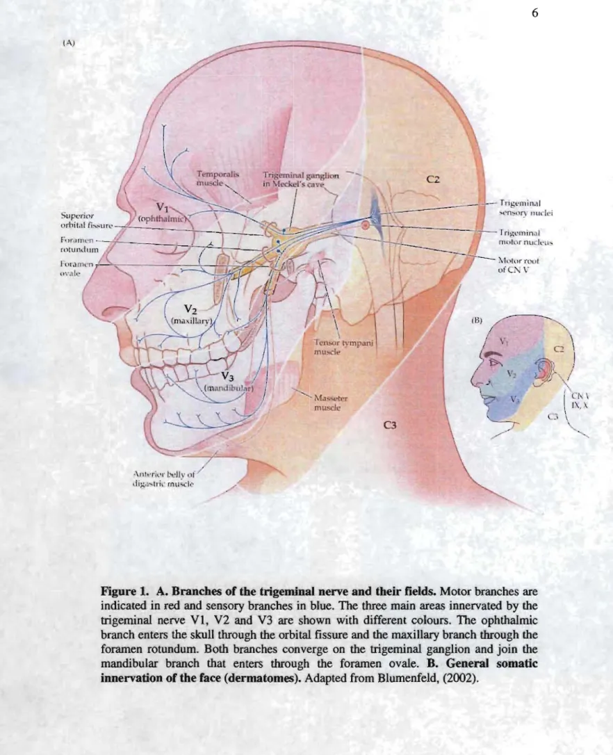
The trigeminal nerve is the largest of the cranial nerves. It is the fifth cranial nerve (nerve V). The name 44trigeminal" cornes from the fact that it has three major branches: ophthalmic (VI), maxillary (V2) and mandibular (V3). These three branches emerge from the trigeminal ganglion (also called ganglion of Gasser) which is the equivalent to the spinal cord ganglia. The trigeminai nerve is a mixed nerve that has both sensory and motor functions. The ophthaimic and maxillar branches are entirely sensory, while the mandibular branch contains both motor and sens ory fibers. The mandibular branch leaves the skull through the foramen ovale (Fig. 1).
2.1.2 The motor branches of the trigeminal nerve
The motor fi bers in the mandibular branch project to the following muscles: masseter, temporalis, medial and laterai pterygoides, tensor veli palatine, mylohyoides, the anterior belly of the digastric and tensor tympani. With the exception of tensor timpani, aU these muscles are involved in mastication (Fig. 1).
(A) ·\nlt-rl,'r l'div ,If dig,,'-tr;,-mu~ -1-' Temporal~ mw;cll' ... Ma ... • r mus...!\.' C3
Figure 1. A. Branches of the trigeminal nerve and their fields. Motor branches are indicated in red and sensory branches in blue. The three main areas innervated by the trigeminal nerve VI, V2 and V3 are shown with different colours. The ophthalmic branch enters the skull through the orbitaI fissure and the maxillary branch through the foramen rotundum. Both branches converge on the trigeminal ganglion and join the mandibular branch that enters through the foramen ovale. B. General somatic innervation of the face (dermatomes). Adapted from Blumenfeld, (2002).
7
In mammals, the anterior digastric and mylohyoid muscles open the jaw, whereas the
temporalis, masseter and pterygoides muscles close the jaw (Blumenfeld, 2002). The motoneuron cell bodies are found in the trigeminal motor nucleus in the brainstem (NVmot). Motoneurons of distinct muscles of the jaw are distributed in distinct locations within NVmot. The ventromedial motoneuron pool contains cells that innervate jaw-opening muscles (digastric and mylohyoideus) whereas the dorsolateral motoneuron pool innervates jaw-closing muscles (temporalis and masseter). The medial- and lateral pterygoides motoneuron pools are located between the other two (Weijs & Datuma, 1981; Jacquin et al., 1983a; Mizuno et al., 1983; Lynch, 1985).
2.1.3 The sensory branches of the trigeminal nerve
Trigeminal sensory fibers are found in all three branches and carry information from epithelial mechanoreceptors (in the skin, hair and mucosa), periodontal mechanoreceptors (in the periodontal ligaments), temporomandibular joint afferents (in the temporomandibular capsule), muscle afferents (primary and secondary muscle spindles, Golgi tendon organs and thermal and nociceptive afferents. Most of the cell bodies of these primary sensory fibers form the trigeminal ganglion (Fig. 1). However, all afferents that innervate the jaw-closing muscle spindles (primary and secondary) and a subpopulation of periodontal afferents have their cell bodies in the brainstem in the trigeminal mesencephalic nucleus (NVmes) (Gottlieb et al., 1984) (Fig. 2).
NVspi _ _ ....
NVspc --T"'-4tl~It
-1-Mesencephalic
trigeminal nucleus, NVmes
Trigeminal motor nucleus, NVmot
Main sensory nucleus, NVsnpr Ophtalmie branch Maxilar branch Mandibular branch Gasserian or trigeminal ganglion
Spinal trigeminal nucleus, NVsp
Figure 2. Sehematie figure depieting trigeminal sens ory (blue) and motor (red) fibers and their associated nuclei (View from a sagital eut aeross the brainstem). The main sensory nucleus (NVsnpr), the spinal nucleus (NVsp), subdivided in three regions (NVspo, NVspi, NVspc) and the mesencephalic nucleus, NVmes receive sensory inputs. NVmes sends efferents to the trigeminal motor nucleus NVmot, that contain motoneuron cell bodies for each group of muscles involved in mastication. Adapted from Cajal, S. Ry, (1909).
The central branches of neurons of the trigeminai ganglion enter the Pons ventrally via the trigeminai sensory root and then bifurcate in ascending and descending branches that are also called the trigeminai tract. Large-diameter (myelinated) primary sensory neurons mediating fine touch and dental pressure travel through the ascending branches of the trigeminai tract and project to the main sensory nucleus (NVsnpr) in the brainstem. Medium and small-diameter (unmyelinated) primary sensory fibers that convey crude touch, pain and temperature sensations enter the lateral Pons through the descending branch of the trigeminal tract (spinal tract) and project to the ipsilateral trigeminal spinal nucleus (NVsp) in the brainstem (Fig. 2).
2.2 THE TRIGE MIN AL SYSTEM
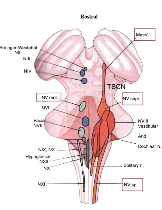
The trigeminal sensory nuclear complex (TSNC) is formed by the NVmes, the trigeminal main sensory nucleus (NVsnpr) and the trigeminal spinal nucleus (NVsp) (Mees sen & Olszewski, 1949; Olszewski, 1950) (Fig. 3). NVsnpr is the brainstem analog of the dorsal colurnn nuclei, whereas NV sp is an extension of the dorsal hom of the spinal cord. NVsp is continuous with NVsnpr and 1 runs caudally to the upper segments of the spinal cord (C2). NVsp is subdivided into nucleus oralis (NVspo) interpolaris (NVspi) and caudalis (NVspc), and NVspo is furtherly subdivided in two cytoarchitectonic distinct regions from rostral to caudal: NVspo y and NVspo ~
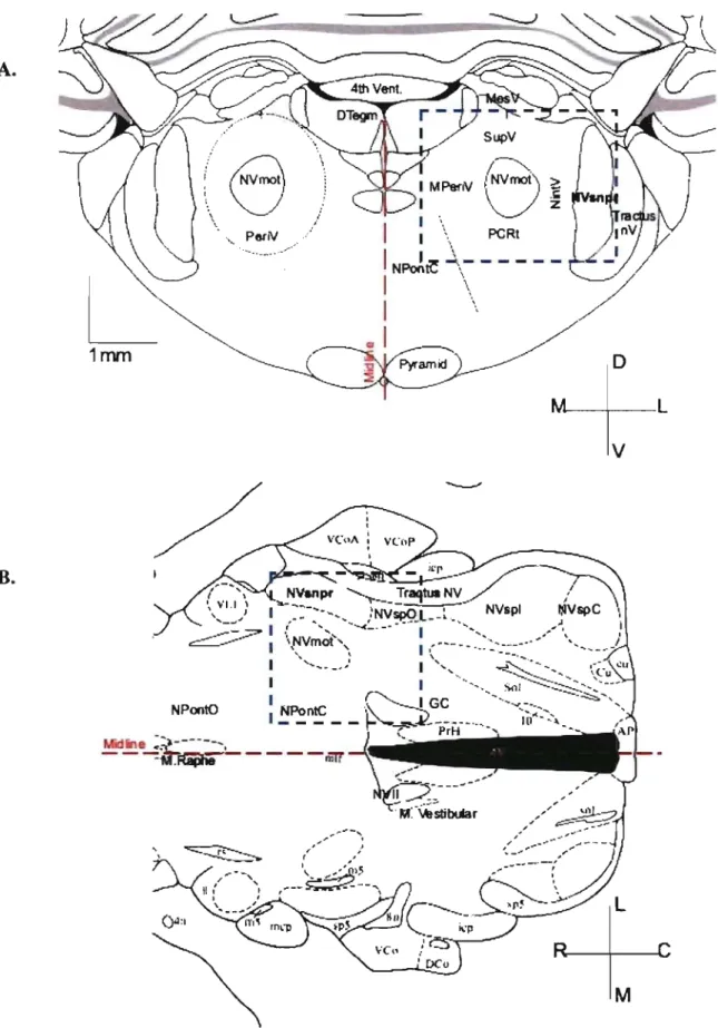
(Eisenman et al., 1963). Both nuclei, NVsnpr and NVsp are bordered laterally by the trigeminal tract and medially by the parvocellular reticular formation (PCRt) (Fig. 4A). NVmes is located dorsoanteriorly to NVmot and lies in the upper to middle regions of the Pons extending from the superior colliculus to the caudal edge of NV snpr. NV mot is located medial to NVsnpr and lateral to nucleus Pontis Caudalis (nPontC) (Fig. 3). A shell of the parvocellular reticular formation (PeriV) surrounds NVmot and is subdivided into several regions: The supratrigeminal nucleus (SupV) dorsal to NVmot, the intertrigeminal region (NintV) between NVmot and NVsnpr, the medial PeriV (mPeriV) located medial to NVmot and PCRt ventral and caudal to NVmot (Paxinos &
Watson, 1982) (Fig. 4A). nPontC lies in the medial reticular formation bordering PeriV (Fig. 4B).
Erdinger-Westphal NIII NIII NIV Hypoglossal NXII NX NXI Rostral _ _ ----\_ NVIII Vestibular And ,...,.+-r- -- Solitary n. Caudal
Figure 3. The trigeminal nuclear complex. Horizontal view of the
brainstem seen from top of cerebellum (The cerebellum itself has been removed and we only see the peduncles and the brainstem structures beneath). We observe how the trigeminal sens ory complex (orange) is organized in relation to other trigeminal nuclei (red, blue, green). Adapted from Kandel et al. (2000).
A.
B.
Pe~'iV 1 1 1 \ \ PCRt 1NPoÂ~
j",-l
'"
1 \L
"
~
1 \,~
.
1
1nvn ~,r , :2 ~ , \ "'1.1] 1~~
I
NPontO 1 1 1 1 I~~n~ __ Midli'le ~;~---, --~M.Râêîh8----mrr
MFigure 4. NVsnpr and surrounding structures A. transverse and
B. horizontal sections of the rat brainstem. In blue, region described
2.3 THE MASTICATORY CENTRAL PATTERN GENERATOR, CPG
2.3.1 Definition
The first attempts to describe the rhythmic process of mastication date from the experiments performed by Sherrington at the beginning of the 20th century. To Sherrington, rhythmic mastication could be explained by an alternate activation of two simple brainstem reflexes, a jaw opening reflex triggered by pressure on the teeth or tactile stimulation of perioral areas, and a jaw-closing reflex triggered by stretching of the jaw closing muscles during the opening. Both reflexes would follow each-other indefinitely until an external command interceded to stop the cycle (Sherrington, 1917) The idea of rhythmic orofacial-movements proper to mastication being patterned by central motor commands rather than as a consequence of pure reflex sensory arcs was put forward by Bremer, (1923). Later, Dellow & Lund (1971) demonstrated that the isolated brainstem can generate rhythmic coordinated activity ressembling mastication even in absence of afferent inputs and postulated the existence of a central pattern generator (CPG) for mastication. A CPG is an assembly of neurons that can produce and maintain a rhythmic pattern of activity in the absence of sens ory feedback or descending central commands because of intrinsic properties and/or connectivity (Rossignol & Dubuc, 1994). CPGs are basic functional elements of essential processes such as respiration (von Euler, 1983), locomotion (Grillner, 1985) and mastication (Lund & Dellow, 1969).
Feldman & Ellenberger (1988) suggested that the respiratory CPG could be subdiv~ded functionally into two different groups of neurons generating separate stages
of the motor pattern. A first CPG component would produce the rhythm of respiration (the timing and length of the cycle); whereas a second component wou Id shape the motoneuron output (duration and amplitude of EMG bursts). Other evidence suggested that this could also be the case for other complex mammalian motor rhythms such as locomotion (Rossignol et al., 2006) and masticatio,: (Lund, 1991 for a review).
In the rabbit (Lund et al., 1984) it was shown that the rhythm and the motoneuron output can be varied independently from each other. Increases in intensity of repetitive stimulation of different points of the cortical masticatory area in anaesthetized preparations could either increase the frequency of the rhythm without modifying the pattern of jaw movement or vice-versa. However, more recent evidence suggest that both functions may be accompli shed by a single population of neurons forming the core of the CPG (Athanassiadis et al., 2005a; Brocard et al., 2006).
15
2.3.2 Localization of the masticatory CPG
It was postulated more than 30 years ago that the fundamental pattern of mastication, consisting of rhythmic opening and closing of the jaws, with associated repetitive. movements of the tongue, cheeks and lips, could be generated by an assembly of neurons in the lower pons and the medulla (Lund & Dellow, 1969; 1971). Early experiments showed that mastication could be obtained in precollicular decerebrate animaIs (Bazett & Penfield, 1922), indicating that descending central commands were not essential for the initiation of the rhythmical motor output.
Complementary sets of experiments using intraoral anesthetics in human subjects (Schaerer et al., 1966), extensive orofacial denervation in rabbits (lnoue et al., 1989) or selective les ions in NVmes (Goodwin & Luschei, 1975), showed that peripheral inputs were not essential for mastication (Lund, 1991 for a review). Dellow and Lund further demonstrated in 1971 that the isolated brainstem alone suffices to generate the typical rhythmic motor output of mastication. Stimulation of the corticobulbar tract in the brainstem of paralyzed and decerebrated animaIs could produce aIternating bursts of activity in jaw-closing and jaw-opening motoneurons and in hypoglossal motoneurons. The rhythmic motor output of mastication did not dependent on the cardiac or respiratory rhythms.
2.3.3 Early models of the masticatory CPG
Nakamura's group was the first to propose a sequential model of the CPG for mastication that involved neurons within the medial bulbar reticular formation (MRF)
.
between the facial motor nucleus (NVII) and hypoglossal nucleus (NXII) (Nozaki, etal. 1986a,b; Nakamura & Katakura, 1995). They suggestèd that the initial step in generation of a masticatory rhythm was the excitation of a group of neurons in the dorsal pole of the paragigantocellular reticular nucleus (dPGC) which receives direct projections from the contralateral cortical masticatory aréa in the guinea pig. They proposed that dPGC neurons project directly to the oral portion of the gigantocellular reticular nucleus, GCo, an area that contains neurons that burst rhythmically in phase with mastication (Fig. 5). After studying the latency of the local field potentials evoked in dPGC and GCo by cortical stimulation, Nakamura's group reported that the masticatory rhythm started with tonie excitation of neurons in dPGC. This area then drove the rhythmic firing of neurons in GCo (Nozaki et al., 1986b). They added a third group of cells in the circuit, a group of intemeurons within the caudal parvocellular reticular formation (cPCRt) located caudal to dPGC and GC, and next to NXII. This last group of cells projected to NVmot to control the trigeminal motor output (Nozaki
... : - . -....
---
...
,.~
...
;;;;
...
-
-
-
-
-
--
-
..
NV
~
••
"
~~
cPCRtFigure 5. Early CPG model. In Nakamura's model, the descending central command travels through the corticobulbar path to the paragigantocellular nucleus and its dorsal portion, dPGC which then aetivate neurons in the gigantocellular nucleus, GCo and GCe, that projeet to the more caudal parvocellular reticular formation, ePCRt. The later aeting as the last relay to the motor nucleus. Adapted from Nakamura & Katakura (1995).
Nozaki et al. (1986a) observed that the CPG for mastication was organized bilaterally in the guinea pig. When the two sides of the caudal pons and medulla were separated, each hemisection was still able to generate an unilateral pattern of mastication. Anatomical evidence showed that commissural axons connect the two half sides of the masticatory CPG (Nozaki et al., 1991; Chandler & TaI, 1986; Landgren et al., 1986; Bourque & KoIta, 2001). Chandler & TaI (1986) also demonstrated that a transection at the level of NXII disconnecting cPCRt from more rostral areas did not abolish mastication indicating that cPCRt was not essential for mastication. On the basis of these observations, Lund (1991) suggested a revis ion of Nakamura's model, and proposed that GCo projected to neurons of the rostral parvocellular reticular formation, PCRt pars a adjacent to NVspo y. However, further evidence indicated that the areas that are essential for rhythm generation are located more rostraly in the brainstem (Lund, 1991 for a review). The elimination of caudal GC by transection does not disrupt mastication in vitro (Kogo et al., 1996; Kogo et al., 1998; Katakura, 1999). Using en bloc brainstem preparations in vitro, Tanaka et al. (1999) showed that the minimal portion of brainstem required to initiate rhythmic masticatory-like motor-output could be reduced to the region between the rostral border of NVmot and the rostral border of NVII. When considering medial to lateral dimensions, the preparation extends 400j..lm from the midline to the lateral border This region inc1udes NVsnpr, NVspo-y, NVmot, PeriV inc1uding PCRt and a portion of NPontC (Fig. 4).
19 The majority of the projections to trigeminal motoneurons arise from these areas, which all show increased c-Fos-like immunoreactivity after bouts of rhythmic mastication (Athanassiadis et al., 2005b), and which all contain neurons with bilateral projections to NVmot indicating that they may participate to the bilateral coordination of the jaw (Mizuno et al., 1983; Landgren et al., 1986; Rokx et al., 1986; Appenteng
& Girdlestone, 1987; Appenteng et al., 1990; Donga & Lund, 1991; Li et al., 1993; Li
et al., 1995; Westberg et al., 1995; Li et al., 1996; KoIta et al., 2000). In addition all of these areas contain neurons that fire rhythmically in phase with the trigeminal motoneurons during cortically induced mastication in the anaesthetized and paralysed rabbits, guinea-pigs and rats (Donga & Lund, 1990; Donga et al., 1990; Inoue et al., 1992; Westberg et al., 1998; Tsuboi et al., 2003).
However, it is unc1ear whether rhythmic firing in these neurons results from intrinsic properties or rhythmic synaptic inputs originating elsewhere. Neurons in Peri V and PCRt do not seem to possess intrinsic rhythm generating properties when studied in vitro (Bourque & KoIta, 2001), suggesting that these areas could be recruited by excitatory or inhibitory inputs rather than participate in the process of generating the masticatory rythm per se. In contrast, NV snpr neurons have membrane conductances that produce plateau potentials. They have intrinsic rhythmic bursting properties as was shown in in vitro experiments in brainstem slice preparation of gerbils and rats (Sandler et al., 1998; Brocard et al., 2006). This feature is found in neurons forming endogenous oscillators observed in other rhythm generating circuits (Calabrese, 1995; Brocard et al., 2006). Additionally, electrophysiological experiments conducted in vitro showed that NVsnpr neurons have· synaptic connections with
.sil
\
ipsilateral NVmot, NPontC, PeriV and PCRt (Arsenault et al., 2004; Athanassiadis et
al., 2005a) and with contralateral NVmot (Donga & Lund, 1991).
In addition, Tsuboi et al. (2003) showed that about one third of the neurons found in dorsal NVsnpr fire rhythmically in phase with trigeminal motoneurons (MNs) during fictive mastication. These evidence suggest that NVsnpr is an area that may contain the core of the masticatory CPG .
21 3. THE MAIN SENSORY NUCLEUS, NVsnpr
3.1 NVsnpr morphology and localization
The NV snpr is a rather compact nucleus with cells of uniform size and shape. It has a kidney-like shape in coronal section and is ovoid in the horizontal plane. It has a rostrocaudal extent of about 1.2mm and is found approximately 4.4 to 5.6mm rostral to the obex in rat. It is located 2.8 to 3.4mm from the midline (Paxinos & Watson, 20,04) and measures 1.8mm from dorsal to ventral (Paxinos & Watson, 2004) (Fig. 4A, B). NVsnpr starts roughly at the same lev el that NVmot in the rostro-caudal axis although it extends more caudally (l400!!m in the cat) (Eisenman et al., 1963; Marfurt &
Rajchert, 1991). NVsnpr ends where the fibres of NVII travel across the medulla and is followed caudally by NVspo y. Classical morphological studies have reported cytoarchitectonic differences between the dorsal (NVd) and ventral (NVv) parts in the cat (Eisenman et al., 1963; Shigenaga et al., 1986a). According to Shigenaga et al., (l986a) NVd has a higher celldensity than NVv. However, this morphological distinction was not observed in the rat (Ide & Killackey, 1985). The average diameter of the cells within NVsnpr ranges from 1OJ..UIl in the rat, and Il to 13J..UIl in platypus and echidna, two australian monotrema (Ashwell et al., 2006) to 15 to 30~m in the cat
(Eisenman et al., 1963). NVsnpr can be distinguished from other nuclei of the trigeminal sensory complex by the high density of small to medium sized round or ovoid neurons. NVsp oralis has conversely large polygonal neurons (22 to 40J..UIl diameter) and its neuropil is interrupted by distinctive rostrocaudal oriented bundles of myelinated axons (Ide & Killackey, 1985).
22
3.2 NVsnpr connectivity
3.2.1. Cortical inputs
Anatomicai and electrophysioIogicai studies conducted by Hernandez-Peon (1955) and BrodaI et al. (1956) showed cortical inputs to NVsnpr in adult cats. These observations were supported by les ion studies of the sensory-motor cortex conducted by Wooisey (1955) and Gobei et al. (1971) who observed degenerating corticofugal endings in NVsnpr using electron microscopy. The corticofugal endings were characterized as small myelinated fibers (1!lm diameter), containing few synaptic vesicles and establishing synapses mainly on dendrites. Few synapses were reported on axons and none on somas. These results confirmed the observations of BrodaI (1956). In 1985, Yasui et al., made HRP injections in the cat motor cortical masticatory area and observed Iabelling in the median NV snpr as one of the projection targets of the descending pathway. Labelling was aiso observed in the dorsal and median portion of NV spo and median NV spi.
3.2.2. Peripheral inputs: Somatotopy of NVsnpr
Primary sensory fibers in the spinal trigeminal tract of nerve V are organized somatotopically in the cat (Marfurt, 1981) and in the rat (Gregg et al., 1973; Jacquin et al., 1983a, b; Shigenaga et al., 1988). Sensory fibers from the ophthalmic division are
found ventrolaterally while those from the mandibular division are found dorsomedially. Fibers from the maxillary division are found between the other two (Fig. 6A). Darian Smith et al. (1963a, b) reported that neurons in NVspo and NVsnpr
in cat conserve the somatotopy of the sensory fibers and form a clear somatotopic map. This consists of an inverted representation of the face in the dorso-ventral aspect of the nucleus, with the mandible region represented dorsally, the ophthalmic region represented ventrally and the maxillary between both (Fig. 6B). Shigenaga et al.
(1986a, b; 1988) studied exhaustively the anatomical and functional projections of the primary afferents in the cat using horse radish and phaseolus vulgaris leucoaglutinin
injections in peripheral nerves innervating oral and facial structures. They confirmed the inverted representation of the face on the dorso-ventral aspect of the nucleus and showed that the dorsal part of NVsnpr is the principal target for intraoral afferents, whereas the ventral part receives both intraoral and facial afferents (mental, infraorbital and frontal nerve afferents). Infraorbital afferents end up in the ventral most part of the nucleus and lingual afferents are located in the dorsalmost part of NVsnpr.
A.
GanglionB.
~~~
..::~
--'~b PonsOphlholmic Moxi/lory Mondibulor
Figure 6. Somatotopic organization of primary atTerents within the trigeminal ganglion and NVsnpr. A. HRP tracing studies reveal that the three trigeminal branches project to distinct areas of NVsnpr in the dorso-ventral axis. Adapted from Kerr et al.
(1968) B. Inverted-face representation. Note that the afferents projecting from the mandible, Md are dorsal while the maxillary, Mx are in the middle of the nucleus. We can additionally see cytochrome oxidase staining of barrel-like spots in the ventral part of the nucleus due to the clustered projections from vibrissa and sensory hair (large black polygons and small black dots, respectively). Adapted from Erzururnlu et al. (2006) and
Arvidsson, (1982) also reported an inverted-face representation in NVsnpr and revealed in addition the localization of vibrissae columns organized in 'barrel-like' s~ructures when observed in transverse sections of the brainstem. Although the
dorso-ventral somatotopic representation of the face in NV snpr has been generally accepted, there is sorne controversy about the organization of NVsnpr in the rostro-caudal axis. Eisenman et al. (1963) found that the majority of peri oral touch cells in the rostral half of NVsnpr had their receptive fields on the lips, while the majority of the cells found in its caudal half had receptive fields located in the hairy skin surrounding the mouth and more remote areas. This group also reported that a larger number of cells with receptive field on whiskers were located on the rostral half of NVsnpr. Other early experiments demonstrated that the face and oral cavity were represented somatotopically but disproportionately in all rostrocaudal levels of the entire sensory trige minaI complex (Kruger & Witkovsky, 1961a; Kruger et al., 1961b; Kruger &
Michel, 1962a, b; Kerr et al., 1968). Shigenaga, (1986a) reported that each area of the anterior face and oral cavity is indeed represented in rostrocaudally oriented columns that run parallel from NV snpr to NV spc. These results were in agreement with the electrophysiological works of Kruger & Michel (1962a) and anatomical works by Arvidsson et al. (1982).
3.2.3. Neurochemical sludies
A number of studies have tried to characterize the neurochemical content of fibers projecting to NVsnpr (Clements & Beitz, 1991; Bae et al., 2000; Waite et al.,
2000). Bae et al. (2000) used HRP-conjugated cholera toxin B-subunit to label primary afferents in the trigeminal ganglion together with immunostaining against glutamate in the trigeminal main sensory complex .. They suggested that large-caliber primary afferent neurons use glutamate as a neurotransmitter. These synapses were subject to pre-synaptic modulation by GABAergic fibers and the authors suggested that this would specially apply for NVsnpr where a higher degree of pre-synaptic control might be required for sharpening spatial somatosensory information. These observations were corroborated by the electron-microscopy studies of Clements & Beitz (1991). Waite et al. (2000) studied anatomical and functional development of the trigeminal sensory complex in rats from age E13 to P6. They found that responses to stimulation of the trigeminal ganglion in NVsnpr neurons were NMDA and AMPA dependent. Additionally GABA-A excitatory responses were also observed and blocked through bicuculline application. GABA-A responses were observed at all ages but were maximal from E20 to Pl, which is consistent with the development of whisker-related patterns (Landers & Zeigler, 2006 for a review).
27
3.2.4 Inputs to NVsnpr from other nuclei in the brainstem
Extracellular injections of biocytin and intracellular recordings in vitro in
conjunction with micro-stimulation were used to show that aIl areas of PeriV, PCRt and NPontC project to NVsnpr monosynaptically (Bourque & Koita, 2001; Athanassiadis et al., 2005a). The majority of responses detected in NVsnpr (80%) could be blocked by DNQX and APV (antagonists of AMPA and NMDA receptors respectively) indicating that these projections are glutamatergic. Fewer stimuli (20%) eiicited IPSPs, and these were sensitive to GABAergic and glycinergic antagonists (Bourque & KoIta, 2001 ; Athanassiadis et al., 2005a). This observation is consistent with immunohistochemical studies conducted in rat and rabbit (Li, et al., 1996; Turman & Chandler, 1994a,b; KoIta et al., 2000) which showed a mixed population of excitatory and inhibitory neurons in PeriV. In the study conducted by Athanassiadis et al. (2005a), NVmot stimulation also resulted in a mixture of excitatory and inhibitory evoked responses in NV snpr. The latency of many NV mot evoked responses fell within the monosynaptic range. Since motoneurons in NVmot lack recurrent axon collaterais and are exclusively cholinergic (Lauterbom et al., 1993; Ichikawa &
Shimizu, 1998; Saad et al., 1999) these responses were attributed to projections from intemeurons within the nucleus (Sessle, 1977; Ter Horst et al., 1990). More recently it has been shown that NVmot contains a mixed population of excitatory (glutamatergic) and inhibitory (GABAergic and glycinergic) intemeurons (Li, et al. 1996; KoIta, et al. 2000; McDavid et al., 2006).
NVsnpr also receive projections from NPontC, and one half of the responses evoked by stimulation of the dorsal NPontC occurred at monosynaptic range (Bourque &
Kolta, 2001; Athanassiadis et al. 2005a). Projections from neurons in NPontC and PCRt of the rat have been also described using HRP and phaseoulus vulgaris leucoaglutinin tracers (Shammah-Lagnado et al., 1987; Ter Horst et al., 1991; Shammah-Lagnado et al., 1992).
3.3. Outputs from NVsnpr
3.3.1 Projections to higher centers: Thalamus
The NVsnpr has been classically described as a sens ory relay to the thalamus. Later it has· been reported that secondary neurons in the nucleus send collaterals to reticular areas such as PeriV (Jacquin et al., 1982; Yoshida et al., 1998; Zerari-Mailly
et al., 2001), NVmot (Yoshida et al., 1994; Buisseret-Delmas et al., 1997; Koita et al., 2000), NVmes or higher areas such as the cerebellum (Huerta et al., 1983; Steindler, 1985). In fact, the most important projections from NV snpr travel to the thalamus in the two reticulothalamic pathways (Blumenfeld, 2002). The first pathway contains axons from NV snpr neurons that carry somesthetic information from the face. It
.
constitutes a large projection arising in the ventral two thirds of the nucleus. The neurons give off ascending axons that ascend with the medial lemniscus, cross the midline to the contralateral side of the brainstem and then project to the ventral-posterior-median nucleus of the thalamus, VPM. This has been confirmed in retrograde studies in the cat (Smith, 1975) and in the dog and pig (Michail &29 Karamanlidis, 1970; Brodai, 1981 for a review). Darian-Smith et al. (1963b) performed electrical stimulation of the contralateral arcuate nucleus of the thalamus in the cat and observed both antidromic and trans-synaptic responses in NVsnpr. Medial-lernniscal neurons were also identified through stimulation, and responses were observed throughout NV spo and the caudal part of NV snpr. These neurons form an homogeneous group located in the dorsolateral part of each nuclei. This data was confirmed by further anatomical studies (Mizuno, 1970; Shigenaga et al., 1979,
1983). The second pathway arises from neurons that convey proprioceptive information, touch and pressure sensation from the oral cavity, including the teeth. These axons arise from cells located in the dorsomedial third of NVsnpr and give off uncrossed axons that travel to ipsilateral VPM in the dorsal trigemino-thalamic tract (Walker, 1939; Carpenter, 1957). Torvik reported half a century ago that the dorsal NV snpr contained ascending axons in the ipsilateral reticular formation that project to the thalamus (Torvik, 1957; Mizuno, 1970). Luo & Dessem (1995) performed anatomical studies in the rat using HRP neurotracer injections in VPM and labelled neurons contralaterally in SupV, NVsnpr, NVspo, NVspi and PCRt. Steindler (1985) obtained similar results in the mouse, and observed important projections to the cerebellar cortex and deep cerebellar nuclei from NVsnpr and NVspi. This was also reported in the rat (Huerta et al., 1983).
y oshida et al. (1998) conducted HRP tracing studies in the c,at in order to determine cell morphology and local projections from cells whithin NV snpr. They found three classes of projecting neurons based on their axonal and dendritic arborisation pattern. Class la neurons were sensory neurons having an ascending stem axon and no branching. This class correspond to the thalamus projecting neurons
reported by Torvik (1957) and Mizuno (1970). Class lIa contained neurons that had both ascending axons and collaterais to lower brainstem nuc1ei, especially projecting to dorsal NV mot. Y oshida et al. suggested that these neurons could be involved intp sens ory discrimination and the jaw c10sing reflex. Class lIb neurons were local circuit neurons that only send collaterais to neighbouring brainstem structures. Neurochemical studies on thalamus-projecting neurons from NVsnpr reported that the majority of them were glutamatergic (Magnus son et al., 1987), although sorne local
branching neurons were GABAergic (Ginestal & Matute, 1993; Avendano et al.,
2005).
3.3.2 Projections to the reticular formation and NVmot
As described above, NV snpr neurons project to nearby reticular nuc1ei such as SupV, PCRt, NintV, mPeriV, and to NVmot (Jacquin et al., 1982; Yoshida et al.,
1998; Zerari-Mailly et al., 2001; Arsenault 1., 2004; Athanassiadis et al., 2005a). In
vitro electrophysiological studies conducted by Athanassiadis in a rat brainstem slice preparation showed that antidromic activation in NV snpr could be obtained through stimulation in the masseteric pool of NVmot and dorsal NPontc (Athanassiadis et al.,
2005a). This is in agreement with the unpublished observations of Arsenault et al.
(2004). They found that stimulation of dorsal NV snpr evokes PSPs in the masseteric pool of NVmot suggesting that dqrsal NVsnpr send direct projections to jaw c10sing motoneurons. PSPs could be also obtained in the digastric pool of NVmot following stimulation of aIl parts of NVsnpr, but the latency of responses elicited by stimulation of the ventral part was much shorter, suggesting that ventral NVsnpr sends direct projections to jaw opening motoneurons. These observations support the findings of Li
31 et al. (1995) who reported that NVsnpr project to jaw-opening motoneurons.
Anatomical studies using rhodamine dextran techniques combined with
immunohistochemistry have shown that at least sorne of the NVsnpr neurons projecting to NVmot are GABAergic and/or glycinergic (Li et al., 1996). Other groups have also reported the existence of glycinergic cells in NVsnpr (Ginestal & Matute, 1993; Turman & Chandler, 1994a; Rampon et al., 1996; Avendano et al., 2005)
aIthough they found more dense labellings in NV spc and NV spi. Employing similar techniques in the rabbit, KoIta et al. (2000) reported the existence of GABAergic and glutamatergic neurons among premotoneurons in dorsal NVsnpr projecting to NVmot. Similar findings were obtained by Turman and Chandler in the guinea-pig (Turman &
3.4 Evidence that NVsnpr may form the core for the CPG for mastication
As described above, sorne NVsnpr neurons project to NVmot and other structures of the lateral and medial reticular formation, and our group has raised the possibility that these cells could be involved in motor processing because they fulfill most of the requirements expected from CPG neurons (Tsuboi et al., 2003; Athanassiadis et al.,
2005a; Brocard et al., 2006). First, they receive massive inputs from the cortex and
sensory afferents (Eisenman et al., 1963; Dellow & Lund, 1971; Tsuru, et al. 1989;
Zhang & Sasamoto, 1990; y oshida, et al. 1998) and have outputs to premotorneurons
and MNs of PeriV, NVmot and NPontC (Inoue et al., 2002; Arsenault et al., 2004;
Athanassiadis et al., 2005a). Second, in vivo studies have shown that about one third of
the neurons in dorsal NVsnpr fire rhythmically in phase with trigeminal motoneurons when fictive mastication is elicited by stimulation of the CMA in paralyzed rabbits (Tsuboi et al., 2003). Third, the expression of c-Fos-like protein, a functional marker
of activity, increases in, this area during fictive mastication (Athanassiadis et al.,
2005a). Fourth, in in vitro brainstem slice preparations, many NV snpr neurons burst
and have plateau properties in the gerbil (Sandler et al., 1998) and in the rat (Brocard et al., 2006). Our group has shown that appearance of these properties coincides with
the emergence of mastication in new-born rats (Brocard et al., 2006). The bursts and
plateaux not only persist in Ca2+ free ACSF (a condition that blocks synaptic
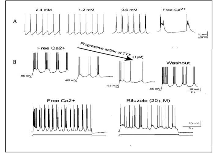
transmission), but are enhanced in these conditions indicating that this pro pert y depends on intrinsic cell-membrane properties (Brocard et al., 2006) (Fig. 7).
2.4 mM 1.2 mM A Free Ca2+ B -65 rnV -65 rnV Free Ca2+ -65 rnV 0.6 mM Free-Ca 2+ 33 20
rnvi
200 ms Washout Riluzole (20 u M) 20 mV 1 55Figure 7. Intrinsic bursting properties of NVsnpr neurons. A. Whole-cell patch-recordings from NV snpr neurons in an in vitro brainstem slice preparation showing bursting
in Ca2+ free conditions. B. Rhythmic bursting depend on a persistent sodic conductance,
INaP, as it is abolished by the local application of TTX or the more specifie antagonist Riluzole. Adapted from Brocard et al. (2006).
Removal of Ca2+ from the perfusing solution results in conversion from tonie firing to repetitive bursting in 80% of neurons (Brocard et al., 2006; KoIta et al., 2007 for a review). Intracellular application of BAPTA, a Ca2+ chelator, did not cause or prevent bursting, suggesting that the initiation of rhythmic bursting in the NVsnpr is related to changes in the extracellular space and not within the cell. Brocard et al. (2006) used a pharmacologieal approach to dissect the ionic mechanisms underlying reCUITent bursting in NVsnpr neurons. The role of small (SK) and big (BK) Ca2+-dependent K+
conductances was assessed with bath application of Apamin and Charibdotoxin respectively. These drugs did not stop the spontaneous bursting of NVsnpr neurons in Ca2+ free ACSF, indicating that neither one of these K+ conductances was essential for
bursting. Blocking K+ conductances with TEA, or the Th CUITent with ZD7288 did not prevent bursting in Ca2+ free ACSF, aIthough the burst frequency was reduced and burst duration increased by TEA. Bath perfusion of tetrodotoxin, (TTX) in Ca2+-free ACSF was used to test whether'Na+ conductances were involved in burst generation. TTX reversibly abolished bursting and plateau potentials at low doses before abolishing action potentials.
Taken aItogether, this data suggested that both the plateaux and the bursts depended on sodium conductances. Riluzole, a specific antagonist of the persistent sodium cUITent, INaP, blocked rhythmic bursting without affecting spikes, indicating that NVsnpr bursts and plateau properties were INaP dependent (Fig. 7). INaP-dependent rhythmic activities have been associated with rhythmic physiological processes such as respiration (Feldman & Smith, 1989; Del Negro et al., 2002; Rybak et al., 2003),
locomotion (Tazerart et al., 2007; Zhong et al., 2007), whisking (Cramer et al., 2007)
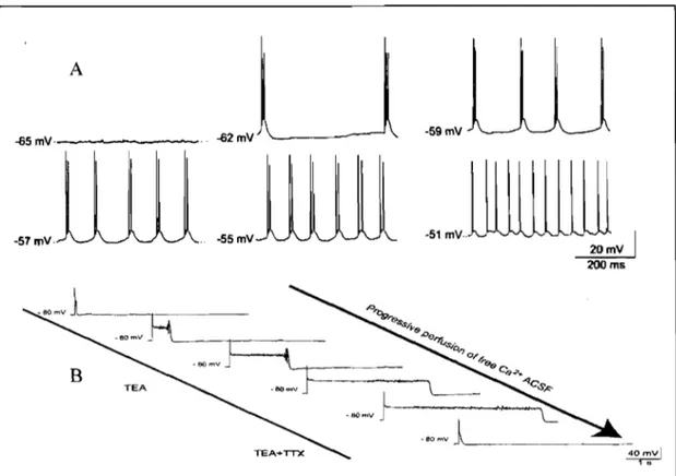
35 the INaP-mediated rhythmic bursting in NV snpr depends on the membrane potential Figure 9 shows a cell that does not burst close to the resting membrane potential (-65mV) but does burst when it is gradually depolarized. Cell firing becomes tonie at potential more depolarizedthan -51mV, indicating that rhythmic firing occurs only within a precise voltage range that correspond to activation and inactivation voltages of INaP (Azouz et al., 1996; Su et al., 2001; Del Negro et al., 2002; Darbon et al., 2004; Brocard et al., 2006). More interestingly, Brocard et al. (2006) also showed that the amplitude and duration of the INaP-mediated plateau that underly bursting depend on [Ca2+]e. An example of this is shown in figure 8.
A -a2mV -59mV -a5mV· -57 mV. -55mV
~1
mvlillUUilll
20mV 200 ms~
~80 .... V J '..
omvl~
_ _
._
B 4o,,:vlFigure 8. Intrinsic properties of neurons in NV snpr A. Rhythmic bursting is linearly dependent with the membrane potential. The cell does not burst close to resting membrane potential (-60mV) but does it when we gradually depolarize the cell. At -51 m V the cell becomes tonie indieating that the rhythmic firing oecurs only within a precise voltage range that is consistent with the activation of INaP. B. The plateau potential has also been isolated pharmaeologieally and depends on INaP conductance. The plateau also depends strongly on [Ca2
l
e• As we see the graduaI perfusion of Ca2+ free ACSF increases burst duration. Adapted from Brocard et al.37
4. THE GENERAL HYPOTHESIS
There is evidence that changes in the intensity of neuronal activity causes important fluctuations in [Ca2+]e in the spinal cord (Somjen, 1980; Murase & Randic, 1983);
cortex (Nicholson et al., 1978; Rusakov & Fine, 2003) and in hippocampal slices (Benninger et al., 1980; Cohen & Fields, 2004) for a review. As pointed out in section 1.1, prolonged (>100ms) repetitive stimulation (10-100Hz) of cortical or sensory fibers is usually required to trigger mastication in ex peri mental animaIs. Therefore, we postulate that the sustained activation of these inputs to NVsnpr causes a drop of [Ca2+]e leading to activation of INaP and to recurrent bursting. A number of mechanisms could be responsible for the local changes in [Ca2+]e: ionotropic receptors such as NMDA, Ca2+-ATPase pumps and Na+/Ca2
+ exchangers. There is evidence that NMDA receptors can contribute to synaptically- evoked [Ca2+]e depletion (Rusakov &
Fine, 2003). Glial cells are also good candidates for [Ca2+]e depletion, and are sensitive
to a number of neurotransmitters, such as glutamate, GABA, acetylcholine, and A TP or ions such as K+ released during neuronal firing (Verkhratsky et al., 1998; Grosche et
al., 1999; Grosche et al., 2002). Other projects being conducted in our laboratory are designed to dissect the contribution of glia and neurons to changes in [Ca2+]e. This study investigates whether long lasting repetitive stimulation of sensory afferents (trigeminal tract) elicits rhythmic bursting in NVsnpr neurons under physiological levels of [Ca2+]e and evaluate the role of NMDA receptors in the process of
38
5. HYPOTHESIS OF THIS STUDY
a) Repetitive but not single activation of sensory inputs to NVsnpr in physiological [Ca2+]e can elicit rhythmic bursting.
b) NMDA receptors do play a role in this process.
6. THE OBJECTIVES OF THIS STUDY WERE:
a) First to confirm that bursting in NVsnpr'neurons depends on age and [Ca2+]e.
b) To test in a brainstem slice preparation, the efTects of single and repetitive stimulation of the sensory tract on the firing pattern of NVsnpr neurons under [Ca2+]e where only few neurons burst spontaneously.
c) Test the efTects of NMDA receptors agonist and antagonist on the firing pattern of NV snpr neurons.
39
SECTION II
40
T. Freund
« Effect of the stimulation of sensory inputs on the firing of neurons of the trigeminal main sensory nucleus in the rat»
A. P. Bernierl, J.P. Lundl,2 and A. Koltal,3
1 Groupe de recherche sur le Système Nerveux Central du FRSQ, Université de Montréal, Montréal, Québec CANADA
2 Faculty of Dentistry, McGill University, Montréal, Québec, CANADA
3 Faculté de Médecine dentaire, Université de Montréal, Montréal, Québec, CANADA
Keywords : central pattern generation, main sens ory nucleus, peripheral stimulation Correspondence:
Dr A.Kolta
Université de Montréal
GRSNC, Pavillon Paul-G-Desmarais c.P. 6128, Suce. Centre-Ville
Montréal, Québec, CANADA H3C 3JT
Number or words
Manuscript without figures: 6333 Manuscript with figures: 13533 Abstract: 247 Introduction: 484 Number of pages 28 Number of figures 12 Tel: (514) 3437112 Fax: (514) 3432111 email:
41
ABSTRACT
Mastication may be triggered by repetitive stimulation of the cortex or of sensory inputs, but is patterned by a central pattern generator (CPG) in the brainstem. This, CPG may lie in the dorsal part of the principal trigeminal sensory nucleus (NVsnpr) where neurons burst repetitively when the extracellular concentration of Ca2+ ([Ca2+]e)
drops (Brocard et al., 2006). Here we examine the effects of repetitive stimulation of sensory afferents in the descending tract on activity of NV snpr neurons recorded extracellularly in vitro under physiological [Ca2+]e (1.6mM). Most spontaneously
active cells had either a tonic or a bursting firing pattern. Stimulation of the tract altered burst duration and/or frequency in bursting cells and firing frequencies in most tonic cells. In 28% of the latter, the firing pattern switched to rhythmic bursting. This effect could be blocked by APV and mimicked by local application of NMDA. Rhythm indices (Sugihara et al., 1995) ca1culated to assess rhythmicity were negative (non-rhythmic) in aIl cases before stimulation and significant (2: 0.01; rhythmic) after stimulation. The mean and median (±SE) bursting frequency were 8.32±0.72Hz and 6±0.5Hz respectively. In 6 cases where firing switched to rhythmic, two units were recorded and cross correlation analysis showed that, in aIl pairs, the units became synchronized after stimulation. Optimal stimulation parameters for eliciting rhythmic bursting consisted in 500ms trains of pulses delivered at 40 - 60Hz. Together, our results show that repetitive stimulation of sensory afferents in vitro can elicit
INTRODUCTION
The basic pattern of mastication is generated by a central pattern ,generator (CPG) located in the brainstem in response to tonic inputs from higher centers or from trige minaI sensory afferents (Dellow & Lund, 1971; Lund & Dellow, 1971). Neither input is essential because CPGs can pro duce repetitive movements in absence of inputs from either the superior centers or sensory afferents (Rossignol et al., 2006 for a review). Tonic stimulation of either type of input can activate the masticatory CPG even in paralysed animaIs (fictive mastication) (Sumi, 1969; Dellow and Lund, 1971; Lund and Dellow, 1971), and this is not unique to mastication, since activation of sensory afferents and descending inputs elicit locomotion (Grillner et al., 1981; Fleshman et al., 1984; Rossignol, 2000). However, the cellular mechanisms by which sustained activation of these inputs is conv,erted into a rhythmic output by the CPG are unknown. Our previous work suggests that the dorsal part of the trigeminal main sensory nucleus (NVsnpr) may form the core of the masticatory CPG (Tsuboi et al., 2003; Athanassiadis et al., 2005a; Brocard et al., 2006; Kolta et al., 2007 for a· review). NVsnpr receives massive inputs from the masticatory area of the cortex and from trigeminal sensory afferents, and neurons of its dorsal part project directly to the trige minaI motor nucleus (Travers & Norgren, 1983; Li et al., 1993; Yoshida et al., 1998; Kolta et al., 2000). The expression of c-Fos (a neuronal marker of activity) increases in neurons of dorsal NVsnpr after bouts of fictive mastication (Athanassiadis
et al., 2005b), and about a third of neurons recorded in there fire rhythmically in phase with trigeminal motoneurons during fictive mastication (Tsuboi et al., 2003). These rhythmical neurons receive inputs from sensory receptors that provide important feedback during mastication: intraoral touch receptors, periodontal pressoreceptors &