The role of Alpha Beta Hydrolase 6 in the neuronal control of
body weight, exercise and anxio-depressive behaviors
Anna Kristyna Franco Flores
Department of Neuroscience
Faculty of Medicine
Thesis for the grade of Master in Science (M.Sc.)
in Neuroscience
August, 2018
©
Anna Kristyna Franco Flores, 2018
ii
Résumé
L'obésité a beaucoup attiré l'attention ces dernières années en raison de son expansion mondiale et de son association à plusieurs maladies. Malgré les énormes efforts déployés par les scientifiques pour comprendre les mécanismes moléculaires et physiologiques de l'obésité, il reste nécessaire de découvrir un traitement efficace. Plusieurs études indiquent que le système corticolimbique est un modulateur potentiel de l'appétit, de l'activité physique et de l'obésité. Le système corticolimbique comprend le noyau accumbens (NAc), une région du cerveau impliquée dans la régulation de la récompense alimentaire, de l'humeur et de l'activité physique. Le NAc est principalement composé de neurones épineux à médiation GABAergique (MSN) qui jouent un rôle important dans le contrôle du bilan énergétique. Les manipulations de MSN par les endocannabinoïdes (eCB) ont une incidence sur la consommation de nourriture et l’humeur, mais des recherches supplémentaires sont nécessaires pour comprendre l’impact de la CBE sur l’activité physique et ses effets gratifiants. Le 2-arachidonoyl-glycérol (2-AG) est l'ebc le plus abondant dans le cerveau. Il est synthétisé dans le neurone postsynaptique afin d'activer le récepteur de cannabinoïde de type 1 (CB1R) situé de manière présynaptique. La modulation de l'accumulation de 2-AG est réalisée par deux enzymes: (1) la monoacyglycérol lipase (MAGL) située au niveau du neurone présynaptique afin de dégrader la masse de la 2-AG, et (2) le domaine α / β-hydrolase de la sérine hydrolase. 6 (ABHD6) localisé au neurone postsynaptique agissant en tant que contrôleur d'accès de 2-AG. Il a été démontré que la suppression du gène ABHD6 protège de l'obésité d'origine alimentaire chez la souris; Cependant, le mécanisme exact de la façon dont ABHD6 exerce cette action reste à élucider. Des travaux antérieurs de notre laboratoire ont révélé que l'inactivation virale de ABHD6 dans les neurones NAc augmente les niveaux de 2-AG, réduit la transmission des entrées neuronales de l'acide gamma-aminobutyrique (GABA), supprime la prise alimentaire et prévient l'obésité induite par l'alimentation. Bien que les interventions CB1R aient montré une efficacité dans la perte de poids, elles étaient notoirement associées à des troubles émotionnels tels que des pensées suicidaires. Par conséquent, l'inhibition de l'ABHD6 pourrait constituer une nouvelle approche thérapeutique de l'obésité sans affecter l'humeur.
Le but de cette étude est d'identifier l'impact de l'inactivation du gène NAc ABHD6 sur le poids corporel, l'alimentation, la course à la roue et les comportements anxio-dépressifs. Le knock-out conditionnel des neurones ABHD6 NAc (ABHD6NAc KO) de souris adultes a été réalisé en utilisant
une approche virale Cre-LoxP. Les souris ABHD6NAc KO ont été générées par des microinjections
iii
aux neurones. Après cela, les souris ABHD6NAc KO et les souris témoins ont été soumises à un
régime riche en graisses ou à une alimentation Control CHOW pendant 12 semaines. Nous avons mesuré le gain de poids corporel, l'apport alimentaire et les changements métaboliques à l'aide de cages métaboliques CLAMS. En conséquence, nous avons donné 5 semaines d'accès aux roues dans leurs cages pour assister aux performances de course sur roues volontaires entre les groupes. Enfin, nous avons évalué les altérations du comportement avec le test de labyrinthe surélevé et plus, le champ ouvert et la nage forcée chez des souris et des contrôles ABHD6NAc KO.
Nos résultats suggèrent que l'épuisement neuronal de l'ABHD6 dans l'NAc réduit le gain de poids corporel évoqué par un régime riche en graisses sans différence significative de consommation de nourriture entre les groupes. De plus, nous démontrons que la perte de fonction neuronale ABHD6 augmentait la dépense énergétique et empêchait l’inactivité physique volontaire de l’alimenter par une alimentation riche en graisses. En outre, nous avons également montré que les souris ABHD6NAc KO ne présentaient pas de comportement anxio-dépressif après une
exposition prolongée à un régime alimentaire riche en graisses. Pris ensemble, ces résultats démontrent que l'inhibition neuronale focalisée de l'enzyme ABHD6 dans le NAc joue un rôle important dans la régulation de l'équilibre énergétique et de l'humeur et peut donc potentiellement fournir une nouvelle stratégie de traitement de l'obésité.
Mots clés: endocannabinoïde, domaine sérine hydrolase α / β-hydrolase 6,
2-arachidonoyl-glycérol, récepteur cannabinoïde de type 1, noyau accumbens, obésité, rouage, comportements anxio-dépressifs.
iv
Abstract
Obesity caught a lot of attention in the last years because of its expansion worldwide and its association with several diseases. Despite the enormous efforts of scientists trying to understand the molecular and physiological mechanisms of obesity, there is still the need to discover an effective treatment. Several studies point to the corticolimbic system as a potential modulator in appetite, physical activity and obesity. The corticolimbic system incorporates the nucleus accumbens (NAc) a brain region involved in the regulation of food reward, mood and physical activity. The NAc is mostly composed by GABAergic medium spiny neurons (MSNs) that are important for the control of energy balance. Manipulations of NAc MSNs by endocannabinoids (eCB) affects food intake and mood however additional research is necessary to understand the impact of eCB on physical activity and its rewarding effects. 2-arachidonoyl-glycerol (2-AG) is the most abundant eCB in the brain, it is synthesized in the postsynaptic neuron to activate the cannabinoid receptor type 1 (CB1R) located presynaptically. Modulation of the 2-AG accumulation is done by two enzymes: (1) monoacyglycerol lipase (MAGL) which is located at the presynaptic neuron to degrade the bulk of 2-AG, and (2) the serine hydrolase α/β-hydrolase domain 6 (ABHD6) localized at the postsynaptic neuron acting as a gatekeeper of 2-AG. ABHD6 gene deletion has been demonstrated to be protective from diet-induced obesity in mice; however, the exact mechanism of how ABHD6 exerts this action is still to be elucidated. Earlier findings from our lab reveal that viral-mediated knockout of ABHD6 selectively in NAc neurons increases 2-AG levels, reduces gamma-Aminobutyric acid (GABA) neuronal input transmission, suppresses food intake and prevents diet-induced obesity. While CB1R interventions showed efficacy for weight loss, they were notoriously associated with emotional disturbances such as suicidal thoughts. Therefore, ABHD6 inhibition could give a novel therapeutic approach for obesity without affecting mood.
The aim of the current study is to identify the impact of NAc ABHD6 gene knockout on body weight, feeding, wheel running and anxio-depressive behaviors. Conditional knockout of ABHD6 NAc neurons (ABHD6NAc KO) of adult mice was accomplished using a viral Cre-LoxP approach.
ABHD6NAc KO mice were generated by bilateral NAc microinjections delivering CRE recombinase
or control GFP in a neuron-specific manner. After, the ABHD6NAc KO and control mice were
subjected to either high fat diet or Control CHOW feeding for 12 weeks. We measured body weight gain, food intake and metabolic changes using CLAMS metabolic cages. Consequently, we gave 5 weeks running wheel access in their cages to see voluntary wheel running performance
v
between the groups. Finally, we evaluated behavioural impairments with the elevated plus maze, open field and forced swim test in ABHD6NAc KO mice and controls.
Our results suggest that neuronal depletion of ABHD6 in the NAc diminishes body weight gain evoked by high fat diet with no significant differences in food consumption between the groups. Furthermore, we demonstrate that ABHD6 neuronal loss of function increased energy expenditure and prevented from voluntary physical inactivity induce by high fat diet feeding. In addition, we also showed that ABHD6NAc KO mice fail to show anxio-depressive behaviors after a prolong
exposure to high fat diet. Taking together, these results demonstrate that focal neuronal inhibition of the ABHD6 enzyme in the NAc plays an important role in the regulation of energy balance and mood and thus potentially can provide a new treatment strategy for obesity.
Key words: endocannabinoid, serine hydrolase α/β-hydrolase domain 6, 2-arachidonoyl-glycerol,
cannabinoid receptor type 1, nucleus accumbens, obesity, running wheel, anxio-depressive behaviors.
vi
Table of contents
Resumé………... ii
Abstract.………...iv
Table of contents………...vi
Tables and figure list………...ix
List of abbreviations....………...xii Dedication.………...xiv Acknowledgements………...xv 1. Chapter 1: Introduction...1 1.1. Obesity...1 1.1.1. Impact...1 1.1.2. Etiology...2
1.1.2.1 Regulation of energy balance...3
1.1.2.1.1 Food intake...3
2.1.2.1.1 Energy expenditure ...4
1.1.3. Brain controls of food intake and energy expenditure...5
1.1.2.1 Hypothalamus and hindbrain...7
2.1.2.1 Extra-hypothalamic control of energy balance: corticolimbic system...9
1.1.4. Brain control of mood...11
1.1.2.1 Anxio-depressive behaviors...12
1.2. Endocannabinoid system...13
1.2.1. Generalities...13
1.2.2. Endocannabinoid system in the brain...15
1.1.2.1 Alpha/beta hydrolase domain 6 (ABHD6) ...17
2.1.2.1 Implications of ABHD6 on energy balance and mood...18
1.3. Objectives...23
vii 2. Chapter 2: Methodology...24 2.1. Animals...24 2.2. Stereotaxic surgery...24 2.3. Diets composition...25 2.4. Metabolic assessment...26
2.4.1. Comprehensive Lab Animal Monitoring System (CLAMS)...26
2.4.2. Echo MRI...27
2.5. Voluntary wheel running...27
2.6. Locomotor activity measurements...28
2.6.1. Amphetamine locomotor sensitization...28
2.6.2. D1 receptor-induced locomotor stimulation...28
2.7. Anxio-depressive behaviors...28
2.7.1. Elevated Plus-Maze ...28
2.7.2. Open Field Test...30
2.7.3. Forced swim Test...30
2.8. Molecular assestments...30
2.8.1. Real-Time quantitative PCR (q-PCR) for ABHD6 depletion…...30
2.9. Statistical analysis...31
3. Chapter 3: Results...32
3.1. Relative mRNA expression of ABHD6 in the NAc...32
3.2. Neuronal ABHD6 depletion in the NAc protects against diet-induced obesity and metabolic changes resulting from high-fat diet...32
3.3. Neuronal ABHD6 depletion in the NAc rescues voluntary physical inactivity phenotype elicited by HFD feeding...33
3.4. Neuronal ABHD6NAc KO does not impact voluntary wheel running on chow diet, however, neuronal ABHD6 depletion in the NAc prevents rebound caused by HFD feeding………...33
3.5. Neuronal ABHD6NAc KO increased locomotor-stimulating effects caused by amphetamine; however, these effects are not related to a higher activation on dopamine D1 receptor...34
3.6. Depletion of ABHD6 from neurons in the NAc attenuates HFD-induced anxio-depressive behaviors...34
viii
4. Chapter 4: Discussion...45
4.1. Effects of ABHD6 loss-of-function within the corticolimbic system on metabolism and physical activity...45
4.2. Lack of ABHD6 in the NAc positive modulates mood behaviors on HFD...48
4.3. Conclusions ...50
4.4. Future directions...52
4.5. References...54
5. Appendix ………...65
5.1. Appendix A: Neuron specific ABHD6 deletion in nucleus accumbens of adult male mice. Images of neurons using the neuronal nuclear protein (NeuN) marker shown in red, Green Fluorescent Protein (GFP) in green, and merge (yellow) in floxed mice injected with AAV2/1h.Synap.HI.GFP.CreWPRES.SV40 at the NAc. (Tobin, Franco et al., unpublished)………...….65
5.2. Appendix B: Liquid chromatography/mass spectrometry (LC/MS) results of the endocannabinoids 2-AG and anandamide in ABHD6NAc KO. Group mean ± SEM, unpaired t test *<0.05 (Tobin, Franco et al., unpublished)……….……65
ix
Figures and table list
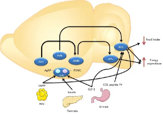
1. Figure 1: Hypothalamic neuronal circuits regulating food intake and energy expenditure. Proopiomelanocortin (POMC) and neuropeptide Y/agouti-related peptide
(AgRP) neurons in the arcute nucleus (ARC) sense the body’s energy and send inputs to other brain regions such as paraventricular nucleus (PVN) which will project to the nucleus of the solitary tract (NTS). The preoptic area (POA), dorsomedial hypothalamus (DMN), rostral raphe pallidus (rRPa), glucagon-like peptide-1 (GLP-1), cholecystokinin (CKK), withe adipose tissue (WAT), gastrointestinal (GI) [1].……….8
2. Figure 2: Overview of the reward system in rodent brain. Dopamine neurons are
located in brain structures called substantia nigra pars compacta (SNc) and the ventral tagmental area (VTA). Nigrostriatal pathway arises from the SNc to striatum; the mesolimbic pathway projects from the VTA to the nucleus accumbens (NAc), and the mesocortical pathway arises from the VTA to the prefrontal cortex [2]. ………..………...10
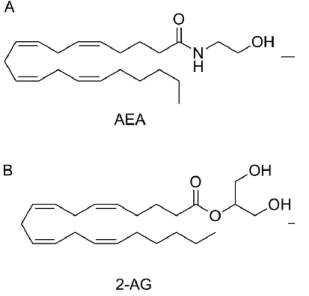
3. Figure 3: Chemical structures of endocannabinoids.
a) Anandamide (AEA) and b) 2-arachidonoylglycerol (2-AG)...14
4. Figure 4: Endocannabinoid synaptic modulation. Pre-synaptic neurotransmitter
release activates the post-synaptic neuron increasing calcium influx and stimulating the EC ligand synthesis acting in the pre-synaptic CB1 receptor. MAGL degrades 2-AG at the presynaptic neuron and ABHD6 catabolizes 2-AG at its site of synthesis. (figure modified
from Velasco et al., [4]
.………...16
5. Figure 5: Modulation of ABHD6 KO in the VMH. ABHD6 KO in the ventromedial
hypothalamus (VMH) regulates 2-AG accumulation and impairs energy metabolism when exposed to homeostatic challenges such as fasting, cold exposure and high-fat diet. SNS, sympathetic nervous system. (Taken from Fisette et al., [3])………..19
6. Figure 6: Mechanism of ABHD6NAc KO to increase 2-AG levels. ABHD6NAc KO enhance
2-AG synthesis that acts on CB1 receptor inhibiting GABA neurotransmission release (figure modified from Velasco et al., [4])……….22
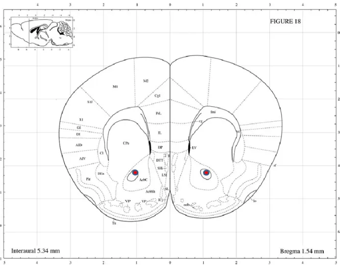
7. Figure 7: Mouse Brain atlas. NAc viral injection coordinates (red) for the ABHD6
x
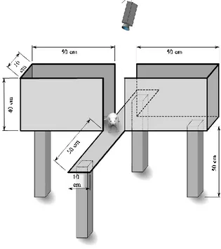
8. Figure 8: Running wheel dimensions. Approximately weight is 110g without batteries.
Each running wheel needed three AAA batteries for proper functioning...27
9. Figure 9: Elevated Plus Maze scheme and dimensions...29
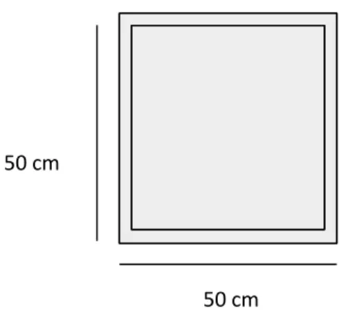
10. Figure 10: Open Field Test scheme and dimensions
...………...30
11. Figure 11: Validation of ABHD6 knockout in the NAc. A) Analysis of proper injections
after surgeries, the three injection coordinates were obtained from three different animals. B) Amplification of ABHD6 mRNA of the three groups of mice was done by q-PCR in real time. The CRE HFD-treated mice showed a significant 40% depletion compared to the GFP CHOW and GFP HFD control groups (n=6-9). Group mean ± SEM; one-way analysis
of variance, Bonferroni post hoc
*<0.01……...36
12. Figure 12: ABHD6NAc KO diminishes diet-induced obesity and metabolic changes resulting from high fat diet. A) Treated mice CRE (n=7) showed a reduction in body
weight gain after 8 weeks on HFD compared to the control GFP (n=6) and B) have no difference on food consumption. CRE mice C) have an increase in energy expenditure during the dark phase and D) an increase in ambulatory activity. E&F) Echo MRI measurements showing no significant difference between groups in fat mass, however there is a significant difference in lean mass on CRE mice compared to GFP. Group mean
± SEM, P*<0.05,
P***<0.001...37
13. Figure 13: ABHD6NAc KO rescues voluntary physical inactivity elicited by HFD feeding. A) Running wheel performance during 5 weeks B) Significant increase in total
running rotations during 5 weeks of voluntary physical activity on CRE (n=7) compared to GFP (n=6). C&D) body weight gain and food consumption during the running performance on HFD. Group mean ± SEM; two-way analysis of variance, Bonferroni post hoc P*<0.05 ...38
14. Figure 14. Neuronal ABHD6NAc KO does not increase running performance on chow fed conditions compared to GFP mice. A) Running wheel rotation during 5 weeks on
chow feeding (control diet) CRE (n=8) vs. GFP (n=8) B) Total cumulative running rotations, C) body weight gain during wheel running and D) cumulative food intake. No significant difference between the groups in any measurement...39
xi
15. Figure 15: ABHD6NAC KO prevents from rebound of HFD feeding but no voluntary wheel running difference was seen during the 3 weeks of wheel access on HFD feeding. A) Body weight gain CRE (n=8) vs. GFP (n=8) on HFD following withdrawal of
running wheels (grey block) and re-introduction of running wheel access (white block) while on HFD. B) HFD food intake during the same period. C) Running wheel re-exposure on HFD feeding D) Total running rotations. Group mean ± SEM; two-way ANOVA analysis
of variance
P**<0.01...40
16. Figure 16. Neuronal ABHD6NAc KO increased locomotor-stimulating effects of amphetamine but not D1R-dependent locomotion. A) Overall amphetamine
sensitization induction. B) Total Locomotion activity obtained by the subtraction of the second low dose from the first low dose CRE vs. GFP (n=6-9) C) Beam brakes data obtained during the effect time of the amphetamine injection D) Total locomotor activity measured in beam breaks from D1 agonist injection and E) Locomotor activity during the D1 effect. Group mean ± SEM, unpaired t test *<0.05……….41
17. Figure 17. Impact of ABHD6NAc KO in neurons on body weight and food intake on a high-fat diet. A) Percentage of body weight gain during 12 weeks on HFD or CHOW, B)
Cumulative food intake during 12 weeks on HFD or CHOW. Group mean ± SEM; two-way analysis of variance, Bonferroni post hoc (n=6-9)……...42
18. Figure 18. ABHD6NAc KO does not increase anxiety-like behaviors caused by HFD feeding. A) Total distance travelled during the EPM test. B&C) Number of entries and time
spent in the elevated plus maze (n= 6-9). D) Total distance travelled during the OFT. E&F) Number of entries and time spent in the center of the open field test. Group mean ± SEM; one-way analysis of variance, Bonferroni post hoc *<0.05 **<0.01...43
19. Figure 19. Depletion of neuronal ABHD6 in the NAc does not affect depressive-like behaviors elicited by HFD feeding. A) Time spent immobile during the last 4 min in the
forced swim test (n=6-9) GFP CHOW vs. GFP HFD and B) Swim velocity from each group since the test started. Group mean ± SEM; one-way analysis of variance, Bonferroni post hoc. P*<0.05 ...44
xii
Tables
I.
Table I. Diet composition...26xiii
List of abbreviations
BMI: Body Mass Index BMR: Basal Metabolic Rate WAT: White Adipose Tissue BAT: Brown Adipose Tissue CNS: Central Nervous System VMH: Ventromedial Hypothalamic ARC: Arcuate Nucleus
PVN: Paraventricular
MC4R: Melanocortin Receptor 4 POMC: Proopiomelanocortin
MSH: Melanocyte Stimulating Hormones AgRP: Agouti Related Peptide
POA: Preoptic Area
SNc: Substantia Nigra pars compacta VTA: Ventral Tegmental Area
NAc: Nucleus Accumbens MSNs: Medium Spiny Neurons HFD: High-Fat Diet
HPA: Hypothalamo-Pitutary-Adrenal THC: Δ9-tetrahydrocannabinol ECS: Endocannabinoid System eCB: Endocannabinoids
CB1: Cannabinoid receptor type 1 CB2: Cannabinoid receptor type 2
GPCRs: G-Protein-Coupled Receptors cAMP: Cyclic Adenosine Monophosphate 2-AG: 2-Arachidonolyl Glycerol
FAAH: Fatty Acid Amide Hydrolase MAGL: Monoacyglycerol Lipase
ABHD6: Alpha Beta Hydrolase Domain 6 DSI: Depolarization-induce Suppression of
Inhibition
DSE: Depolarization-induced Suppression
of Excitation
VGCCs: Voltage-Gated Calcium Channels TRPV1: Vanilloid Receptor Type 1
µ: Mu
CLAMS: Comprehensive Lab Animal
Monitoring System
EPM: Elevated Plus Maze OFT: Open Field Test FST: Forced Swim Test
ABHD6NAc KO: Alpha Beta Hydrolase
Domain 6 nucleus accumbens knockout LC/MS: Liquid Chromatography-Mass Spectrometry
GFP: Green Fluorescent Protein NeuN: Neuronal Nuclear protein
xiv
To my parents; Anna K. Flores and Fernando D. Franco for always believing in me and loving me for who I am. To my love and best friend Leonel Ramos, whose support and love was unconditional.
xv
Acknowledgements
First, I would like to thank God because His hand was never apart from me and I know he gave me the tools and skills to accomplish this mission. I will be forever grateful.
I would like to thank my advisor, Dr. Stephanie Fulton, whose patience and work was remarkable during these two years, she encouraged and guided me whenever I felt lost and she believed in me during my darkest moments. Stephanie gave me the opportunity to prove to myself that I can succeed in science and I will always be thankful for all of her support.
I also want to thank all the members in the Fulton and Alquier Lab, for all their help during these two years. They helped me to develop protocols, analyze data and learn new techniques. I also appreciate the motivation as they were very enthusiastic inside and outside the laboratory. I found in them very good friends who made the work environment more comfortable.
Many thanks to the members of my committee: Dr. Gareth Lim and Dr. Pierre-Paul Rompré for all the constructive criticism and guidance during my master´s thesis. I really appreciate your time, patience and knowledge.
To my family, Fernando Franco, Anna K. Flores, Anette Franco, Alexa Franco, Guillermo Franco and Milly Franco. For all the love and faith they put in me. It has been a long way and I couldn’t definitely make it possible without them. Their love followed me all this time, in good and bad moments they were always by my side.
To my courageous boyfriend Leonel Ramos who never doubted my dreams. I could feel his support from far. I want to thank him for the days and nights he listen to me talk about my work and for pushing me to be the best version of myself.
Finally, I would like to thank my alma mater and my country. I am so proud to be Mexican, because we fight for our dreams, we embrace our cause and we always find our way to success.
Thank You,
1
1. Chapter 1: Introduction
1.1 Obesity
1.1.1 Impact
Obesity has become one of the most important health problems in today’s age and has been characterized as a global epidemic [5]. Obesity is caused by genetic, social and environmental factors and can affect people of all ages, sex and ethnicity. Obesity increases the probability to develop chronic diseases such as type 2 diabetes, cardiovascular disease, some types of cancer, mood disorders (depression, anxiety), sleep and respiratory disorders and, in worst cases, death [6], [7].
An overweight person is considered to have a body mass index (BMI) between 25 and 29.9, while an obese person has a BMI of 30 or higher [8]. BMI is a value obtained by dividing a person's weight (kg) by the square of his height (m) [9]. Approximately half a billion of the world`s population is now considered to be overweight or obese [10]. Statistical projections estimate that 20% of the world’s population will be obese and 38% will be overweight by 2030 [11]. It was initially believed that this problem only affected developed countries; however, the problem has also emerged in developing countries in Central and South America [12].
With the rise in prevalence of these metabolic diseases (obesity, diabetes, etc), there is an augmentation in health costs related to preventative measures for obesity and to treat its comorbidities. Although there is a lower incidence of people with obesity in Canada compared to the U.S. there has been a significant increase during the last decade [13]. In 2010, $6 billion – 4.1 % of Canada’s total health care budget – was spent towards obesity-related issues [14]. Some psychiatric disorders such as depression can predispose individuals and influences the development of obesity. It is thought that this could be a counter-balancing effect induced by the negative emotions caused by overeating [6].
There is an urgent need to find a treatment that can decrease obesity and its comorbidities. Currently, there are dietary interventions or surgical procedures that reduce around 60 to 80 percent of weight [15]; however, it becomes difficult to maintain long-term results from these approaches despite the efforts of both patients and physicians. Even more, repeated experiences of failure to lose weight can trigger psychological distress in patients, which further contributes to the problem. The common treatment consists in changing bad food habits (diet) and increase
2
physical activity. This can also be accompanied by medications that help to decrease food consumption or decrease fat absorption. If those strategies do not work, physicians may recommend surgical options such as gastric balloon or bariatric surgery [16]. These procedures reduce the stomach volume or length of the intestines, leading to feel early satiety. Multiple side effects can emerge during and after these surgical interventions, which is why we need to investigate other potential new treatments to stop or minimize the “globesity” expansion [17]. In this chapter we are going to explain the etiology of obesity along with a literature review of the brain areas implicated in the regulation of the energy homeostasis and the consequences of disruptions of these brain pathways. Finally, we are going to explain the importance of the endocannabinoid system and its impact on energy balance and body weight.
1.1.2 Etiology
As discussed above, obesity is defined as an augmentation or excess of adipose tissue in the body. This increase of fat mass can affect body homeostasis by altering metabolism. Obesity has a complex etiology arising from social, cultural, environmental, hormonal and genetic factors. Many genetic markers have been implicated in the development of obesity [18], [19], for example rare monogenic mutation such as the leptin gene [20]. There is a clear genetic susceptibility between populations that can determine which are more prone to become obese despite the environmental circumstances [6], [21]. Genes linked to obesity mainly affect metabolism, body fat composition, food intake and energy expenditure. Although, there are few sporadic mutations that result in the development of obesity, predisposition appears to be inherited from parents.
There are also gender differences related to body weight gain. In the third national health and nutrition examination survey from the United States (1988-94), found a higher prevalence of obesity in women (25% U.S. women) compared to men (20% U.S. men) [21]. It is now known that women have more trouble losing weight compared to men, although the mechanism underlying this is not well understood [22][21]. Moreover, maternal obesity can give a predisposition to the baby to develop the disease. Some interesting data from Udall et al; suggests an interaction between maternal and neonatal obesity. Weight gain during pregnancy has been associated with an increase in subcutaneous fat in neonatal humans [23]. Broadney et al., also highlights the importance of parental health, specifically in the mother, where maternal obesity influences comorbidities such as inflammation and alterations in immune responses in children [24].
3
The two most recognized causes of the massive increase of obesity around the world are: high caloric intake and the lack of physical activity (sedentary lifestyle). This can be translated as an imbalance or disruption in energy balance between calories consumed and calories expended. In developed countries there is a relationship between low levels of physical activity and obesity. For example, a study in the United Kingdom showed no correlation between energy intake and the prevalence of obesity, although they did find a tight relationship between the amount of immobility (due to T.V. viewing and lack of walking) and weight gain [26]. In addition, gut flora has been shown to differ between lean and obese humans which can affect energy balance [25] . Therefore, obesity is the result of many environmental and biological factors, with their overall contribution accounting for the dramatic increase of obesity worldwide.
1.1.2.1 Regulation of energy balance
To understand the mechanisms behind obesity we must first understand the concept of energy balance. Energy balance is the equilibrium between the amount of food ingested and energy expended. There are multiple molecules from the gut and both, the periphery and the central nervous systems that contribute to maintain energy balance (and imbalance) will be discussed in the following sections.
1.1.2.1.1 Food intake
The world has increase diversity in food supplies. Nowadays, supermarkets offer a variety of palatable foods with high content of sugars, salts and fat. The 24H accessibility to a variety of palatable food can be a strong contributor to the increase of fat storage seen in the world. Additionally, reward and emotional processes can interfere with these needs by both increasing or decreasing appetite and physical activity beyond homeostatic mechanisms. The experience of eating palatable food produces positive or negative feelings and can influence our willingness to approach or avoid those foods again. Positive, rewarding, or pleasurable effects, also known as hedonic effects, is one of the major causes of overeating and the development of obesity [27]. Palatable food stimulates appetite and delay satiety which increase energy intake [28]. Food macronutrients can play an important role in satiety; fat for example does not have a good satiety time period contrary to proteins that have a better impact in our body. Surprisingly, satiety does not come from the food portion or meal size but from the nutritional content that is consumed [29].
4
Many studies have focused on high fat diets as a possible cause of obesity. Indeed, Golay et al., showed that fat induces overconsumption and weight gain through its low satiety properties and high caloric density, thereby increasing fat storage [29]. In some cases, socioeconomic status can influence the choice for low cost, high calorie foods. Therefore, it is important to establish a balance diet that provides us the necessary nutrients to feel satisfied at each meal.
Moreover, TV advertisement can strongly influence meal preference including sweetened cereals, sweetened beverages and salty snacks; which leads to an energy imbalances that enhance weight gain and consequently obesity [30]. The accessibility of high caloric food gives reasonable foundations to blame obesity expansion. However, the prevalence of obesity and the global epidemic seen in this century cannot be only attributed to one factor, but several such as environmental, social, genetic, cultural and sedentary lifestyle.
2.1.2.1.1 Energy expenditure
Organisms need locomotor activity to function and provide necessities like food, reproduction and protection from predators that involve interactions with the environment [31]. Energy expenditure is the amount of energy that a single person needs to perform actions such as breathing, walking, eating and others. Total energy expenditure will be the total number of calories burned each day [32]. Currently, one strategy for treating obesity is to increase energy expenditure which is dependent on internal (e.g., temperature, metabolism, circadian rhythms, hormones) and external factors (e.g., weather, exercise, etc.). Energy expenditure is distinguished by three key components: basal metabolic rate (BMR), adaptive thermogenesis and physical activity. BMR is the minimal energy required to maintain bodily functions; i.e. energy under normal conditions when there is no extra demand for defensive adaptations such as cold-warm exposures, after meal digestions or in rest [33]. Adaptive thermogenesis is activated during chronic cold or heat exposures. At the cellular level, mitochondrial proton uncoupling from ATP production will be needed to release that energy as heat [34].
As a major component of energy expenditure, physical activity contributes considerably to whole-body energy balance. It can be divided into spontaneous and voluntary physical activity. Spontaneous physical activity is a low intensity and unplanned physical activity (e.g., standing, ambulating, talking and fidgeting). In rodents spontaneous physical activity can be measured by the activity in their home cage after the acclimation phase [35]. Voluntary physical activity in
5
contrast is defined by the willingness to increase locomotor activity and can vary in intensity and duration (e.g., sports). In humans, motivation to engage in voluntary exercise can be attributed to many internal or external factors. Physical activity can provide a rewarding effect and it can even become addictive [31]. In rodents the most effective way to measure voluntary physical activity is by the introduction of the wheel running. There are a number of studies using wheel running to assess the rewarding effect of voluntary physical activity in rodents and the underlying neurobiologically [36][37]. Moreover, running wheels are a useful tool to examine the association of physical activity to energy balance and obesity, however there are still a few challenges to fully understand the neuronal mechanisms affecting voluntary physical activity and its beneficial impact on diabetes and obesity.
The means by which the body maintains and achieves homeostasis are key to understanding for developing therapeutic approaches for reducing obesity. While some strategies appear to be promising in theory, there is no effective treatment to stall the increasing yearly rates of this epidemic.
1.1.3 Brain controls of food intake and energy expenditure
The brain integrates different circulating signals such as nutrients, gut-derived hormones and adiposity signals to regulate food intake and energy expenditure. In 1953, Kennedy et al. proposed a model of energy homeostasis where inhibitory signals generated by body fat stores act in the brain to reduce food intake [38]. If body fat decreases, peripheral signals are decreased and thus a demand for food intake is increased. Being among the first to describe a potential mechanism whereby periphery signals could control energy balance via their actions in the central nervous system (CNS), it had many shortcomings especially an explanation on how individual meals modulate inhibitory or excitatory signals to the brain. Fortunately, Gibbs and Smith proposed two decades later that meals can generate signals from the gastrointestinal tract which can act on the brain giving a feeling of satiety and to indicate the meal is finished [39]–[41]. Now we know that it is more complicated than that; metabolism implies neuronal control by the interaction with other organs such as gastrointestinal tract, liver, pancreas, white adipose tissue (WAT) and brown adipose tissue (BAT). Even more, the brain controls specific functions of these organs via the autonomic nervous system (responsible for the control of unconscious body functions like breathing) and endocrine mobilization [33].
6
Insulin was the first hormonal signal discovered to be implicated in the modulation of body weight at the CNS [42]. Studies using transgenic ob/ob mice (which had a mutation on the gene encoding leptin, a hormone secreted by adipocytes) gave insight that a hormone can be implicated in hyperphagia and obesity and could be mostly implicated in adipocyte signalling. Circulating leptin levels are in correlation with the amount of body fat content [43]. In the CNS, there are receptors for both leptin and insulin in hypothalamic neurons and they are known to be important in modulation of energy intake. Insulin and leptin act on two different groups of neurons in the hypothalamic arcuate nucleus (ARC) to give negative feedback for food intake and energy balance. More precisely, insulin and leptin act on proopiomelanocortin (POMC) neurons by stimulating the secretion of anorexic neuropeptide and on neuropeptide Y/ agouti gene-related protein (NPY/AGRP) neuron, therby decreasing the expression of NPY [44]. Some studies have found that leptin can have a greater impact than insulin in the CNS control of energy homeostasis [45]. In this section we are not going to give much detail about it, however, a good example is that lack of leptin causes severe obesity with hyperphagia that persists despite high insulin levels. Contrarily, insulin deficiency does not cause obesity having an impact in different critical roles on metabolism [33]. Thus, both leptin and insulin participate in the CNS control over energy homeostasis and disruption of these mechanisms in the development of obesity are focal points of research.
Energy expenditure is the other important aspect that regulates body weight and maintains a healthy life. Information regarding energy stores is sent to specialized neurons in the hypothalamus and in the brainstem [1]. Kennedy, among others, observed that rats that had ventromedial hypothalamic (VMH) lesions showed an increase in body weight and adiposity tissue even though they were food restricted, making clear that hypothalamus controls both energy intake and energy expenditure [46]. More specifically, a hypothalamic region called the arcuate nucleus (ARC) plays an important role in mediating leptin’s effect on locomotor activity. In one study, leptin receptor null mice showed normalized locomotor activity after restoration of leptin signalling in ARC POMC neurons. Therefore leptin signaling is an important modulator to increase locomotor activity [47]. Thermogenesis is another important component in energy expenditure, it is dependent on BAT and it maintains body temperature created by internal factors. More information about thermogenesis and its regulation will be discussing in the following section.
7 1.1.2.1 Hypothalamus and hindbrain
The hypothalamus is a brain region involved in homeostatic regulation (e.g. hunger, body temperature, sleep, etc), as well as energy intake and expenditure [48]. The hypothalamus participates in the organism’s adaptation to environmental changes due to its high levels of plasticity. In the past decade, literature regarding the neuronal wiring and intercellular signalling of different peripheral hormones to the hypothalamus has grown exponentially [49]–[51]. For instance, lesions in the paraventicular nucleus (PVN), the ARC and the ventromedial nuclei of the hypothalamus lead to hyperphagia and obesity due to its neuronal integration that modulates energy balance [52]. In the next paragraphs we are going to explain with more detail some of the molecules involved in how the hypothalamus can regulate feeding and contributes to an important part of the increasing obesogenic population in the world.
A small section of the hypothalamus called ARC plays a critical role in the regulation of metabolism. Melanocortin receptors are found in the ARC, but it wasn’t until some studies deleted the melanocortin receptor 4 (MC4R) in animals that they saw a significant difference in body weight [53]. Subsequently, researchers discovered that the melanocortin system is the neurocircuitry connecting appetite and neuroendocrine signalling to regulate metabolism. The generation of melanocortin ligands is done by proopiomelanocortin (POMC) neurons. In the CNS, POMC generates melanocyte stimulating hormones (MSH), α-MSH, β-MSH and γ –MSH that have high affinity (agonist) for the melanocortin receptors MC1R, MC3R, MC4R and MC5R [54]. They also found endogenous antagonists: Agouti and Agouti related peptide (AgRP) that interact with specific melanocortin receptors that interfere with MSH activity. Distribution of AgRP and POMC neurons can have similar communication to other brain areas, but it is believed that only POMC neurons have projections to the brainstem. These neuronal populations have opposite effects to their hypothalamic counterparts in the maintenance of energy homeostasis; which is important as POMC neurons (via MSH) inhibit food intake, while AgRP neurons increase food intake [52]. There are diverse new techniques (e.g. DREADD, KO mice, optogenetics) that facilitate specific neuronal control of POMC and AgRP activity, proving the importance of a balance between their connections. Long term AgRP neuronal loss results in severe aphagia and subsequient death [55] while mice POMC deficiency causes hyperphagia and obesity [56], [57]. Furthermore, POMC and AgRP neurons respond to leptin, insulin [58], ghrelin, serotonin and glucose. Their signalling results in the generation of melanocortin receptors to modulate food intake, glucose metabolism, energy expenditure and others (Figure 1).
8
Figure 1: Hypothalamic neuronal circuits regulating food intake and energy expenditure.
Proopiomelanocortin (POMC) and neuropeptide Y/agouti-related peptide (AgRP) neurons in the arcute nucleus (ARC) sense the body’s energy and send inputs to other brain regions such as paraventricular nucleus (PVN) which will project to the nucleus of the solitary tract (NTS). The preoptic area (POA), dorsomedial hypothalamus (DMN), rostral raphe pallidus (rRPa), glucagon-like peptide-1 (GLP-1), cholecystokinin (CKK), withe adipose tissue (WAT), gastrointestinal (GI) [1].
Energy is expended during physical activity, basal metabolism and adaptive thermogenesis. The ARC is considered a key site for mediated locomotor activity induced by leptin signalling, but there are other brain areas that also contributes to locomotor behaviors. In a thermoregulatory perspective, heat must be generated to maintain body temperature or in response to the energy given by food intake. The preoptic area (POA) regulates the neuronal circuit of sympathetic BAT outflow by sending inputs to the premotor neurons in the rostral raphe pallidus. Moreover, administration of MC3R and MC4R agonists can stimulate BAT activity - meaning hypothalamic melanocortin system has a regulatory response in BAT thermogenesis [1]. In this regard,
9
endocrinal signalling by insulin, leptin, and GLP-1 can modulate BAT outflow: it has been shown that insulin and leptin act on POMC neurons to promote white adipose tissue browning and enhance energy expenditure. Since WAT is reduced during obesity, manipulation in the homeostatic mechanism to increase WAT could potentially prevent diet-induced obesity [59].
2.1.2.1 Extra-hypothalamic control of energy balance: corticolimbic circuits
There are extra-hypothalamic structures that play a very important role on the control of energy balance also known as the corticolimbic circuits. If we wonder why we prefer a cookie over broccoli, the answer lies in neural circuitry that is activated by these palatable foods. There are some foods that cause more pleasurable or rewarding effects triggering a higher release of the neurotransmitter called dopamine within the reward system. Dopamine’s first precursor is tyrosine, which is converted to L-dopa to be catabolized as dopamine [60]. There are many physiological dopaminergic actions, which are mediated by G protein-coupled receptor subtypes. There are 5 different receptor subtypes: D1-like receptor (D1A-D and D5) that activates adenylyl cyclase by Gs protein and D2-like receptor (D2, D3 and D4) that inhibits adenylyl cyclase and activates K+ channels by inhibitory G protein. Both receptors’ activity influence behavior, increasing or inhibiting reward depending on the brain area [2], [61]. Even though dopamine only accounts for 1% of the total neuronal population of the brain, it is involved in many brain areas associated with motor, memory, reward, and other functions. It has also been demonstrated that dopamine is involved in the hedonic component of reward [2], [62].
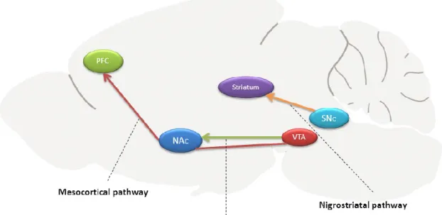
Reward is very important for survival and it is implicated in basic processes like eating, drinking, and sexual reproduction, as well as other pleasurable behaviors such as gambling. The reward system is activated in response to these activities and increases the chances of seeking these behaviors again. Reward can be defined as the willingness to work (spend time, energy and effort) for any goal or behavior from which we have previously derived pleasure [63]. Therefore, a rewarding situation has an impact on motivation. Because of these seeking-behaviors, scientist proposed a neuronal signalling pathway which processes reward such as the dopamine system. Studies revealed that dopaminergic neurons are localized in the mesencephalon, diencephalon and the olfactory bulb. As seen in Figure 2, dopamine neurons located in the midbrain structures substantia nigra pars compacta (SNc) and ventral tegmental area (VTA) project to the striatum (composed by caudate nucleus, putamen, ventral striatum and nucleus accumbens (NAc)), and
10
the dorsal and ventral prefrontal cortex. There are additional projections, which influence reward, but they will not be discussed in this thesis.
Figure 2: Overview of the reward system in rodent brain. Dopamine neurons are located in
brain structures called substantia nigra pars compacta (SNc) and the ventral tagmental area (VTA). Nigrostriatal pathway arises from the SNc to striatum; the mesolimbic pathway projects from the VTA to the nucleus accumbens (NAc), and the mesocortical pathway arises from the VTA to the prefrontal cortex [2].
In this study, we are interested in the mesolimbic pathway that projects from the VTA to the NAc as it has been shown to be implicated in the motivations for natural rewards, locomotor activity and mood. The NAc is a large structure that has two sub-regions: the core and the shell. NAc core and shell execute different brain functions based on the inputs received [64]. The NAc shell has been well implicated in the activation of dopamine transmission during substance abuse such as cocaine, amphetamine, THC, MDMA and nicotine [65]. The NAc core, on the other hand, is involved in reward-related approaches such as the Pavlovian-instrumental transfer, as well as cognitive and motor functions [66]. Together the shell and core regulate, motivation, addiction,
11
reinforcement learning, mood, etc [64]. Approximately 95% of neurons in the NAc are GABAergic medium spiny neurons (MSNs) that express either D1-type or D2-type receptors. There are also cholinergic and GABAergic interneurons. From the NAc core, the GABAergic MSNs projects to other subcortical areas such as the globus palladus and the substantia nigra [67], [68]. A well -supported idea about the NAc dopamine system is that it could be an interface between motivation and action, meaning that it can play an important role in the motivation to work for palatable food but not necessarily in reward perception [69]. Also, dopamine is implicated in regulating voluntary physical activity. The difference between spontaneous and voluntary physical activity can be summarized that the second needs a purpose and motivation to do it while spontaneous activity is just an involuntary movement that the body creates.
In mice research evaluating the dopaminergic system in the control of voluntary physical activity,
Rhodes et al. showed that administration of D1-like antagonist reduced wheel running compared
to control mice. In contrast, D2-like antagonist had no effect between control and treated mice suggesting that D1-like receptor is a possible modulator of wheel running performance [70]. Studies have shown that increased dopamine activity can be dependent on physical activity, but it is not clear if dopamine is acting as an independent variable of physical activity levels or if physical activity is affecting dopamine functioning [71]. Therefore, it is important to investigate the implication of NAc dopamine system in the regulation of voluntary physical activity, especially D1-like receptor that has been shown to modulate these effects.
1.1.4 Brain control of mood
There are many neuronal networks implicated in emotional behaviors. The limbic system controls mood, memory, and others. The limbic system is located between the brainstem and the two hemispheres and involves the PFC, amygdala, hippocampus and the ventromedial parts of the basal ganglia. In fact, these areas provide some evidence of how their specific neuronal networks controls mood such as fear, anxiety, depression, etc. The amygdala, in particular, is responsible for modulating our perception and reactions to aggression and fear; functional magnetic resonance imaging studies in humans have found that fearful faces and fear conditioned cues activates the amygdala region [72]. Emotions and memory are very closely related; therefore, the hippocampus is an important region that stores information in a long- and short-term memory. The hippocampus is necessary to create new memories but also to store old memories. Moreover, disturbances in this brain region appears to have psychological consequences such as
12
schizophrenia and severe depression [73]. The mesolimbic dopamine areas such as NAc and VTA structures are important for reward and emotional states such as depression, anxiety and stress. Dysfunctions within these areas can induce disturbances on emotional and cognitive disorders such as depression and anxiety [74]. As we discussed in the previous section, the NAc is implicated in the control of food-motivated behaviors, in locomotor activity and mood disorders. NAc integrates inputs from diverse areas, making it one of the major controls of motivation, reward, stress, and mood. Deep brain stimulation in the NAc decreased behavioral responses of depression and anxiety in patients that were resistant to pharmacotherapy treatment, indicating a potential alternative therapy for mood disorders [75].
1.1.2.1 Anxio-depressive behaviors
Obesity is implicated in CNS impairments such as mood disorders. It has been shown that obesity increases the incidence of depressive symptoms and that depression could lead to the development of obesity and its associated complications; Luppino et al., showed that the obese population has a 55% increased chance of developing depression and that individuals that suffer from diabetes have a doubled incidence of depression compared to healthy controls. This relationship could be due to poor food choices or lack of physical activity associated with depression and obesity [76]. Mood states like anxiety and depression can also be altered by food choices and energy metabolism. For example, people with negative emotional states tend to eat palatable foods (chocolate or ice cream) to alleviate these feelings. Palatable food consumption induces short-term positive emotions while chronic consumption of these foods will increase adipose tissue leading to anxio-depressive behaviors [77]. It is believed that there is a bidirectional relationship between mood disorders and obesity, however the nature of this association is still poorly understood. Sharma et al., showed that chronic high-fat fed mice resulted in molecular adaptations in NAc and expressed anxio-depressive behaviors in mice [78]. Additionally, Décarie-Spain et al., showed that saturated high-fat diet (HFD) fed mice produces obesity and hyperleptinemia (higher amounts of leptin in the bloodstream); triggered anxiety-like behaviors, peripheral inflammation and multiple pro-inflammatory signs in the NAc including gliosis (glial cell inflammation), enhance of cytokine expression and NFkB transcriptional activity. A saturated HFD also decreases in NAc dopaminergic tone and function as well as perturbations in hypothalamo-pitutary-adrenal (HPA) axis [79]. These data suggests that the NAc is important in anxio-depressive behaviors caused by saturated HFD [78]. Although more information is needed to
13
characterize the importance of the NAc in anxio-depressive behaviors, it appears the NAc could be a critical structure to investigate HFD-induced changes in metabolism, decrease of voluntary physical exercise, and comorbid anxio-depressive behaviors.
1.2 Endocannabinoid system
1.2.1 Generalities
For decades’ cannabis or marijuana has been used by humans as a recreational drug. There is evidence that shows the use of cannabis in the second millennium BC in the Assyrians culture. Different cultures in the Middle East, Europe, Africa and others were engaged for the positive effects of cannabis such as euphoria, feelings of well-being, and its medical use. Its wide use in these countries has continued ever since and it has had a world impact that now is the longest recorded drug in history of human use [80]. Although in ancient cultures the use of cannabis not only was for recreational purposes but also medical (for example, as a pain killer, anxiety-relieving and sleeping disorders), little was known about the molecular actions of cannabis within the body. It was until 1964 when Dr. Mechoulam discovered the psychoactive product of cannabis (also known as marijuana) Δ9-tetrahydrocannabinol (THC) [81]. The discovery of THC brought the possibility to create synthetic cannabinoid-like compounds with different chemical structures or different arrangement. These compounds where divided in three classifications: classical, non-classical and aminoalkylindol. After all the research trying to understand how THC exerted its effect within the body among the different synthetic analogs (plant derivate and synthetic generated), researchers discovered endogenous cannabinoid-like compounds in our bodies called the endocannabinoid system (ECS).
ECS is widely distributed in the mammalian tissues and it is known for its properties regulating cardiovascular, nervous and immune system inside the cells to maintain body homeostasis [81], [82]. The endocannabinoid (eCB) mechanism of action was not known until the 19th century when
Dr. Howlett published the first data indicating that eCBs inhibited the formation of adenylate cyclase, an enzyme important for the production of cyclic adenosine monophosphate (cAMP) within the cell. Not long time after the same laboratory showed the existence of binding sites in the brain [83] and shortly the first cloning of cannabinoid receptor was done [84]. Today, researchers have identified two cannabinoid receptors: cannabinoid receptor type 1 (CB1) and cannabinoid receptor type 2 (CB2). The CB1 receptor is mostly expressed in the brain and it was
14
known to be the “brain receptor”, however, it can be also present in some peripheral organs. On the other hand, CB2 is abundant in immune cells, astrocytes and microglial [85]. Both receptors are 7-transmembrane domain macromolecules of the “G-protein-coupled” (GPCRs) [86].
The eCB’s, in the other hand, are lipophilic lipid compounds that are produced on-demand; the two major ligands are: N-arachidonoylethanolamide or anandamide and 2-arachidonolyl glycerol (2-AG). The chemical structures of the ligands are described in figure 3 [87]. These two ligands have been investigated in great detail and showed not to be conventional neurotransmitters. Surprisingly, their mode of actions differ from standard neurotransmitters as they act mostly pre-synaptic instead of post-pre-synaptic to inhibit neurotransmitter release [83]. The enzymes responsible of the brake-down of the two ligands are the fatty acid amide hydrolase (FAAH), the monoacyglycerol lipase (MAGL) and the alpha beta hydrolase domain 6 (ABHD6) who was been recently discovered [88]. The purpose of this study is to focus more in the enzyme ABHD6
as it has been shown to play an important role in metabolism.
Figure 3: Chemical structures of endocannabinoids: a) Anandamide (AEA) and b)
2-arachidonoylglycerol (2-AG) [89].
In general, ECS are important for the body homeostasis. The ongoing research on the ECS has brought new therapeutic approaches in diseases like epilepsy, cancer, hypertension, diabetes, obesity and anxio-depressive behaviors. However, there is still lack of information regarding the molecular bias and mechanism of how ECS is helping the body to maintain its balance. In this section we are going to give evidence that point to the ECS as a mediator in the
15
brain and how it can be linked to obesity, physical activity and anxio-depressive behaviors, but more important, we are going to evidence how ECS could be thought as a potential therapeutic approach for obesity. The novel discovery of the ABHD6 enzyme (which constitute a part of the ECS) can be thought as a potential therapeutic approach for body weight loss, exercise and mood; important targets for decreasing obesity in the world without using an invasive procedure such as surgical interventions.
1.2.2 Endocannabinoid system in the brain
The central nervous system is composed by the brain and spinal cord [90], [91]. As discussed above, the endocannabinoid system is present in the CNS to act as a synaptic modulator. Although CB1 receptor was originally believed to be only expressed in the CNS, now we know that it is present in numerous peripheral organs. CB1 is the most abundant receptor in the brain and both anandamide and 2-AG have high affinity for this receptor [92]. There are several brain regions with high density in CB1 receptor such as basal ganglia, substantia nigra, globus pallidus, cerebellum and hippocampus [93], [94]. Because endocannabinoids are lipid-based compounds and the brain is surrounded by aqueous solution it is easy to have an interaction between intracellular environments for lipid messengers [95].
One well-known endocannabinoid mechanism is that they are synthesized “on demand”, which differentiate them from classical neurotransmitters that are constantly synthesized and then stored in synaptic vesicles [96]. In this regard, they modulate synaptic transmission by inhibiting the release of many excitatory and inhibitory neurotransmitters. Therefore, new definitions were implemented to describe that eCBs mediates short-term synaptic plasticity, also known as depolarization-induce suppression of inhibition (DSI) and depolarization-induced suppression of excitation (DSE). Not long time after scientist demonstrated that they also modulate long-term plasticity[95], [97]. On the other hand there has been some evidence that ECS has been implicated in synapse formation and neurogenesis [98]. Therefore, there is a clearly eCB synaptic modulation that can modify brain networks in certain circumstances.
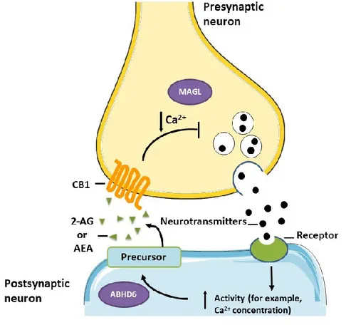
The eCB signalling is very complex system as they do not act as standard neurotransmitters; however, the retrograde signalling that many studies have found is that neuronal post-synaptic stimulation (induced by GABA or glutamate) will activate voltage-gated calcium channels (VGCCs) elevating intracellular calcium that will stimulates 2-AG synthesis. 2-AG will be release
16
to the synaptic clef to act in the CB1 located at the pre-synaptic neuron in a retrograde manner (showed in Fig 4). In contrast, studies have found a non-retrograde signalling in which neuronal modulation involves the transient receptor potential vanilloid receptor type 1 (TRPV1) or CB1 receptor located at the post-synaptic neuron [83].
Figure 4: Endocannabinoid synaptic modulation. Pre-synaptic neurotransmitter release
activates the post-synaptic neuron increasing calcium influx and stimulating the EC ligand synthesis acting in the pre-synaptic CB1 receptor. MAGL degrades 2-AG at the presynaptic neuron and ABHD6 catabolizes 2-AG at its site of synthesis. (figure modified from Velasco et al., [4]).
Moreover, eCB modulates synaptic strength, and depending on the brain region can induce neuronal changes in memory, locomotor activity, food related behaviors and pain [99]. The enzymes MAGL and ABHD6 play an important role in the modulation of synaptic strength. Many studies focus more on MAGL because it was known to be the “traditional” 2-AG breakdown but now growing evidence is pointing to ABHD6 as an important modulator of “fine tunes” in synaptic
17
plasticity. Therefore, in the next sections we are going to discuss about ABHD6 and the findings on synaptic plasticity and metabolism.
1.1.2.1 Alpha/beta hydrolase domain 6 (ABHD6)
As discussed above, there are many different enzymes important for the degradation of 2-AG. MAGL and FAAH were the first ECS enzymes to be studied in 2-AG degradation. In fact, there are plenty of pharmacological and genetic studies about these two enzymes, however; recent studies discovered new enzymes from the monoglycerol lipase family such as ABHD6 and ABHD12. We are interested in studying the enzyme ABHD6 because it is believed that it plays an important role in the pathogenesis of obesity. Some studies accomplish ABHD6 deletion in adipose tissue and saw a protective effect from metabolic syndrome and insulin resistance [100]. ABHD6 has been also detected in different tissues including liver, kidney, pancreatic islets and the brain [101]. However, the mechanisms of its physiological functions in the brain are still unknown.
ABHD6 is monoglycerol lipase that acts endogenously to decrease 2-AG accumulation and thereby signaling at CB1 receptor in the CNS. Marrs et al., demonstrated that ABHD6 played an important role controlling the accumulation of 2-AG which was surprising because most of the monoglyceride lipase inhibition, exhibit a strong increase in 2-AG levels while ABHD6 inhibition caused minor changes in 2-AG levels [102].This minor activation of CB1 receptor caught a lot of attention in the recent years and some studies showed that pharmacological ABHD6 inhibition can have neuro-protective effects in several diseases such as traumatic brain injury, epilepsy, and cancer [103],[104]. The enzyme MAGL is responsible for approximately 85% of 2-AG hydrolysis and it is localized in the presynaptic axon terminal, same site as the CB1 receptor. In contrast ABHD6 hydrolyses the small quantity of approximately 4% of 2-AG in the brain and its localization is in the post-synaptic neuron [105]. Thus, ABHD6 acts to guard the accumulation of 2-AG and thus mediate CB1R signal transduction by reducing 2-AG. Furthermore, Marrs et al., demonstrated that ABHD6 was expressed in glutamatergic neurons, some GABAergic interneurons and astrocytes modulating synaptic plasticity but nor in microglia. Interestingly, ABHD6 was detected in mitochondrial fraction of microglial cell line BV-2 and its inhibition reduced 2-AG hydrolysis. Furthermore, ABHD6 acts as a modulator of synaptic plasticity and it has been shown that inhibition increased the induction of long-term synaptic depression by CB1 receptor but not short-term depression [105]. Inactivation of ABHD6 expression in neurons using the
18
molecular tool CRISPR/cas9 significantly increases excitatory neurotransmission while overexpression of ABHD6 reduces glutamate currents and even more. Wei et al., demonstrated that ABHD6 negatively regulates the synaptic function of AMPA receptors [101]. Thus, all this evidence expands our understanding of the importance of ABHD6 for many neuronal targets and integrates molecular mechanism necessary for new therapeutic approaches. In general ABHD6 looks to have an important action in the regulation of synaptic plasticity in different types of neurons, however, its specific actions in brain reward areas need to be defined.
2.1.2.1 Implications of ABHD6 on energy balance and mood
The eCB AEA and 2-AG have been implicated in energy balance and body composition. In humans, Côté et al., demonstrated a relationship between eCB 2-AG and cardio-metabolic risk factors, showing a correlation in levels of 2-AG and BMI in obese men [106]. In the other hand, a few rodent studies showed that 2-AG levels increase after 18 hours of food deprivation and decrease right after food intake. 2-AG hypothalamic changes give the understanding that endocannabinoids can act as a pro-orexigenic mediator in this area. More studies showed similar effects when rodents are following a dietary restriction for a long period, reducing hypothalamic 2-AG levels while overweight rodents exhibit higher levels [107]. Interesting, the mice of Matias
et al. were exposed to different high fat diets for different periods of time and then there was an
evaluation of AEA or 2-AG where they measured their kidneys, hearts, skeletal muscle and thyroids. They found that obesity can cause dysregulation of the ECS in different organs important for endocrine functions and energy expenditure; these proposed important roles for the ECS in the control of metabolic, endocrine and cardiovascular functions [108]. In addition, the 2-AG
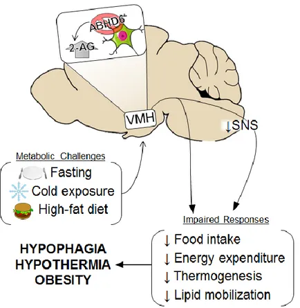
concentration levels can be controlled by ABHD6; Fisette et al., 2016 showed that ABHD6 deletion in the VMH neurons increase 2-AG levels compared to the control group. ABHD6
VMH KO mice exhibited disturbances in metabolic challenges such as impairment in feeding response to fasting, increased susceptibility to hypothermia, resistance to weight loss when transitioned from high fat diet to regular chow and higher prevalence in high fat diet-induced obesity (see Figure 5) [109]. Altogether, there is evidence to suggest that ABHD6 in the VMH plays an important role in energy metabolism.
19
Figure 5: Modulation of ABHD6 KO in the VMH. ABHD6 KO in the ventromedial
hypothalamus (VMH) regulates 2-AG accumulation and impairs energy metabolism when exposed to homeostatic challenges such as fasting, cold exposure and high-fat diet. SNS, sympathetic nervous system. (Taken from Fisette et al., [3])
Physical activity has a great impact maintaining body homeostasis. In athletes scientific investigation showed that exercise gives states of consciousness and in the 1960s they discovered some psychological changes induced by prolonged physical activity. In the last decades these effects were better described as “runners high” which gather states of happiness, elation, feelings of peace, well-being and pain relief [110]. However, the lack of scientific information or evidence about the “runners high” lead to the question: Do these effects really exist? and Why haven’t all athletes experienced it? One of the hypotheses for the mechanism of the “runners high” goes to an opioid point of view. Opioids are well known to induce euphoria and analgesia thus some of the effects caused by physical activity could be related to opioid activation; however, the “endorphin-related hypothesis” didn’t last because it did not explain other problems
![Table I. PALM diet composition (edited from Decarie et al) [78]](https://thumb-eu.123doks.com/thumbv2/123doknet/2040787.4730/41.918.274.649.138.841/table-palm-diet-composition-edited-from-decarie-et.webp)
![Figure 8: running wheel dimensions [121]](https://thumb-eu.123doks.com/thumbv2/123doknet/2040787.4730/42.918.140.735.845.1003/figure-running-wheel-dimensions.webp)