Pépite | Développement d’un modèle microfluidique in-vitro d’intérêt sur le plan physiologique pour l’étude et le suivi des interactions entre le foie et les cellules cancéreuses du pancréas
143
0
0
Texte intégral
(2) Thèse de Mathieu Danoy, Lille 1, 2017. “センスは時代を先駆ける。. 技術はその後について来るんだ”. ジョヴァンニ・バッチスタ・カプロニ、「風立ちぬ」. “Inspiration unlocks the future. Technology will catch up” Giovanni Battista Caproni, The Wind Rises. 1 © 2017 Tous droits réservés.. lilliad.univ-lille.fr.
(3) Thèse de Mathieu Danoy, Lille 1, 2017. Table of Contents. List of Figures….………...…………………………....…....…...…….………………… 06 Abstract English ….…………………………………………………..........….…………….………………… 10 French ….…………………………………….………..........….……...………….………………… 12. Chapter 1 General Introduction 1.1 The Origins of Cancer Metastasis ………………………………..…………………… 21 1.1.1 Development of the Cancer in the Primary Site …………………..……………. 21 1.1.2 Implications of the Development of Cancer …………………………..……….. 22 1.1.3 Migration of Cancer Cells through the Metastasis Process …………..………. 23 1.1.4 Metastatic Distribution Patterns ……………………………………...…...……. 24 1.2 Main Issues in Assays for Cancer Metastasis ……….……...………………....….…... 27 1.3 Current Strategies in Cancer Metastasis Models ……….……...…………....….…... 28 1.3.1 Previous research on in-vivo models ……………………………………………. 28 1.3.1.1 Choice of the animal model ………………………………………….……. 28 1.3.1.2 Syngeneic & Xenograft models ……………………………..………….…. 29. 2 © 2017 Tous droits réservés.. lilliad.univ-lille.fr.
(4) Thèse de Mathieu Danoy, Lille 1, 2017. 1.3.1.3 Experimental & Spontaneous metastasis models ……………...…………. 31 1.3.1.4 In-vivo metastasis characterization & imaging models ……………...…... 32 1.3.2 Previous research on in-vitro models ………………………………….……..…. 35 1.3.2.1 Generalities ……………………………………………………………...…. 35 1.3.2.2 2D in-vitro assays ………………………………………………………..…. 37 1.3.2.3 3D in-vitro assays …………………………………………………………... 40 1.3.2.4 Microfluidic in-vitro assays ……………………………………..…………. 43 1.4 Remaining Issues of the Current Models ……….……...……………...………….…... 45 1.5 Objectives and Approach of the Thesis ……….……...……………….……...….…... 46 References ………………………………………………………………...………...……… 49. Chapter 2 Modeling of the liver microvasculature in static conditions by physiologically-relevant coculture 2.1 Introduction ……………….….……………………………………...………………… 61 2.2 Objectives ……………………………………...…………………………...…………... 63 2.3 Materials and methods ………………...………………………………………………. 64 2.3.1 Routine cell culture ………………………………………………....……………. 64 2.3.2 Establishment of the hierarchical coculture …………………..………….……. 65 2.3.3 Cytoplasmic fluorescent staining …………………..……………………...……. 67 2.3.4 Measurement of the production of Albumin and VEGF ………………………. 67 2.3.5 Cells adhesion assay …………………..………………………………….………. 68 2.3.6 Live Immunostaining ………………………………………………..…..………. 68 2.3.7 TNF-α induced cell activation …………………..………..…………………..…. 69 2.3.8 Statistical Analysis …………………..………………………………………..…. 70. 3 © 2017 Tous droits réservés.. lilliad.univ-lille.fr.
(5) Thèse de Mathieu Danoy, Lille 1, 2017. 2.4 Results ……………………………………………………………………..…………… 70 2.4.1 Cellular morphology in the different culture conditions …………………..…. 70 2.4.2 Hepatocytes function and cross-talk with other cells in coculture …….…..…. 70 2.4.3 Quantification of the adhesion of pancreatic cancer cells in the different culture conditions …………………………………………………………....………..…. 73 2.4.4 Influence of coculture on common liver endothelial markers ……………...…. 74 2.4.5 Inflammatory stimulation with TNF-α …………………..………………..……. 77 2.4.6 Immune status of the culture …………………..…………………………..……. 78 2.5 Discussion …………………………………………………………...…………...……... 80 2.6 Conclusion ……………………………………………………………...……...…...…... 82 References ………………………………………………………………...…………...…… 84 Supplementary Information …………………………………………………………....…. 92. Chapter 3 Modeling of the liver microvasculature in dynamic conditions by physiologically & physically-relevant coculture 3.1 Introduction ……………….….………………………………………...………....…… 99 3.2 Objectives ……………………………………...…………………………………….... 101 3.3 Materials and methods ………………...………………………….…………...……... 102 3.3.1 Design and fabrication of the biochip for hierarchical coculture of the liver microvasculature …………………..……………………………………..……. 102 3.3.2 Cell culture …………………..…………………………………………….……. 103 3.3.3 Isolation of primary rat hepatocytes …………………..………………………. 104 3.3.4 Establishment of the co-culture in the biochip ……………………..…………. 105 3.3.5 Viability assay …………………..………………………………………...…….. 107. 4 © 2017 Tous droits réservés.. lilliad.univ-lille.fr.
(6) Thèse de Mathieu Danoy, Lille 1, 2017. 3.3.6 Measurements of albumin levels …………………..…………..………………. 107 3.3.7 Assessment of the influence of coculture on pancreatic cancer cells migration 108 3.3.8 Live immunolabeling …………………..…………………………………….…. 108 3.4 Results ……………………………………………………………………..………..… 109 3.4.1 Viability of the cells in the biochip …………………..…………………………. 109 3.4.2 Effects of the coculture on the pancreatic cancer cells ………………………... 111 3.4.3 Expression of hepatic and endothelial markers in the device …………..……. 116 3.5 Discussion …………………………………………………………...……...……..…... 118 3.6 Conclusion ………………………………………………………......……..……...…... 120 References ……………………………………...……………………...…..……………… 122 Supplementary Information …………………………………………...……………...…. 129. Chapter 4 Conclusions & Prospects 4.1 Conclusions ……………….….……………………………...…………………..…… 134 4.2 Prospects ………………………….…...…………………………..………………...... 136 4.2.1 Toward a more complete in-vitro representation of the liver ……………………. 136 4.2.2 Toward a device for real-time observation of the cancer cells extravasation ..…. 137. Acknowledgements Acknowledgements ….………………………………..........….…………….…………… 139. List of publications and presentations Publications ….………………………………..........….…………………….…………… 141 International conferences……………………………........….…………….……...…...… 141 Domestic conferences ….……………………………..........….…………….……..…...… 142. 5 © 2017 Tous droits réservés.. lilliad.univ-lille.fr.
(7) Thèse de Mathieu Danoy, Lille 1, 2017. List of Figures Fig. 1-1: The Hallmarks of Cancer. ………………………………………………………….………………….. 22 Fig. 1-2: Probability (%) of development of an invasive cancer in the United States between 2009 and 2011 sorted by age and sex. …………………………………...………………………………………………………….….. 23 Fig. 1-3: The metastatic process from the primary site to the secondary site. ………………………………….. 24 Fig. 1-4: Examples of metastatic pattern which cannot be explained by the pattern of blood flow in the body. .. 26 Fig. 1-5: Steps and effects in the development of (A) spontaneous cancer (B) transplanted cancer. ……….….. 30 Fig. 1-6: Influence of the presence of the originally transplanted tumors on the growth of its metastasis and of the related angiogenesis (Blue staining: tumors, brown staining: new vessels). …………………….………..…….. 33 Fig. 1-7: Imaging of fluorescent protein-expressing cancer cells metastasis in mice. (A) Whole body image (B-I) Details of bones metastasis. ……………………………………………………………………..………………. 34 Fig. 1-8: (Left) Imaging of fluorescent protein-expressing cancer cells after injection in the adrenal gland, scale bar 100 μm. (Right) After colonization in the brain, scale bar 80 μm. ……………………….…………………. 35 Fig. 1-9: (Left) Migration of endothelial cells (Green Staining) towards Tumors (Red staining) during angiogenesis and driven by hypoxia, scale bar 50 μm (Right) Migration of bladder carcinoma (Arrow) through the. endothelial. barrier. (Green. staining),. leaving. the. latter. damaged,. scale. bar. 50. μm.. …………………………………………………………………………………………………………………... 36 Fig. 1-10: (Left) Scanning Electron Microscopy images of the adhesion and migration of melanoma cells on endothelial cells, scale bar 5 μm (Right) Transmission Electron Microscopy images of the same phenomenon, scale bar 2 μm. …………………………………………………………………………………………………... 38. Fig. 1-11: (A) Time-lapse sequence of epithelial cells, covering the wound after the scratch assay was performed, scale bar 20 μm (B) Same phenomenon observe with actin staining, scale bar 100 μm (C) Process of a basic scratch assay. …………………………………………………………………………………………………..... 39 Fig. 1-12: In-vitro transendothelial migration assay in a Boyden chamber. ……………………………………. 40 Fig. 1-13: Migration of breast cancer cells, triggered by coculture with fibroblasts. ……………………..…….. 41 Fig. 1-14: The coculture of different types of cells induces different pattern of migration. In controls, no migration of cells was specifically observed. Angiogenesis of the endothelial cells (HMVEC) was detected as the cells migrated toward the breast cancer cells (MTLn3). However, brain cancer cells (U87MG) and pericytes (10T1/2) were. observed. to. migrated. toward. the. endothelial. cells. by. chemoattraction.. …………………………………………………………………………………………………..………………. 44. 6 © 2017 Tous droits réservés.. lilliad.univ-lille.fr.
(8) Thèse de Mathieu Danoy, Lille 1, 2017. Fig. 1-15: (A) Formation of a microvascular network of endothelial cells. (B) Extravasation events observed in the same microvasculature. (C) Quantification of tumor cells migration ………………………….........….….... 45 Fig. 2-1: Custom-designed culture well-plate, inspired from the 96 well-plate format. The PDMS membrane was clamped between the bottom frame (metal) and the main frame (polycarbonate). The described hierarchical coculture model was established in the plate as shown. …………………………………………....………….... 66 Fig. 2-2: Confocal imaging of the cell layers (HUVECs in green and LX-2 in red) on Day6 in the culture conditions at different magnification: 40X(A-F) and 5X(G-H). …………………………..………………………………... 71 Fig. 2-3: Production of albumin in the different cultures conditions. Data represents the mean ± SE, Differences with P < 0.05 (*), P < 0.01 (**) and P < 0.001 (***) were considered to be significantly, highly or extremely different. Data are issued from 3 independents experiments. …………………………………………..……….. 72 Fig. 2-4: Detected concentrations of VEGF in the different cultures conditions, in rat hepatocytes and culture medium control. Data represents the mean ± SE. Data are issued from 3 independents experiments. ………………………………………………………………………………………………………..…………..73 Fig. 2-5: Adhesion of MiaPaCa-2 in the different culture conditions. Data represents the mean ± SE, Differences with P < 0.05 (*), P < 0.01 (**) and P < 0.001 (***) were considered to be significantly, highly or extremely different. Data are issued from 3 independents experiments. ……………………………………….………….. 74 Fig. 2-6: Immunostaining of ICAM-1 (Magenta, A-D), DAPI (Blue, E-H) (Day 6) and of VAP-1 (Green, I-L), DAPI (Blue, M-P) (Day 6) obtained by confocal imaging. ……………………………………………………... 75 Fig. 2-7: Immunostaining of Stabilin-1 (Magenta, A-D) and DAPI (Blue, E-H) (Day 6) obtained by confocal imaging. ……………………………………………………………………………………………..………….. 76 Fig. 2-8: Immunostaining of LYVE-1 (Red, A-D) and DAPI (Blue, E-H) (Day 6) obtained by confocal imaging. ………………………………………….………………………………….…………………………...……….. 76 Fig. 2-9: Immunostaining of ICAM-1 (Magenta, A-D) and DAPI (Blue, E-H) (Day 6) obtained by confocal imaging after TNF-α stimulation. ……………………………………………………………………………….. 77 Fig. 2-10: Adhesion of MiaPaCa-2 in the different culture conditions with or without TNF-α stimulation. Data represents the mean ± SE, Differences with P < 0.05 (*), P < 0.01 (**) and P < 0.001 (***) were considered to be significantly, highly or extremely different. Data are issued from independents experiments. ………..………... 78 Fig. 2-11: Adhesion of HL-60 in the different culture conditions with or without TNF-α stimulation. Data represents the mean ± SE, Differences with P < 0.05 (*), P < 0.01 (**) and P < 0.001 (***) were considered to be significantly, highly or extremely different. Data are issued from 3 independents experiments. …………………………………………………………………………………………………………..………. 79 Fig. 2-12: Confocal imaging of Celltracker stained TMNK-1 on Day6 in coculture with HepG2 and LX-2. Highlighted in red are zones in which the green stained TMNK-1 could not be detected while darker cells (HepG2) in the transmission image were observed at the same position. ………………………………….………..…….. 92. 7 © 2017 Tous droits réservés.. lilliad.univ-lille.fr.
(9) Thèse de Mathieu Danoy, Lille 1, 2017. Fig. 2-13: Confocal imaging of Celltracker stained TMNK-1 on Day6 in different coculture conditions with HepG2, LX-2 or none. ………………………………………………………………………...……………….... 93 Fig. 2-14: Confocal imaging of actin stained TMNK-1 and HUVECs on Day6 in monoculture over collagen gel. While HUVECs formed an even monolayer, the TMNK-1 layer exhibited dense 3D aggregates and zone with fewer cells. …………………………………………………………………………………………………….... 94 Fig. 2-15: Adhesion of BxPc-3 in the different culture conditions. Data represents the mean ± SE. ……..…….. 95 Fig. 2-16: Adhesion of THP-1 in the different culture conditions. Data represents the mean ± SE. ………...….. 96 Fig. 2-17: Adhesion of THP-1 in the different culture conditions with or without TNF-α stimulation. Data represents the mean ± SE. ……………………………………..…………………………….….……………….. 96 Fig. 2-18: Optical microscopy images of the cross-section of the indicated cultures conditions after HE staining. ………………………………………………………………………………………………………………....... 97 Fig. 3-1: Geometry of the microfluidic device with three channels: bottom for hepatocyte culture, center for pericytes in gel and top for endothelial cell culture and perfusion. …………………………………………….. 102 Fig. 3-2: Shear stress in the top channel of the device for a perfusion rate of 10 µL/min. ……………………... 103 Fig. 3-3: Perfusion circuit including the peristaltic pump, the bubble trap, the biochip and the PTFE tubing.… 107 Fig. 3-4: Confocal imaging of the calcein labeled cells in the biochip channels. ………………………….....… 109 Fig. 3-5: Secretions of albumin in the perfused culture medium over the course of the experiment. ………..… 111 Fig. 3-6: Microscopy images of the coculture biochip including hepatocytes, pericytes embedded in gel and endothelial cells on Day 2 (A), 3 (B), 4 (C) and 5 (D). Details of the endothelial cells on Day 2 and 5. Morphology of some discernable cells are highlighted in blue. ……………………………..……………………………..… 112 Fig. 3-7: Microscopy images of the monoculture biochip including endothelial cells on Day 2 (A), 3 (B), 4 (C) and 5 (D). Details of the endothelial cells on Day 2 and 5. Morphology of some discernable cells are highlighted in blue. ……………………………………………………………….……………….………………………... 113 Fig. 3-8: Microscopy images of the coculture biochip including hepatocytes, pericytes embedded in gel, endothelial cells and pancreatic cancer cells on Day 6 (A), 7 (B), 8 (C) and 9 (D). The cancer cells can be observed in the red fluorescence channel in both the channel and at the interface. Aggregates that did not cover the whole channel were highlighted (in yellow here) to monitor their movement in the channel. ………………….…….. 114 Fig. 3-9: Microscopy images of the coculture biochip including endothelial cells, gel and pancreatic cancer cells on Day 6 (A), 7 (B), 8 (C) and 9 (D). The cancer cells can be observed in the red fluorescence channel in both the channel and at the interface. Aggregates that did not cover the whole channel were highlighted (in yellow here) to monitor their movement in the channel. ……………………………………………………...….….……….. 115 Fig. 3-10: Measurement of the movement of the pancreatic cancer cells in the top channel for both the coculture biochip and the control monoculture biochip. …………………………..………………………….………….. 115. 8 © 2017 Tous droits réservés.. lilliad.univ-lille.fr.
(10) Thèse de Mathieu Danoy, Lille 1, 2017. Fig. 3-11: Immunostaining of albumin (Green, A) and transmission image (B) obtained by confocal imaging. 116 Fig. 3-12: Immunostaining of ICAM-1(Magenta) with DAPI (Blue) and labelled cancer cells (Red) in the coculture (A) and monoculture (C) biochips and corresponding transmission images (B, D) obtained by confocal imaging. ………………………………………………………………………..…………………….……...… 117 Fig. 3-13: Immunostaining of Stabilin-1(Magenta) with DAPI (Blue) and labelled cancer cells (Red) in the coculture biochips (A) and corresponding transmission image (B) obtained by confocal imaging. …...………. 117 Fig. 3-14: Comsol Multiphysics simulations of channel gel filling with the previously described parameters. During the filing (A) and after the filling (B). The gel is represented in blue and the air in red. ………………. 129 Fig. 3-15: Microscopy images of the device during the filling of the middle with gel (A) and after gelation for 1h30 at room temperature (B). ……………………………….……………………..………………………….. 130 Fig. 3-16: Microscopy images of the device filled with beads (Accumulations of beads indicated by the red arrows). …………………………………………………….…………………………………………………………… 131 Fig. 3-17: Microscopy images of hepatocytes seeded in the bottom channel after 4 hours of adhesion. The middle channel is filled with pericytes embedded hydrogel. ………………………………………………….………. 132 Fig.4-1: Proposed improved hierarchical coculture model in wells with Kupffer cells. …………………..….. 137. 9 © 2017 Tous droits réservés.. lilliad.univ-lille.fr.
(11) Thèse de Mathieu Danoy, Lille 1, 2017. Abstract English. The cancer metastatic process and its understanding have been a major topic of interest for researchers in the past. Using in-vitro models in both standard culture conditions and in microfluidic devices, we investigated the feasibility of such models in the representation of the physiological in-vivo situation. We developed a hierarchical coculture model in PDMS plates, composed of hepatocytes, pericytes and endothelial cells. In different culture conditions, the influence of the different cells composing the model on the adhesion of cancer cells and promyeloblastic cells was investigated. Cross-talk between the different types of cells in the model was highlighted as a change in the cells’ secretion and phenotypes was observed. The coculture of the three types of cells also exhibited to a certain extent a regulation of an inflammation voluntarily provoked by stimulation with an inflammatory cytokine. This yet to be reported mechanism was observed on both the endothelial cells’ phenotype and on the adhesion of cancer cells and promyeloblastic cells. To reproduce the in-vivo blood flow and shear stress to which the endothelial cells and the adhering cells are subjected, the model was then transferred into a microfluidic biochip. The device was composed of three channels, separated by micropillars and which could be filled independently one from another. Pericytes embedded in a hydrogel, hepatocytes, endothelial cells and finally pancreatic cancer cells could be inserted successively to reproduce the in-vivo hierarchical situation. Cells in the three channels were found to be viable and the hepatocytes to produce albumin though the culture. Stabilin-1, a common liver endothelial 10 © 2017 Tous droits réservés.. lilliad.univ-lille.fr.
(12) Thèse de Mathieu Danoy, Lille 1, 2017. marker, as well as ICAM-1, a marker related to cellular adhesion and to inflammation were found to be expressed. By performing a control experiment with only endothelial cells, gel and pancreatic cancer cells, the influence of the presence of hepatocytes and pericytes was investigated. It was found that pancreatic cancer cells were attracted by the cells in other channels while they migrated to low flow and high shear stress areas when hepatocytes and endothelial cells were not present. The established models lay the bases for more complex and relevant systems that could complement their in-vivo counterparts in the drug discovery process.. 11 © 2017 Tous droits réservés.. lilliad.univ-lille.fr.
(13) Thèse de Mathieu Danoy, Lille 1, 2017. Résumé Français Titre en français : Développement de modèles in-vitro microfluidique d’intérêt physiologique pour le suivi des interactions entre les cellules cancéreuses pancréatiques et le foie. Le développement d’un cancer dans un tissu se caractérise par la croissance incontrôlée de cellules mutées. La mutation de ces cellules peut quelques fois s’expliquer par l’hérédité ou par une exposition à un environnement spécifique ou peut quelques fois ne pas s’expliquer du tout. Une fois le cancer développé dans son organe d’origine, son site primaire, il peut avoir accès à la circulation sanguine par le procédé communément appelé angiogenèse. Dans la circulation sanguine, les cellules cancéreuses vont ainsi être disséminée dans le reste du corps en suivant le principe de « la graine et du sol ». En effet, des profils de disséminations des cellules cancéreuses peuvent être observés. Ainsi, le cancer du sein a tendance à métastaser dans le cerveau ou aux os alors que le cancer du poumon aura aussi tendance à métastaser dans le foie. Il est cependant intéressant de remarquer que nombre de cancers ont tendance à métastaser dans le foie et que le traitement devient très difficile une fois le foie atteint. Afin de trouver et tester de nouveaux traitements, un procédé, passant par des tests précliniques sur animaux, puis des tests cliniques sur humains a été défini. Cependant, le problème éthique posé par l’usage intensif d’animaux ainsi que le manque d’efficacité du procédé ont motivé le développement de nouveaux modèles in-vitro d’étude pour la migration des cellules cancéreuses. Ces modèles posent cependant toujours un certain nombre de problème qui doivent être résolus avant leur utilisation systématique par l’industrie. Ils sont en effet incomplets et ne représentent qu’une situation donnée où beaucoup de variables sont ignorées. 12 © 2017 Tous droits réservés.. lilliad.univ-lille.fr.
(14) Thèse de Mathieu Danoy, Lille 1, 2017. De plus, ils restent souvent assez difficile d’accès étant donné que l’opérateur doit être formé à son utilisation. Néanmoins, depuis plusieurs dizaines d’années, de nombreux efforts ont été fait dans la mise en œuvre de ces modèles et dans leur perfectionnement. Les premiers modèles d’étude 2D ont simplement cherchés à comprendre le mécanisme d’adhésion des cellules cancéreuses sur d’autres couches de cellules ou la façon dont les cellules cancéreuses migrent après qu’une partie d’entre elles aient été grattées du substrat de culture. Des systèmes plus complexes, dont font parties les chambres dites de Boyden, étudient la migration de cellules à travers une membrane, qui peut elle aussi être couverte d’un autre type cellulaire, d’une chambre de culture à une autre. Le mécanisme étudie lors de ce phénomène est plus communément appelé la chimiotaxie. Cependant, le corps humain et les tissus sont des environnements en trois dimensions, souvent riches en matrices extracellulaires et où les cellules possèdent des voisins dans chaque direction. Dans l’optique de la reproduction de cet environnement, des modèles de migration de cellules dans des gels ou des études dans des sphéroïdes ont été menées. La complexité croissante de ces modèles a fourni des résultats très encourageants dans le domaine des modèles in-vitro mais, manque toujours d’une caractéristique fondamentale du corps humain, la circulation sanguine. Le désir de reproduire l’effet de la circulation sanguine sur les cellules ainsi que le développement des nanotechnologies a poussé le développement des dispositifs micro fluidiques en tant que réponse. Ces nouveaux outils permettent en effet de reproduire au mieux les tailles présentes dans le corps humain ainsi que les flux et les contraintes de cisaillement auxquels sont soumises les cellules. Les effets de ceux-ci sur les cellules endothéliales ont par exemple ainsi pu être facilement observes notamment en termes d’orientation, de morphologie et solidité des jonctions intercellulaires. La migration de cellules provoquée par des gradients de facteurs de croissance précisément contrôlés a aussi pu être observée et des modèles 13 © 2017 Tous droits réservés.. lilliad.univ-lille.fr.
(15) Thèse de Mathieu Danoy, Lille 1, 2017. complexes de réseaux vasculaires dans des gels ont pu être formes. Cependant, l’environnement cellulaire et physique de la migration des cellules cancéreuses reste toujours assez mal représenté. Dans le premier cas, il est possible d’inclure de plus en plus de types cellulaires dans les cultures et d’augmenter étape par étape la complexité du modèle. Dans le second, l’environnement riche en matrice extracellulaire peut être reproduit de manière de plus en plus précise et la reproduction de conditions de culture dynamiques peut être faite de par l’utilisation de système microfluidiques. Dans cette thèse, nous avons choisi de combiner les deux approches qui ne l’ont été pour l’instant que dans de rares cas. Dans un premier temps, nous avons réalisé un modèle de coculture hiérarchique en puits visant à reproduire la microvasculature du foie. Ce modèle, inspiré de la structure in-vivo du foie, est composé d’hépatocytes, de péricytes et de cellules endothéliales. Les hépatocytes sont les cellules qui composent la majorité du foie. Leur rôle est divers mais peuvent être notés la synthèse de nombreuses protéines ou la détoxification comme fonctions majeures. Les péricytes sont des cellules que l’on trouve le long de la microvasculature du foie. Ces cellules prennent souvent la forme d’étoiles dans le foie d’où le nom de cellules stellaires qui leur est parfois attribué. Elles sont impliquées de façon notable dans le stockage de la vitamine A quand elles sont dans leur état normal. Cependant, une fois activées suite à un dommage au foie ou à un état inflammatoire, elles produisent en quantité de la matrice extracellulaire et mènent à la cirrhose. Enfin, les cellules endothéliales sont celles qui composent les parois des vaisseaux sanguins et notamment de la microvasculature du foie. Dans celle-ci, ces cellules présentent un phénomène appelle la fenestration, des petites ouvertures permettant les échanges entre le sang circulant et les cellules du foie. L’état de ces cellules est primordial dans tout modèle d’interaction entre cellules cancéreuses et le foie car ce sont celles qui sont directement en contact avec les cellules cancéreuses lors de leur adhésion et leur migration.. 14 © 2017 Tous droits réservés.. lilliad.univ-lille.fr.
(16) Thèse de Mathieu Danoy, Lille 1, 2017. Notre modèle est donc composé de trois différentes couches de cellules. Tout d’abord, les hépatocytes sont ensemencés de façon à former une unique couche dense. Ensuite, les péricytes sont incorporés en fine couche dans du collagène afin de reproduire l’espace de Disse présent in-vivo. Afin de ne pas être confronté au ménisque de gel formé par la tension de surface dans les puits de petite taille, une plaque spécifique avec une structure en forme de marche a été utilisée. De plus, ces plaques de culture produites spécialement pour ce projet possèdent un fond en PDMS (polydiméthylsiloxane) afin de faciliter l’oxygénation dans la culture et notamment afin de préserver la fonction des hépatocytes comme les recherches de notre groupe l’ont précédemment démontré. Enfin, les cellules endothéliales sont ensemencées en unique couche dense afin de ne laisser aucun trou. Après quelques jours de culture, les cellules cancéreuses du pancréas sont ajoutées et leur adhésion sur une durée limitée est évaluée. Afin d’évaluer l’influence de chaque type cellulaire composant le modèle, différentes conditions de culture, variant la présence ou l’absence de chaque type cellulaire ont été mises en œuvre. De plus, afin de juger de la nécessité de l’utilisation de plaques de culture avec un fond en PDMS, des contrôles dans des plaques de culture standards en polystyrène ont été fait. Le test d’adhésion des cellules cancéreuses se faisant au jour 6 à compter du début de la culture des hépatocytes, il est important de vérifier de la bonne viabilité et fonction des cellules du modèle. Dans toutes les conditions de culture, les cellules endothéliales ont formé une couche complète ne laissant apparaitre aucun trou. Les péricytes ont quant à eux proliféré et ont adopté leur morphologie typique de cellules stellaires. Afin de vérifier la bonne fonction des hépatocytes, la mesure de l’albumine produite par ceux-ci est un très bon premier indicateur. Dans notre modèle, la concentration d’albumine détectée dans le milieu de culture s’est trouvée être plus importante après l’ajout des cellules endothéliales et accentuée par l’ajout de pericytes. Cela peut être interprété comme un effet positif de la présence des autres types cellulaires sur le maintien de la fonction hépatique. De plus, il est connu que les hépatocytes produisent en 15 © 2017 Tous droits réservés.. lilliad.univ-lille.fr.
(17) Thèse de Mathieu Danoy, Lille 1, 2017. quantité des facteurs de croissance pour cellules endothéliales vasculaires. Ces facteurs, détectés en culture d’hépatocytes seuls, ne l’ont plus été lors de la coculture avec les cellules endothéliales, indiquant une consommation probable des facteurs par ces dernières. Ces résultats illustrent bien que les différents types cellulaires inclus dans le modèle sont capables d’interagir entre eux et prouve la nécessité de modèle de culture plus complets. Etant donné que l’objectif du modèle est d’étudier les interactions entre les cellules cancéreuses du pancréas et le foie, les cellules endothéliales, qui sont en contact direct avec les premières ont été en partie caractérisées. Diffèrent marqueurs endothéliaux relatifs à l’adhésion des cellules, à l’inflammation ou des marqueurs spécifiques à différentes cellules du foie ont été observés par immunomarquage. Les marqueurs relatifs à l’inflammation ont été trouvé moins exprimés dans le modèle de coculture complet que dans les autres conditions alors que la tendance inverse a été observée par un marqueur relatif aux cellules vasculaires du foie. Au niveau de l’adhésion des cellules cancéreuses, cela s’est traduit par une diminution de celle-ci en fonction de l’augmentation de la complexité du modèle. Globalement, cela peut être interprété par le fait que la coculture amène les cellules endothéliales à un état plus mature et moins inflammé et que, étant donné que l’adhésion des cellules cancéreuses est connue pour être très liée à l’inflammation des tissus, cela s’est traduit par une diminution de l’adhésion en coculture. Afin de tester la robustesse du modèle à l’inflammation, différentes conditions de culture ont été soumises à une stimulation par une cytokine. Dans le cas de la culture de cellules endothéliales seules, la réponse inflammatoire s’est trouvée être importante. Dans le cas du système de coculture complet, la réponse du système s’est trouvée être plus faible en termes de marqueurs relatifs à l’inflammation. Cela s’est traduit en termes d’adhésion de cellules cancéreuses par une forte augmentation de celle-ci après stimulation en culture de cellules endothéliales seules mais par aucun changement après stimulation dans le modèle de coculture 16 © 2017 Tous droits réservés.. lilliad.univ-lille.fr.
(18) Thèse de Mathieu Danoy, Lille 1, 2017. complet. L’hypothèse principale pour expliquer ce phénomène est qu’en coculture d’hépatocytes et de péricytes, il est connu que la production de facteurs de croissance hépatocytaires est stimulée et que ces facteurs, ayant un effet anti-inflammatoire notable, ont affectés les cellules endothéliales pour autoréguler leur état inflammatoire. Les résultats de cette première partie de la thèse montrent l’importance de modèle de culture complets au niveau cellulaire et physique afin de reproduire le mieux possible des phénomènes observes invivo. Dans la seconde partie de la thèse, le modèle précédemment établi a été transféré dans un système microfluidique pour la culture. Ce transfert a pour but de reproduire au mieux la situation physiologique de l’in-vivo en réduisant notamment la tailles de espaces de matrice extracellulaires et en incluant un flux reproduisant des valeurs de forces de cisaillements semblables à celles observées in-vivo. Le dispositif microfluidique, conçu en PDMS, se compose de trois canaux parallèles séparés par des micro-piliers. En utilisant la tension de surface, il est possible de remplir un des canaux indépendamment des autres. Ainsi, le canal du milieu a tout d’abord été rempli d’un hydrogel résistant, composé de collagène, d’acide hyaluronique et dans lequel des péricytes ont été ensemencé. Après durcissement de cet hydrogel, chacun des canaux du haut ou du bas peuvent être perfusés de manière indépendante et diffèrent types cellulaires peuvent y être ajoutés. Les hépatocytes ont ainsi ensuite été ajoutés dans le canal du bas et les cellules endothéliales dans le canal du haut. Une fois la coculture établie, le dispositif microfluidique a été perfusé à l’aide d’une pompe péristaltique connectée par des tubulures limitant l’adhésion de facteur de croissance et incluant un réservoir empêchant la formation de bulles dans le circuit. Après six jours de culture, les cellules cancéreuses ont été insérées avec minutie dans le canal du haut, en contact avec les cellules endothéliales et leurs mouvements ont été monitorés sur quatre jours. Après dix jours de culture au total, la viabilité dans tous les canaux 17 © 2017 Tous droits réservés.. lilliad.univ-lille.fr.
(19) Thèse de Mathieu Danoy, Lille 1, 2017. a été évaluée par un marquage avec de la calcéine. Malgré la difficulté posée par les nombreux lavages nécessaires lors des marquages, les cellules dans chacun des canaux ont été marquées en tant que cellules viables. Afin d’évaluer le maintien de la fonction hépatique, la concentration d’albumine dans le système perfusé a été mesurée tout le long de l’expérience. Une concentration d’albumine a été détectée tout au long de la culture. Avant l’ajout des cellules cancéreuses, une diminution progressive de la production d’albumine dans le système a été détectée. Cependant, l’ajout de ces cellules a entrainé une forte augmentation de la production avant une nouvelle baisse progressive. Cela peut s’expliquer par les nombreuses interactions entre cellules du pancréas et du foie qui ont été reportées dans la littérature. Ces résultats n’ont cependant pas être reportés ou étudiés jusque lors avec des cellules cancéreuses pancréatiques. L’influence de la présence des hépatocytes et des péricytes a été étudiée en variant les conditions de culture et en préparant des dispositifs de contrôle ne contenant que des cellules endothéliales, du gel et des cellules cancéreuses pancréatiques. Dans les dispositifs comportant le modèle de coculture complet, il a été observé que les cellules cancéreuses avaient tendance à migrer dans le canal du haut, vers l’interface avec le gel et donc vers les autres types cellulaires. Donc les dispositifs de contrôle, l’effet inverse a été observé alors que les cellules cancéreuses migraient vers le mur opposé où le flux est plus faible mais les forces de cisaillement plus fortes. Cela peut être explique par des effets de chimiotaxie connus pour les cellules cancéreuses et tout à fait en accord avec la théorie de Paget. En termes de marqueurs, les cellules endothéliales ont exprimé dans les deux conditions des marqueurs inflammatoires relatifs à l’adhésion autour des cellules cancéreuses en contact direct. Un marqueur spécifique du foie a aussi été détecté dans le modèle de coculture en accord avec les résultats de la partie un.. 18 © 2017 Tous droits réservés.. lilliad.univ-lille.fr.
(20) Thèse de Mathieu Danoy, Lille 1, 2017. Les modèles établis lors de ces travaux de thèse posent les bases pour de nouveaux modèles d’études des interactions entre le foie et les cellules cancéreuses encore plus complexes. Une fois ces modèles assez complets et satisfaisants des critères de l’industrie, ils pourront être utilises en complément de l’in-vivo lors des procèdes de test de nouvelles drogues et permettront d’obtenir des résultats moins couteux, plus rapides et plus efficaces.. 19 © 2017 Tous droits réservés.. lilliad.univ-lille.fr.
(21) Thèse de Mathieu Danoy, Lille 1, 2017. Chapter 1. General Introduction. Chapter 1 General Introduction. 20 © 2017 Tous droits réservés.. lilliad.univ-lille.fr.
(22) Thèse de Mathieu Danoy, Lille 1, 2017. Chapter 1. General Introduction. 1.1 The Origins of Cancer Metastasis 1.1.1 Development of the Cancer in the Primary Site. Cancer is a disease which can be characterized by the fast proliferation rate of mutated cells in healthy tissues. [1]. . Foulds et al. described the tumor (i.e. abnormal growth of tissues). progression as the irreversible change of one or more of the characteristics of a group of cells which leads to the development of a tumor in a primary site. They described this change as a phenomenon which can be triggered by chemical exposure, infectious agents, hormonal stimulation or even spontaneously. The phenomenon is also known to be subjected to a certain age dependency, following tendencies which vary in the different organs in which the tumor develops [2]. Types of cancer in the body vary widely but can be defined by six alterations in the cell physiology [3,4]. Hanahan et al. described the set of abilities that the cancer cells acquire during their development (Fig. 1-1). In details, cancer cells should be able to produce their own growth signaling to become independent from the surrounding environment. The cells will also need to evade the antigrowth signals which are usually produced in healthy tissues, to free themselves from the programmed cell apoptosis and to be able to reproduce themselves indefinitely. Finally, the cells, or aggregates of cancer cells will be required to develop their own vasculature to be alimented in oxygen and nutrients by the process that is called angiogenesis. This last phenomenon is also the trigger to cancer cells invasion and to metastasis which will allow the colonization of close and distant tissues.. 21 © 2017 Tous droits réservés.. lilliad.univ-lille.fr.
(23) Thèse de Mathieu Danoy, Lille 1, 2017. Chapter 1. General Introduction. 1.1.2 Implications of the Development of Cancer Cancer has been a major subject of interest for many years because of the high number of death related to it and as it remains the second cause of death in the United States. [5]. .. Especially, the probability to develop an invasive cancer has been established to 1 in 2 for males and 1 in 3 for females (Fig. 1-2) enforcing the global interest in the disease.. Fig. 1-1: The Hallmarks of Cancer [3].. While the occurrence of cancer did, in certain cases remain stable and in other cases, progress, the survival rate did, in general, raise drastically due to the evolution of the different treatments and surgical possibilities for patients. However, some cancers remain extremely deadly, especially for the ones, such as pancreatic cancer, which have tendencies to develop in distant sites in a process called metastasis.. 22 © 2017 Tous droits réservés.. lilliad.univ-lille.fr.
(24) Thèse de Mathieu Danoy, Lille 1, 2017. Chapter 1. General Introduction. 1.1.3 Migration of Cancer Cells through the Metastasis Process The metastatic process can be detailed in different steps that will lead to the growth of a secondary tumor in close or distant tissues. [6-8]. . Briefly, due the development of the. vascularization network of the primary tumor during angiogenesis, the cancer cells have access to the bloodstream (In the process is called “Intravasation”) and can reach distant sites in a very inefficient manner [9] as most of the cell are destroyed in circulation. Once a secondary site is reached, the cancer cell will be able, under certain circumstances, to migrate into the tissues (In the process called “Extravasation”) and to form a secondary tumor which, in turn, will develop its own vascular network (Fig. 1-3).. Fig. 1-2: Probability (%) of development of an invasive cancer in the United States between 2009 and 2011 sorted by age and sex [5].. 23 © 2017 Tous droits réservés.. lilliad.univ-lille.fr.
(25) Thèse de Mathieu Danoy, Lille 1, 2017. Chapter 1. General Introduction. Fig. 1-3: The metastatic process from the primary site to the secondary site [8].. 1.1.4 Metastatic Distribution Patterns Several theories have been elaborated regarding the repartition of cancer metastasis in the different secondary sites observed in-vivo [10]. The first theory is effective in most cases for metastasis which could be found close to the primary tumor site. It is supposed that the cancer cells, after exiting the primary site, will, due to their size and mechanical properties, stop in the first capillary bed which is encountered. In such cases, the cell would be blocked in vessels of small size and cause an embolus. However, some metastasis in distant sites cannot be explained with the latter theory. Stephen Paget hypothesized, after in-vivo observations, the theory of the “Soil and Seed”. [11]. which details that the properties and characteristics of the primary tumor and the microenvironment of the secondary site are determinant in the distribution of the metastasis in the body. Indeed, while it was hypothesized that the cancer cells would migrate after causing an embolus in the first capillary bed encountered, in-vivo experiments on animals showed that, 24 © 2017 Tous droits réservés.. lilliad.univ-lille.fr.
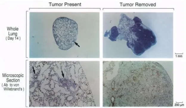
(26) Thèse de Mathieu Danoy, Lille 1, 2017. Chapter 1. General Introduction. in some cases, there was no trace of embolus close to secondary sites of metastasis. [12]. .. Moreover, some tissues such as the heart, the kidneys, the intestine and the muscles which account for a great part of the blood output in the body, are not found to be common sites of metastasis and are rarely colonized [10]. The site specific and non-blood flow dependent colonization of metastasis in the body has been explained since Paget’s hypothesis [13]. The interactions between the endothelial cells of the secondary site and the cancer cells have been defined by the fact that the cell-surface receptors of the endothelial cells in each organ are different and that the cancer cells respond to local growth factors which are able to stimulate their migration into the tissues. This hypothesis, made from in-vivo observations, could be verified, in some cases, in in-vitro models. [14]. . Auerbach et al. have shown that the adhesion of several types of cancer. cells on endothelial cells extracted from different sites exhibited in most cases, the same patterns observed previously. This proves that without even taking in account the different growth factors produced by the cells in the vicinity of the endothelial cells, the cell specific cell-surface markers expressed would explain, in most cases, the distribution of the metastasis in non-random sites. Since then, number of molecules have been analyzed and their correlation with organspecific metastasis has been defined [15]. Metastasis specific to the liver, the brain, the bones, and other major sites have been partially eluded but the complexity of the phenomenon and the discrepancies of the data obtained by different groups have not lead to the identification of specific targets for therapy. However, as the liver is one of the most common distant site for metastasis which cannot be explained by the pattern of blood flow in the body (Fig. 1-4), it has been a therapeutic focus for many years and many trials have been performed.. 25 © 2017 Tous droits réservés.. lilliad.univ-lille.fr.
(27) Thèse de Mathieu Danoy, Lille 1, 2017. Chapter 1. General Introduction. Fig. 1-4: Examples of metastatic pattern which cannot be explained by the pattern of blood flow in the body [10].. Historically, therapies toward cancer metastasis includes resection (i.e. the surgical removal of an organ, in this case, host of the tumor), radiation therapy and chemotherapy. However, in the case of the liver, results from resection have been quite mixed and unsuccessful [16]. . The survey performed by Foster indicated that, because there are much difficulties to. perform the resection of parts of the liver, the mortality during the operation remained at 11%. Moreover, the survival rate after 5 years was found to be close to zero, strongly putting in perspective the usefulness of the operation. The lack of therapeutic success and the continuing high mortality rate of the primary cancers that metastases in the liver have led to the development of new strategies either acting directly on the adhesion of the cells in the vasculature or on their migration into the tissues.. 26 © 2017 Tous droits réservés.. lilliad.univ-lille.fr.
(28) Thèse de Mathieu Danoy, Lille 1, 2017. Chapter 1. General Introduction. 1.2 Main Issues in Assays for Cancer Metastasis The increase of regulations toward animal experiments has made the research for an alternative and by extension, the development of in-vitro assays, a hot topic for the past few years. Indeed, in addition to stricter guidelines in terms of protocol approval and of formation of the individuals performing the experiment, the rule of the “3R” has been set for example in Japan [17]. This rule compels the researchers to find alternative methods (Replacement) to their experiments on animals, to reduce the number of animals used (Reduction) and to reduce the pain inflicted to them (Refinement). While the development of in-vitro models is strongly encouraged by this rule, both invivo and in-vitro methods have their advantages and inconvenient. The simplicity of in-vitro models allows to decrease the number of variables and the fluctuations in the results. [7]. .. However, those models do, by definition, not represent the exact in-vivo situation and need to be compared in in-vivo situation while limiting the use of animals. The skills required to perform in-vitro assays are also different from those of their main targeted users, the clinicians and they are still laborious to completely characterize. In-vivo studies then appear as a “gold standard” for assays of cancer metastasis but they also have their limits. For example, assays for drug screening should be designed for a human patient. Many trials which have been led from the animal stage to the human trial stage have been seen to fail as the behavior of the species are generally different. Ethics concerns regarding animal experiments might also be raised regarding in-vivo assays. Moreover, and more importantly, in-vivo assays do give a general information on the final output of an assay. The metastasis might or might not have occurred but the specific reason remains unknown. [7]. . This is due to the lack of technic,. allowing a direct real-time in-vivo observation of the phenomenon and which would permit to conclude on the specific mechanism involved in the assay.. 27 © 2017 Tous droits réservés.. lilliad.univ-lille.fr.
(29) Thèse de Mathieu Danoy, Lille 1, 2017. Chapter 1. General Introduction. 1.3 Current Strategies in Cancer Metastasis 1.3.1 Previous research on in-vivo models 1.3.1.1 Choice of the animal model Murine models have been extensively used for in-vivo metastasis assays and have been challenged in only a few cases by companion animal models. [18,19]. . The use of rat and mouse. for in-vivo experiments is indeed favored by a relatively low cost, the high degree of development of the assays, the possibility to genetically engineer mice and the lower ethical concern of experimenting on those small animals [18]. However, while being a primary source of information regarding cancer metastasis, those models, often issued from inbred population and raised in environmentally-controlled laboratories, may not be the most representative models for cancer in the human which, unlike in murine species, appears spontaneously and is affected by the environmental exposure of the patient [19]. Companion animals (dogs and cats), may give an alternative to murine models and have already been used in preclinical trials [20]. The advantages of pets as a model are numerous. As companions, they are often exposed to the same environment, source of carcinogenesis, as their owner and, for example, the exposure of the later to asbestos has been related to the occurrence of cancer in their pet animals. [21]. . Those models are also obtained from natural outbreeds (in. opposition with murine models), are subjected to spontaneous metastasis as it is the case in human, exhibit higher incidence rates of cancer than humans, allowing studies over large population and with a rate of progression higher than in humans [18]. Finally, in terms of size, they are more comparable to human and similarities in genes responsible for cancer have been found in canine models and humans [22]. However, the use of these models is often limited by the lack of reagents, such as antibodies available for analysis. [19]. and ethical concerns. While. both murine and companion animal models provide different set of information regarding 28 © 2017 Tous droits réservés.. lilliad.univ-lille.fr.
(30) Thèse de Mathieu Danoy, Lille 1, 2017. Chapter 1. General Introduction. cancer progression, it is important to note that one model is not to be taken as better than the other and that different models are able to answer different questions regarding the same phenomenon.. 1.3.1.2 Syngeneic & Xenograft models Murine models can be devised in two distinct categories, syngeneic or xenograft. Syngeneic models refer mostly to assays involving cancer cells of the same genetic background as the host. [18]. . Most of these models are designed by using murine models, with the. disadvantages previously described. However, as both the cancer cells and the host are of the same origin, interactions between the cells and the tumor microenvironment can be studied in detail. Especially in those models, the influence of proteins added to the extracellular space [23] or of the modification of the tumor environment. [24]. have been studied and the possibility to. stimulate metastasis shown. More interestingly, those models have also been used to test the effects of different tumor-inducing chemicals [25]. While the same oncogenes were found in the tumor, different mutations of the cells were noticed in each case. In this case, the model allowed to understand that in response to different exposures, different pathways of mutations were activated but tumors containing the same oncogenes could be finally formed, highlighting the diversity of initiating agents for the cancer cascade. The tumors formed by chemical exposure can, in most cases, be transplanted in other animals after extraction. [26]. . The character of the. cells can be preserved up to several transplantations, allowing to perform several assays with cells of the same origin, behavior and genetic expression and reducing drastically the lack of reproducibility in in-vivo assays. Xenograft models correspond to the transplantation of human tumor cells in immunodeficient mice. The animal host must be immuno-depressed to prevent any rejection of the 29 © 2017 Tous droits réservés.. lilliad.univ-lille.fr.
(31) Thèse de Mathieu Danoy, Lille 1, 2017. Chapter 1. General Introduction. transplanted tissue. This type of model is the most widely used nowadays by cancer researchers. Especially, the transplantation of human breast cell lines into different types of mice has been extensively characterized and the difference of progression in different mutations of mice allowed to understand more in details the immunobiology of breast cancer [27]. Those models allow to follow, for extended period, the progression of cancer in the host [28] and provide useful information on what might be the development of those cancers in human. As the mice used are immuno-deficient, the interactions between the immune system and the tumor cannot be studied. To solve this problem, models including both human immune cells and human tumor cells in an in-vivo model have been established [29]. However, those models using transplant of human cells still have limits. It has been suggested that in terms of angiogenesis and tumor growth, tumors that have been transplanted and tumor that developed spontaneously behave differently [30,31].. Fig. 1-5: Steps and effects in the development of (A) spontaneous cancer (B) transplanted cancer [30].. Xenograft are proposed to be less dependent on the regulation of angiogenesis in the host and their vascular network suggested to be rapidly developing independently of the host. In 30 © 2017 Tous droits réservés.. lilliad.univ-lille.fr.
Figure
![Fig. 1-1: The Hallmarks of Cancer [3] .](https://thumb-eu.123doks.com/thumbv2/123doknet/3684155.109201/23.892.286.607.349.731/fig-hallmarks-cancer.webp)
![Fig. 1-2: Probability (%) of development of an invasive cancer in the United States between 2009 and 2011 sorted by age and sex [5]](https://thumb-eu.123doks.com/thumbv2/123doknet/3684155.109201/24.892.113.787.507.848/fig-probability-development-invasive-cancer-united-states-sorted.webp)
![Fig. 1-3: The metastatic process from the primary site to the secondary site [8] .](https://thumb-eu.123doks.com/thumbv2/123doknet/3684155.109201/25.892.251.639.106.463/fig-metastatic-process-primary-site-secondary-site.webp)
![Fig. 1-4: Examples of metastatic pattern which cannot be explained by the pattern of blood flow in the body [10]](https://thumb-eu.123doks.com/thumbv2/123doknet/3684155.109201/27.892.115.806.113.344/fig-examples-metastatic-pattern-explained-pattern-blood-flow.webp)
+7
![Fig. 1-5: Steps and effects in the development of (A) spontaneous cancer (B) transplanted cancer [30]](https://thumb-eu.123doks.com/thumbv2/123doknet/3684155.109201/31.892.196.700.588.880/fig-steps-effects-development-spontaneous-cancer-transplanted-cancer.webp)
![Fig. 1-7: Imaging of fluorescent protein-expressing cancer cells metastasis in mice. (A) Whole body image (B-I) Details of bones metastasis [42]](https://thumb-eu.123doks.com/thumbv2/123doknet/3684155.109201/35.892.300.590.110.609/imaging-fluorescent-protein-expressing-cancer-metastasis-details-metastasis.webp)
![Fig. 1-9: (Left) Migration of endothelial cells (Green Staining) towards Tumors (Red staining) during angiogenesis and driven by hypoxia, scale bar 50 μm [47] (Right) Migration](https://thumb-eu.123doks.com/thumbv2/123doknet/3684155.109201/37.892.117.800.547.826/migration-endothelial-staining-tumors-staining-angiogenesis-hypoxia-migration.webp)
![Fig. 1-13: Migration of breast cancer cells, triggered by coculture with fibroblasts [65]](https://thumb-eu.123doks.com/thumbv2/123doknet/3684155.109201/42.892.193.757.637.1057/fig-migration-breast-cancer-cells-triggered-coculture-fibroblasts.webp)
![Fig. 1-14: The coculture of different types of cells induces different pattern of migration [75]](https://thumb-eu.123doks.com/thumbv2/123doknet/3684155.109201/45.892.314.577.492.790/coculture-different-types-cells-induces-different-pattern-migration.webp)
Documents relatifs
