Clonage et caractérisation des protéines liant
l’élément de réponse à l’insuline (IREBP) du gène de
I’angiotensïnogène chez le rat
Par Chih-Chang Wei
Programme de sciences biomédicales
Faculté de médecine
Thèse présentée à la faculté des études supérieure
En vue de l’obtention du grade de docteur ès sciences (Ph.D)
En sciences biomédicales
Avril 2007
L
J
de Montréal
Direction des bibliothèques
AVIS
L’auteur a autorisé l’Université de Montréal à reproduire et diffuser, en totalité ou en partie, par quelque moyen que ce soit et sur quelque support que ce soit, et exclusivement à des fins non lucratives d’enseignement et de recherche, des copies de ce mémoire ou de cette thèse.
L’auteur et les coauteurs le cas échéant conservent la propriété du droit d’auteur et des droits moraux qui protègent ce document. Ni la thèse ou le mémoire, ni des extraits substantiels de ce document, ne doivent être imprimés ou autrement reproduits sans l’autorisation de l’auteur.
Afin de se conformer à la Loi canadienne sur la protection des renseignements personnels, quelques formulaires secondaires, coordonnées ou signatures intégrées au texte ont pu être enlevés de ce document. Bien que cela ait pu affecter la pagination, il n’y a aucun contenu manquant. NOTICE
The authoc of this thesis oc dissertation has granted a nonexclusive license allowing Université de Montréal to reproduce and publish the document, in part or in whole, and in any format, solely for noncommercial educationaT and research purposes.
The author and co-authors
if
applicable retain copyright ownership and moral rights in this document. Neither the whole thesis or dissertation, nor substantial extracts from it, may be printed or otherwise reproduced without the author’s permission.In compliance with the Canadian Privacy Act some supporting forms, contact information or signatures may have been removed from the document. While this may affect the document page count. it does flot represent any loss of content from the document.
Université de Montréal
Faculté des études supérieure
Cette thèse intitulée
Clonage et caractérisation des protéines liant
l’élément de réponse à l’insuline (IREBP) du gène de
l’angiotensinogène chez le rat
Présenté par:
Chih-Chang Wei
a éte évaluée paré un jury composé des personnes suivantes:
Dw. Johanne Trembly
Président rapporteurDt. John S.D. Chan
Directrice de rechercheD’. Marie-Josée Hébert
Membre du juryDte. Tomoko Takano
Examinateur externe
RÉSUMÉ
De plus en
plus d’études
démontrent que l’hyperglycémie et
l’activation du système rénine-angiotensin (RAS) sont des facteurs de
risque majeurs dans la pathogénèse de la néphropathie diabétique (DN).
L’existence d’un RAS local intrarénal est maintenant bien acceptée. Toutes
les composantes du RAS sont exprimées dans les cellules du tubule
proximal du rein. L’angiotensinogène est le seul substrat du RAS et est
exprimé principalement dans les RPTC; il est ensuite converti en Ang li
(forme biologiquement active), par la rénine rénale et l’enzyme convertrice
d’angiotensine (ACE). Les niveaux d’Ang li et l’expression génique du RAS
sont augmentés dans le diabète, indiquant que le RAS intrarénal joue un
rôle important dans la progression de DN.
Le(s) mécanisme(s) de
régulation de l’expression génique d’AGT dans le rein par un milieu élevé
en glucose ainsi que l’insuline, demeurent cependant mal compris.
Afin d’identifier et des cloner ces protéines ainsi que définir leur action
sur l’expression génique d’AGT, les protéines nucléaires extraites des
IRPTC ont été séparées par électrophorèse en deux dimensions. Les
protéines positives ont été hybridées par Southwestern Blot, identifiées par
spectrométrie de masse et enfin confirmées par Western Blot. Nous avons
identifé deux protéines nucléaires de 48-kD et de 70-kD identiques aux
ribonucléoprotéines hétérogènes F (46-kDa hnRNP F) et hnRNP K (65-kDa
hnRNP K). Les cDNA de hnRNP F et hnRNP K ont été clonés des IRPTC
par RT-PCR et ensuite exprimés dans des clones bactériens.
Les
recombinants bactériens de hnRNP F et hnRNP K sont liés à l’élément de
réponse à l’insuline (IRE)-AGT du rat, comme démontré par essai de
retardement sur gel (GMSA) et immunoprécipitation de la chromatine
(ChIP). L’addition d’anticorps polyclonaux contre hnRNP F résulte en un
supershift dans le GMSA. La transfection transitoire de cDNA de hnRNP F
ou hnRNP Kdans des IRPTC a inhibé l’expression AGT au niveau mRMA
et protéine. D’autre part, le knockdown de hnRNP F ou hnRNP K par small
interference RNA augmente l’expression de AGT au niveau mRNA (et
protéine?) dans les IRPTC. De plus, hnRNP F interagit avec hnRNP F dans
les essais de pulldown et de Co-IP
La co-transfection de hnRNP F et
hnRNP K inhibent l’expression génique d’AGT au niveau mRNA et protéine.
Un mileu élevé en glucose stimule hnRNP F ou hnRNP K tandis que
l’insuline inhibe leur expression dans les IRPTC in vitro et des RPTC de rat
in vivo. Nous avons établi des tranfectants stables d’IRPTC surexprimants
hnRNP F et des souris transgéniques surexprimant hnRNP F de façon
spécifique dans les RPIC in vitro et in vivo. Nous avons démontré que la
surexpression de hnRNP dans les RPTC peut prévenir la stimulation par le
glucose (25mM) d’AGI ainsi que de growth factor-31 (TGF-31) au niveau
mRNA et protéine, ainsi que l’hypertrophie cellulaire (ie: contenu total en
protéine de la cellule,
incorporation [3H]-Leucine et expression protéique
de
p27kinf)De plus, les souris transgéniques surexprimants HnRNP F
démontrent une atténuation de l’expression d’AGT et du récepteur TGF-31
Il au niveau mRNA ainsi qu’une diminution de l’expression de la protéine
p27’) et du ratio protéine/ADN dans les RPTC de souris diabétiques
induite au streptozotocin. Ces observatioms suggèrent qu’AGT est
modulée par HnRNP F et HnRNP K via la liaison avec l’IRE, action aussi
régulée par le glucose et l’insuline. Nous résultats démontrent aussi que
HnRNP F ou K pourrait jouer un rôle protecteur ou modulateur dans la
prévention de l’hypertrophie des RPTC dans le diabète et ses mécanismes
sont médiés via l’atténuation de l’expression génique d’AGT intrarénal et
de la voie de signalisation TGF-E31
in vitro
and
in vivo.
Mots-clé: angiotensinogène, hnRNP F, hnRNP K, glucose, Insuline,
élément de réponse à l’insuline, souris transgéniques, rein, hypertrophie
ABSTRACT
Accumulating evidence has demonstrated that hyperglycemia and
renin-angiotensin system (RAS) activation are major risk factors in the
pathogenesis of diabetic nephropathy (DN). The existence of a local
intrarenal RAS has now been well accepted. AIl components of the RAS
are expressed in renal proximal tubular cells (RPTCs). Angiotensinogen
(AGT) is the sole substrate in the RAS and is expressed predominantly in
RPTCs and converts into biologically-active Ang II by renal renin and
angiotensin converting enzyme (ACE). Intrarenal Ang Il levels and RAS
gene expression are elevated in diabetes, strongly indicating that intrarenal
RAS activation plays an important role in the progression of DN. The
mechanism(s) of regulation of intrarenal AGT gene expression by high
glucose and insulin remain, however, incompletely understood. Previously,
our lab has demonstrated that an insulin-responsive element (IRE) in rat
AGT gene promoter that binds to two nuclear proteins with apparent
molecular weights of 48 and 70 kD from rat immortalized RPTCs (IRPTC).
The expression of
two
nuclear proteins in IRPTCs was up-regulated and
down-regulated by high glucose and insulin, respectively. To identify and
clone these proteins and to define their action on AGT gene expression,
nuclear proteins from
IRPTC were separated by 2-dimentional gel
electrophresis, positive proteins were detected by Southwestern blotting
and identified by mass spectrometry and subsequently confirmed by
Western blotting. We identified that the 48-kD and 70-kD nuclear protein
were identical to 46-kD and 65-kD heterogenous ribonucleoprotein F
(hnRNP F) and hnRNP K. HnRNP F and K cDNAs were then cloned from
IRPTC by reverse transcriptase-PCR and expressed in bacteria. Bacterially
expressed recombinant hnRNP F and K bound to rat AGT-IRE, as revealed
by gel mobility shift assay (GMSA) and chromatin immunoprecipitation
assay. The addifion of polyclonal antibodies against hnRNP F yielded a
supershift in GMSA. Transient transfection of hnRNP F or hnRNP K cDNA
in IRPTC inhibited AGT mRNA and protein expression. In contrast,
knockdown hnRNP F or hnRNP K gene expression by small interference
RNA enhanced AGT mRNA expression in IRPTC. Moreover, hnRNP F
interacted with hnRNP F in pulldown and co-immunoprecipiation assays.
Co-transfection of hnRNP F and hnRNP K further suppressed AGT mRNA
expression. Hyperglycemia stimulated and insulin inhibited hnRNP F and
hnRNP K expression in IRPTCs in vitro and rat RPTCs in vivo. We have
established stable IRPTC transfectants overexpressing hnRNP F and
transgenic mice overexpressing hnRNP F specifically in RPTCs
in vitro
and
in vivo.
We demonstrated that overexpression of the hnRNP F in RPTCs
prevented the high-glucose stimulation of AGT and transforming growth
factor-f31 (TGF-1) mRNA and protein expression as well as cellular
hypertrophy, i.e., total cellular protein contents, [3H]-Leucine incorporation
and
p27kIPlprotein expression). HnRNP F transgenic mice displayed an
attenuation of AGT and TGF-31-receptor II mRNA expression as well as a
decrease of celi and nuclear volume,
27kiP1protein expression and
protein/DNA ratio in RPTCs in streptozotocin-induced diabetic mice. Taken
together, these data suggest that AGI was modulated by hnRNP F and
hnRNP K through the binging with IRE and this was also regulated by
glucose and insulin. Our findings demonstrate that hnRNP F or K may play
a protective or modulatory role in preventing RPTC hypertrophy in diabetes
and its underlying mechanism is mediated via attenuation of intrarenal AGI
expression and TGF-131 signaling
in vitro
and
in vivo.
Key words: Angiotensinogen, hnRNP F, hnRNP K, glucose, Insulin,
insuli n-responsive element, transgenic mice, kidney, hypertrophy.
Table of Contents
Résumé
iii
Abstract
vi
Table of Contents
ix
List of Tables
xiv
ListofFïgures
xv
List of Abbreviations
XviAcknowledgments
xxi
Chapter I
—Introduction
1.1 Anatomy and Function cf the Kidneys
2
1.2 End-Stage Renal Disease (ESRD)
3
1.2.1 EpidemiologyofESRD
3
1.2.1.1 Incidence ofESRD
3
1.2.1.2 Prevalence of ESRD
4
1.2.2 Health Care Cost of ESRD
5
1.2.3 Causes of ESRD
5
1.2.4 Diabetes and Renin-Angiotensin System (RAS)
7
1.2.5 Diabetic Nephropathy
8
1.2.6 CeIlular Hypertropathy in Diabetic Nephropathy
8
1.2.7 Hyperglycemia and Kidney Injury
10
1.3TheRAS
11
1.3.1 Biosynthesis cf RAS Members
12
1.3.2 Historical Views of RAS
17
1.4 Local RAS
.21
1.4.1 Discovery of local RAS
22
1.4.2 Local RAS in the Kidneys and Its Function
23
1.5 Activation of the Local Renal RAS in DN
25
1.5.1 Ang in DN
25
1.5.2Ang Receptors in DN
27
1.5.3Ang ll-Induced DN
27
1.6 Clinical Trials of RAS Blockages in Diabetic Patients
28
1.6.1 Clinical Trails ofACEi in Diabetic Patients
29
1.6.2 Clinical Trails ofARBs in Diabetic Patients
33
1.7 Gene Regulation ofAGT
34
1.7.1 AGT Regulated by Hormonal Factors
35
1.7.2 AGI Expression in Diseases
36
1.7.3lranscriptional Regulation of AGI
37
1.8 Insulin and AGI
38
1.8.1 Insulin Signaling
38
1.8.2 Insulin Effect on Gene Regulation
41
1.9 Heterogenous Nuclear Ribonucleoprotein Family (hnRNP)
45
1.9.1 Structure of the hnRNP Family
46
1.9.2 Transcriptional Function of the hnRNP Family
47
1.9.3hnRNPF
49
1.9.4 hnRNP K
50
1.10 Mouse Models in Diabetes Research
52
1.10.1 Characterization 0f Diabetic Nephropathy
in Humans and Mice
53
in Research of Diabetic Nephropathy
.55
1.11 Hypothesis andAims of These Studies
58
Chapter 2
—Article I
Heterogenous nuclear ribonucleoprotein F modulates
angiotensinogen gene expression in rat kidney proximal tubular ceils.
2.1 Abstract
63
2.2 Introduction
64
2.3 Materials and Methods
66
2.4 Results
76
2.5 Discussion
81
2.6 Acknowledgements
87
2.7 References
88
2.8 Figures
106
Chapter 3
—Article 2
Heterogeneous nuclear ribonucleoprotein K modulates
angiotensinogen gene expression in kidney cells.
3.1 Abstract
119
3.2 Introduction
120
3.3 Materials and Methods
122
3.4 Results
133
3.5 Discussion
137
3.7 References
.143
3.8 Figures
157
Chapter 4— Article 3
Heterogenous nuclear ribonucleoprotein F overexpression attenuates
angïotensïnogen gene expression and renal proximal tubular ceil
hypertrophy in transgenic mice.
4.1 Abstract
169
4.2 Introduction
170
4.3 Materials and Methods
171
4.4 Results
177
4.5 Discussion
182
4.6 Acknowledgements
187
4.7 References
187
4.8 Figures
192
Chapter 5— Discussion
5.1 Protein-DNA interaction
203
5.1.1 General prospecting technologies to elucidate Protein-DNA
interaction
204
5.1.2 Advantages and disadvantages of 2D gel electrophoresis
combined with MS
207
5.2 Advantages and disadvantages of IRPTCs
210
Expression
.212
5.4 Renopathology Significance of Suppression AGI Gene
Expression via hnRNP F or hnRNP K in Hyperglycemia
216
5.5 Physiological Significance of Suppressing Local RAS
Gene Expression via hnRNP F and hnRNP K in Diabetes
220
5.6 Conclusion
223
Chapter 6 —Perspectives of Research
6.1 Correlation between hnRNP F/hnRNP K and RAS
Gene Expression in Diabetic Animais in vivo
226
6.2 Identification ofTranscriptional Factors that lnteract or
Associate with hnRNP F and hnRNP K in vitro
227
6.3 Gene Chip Microartays to ldentify the Downstream Genes that are
Differentiaily Regulated by hnRNP F and hnRNP K in RPTCs
228
6.4 Potential Gene Therapy with hnRNP F and hnRNP K
229
Chapter 7— References
List of Tables
Table I
Proposed mechanisms of angÏotensin Il effects in
diabetic nephropathy
Table 2
Receptors for angiotensin related peptides
Table 3
Clînîcal evïdence for prevention of diabetes with
ACE inhibïtors
Table 4
Clinical evidence for prevention of diabetes with
ARBs
Table 5
Insulîn response sequenceslelements
Lïst of Figures
Figure 1 Incident ESRD RRT patients, selected countries, from 1998-2003 (Crude Rate PMP)
Figure 2 Prevalent ESRD patients at year-end, Canada 1990-2004 (number)
Figure 3 Distribution of incident ESRD patients by primary diagnosis, Canada, 1993-2004
Figure 4 Enzymatïc pathway of bioactive angiotensins generation
Figure 5 Structure ofATI and AT2 receptors
Figure 6 Tubular and interstïtial formation, secretion and uptake of angiotensin
Figure 7 Insulin signaling pathway and its cellular function Figure 8 General structural domains of the hnRNP family Figure 9 Structure of hnRNP F
Figure 10 Interaction of hnRNP K with TATA-Box Binding Protein (TBP)
Figure 1f HnRNP K overexpression prevents cellular hypertrophy in high glucose
Figure 12 Endogenous hnRNP F and K mRNA expression in dïabetic animal models
Lïst of Abbreviations
2D gel
2-dimentional gel
ACC2
Acetyl-Co-A Carboxylase-2
ACE
Angiotensin-converting enzyme
ACEI
Angiotensin-converting enzyme inhibitor
AGCEI
AGT core promoter element 1
AGCFI
AGT core promoter binding factor 1
AGI
Angiotensinogen
ALLHAT
The Antihypertensive and Lipid-Lowering treatment
to prevent
Heart Attack trial
ALPINE
The Antihypertensive treatment and Lipid Profile In a
North of Sweden Evaluation
Ang
Angiotensin
AP-1
Activator protein 1
APA
Aminopeptidase A
APN
Aminopeptidase N
Apo-ClIl
apolipoprotein CIII
ARBs
Angiotensin II receptor blockers
ASCOT
The anglo-scandinavian cardiac outcomes trial
ATI
Angiotensin II receptor type 1
AT2
Angiotensin II receptor type 2
CAPPP
The Captopril Prevention Project
CbI
Casitas B-lineage lymphoma
CHARM
The Candesartan in Heart failure—Assessment of
Reduction in Mortality and morbidity
CIHI
Canadian Institute for Health Information
CVD
Cardiovascular disease
DAG
Diacylglycerol
DREAM
The Diabetes Reduction Approaches with ramipril
and rosiglitazone Medications trial
ECM
Extracellular matrix
ERK
Extracellular signal-regulated kinase
ESRD
End-stage renal disease
FAS
Fatty acid synthesis
FOXOI
Forkhead transcription factor box 01
G6Pase
Glucose-6-Phosphatase
GLUT4
Glucose transporter 4
Grb2
Growth factor receptor binder-2
Gsk3
Glycogen synthase kinase 3
GSK3f3
glycogen synthesis kinase 3
HCTZ
Hydrochlorothiazide
HNFI
Hepatic nuclear factor 1
HNF3
Hepatic nuclear factor 3
hnRNP
Heterogeneous nuclear ribonucleoproteins
HOPE
The Heart Outcomes Prevention Evaluation
1DDM
Insulin-dependent diabetes mellitus
IEF
Isoelectric focusing
IGF
insuiin-like growth factor
IGFIR
IGF receptor 1
1P3
1,4,5-inositol triphosphate
1PG
immobilized pH gradient
IR
insuiin receptor
IRE
Insulin-responsive element
IRPTC
Immortalized renal proximal tubular ceil
1RR
1R-related receptor
IRS
insulin receptor substrates
JNK
]un (N)-terminal-kinase
LIFE
The Losartan Intervention For Endpoint
reduction in hypertension study
MALDI
Matrix-assisted laser desorptionhionization
MAPK
Mitogen activated protein kinase
MMP-1
Matrix metalioproteinase-1
MS
Mass spectrometry
mSOS
Mammalian son of seven less
mTOR
Mmmalian target of Rapamycin
N1DDM
Non-insulin-dependent diabetes meilitus
p70S6Kp70 ribosomal S6 kinase
PDE3b
Phosphodiesterase 3b
PDK
Posphoinositide-dependent kinase
PEPCK
Phosphoenoolpyruvate carboxykinase
PH
Pleckstrin homoiogy
PI
Posphatidylinositol
pi
isoeiectric point
P13K
Phosphatidylinositol 3-kinase
PKC
Protein kinase C
PRA
Plasma renin acfivity
PTK
Protein tyrosine kinase
RAAS
Renin-ang iotensin-aldosterone system
RAS
Renin-Angiotensin System
RBD
RNA binding domain
RPTC
Renal proximal tubular ceil
RRM
RNA recognition motif
RRT
Renal replacement therapy
SCOPE
The Study on COgnition and Prognosis in the Elderly
Set
Serine
SH2
Src homology 2
SHC
Src homology collagen
SHR
Spontaneously hypertensive rats
SOLVD
The studies of left ventricular dysfunction
SRE
Sterol response element binging protein-J
SREBI
Sterol response element binging protein-1
STZ
Streptozotocin
TAT
Tyrosine aminotransferase
TBP
TATA box binding protein
TGF-f3
Transforming growth
factor-TOF
Time-of-flying
TTF2
Tyroid transcription factot-2
VALUE
The valsartan antihypertensive Iong-term use
eval uation
ACKNOWLEDGEMENTS
I wouid like to express my gratitude to ail those who gave me the
possibiiity to compiete this thesis. Especialiy, I am deepiy indebted to my
supervisor Prof. Dr. John S.D. Chan whose help, stimulating suggestions
and encouragement helped me in ail the time of research for and writing of
this thesis.
1.1 Anatomy and Function of the Kidneys
In humans, the kidneys are two bean-shaped organs, each about the
size of a fist and Iocated in the posterior part of the abdomen, on each side of the spine. The right kidney sits just below the liver, and the Ieft kidney, below the diaphragm, adjacent to the spleen. In a normal human aduit, each kidney weighs 150 grams and is about 10 cm long, 5.5 cm wide and 3 cm thick (1; 2).
Both kidneys are surrounded by 3 layers. 1. The renal capsule covers the outer surface of the entire organ. 2. Fat keeps them in place and surrounds the renal capsule. 3. The renal fascia is a dense, fibrous outer layer that also secures the kidneys to the posterior abdominal wall and surrounding structures (3). The kidneys themselves are made up of 2 layers, the cortex and the medulla. The cortex is the outer layer, and the medulla, the inner layer. Wthin the medulla are 8-18 triangular structures, the renal pyramids. The tips of the pyramids are referred ta as renal papillae. The renal cortex and the pyramids together make up the parenchyma, which consists of approximately 1.25 million nephrons, the functional units cf the kidneys that produce urine and help regulate blood composition (1; 4).
The kidneys filter wastes (such as urea) from the blood and exctete them, along with water, as urine. The nephrons consist of renal tubules and renal corpuscles. The tubules are approximately 50 mm in length and comprise the convoluted tubules (proximal and distal) and loop 0f Henle (4). The proximal tubule, as a part of the nephron, can be divided into an initial convoluted portion and a following straight (descending) portion. Fluid in
filtrates entering the proximai convoluted tubule is reabsorbed into the peritubular capillaries, inciuding approximateiy two-thirds of filtered sait and water (65%) and ail (100%) filtered, organic solutes (primarily glucose and amino acids). These functions of the renal tubules are performed by a number of transporters and channels expressed at the apical and basolateral membranes of the tubular cells (5).
1.2 End-Stage Renai Disease (ESRD)
ESRD (or kidney failure) is a slow, progressive loss of kidney functions caused by inherited disorders, prolonged medical conditions or the long-term use of medications (6; 7). If untreated, it is irreversible and fatal. Renal replacement therapy (RRT), which includes hemodialysis, peritoneal dialysis and renal transplantation, is the major treatment available since the 1960s for ESRD patients (8). ESRD has become a serious medical and economic health problem in developed countries, including Canada (9; 10).
1.2.1 Epidemiology of ESRD
The Canadian lnstitute for Health Information (CIHI) is the national information system that provides timely, accurate and comparable health information on ESRD and other diseases in Canada.
1.2.1.1 Incidence of ESRD
The incidence of ESRD represents the number cf persons newly diagnosed with the disease in a specific population in a given time period. It
is useful in medicai and epidemiological research examining the causes of
disease and the differences in subpopulations affected by these causes. The incidence of ESRD in Canada and other countries during 1998 to 2003
4
and this number continues to tise every year. If we examine the data in more detail by age group, the rate among persons aged 19 years or younger was relatively stable, and decreased by 5% in young adults (20 to 39 years old). However, it increased by 32% among those between the ages of 40 to 59 years, and by 17% in people over 60 years (11).
400 35t;. ---_}__—- _______-- — T)—-— --•—-—— — 300 —--- -, Q. S 70 -—---— - _:_— C - - — 200. --2 100 — -•----_ •--50 . —— o —----___________________________________________________________ 19913
I
1999 2000 2001 2002 20C3 ---__ —-—- Cncl 41 150 150 158 19 •- At,j 86 1 92 9493 — -1.- --- 140J
50 149 160j
170J
172 »13c.,-,- 48 413 175 1133 174 186 234 231 250 250 254 254 --.2-- 135 j- 152 147 167 153 137 127 125 120 126 124 121 —.2—United 9tts - 300] 321 -- 325 32 336 331 YerSource Canadien Organ Replacement Register, CIHI
Figure 1. Incident ESRD RRT patients, selected countries, from 199$-2003
(Crude Rate PMP)
1.2.1.2 Prevalence of ESRD
ESRD prevalence represents the number of persons in a specific population who have ESRD at a given time point. Figure 2 shows that ESRD prevalence increased dramatically, by almost 2-fold from 1990 to 2004 in Canada. At the end of 2004, 18,827 patients were on dialysis and 12,099 were living with a functioning kidney transplant, for a total of 30,924 Canadians with kidney failure registered in the CIHI (11).
Source Canadian Organ Replacement Register, Canadian Institute for Health Information Ç2OŒ)
Figure 2. Prevalent ESRD patients at year-end, Canada,1990-2004 (Number)
1.2.2 Health Care Costs of ESRD
The health care costs incurred by dialysis and transplantation are enormous. The average cost of peritoneal or hemodialysis is
$50,000-75,000 per patient year(12; 13). At least 1 billion Canadian dollars were spent to maintain the quality of life of ESRD patients in last 5 years. Since its incidence and prevalence have increased every year in Canada, the economic burden of ESRD in Canada has become a serious probtem. Effective and economic treatment as well as prevention of ESRD onset are major ways of reducing its clinical incidence and could resuit in considerable cost savings for the Canadian public healthcare system. 1.2.3 Causes of ESRD
According to the CIHI, there are several major causes of ESRD (Figure 3). Diabetes and hypertension are the number J and 2 causes cf
35fJ 25JŒI ; 2OXJ E iojjœ o 1990 1991 1992 1993 1994 1996 1996 1997 1998 1999 2000 2001 2002 2003 2004
kidney disease, respectively, in the USA and Canada. Besides this, kidney failure patients aged 65 and older had the highest overali rate of diabetes between 1995 and 2004, more than doubling from 124 per million in 1995 to 270 per million in 2004.
Unknin
Other
Diibetes
Diabetes is the leading cause of ESRD, accounting for more than 40%
of ail registered ESRD patients in Canada, up from 25% 10 years ago, a
paftern that is also being encountered in the USA. In addition, it is noteworthy that diabetes types account for these increases. While the number of ESRD patients with type 1 diabetes declined from 526 in 1995 to 303 in 2004 (down 42% in 10 years), the number of patients with type 2 diabetes more than tripied over the same period, from 540 to 1,836. Investigating effective ways of reducing the prevalence of diabetes couid
Vascular disease P(D Dmg Pyelonephribs Glomerulo nephrltls Q03 04 90 Q8 e97 Q0 00 2000 2001 2002 2003 2004
Source: Canadian Organ Reptacement Register, Canadian Institute for Health Information (2006)
Figure 3. Distribution of incident ESRD patients by primary diagnosis, Canada, 1993-2004
heip decrease the devastating health consequences associated with it and ESRD.
1.2.4 Diabetes and the Renin-Angiotensin System (RAS)
Diabetes is a chronic disease characterized by hypergiycemia and complications 0f multiorgan dysfunction, i.e. the kidneys, heart, brain and eyes. In Canada, ovet 2 miflion people have diabetes, of which there are 3 main types: type 1 (insulin-dependent diabetes mellitus, IDDM), type 2 (non-insulin-dependent diabetes mellitus, NIDDM), and gestational diabetes (caused by the hormones of pregnancy). The most common types are type 1 and type 2, with 10 to 21% of diabetes patients developing kidney disease (11).
Considerabie evidence suggests that the intrarenal RAS plays an important role in diabetic nephropathy (DN). Landmark studies by Anderson et al. (14) have revealed the superiority of angiotensin-converting enzyme (ACE) inhibitors (ACEi) in haiting the progression of renai disease. Conversely, chronic infusion of angiotensin
(Ang) II produces marked giomerular and tubulointerstitial injury in the
kidneys (15-17). These studies suggest that Ang II is pivotai in the pathophysiology of chronic renai disease. However, it is difficuit to separate the effects of blood pressure (BP) reduction from these agents in whole animais and humans. This is because patients with overt DN generaily develop systemic hypertension, an adverse factor in ail progressive renai diseases and especially SO in DN. b elucidate the non-hemodynamic
challenge for researchers. Furthermore, there is close interaction between the RAS and transforming growth factor-beta (TGF-3) signaling (18-20). In fact, many profibrotic effects ofAng Il are mediated by TGF-3 stimulation. 1.2.5 Diabetic Nephropathy
DN, a chronic renal disease characterized by glomerulosclerosis,
fibrosis of the tubulointerstitial microenvironment, and tubular atrophy (21),
is due to Iongstanding DM. The deposition and decreased turnover of
extracellular matrix (ECM) proteins, including fibronectin, collagen types I,
III, and
IV,
are important components of the scarring observed during theevolution of glomerulosclerosis and tubulointerstitial fibrosis (22). TGF-f3 serves as a paradigm of profibrogenic cytokines (23). Its function in the renal pathophysiological process has been investigated by Border et al. (24).
DN is one of the most significant long-term complications in terms of
morbidity and mortality for individual patients with diabetes. Although both type 1 and type 2 DM lead to ESRD, the majority of patients are those with
NIDDM. In a prospective study in Germany, the 5-year survival rate was
less than 10% in the elderly population with type 2 diabetes, and no more than 40% in the younger age group with type 1 diabetes. The exact cause of DN is unknown, but various postulated mechanisms are hyperglycemia (causing hyperfiltration and renal injury), advanced glycosylation products, and the activation of cytokines (8; 25; 26).
Diabetic nephropathy is typified by renal hypertrophy, increased
glomerular filtration rate (GFR), and increased urinary albumin excretion
(27; 28). It has been presumed that early hypertrophic changes in the renal
ceNs are integral to the subsequent development of more severe renal
histopathologic changes. Nevertheless, the mechanisms initiating and
regulating the functionai changes in renal ceNs remain incompietely
characterized. The proximal tubuie is uniquely susceptible to a variety of
metabolïc and hemodynamic factors associated with diabetes. Renal
function and prognosis correlate better with structural lesions in the tubuB
and cortical interstitium than with classical glomerular changes of diabetic
nephropathy (29). The proximal tubules show a variety of poorly
characterized changes, which have Ied to the notion that tubular injury
represents a “final common pathway” for proteinuric renai injury. However,
tubular hypertrophy, reduced organic ion transport, and other tubular
changes are already apparent before the onset of proteinuria in diabetes. It
seems reasonable to assume that cytokines and polypeptide growth
factors including inhibitory factors such as TGF-beta induce in concert,
rather than as single factors, tubular hypertrophy (30).
Tubuiar celis can undergo hyperplasia or hypertrophy,
two
totally
different growth responses. Hyperplasia with mitogenesis of tubular cells
plays a central role in the regeneration of functional tubular epithelium
subsequent to acute tubular necrosis. The molecular mechanisms how
mitogenic signais are transduced to the nucleus are relatively welI
characterized. In contrast to the hyperplasia, celiular hypertrophy is less
well understood. Hypertrophic celis are arrested in the Gi-phase of the cell
cycle and increase their size, protein and RNA content, but do not replicate their DNA normally. Such an eniargement of tubular cefls often occurs in more chronic situations of renal damage in which remnant nephrons adapt their function to the increasing need. However, evidence exists that hypertrophic tubules are finaliy joined into the process of maladaptation of renai function leading to tubular atrophy, interstitial scarring, and progression of renai disease (31).
1.2.7 Hyperglycemia and Kidney lnjury
Ihe critical role of hyperglycemia in the genesis of DN has been
established by ceil culture studies, experimental animal models, and clinicai triais. Certain cytokines and growth factors have been identified as likely mediators of the effects of high ambient glucose on the kidneys, and prominent among them is TGF-, a prototypical hypertrophic and fibrogenic cytokine. TGF-p overexpression has been demonstrated in the glomerular and tubuiointerstitial compartments of experimental diabetic animais (32). The TGF- receptor signaling system is also triggered, as evidenced by the upregulation of TGF-f3 type 2 receptors and activation of the downstream Smad signaling pathway (33). Treatment of diabetic mice with neutralizing
anti-TGF-13 antibodies prevents the development of renal hypertrophy,
mesangial matrix expansion, and the decline in renal function. Antibody therapy also reverses the estabiished lesions of diabetic glomerulopathy (34). TGF-3 may also contribute to the cellular hypertrophy found in petsons with DN. Ceil culture studies suggest that high-glucose concentrations stimulatethe hypertrophy of proximal tubular and mesangial celis (35; 36) as weIl as the production of matrix moiecules, such as
fibronectin and collagens in these ceNs, and even in epithelial, endothelial, and interstitial-fibroblastic cells (37-39). Such investigations strongly support the hypothesis that overactivity of the TGF-3 system in the kidneys
is a crucial mediator of diabetic renal hypertrophy and mesangial matrix
expansion.
Hyperglycemia may activate protein kinase C (PKC) (40; 41), which may contribute to renal disease and other vascular complications of diabetes. Hyperglycemia may also activate Ang II, which is the bioactive peptide of the RAS, and has hemodynamic and non-hemodymic effects in the kidneys, as shown in Table 1.
Table 1. Proposed mechanisms ofAng II effects in DN
Hemodynarnic effects Non-hemodynamiceffects
Systemic hypertension Induction of renal hypertrophy and ceil proliferation Systemic and renal vasoconstriction Stimulation ofextracellular maffix synthesis
lncreased glomerular capillaiy pressure Inhibition of extracellular matrix degradation
and pemieal,ility
Stimulation ofcytokine (e.g., TGF-,VEGF)
Mesangial celi contraction leading to production reduction of tue filtration surface area
Stimulation of superoxide production
Modited from Ref. 21
1.3 The
RAS
Ihe RAS, or the renin-angiotensin-aldosterone system, is an extracellular hormonal system that plays an important role in regulating blood volume and systemic vascular resistance, which together influence cardiac output, arterial pressure, and electrolyte homeostasis (42). In this tightly-regulated system, physiological regulators of BP and fluid balance induce the production of potent vasoactive Ang peptides by the sequential proteolysis of angiotensinogen (AGT) prohormone. AGI is the only known
precursor of Ang peptides, whose circulating concentrations influence the tonic activity of the RAS (43). AGI abundance is controlled at the transcriptional level by hormonal and celi type-specific regulators (44; 45).
AGI has been identified as a non-inhibitory member cf the serine proteinase inhibitor (serpin) family. Although the most abundant source of plasma AGT is the liver, Northern blotting and reverse transcription-polymerase chain reaction (RT-PCR) techniques have confirmed AGT mRNA expression in a wide range of tissues, including the kidneys, brain, vascular tissue, adrenai glands, placenta, and leucocytes
(46; 47). The sequencing of rat (r) and human (h) AGI genes has
increased our understanding of this protein and its role in the physiology and pathogenesis of human disease (48; 49). Early observations on the reguiation of AGI are now explicable at the molecular level, with identification of the core promoter, the hormone, acute phase responsive elements and tissue-specific enhancers (45). The role of AGI in DN has been studied.
Ang li, the most important biologically-active end product of the RAS,
is an extremeiy potent vasoconstrictor and a major determinant of sait and
water homeostasis. lt is thought to act as both a circulating hormone and paracrine factor. However, the physiological significance of paracrine Ang II is stiil flot weii-understood.
1.3.1 Biosynthesis of RASMembers
The RAS is composed of the precursor moiecule AGT a peptide related to the family of serpins. A unique substrate for the circulating
protease renin (EC 3.4.23.15), AGT is derived mainiy from juxtagiomerular celis of the kidneys, and aiso from many other tissues and organs (46; 47; 50). Figure 4 illustrates the biosynthesis pathway of the
RAS.
Prorenin, the inactive precursor of renin, circulates in human plasma in
excess of renin, at up to 100 times higher concentrations (51). Studies in
transgenic (1g) animais displaying prorenin expression show that prorenin
activation at tissue sites might invoive proteolytic removal of its prosegment
(52-54). Alternativeiy, activation could occur in a non-proteoiytic manner, for
instance, through binding to a receptor. lndeed, Ichihara et ai. have
proposed recentiy that human prorenin has so-called gate and handie
regions for non-proteolytic activation through its putative receptors (55).
Nguyen et ai. have cloned a prorenin receptor, as prorenin binding to it
aiiows prorenin to become fuiiy active enzymatically in a non-proteolytic
manner (56). It has been reported recently that prorenin binding to the
prorenin receptor plays a pivotai role in the deveiopment of DN by a
mechanism that involves the receptor-associated prorenin system (55; 57).
AGT hydrolysis by renin is rate-limiting for the whole system and resuits in
production of the peptide Ang I, which is converted to the peptides Ang Il
and Ang III by ACE (EC 3.4.15.1) and aminopeptidase A (APA) (EC
3.4.11.7), respectiveiy. These peptides act on 2 membrane-bound Ang II
receptors
(AT1
and
AT2)
belonging
to
the
7-transmembrane
G-protein-coupled receptor family.
Angiotensinogen (AGT)
(Asp-Arg-VaI-Tyr-Ite-His-Pro-Phe-Leu-Leu-VaI-Tyr-Ser-proteln) Renir, Non-Ren4n pathway P.nglotensln I 4 0G 4 (1-10) ‘%47-C2PathwayAng IotnsIn 1- Afl;es
A
Anglotensln 1- 7 Anglotensin III
(2- 8)
j
Arn4nopeptidase N Anglotensln 1- 5 Anglotensln IV (3- 8)f
Amnopep0dase O Anglotensin V (3- 7)Figure 4. Enzymetic pathway of bioactive angiotensins generation.
Besîdes the circulating renin of renal origin, there are at least 2 categories of tissue renin-like enzymes (isorenins) (58). Ihe first category
is represented by tissue cathepsins (G, D, and E), which are lysosome
serine-proteases, ensuring local capacity for the lysis of AGI to Ang I, as the common precursor of bioactive Angs. While cathepsin G is present in human neurotrophils and activates circulating prorenin (59; 60), cathepsin
D has been found in the heart and vascular smooth muscle and stimulates
myocyte growth (61). In turn, cathepsin E is involved in Ang I and endothelin biosynthesis (62). Ihe second category of tissue renin-like enzymes is represented by tonin (59), a cytosolic serine-protease that allows direct Ang II synthesis from tissue AGI.
Classical ACE1, a zinc metallopeptidase Iocated predominantly at the
luminal surface of the pulmonary vascular endothelium, cleaves the histidylleucine dipeptide from inactive Ang I, producing active Ang 11(63). Being a dipeptidylcarboxypeptidase, ACE1 is rather nonspecific in the way
it
hydrolyzes several substrates, including bradykinin and opioid peptides(64; 65).
Ang II is detected in plasma and tissues, even when ACE1 is inhibited, indicating a role for other enzymes in the conversion of Ang I to Ang II. In the human heart, for instance, chymase is the major Ang Il-forming enzyme (66). It belongs to the family of myocardial cathepsins with Ang-forming
activity. Unlike the converting enzyme present mainly in the atria and
endothelial cells, chymase has a predominantly ventricular Iocalization and does not inactivate bradykinin (67).
Recently, a new homologous ACE, ACE2, has been discovered in the heart, kidneys, liver, and intestine (68). This new converting enzyme induces hydrolysis of the Iast amino acid of Ang I, producing Ang 1-9, an inhibitor ofclassicalACEl and a precursor of vasodilating Ang 1-7. Ang 1-9 thus limits the formation and actions of Ang II, contributing to autoregulation of the local circulation (69). In addition, the proteolytic activity ofACE2 is 400 times higher with Ang II as substrate than Ang 1(70). ACE2 may function as a tissue-specific, negative feedback regulator 0f the activated RAS, e.g. by vasodilation mediated by Ang 1-7 and bradykinin (71). The actions of both ACE1 and its homologue ACE2 seem to be more complex than originally believed. The precise interplay between ACEI, ACE2 and their substrates and by-products is stili unclear.
Angiotensinases are peptidases that generate active or inactive Ang peptides, altering the ratio between their bioactive forms. Ang II is converted to Ang III by APA, whereas Ang III is inactivated by aminopeptidase N (APN) (72; 73). Endopeptidases use Ang I as a
substrate to form Ang 1-7 (74; 75). Tissue carboxypeptidases and proteolytic enzymes (pepsin, trypsin, chymotrypsin, etc.) also contribute to the degradation of bioactive Angs to inactive fragments (76).
The major biological actions of Ang Il are mediated by 2
weII-characterized receptors, type 1 (AT1) and type 2 (AT2) (Table 2) (77).
In aduit tissues, the Ail receptor is widely distributed in the vasculature,
kidneys, adrenal glands, heart, liver, and brain (50; 78). The AT2 receptor is widely distributed in the fetus and then expressed minimally in adults. In healthy aduits, the AT2 receptor is present only in the adrenal medulla, uterus, ovaries, vascular endothelium, and distinct brain areas (50; 78).
Table 2. Receptors for angiotensin related peptÏdes
Type Ligand Distribution Function
Ail Ang Il Widespread, Sait and water
(kidneys, blood homeostasis, vessels, etc.) blood pressure
AT2 Ang li Fetal tissue, Apoptosis,
adrenal, ovaries, growth inhibition, brain, endothelium vasodilation
Mas Ang l-7 Kid neys, heart Blood flow,
renai Na
AT4 Ang III Brain, heart, Blood 110w,
Ang IV adrenal, kidneys, renal Na,
blood vessels memory
Ang receptors share several similarities. They are both G-coupled polypeptides that contain approximately 360 amino acids with 7 ceIl
membrane-spanning regions (79; 80). Both genes share sequence
homology of approximately 30% (Figure 5) (81). As a resuit, they are functionally distinct and employ different signal transduction pathways (80; 82). For example, the Ail receptor mediates the hemodynamic actions,
17
endocrine functions, and mitogenic effects of Ang ii in the kidneys (82; 83), whereas the AT2 receptot regulates fetal organ deveiopment and exerts both vasodiiatory and antiproliferative effects (84; 85). The AT2 receptor ïnteracts with and modulates the actions perpetuated by the Ail receptor,
likely antagonizing many of them. For instance, AT2 receptor deletion in
mice treated with a nitric oxide synthase inhibitor is associated with hypertension as well as reduced sodium and water excretion (86). In contrast, mice with intact AT2 receptors remain normotensive and maintain normai sait and water homeostasis.
AT1
1 7-transmembrane domain 35gNP_œ0677. 1
AT2
1 domai n 363NP_œœ77.2
Figure 5. Structure of ATI and AT2 receptors.
1.3.2 Historical View of the RAS
After 1898, when Tigerstedt and Bergman discovered a substance in both watery and alcoholic extracts of the rabbit renal cortex, renin was found. ihis is seen as one of the great advances in physiology, although it had liftle impact at that time (87). Now, more than a century later, the fuii physiological and pathoiogical significance of renin or even the whoie
RAS
is no longer doubted by medical scientists.Ciassically, the
RAS
has been considered to be a hormonal circulatingan important role in the maintenance of BP and electrolyte balance. Since the identification of the classical enzyme cascade that produces active Ang
Il in circulating blood, experimental support has accumulated in favour of
renin-like activity within many tissues and organs (88). Thus, the notion of the extrarenal RAS was established, leading to the concept of the RAS as a unitary hormonal system, with circulating and tissue components.
During the last decade, it has been established that, apart from its classical actions, Ang Il has various other effects induced by direct impact on its receptors or via the local actions 0f Ang metabolites. Ihus, the RAS has been postulated to be a complex humoral system with endocrine, paracrine, autocrine and intracrine properties (89).
1.3.3 General Properties of RAS Members
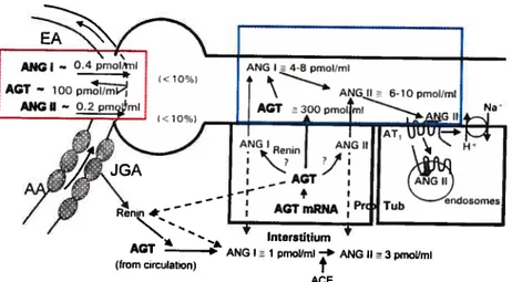
The contributions cf the RAS to the reg ulation cf arterial pressure and the physiopathology of hypertension have long been recognized. It is important to understand that circulating AGT concentrations are generally quite high, being more than 1,000 times greater than plasma Ang I and Il concentrations (90-92). Figure 6 depicts the representative plasma AGT concentrations in rats, expressed as pmol/ml, while Ang I and Ang li concentrations are expressed as fmol/mI, indicating that active Ang Il concentrations in plasma are a small fraction of Ang Il available in the form
of AGI (91). Therefore, even small relative changes in the rates of Ang I
and Ang Il generation may make large absolute differences in circulating concentrations. Nevertheless, changes in AGI synthesis and release occur slowly and, thus, their effects are flot as dynamic as the effects cf changes in plasma renin concentration (44; 93).
Figure 6. Tubular and interstitial formation, secretion and uptake 0f angiotensin (Ref. 91).
As discussed above, Ang Il exerts its effects via Ang receptors, which trigger intracellular signaling processes. In response to Ang II stimulation, the Ail receptor interacts with various heterotrimeric G proteins, inducing the phosphorylation of tyrosine kinase (94). ihe intracellular signaling processes are multiphasic, leading to various biological actions. AT1 receptor activation results in increased protein tyrosine phosphorylation and mitogen-activated protein kinase (MAPK) Activation. ihese processes are associated with growth factors, cytokines, and inflammatory agents.
ihus, in addition to its potent vasoconstrictor actions, Ang Il has
proliferative and pro-inflammatory properties (95).
One of the earliest detectable events arising from Ang Il stimulation is
Ail signaling through phospholipids with the involvement of
phospholipase C (PLC), PLD and PLA2. While the stimulation of PLC induces rapid 1 ,4,5-inositol triphosphate production and diacylglycerol
(DAG) release, PLD hydrolyzes phosphatydylcholine to phosphatidic acid
and choline. PLA2 is responsible for arachidonic acid release from celi
Interstjtium
AGT ANG I 1 pmol/mI * ANG II
3pmollml
(from Circulation)
f
membrane phospholipids (94). ihe phospholipid-derived second messengers produced by Ang ll-activated PLs induce multiple subsequent events (Ca2+ release from the sarcoplasmic reticulum, PKC activation by
DAG, eicosanoid generation, etc.).
The Ai2 receptor may counteract some of the actions of the Ail receptor. Found in large amounts in fetal tissues, the Ai2 receptor is considered to be a factor that stimulates growth and development. Preferentially expressed by various secretary celis, it is activated by both circutating and locally-generated Angs (96). As already noted, AT2 has antagonistic properties, being involved in local blood flow control as a vasodilating mechanism (97).
It has been demonstrated that Ang Il exerts opposite effects on the
growth cf certain ceils within blood vessels by binding to either the Ail or Ai2 receptor. Ang li induces celi proliferation by activating its Ail receptor and inhibits cell growth by stimulating the A12 receptor in different cell types (98). ihe growth-suppressing effects mediated by the AT2 receptor do flot involve repression cf fos and c-jun gene expression, but are evoked through the inhibition of cyclin Dl expression and cyclin Dl-associated kinase activity (99). Furthermore, Ai2 receptor stimulation modulates Ail receptor-mediated vasoconstriction, and altered Ai2 receptor function may contribute to an exaggerated vasoconstrictor action of Ang lI (97). Apoptosis is another biological phenomenon which seems to be triggered
by the stimulation cf Ai2 teceptors. Elevated A12 receptor levels have
been observed in the brain of rats after global ischemia, eventually leading to delayed neuronal cell death (100).
of its active fragments, represented by Ang III [2-8], Ang IV [3-8], Ang V [3-7] and Ang l-7 (Figure 4). ihe heptapeptide Ang III has physio-pharmacological effects similar to those of Ang Il (58; 89; 101-104), mediated by their common receptors, Ail and Ai2 (103). Ang IV is involved in blood flow regulation, exploratory behavior, learning, memory, and neuronal development (103). It has been demonstrated that the inhibitory influence of Ang li via Ail, and a facilitory role of Ang IV via its receptor Ai4, in the neuronal firing rate, associative and spatial learning (105).
Ang l-7 produces vasodilation, diuresis and antiproliferative reactions through a specific receptor (Mas). The vasodilating effects of Ang 1-7, more intense in vessels with an intact endothelium than in those with the endothelium removed, are accompanied by the release of nitric oxide (NO), kinins, and prostaglandins (76). ihrough inhibition of the pressor and proliferative actions of Ang Il, Ang 1-7 behaves like e true counteracting factor. In agreement with these data, possible balance has been hypothesized between the vasoconstrictor and hypertensive properties of Ang Il and the actions of Ang 1-7 (69). Ang 1-7, one of the active components of the RAS, is involved as a possible counter-regulatory factor
in the vasoconstrictor effects ofAng II.
1.4 The Local RAS
Over the last 3 decades, it has become increasingly clear that bioactive Ang peptides can be generated, not only in the systemic circulation, but also as local hormones in several tissues and organs. Since
numerous investigations have shifted their focus from its endocrine to its autocrine/paracrine role in specific tissue functions, such as tissue growth and differentiation.
The localization and expression of key RAS components, notably AGT and renin, which are mandatory for the presence of tissue RAS, have been reported in a wide range of tissues (47; 106; 107). The expression of local RAS components in tissues such as the brain, heart, kidneys, adrenals, and gonads has led to the proposition that these components may either potentiate systemic functions, or have entirely separate activities meeting the specific needs of individual tissues (107-109). There is accumulating evidence that changes in tissue/organ-specific RAS may be associated
with the pathophysiology of respective tissue/organ functions, giving rise to
the possibility that drugs acting on tissue RAS could ameliorate some of these disorders (110).
1.4.1 Discovery of the Local RAS
Evidence accumulated over decades has expanded significantly by more recent findings that have increased the complexity of the RAS. Different Ang receptors and signal transduction pathways have been characterized. Moreover, additional truncated peptides, such as Ang 1-7, have been identified, and alternative Ang Il formation by non-renin and non-ACE pathways has been reported. These resuits have modified our view of the RAS to the concept of “local” or “tissue” RASs. This concept is based on flndings of RAS components in “unlikely” places (such as “kidney enzyme” renin in the brain), where endocrine actions of the system cannot
be explained (111-113). This, in turn, has leU to new hypotheses and functional concepts of local RAS actions based on the tissue synthesis of Ang Il.
The overexpression of RAS genes in Tg mice and rats as well as the knockout of these genes in mice have fostered detailed studies on functions of the local RAS (113). It has also become increasingly clear that these systems are flot isolated entities, but can interactwith the endocrine RAS as well as other peptide systems (such as the endothelin system) at multiple levels.
Early controversies surrounding the novel concept of tissue RAS arose from the question of local synthesis versus uptake from the circulation. A case in point is the controversy between investigators after the demonstration 0f renin expression in the heart (75; 114) and those questioning local renin synthesis. This controversy was aftributed to the fact that renin mRNA in the heart could only be demonstrated inconsistently, suggesting that studies measuring cardiac reninwere biased by artifacts from contamination with plasma renin or active renin uptake from the circulation. The issue should not threaten the concept of the local RAS, since either mechanism could contribute to local AGT synthesis and actions. Modem concepts of the tissue RAS are therefore function-oriented.
The multiple intrarenal actions of Ang Il are based on its predomînantly local synthesis and the presence of Ail receptors within glomerular vessels and various tubular segments. A density gradient of these receptors has been demonstrated by their much higher amounts in superficial than in juxtamedullary glomeruli. Ang receptors can be found mostly in the basal membrane of the tubular epithelium and mesangial cells.
ihis distribution provides the contractile effect of the peptide on mesangial
cells and the control of proximal sodium and water reabsorption. Proximal tubular sodium transport is stimulated by enhanced sodium/hydrogen ion antiporter activity on luminal membranes (115). lncreased sodium/potassium pumps and sodium/bicarbonate co-transporters on the basolateral membranes of proximal tubular celis (PiCs) also enhance sodium reabsorption (116; 117). Ang Il augments sodium reabsorption through actions on sodium/potassium pumps in the medullary thick ascending limb and epithelial sodium channel of cortical collecting tubules
(117; 118). At the functional level, Ang li contributes to control of the
glomerular filtration rate (GFR).
Aside from its hemodynamic effects, Ang Il promotes other processes
in the kidneys. It perpetuates the production of nephrotoxic reactive oxygen
species (ROS), and stimulates celI proliferation as well as tissue remodeling by enhancing the synthesis of profibrotic cytokines and growth factors. ihe overexpression of chemokines, chemotactic factors, and celi adhesion molecules contributes to abnormal cellular proliferation and renal fibrosis. Collagen deposition is also enhanced through the inhibition of proteases that normally function to degrade abnormal tissue proteins (119).
Together, these effects increase the development of glomerulosclerosis and tubulointerstftial fibrosis (119; 120).
At the same time, the vasa recta and peritubullary capillaries, being
most sensitive to the vasoconstricting action of Ang Il, contribute to both tubulo-glomerular feedback and the control of water and ion elimination. Ang II participates in the long-term regulation of arterial BP through such volume regulatory mechanisms.
1.5 Activation of the Local Renal RAS in DN
Sustained interest in dissecting the pathophysiology of DN has leU many to focus on renal hemodynamics. One factor that has fueled this interest is the unequivocal benefit derived from ACE inhibition in DN. Baseline plasma renin activity is found to be reduced in type 2 diabetic patients. The renovascular response to an Ang antagonist is substantially greater than normal, indicating intrarenal
RAS
activation flot reflected in plasma levels (121). Thus, investigators are searching for factors that evokeRAS
activation in diabetes. Hyperglycemia is a logical candidate (122-124).1.5.1 AnginDN
The beneficial effects of ACEi or Ang receptor blockade in the prevention of diabetic renai disease suggest that Ang Il is a major mediator
of progressive renal injury (25; 125-127). However, measurement of the
activity of circulating
RAS
components largely points to suppression in DN (121). These observations indicate local renal tissue activation of theRAS
or increased intrarenal sensitivity to Ang II, especially at Ail receptors, in
DN.
mRNA and protein levels of renin are elevated at the onset of DN in spontaneously or streptozotocin (SIZ)-induced diabetic rats (128). In early
DN, proximal tubule renin mRNA is signiflcantly upregulated, with no
change in AGT or ACE mRNA (129). Ihis process is reversed by insulin therapy. In addition, an increase in intrarenal AGI mRNA has been reported in diabetic rats, implying heightened AGI synthesis by proximal tubules (128; 130). In our Iab, we have discovered that hyperglycemia stimulates AGI synthesis in a concentration-dependent manner in association with augmented AGT mRNA expression (131). A
glucose-response element has been located on the AGI gene promoter that might mediate this effect (129). Recently, we also found that hyperglycemia increases AGT gene expression via ROS in rat proximal tubules, providing evidence of intrarenal RAS activation in DN (132).
In diabetic rat and human kidneys, ACE1 is redistributed towards
glomeruli and the renal vasculature but away from the proximal tubules (130; 133). These studies suggest that glomerular ACE mediates nephron
injury, possibly by increasing local intraglomerular Ang II formation. In
cultured rat mesangial cells, high-glucose levels stimulate Ang II production
in a concentration-dependent manner in association with augmented IGF-13 production (134). Nevertheless, whole-kidney Ang II levels in
unchanged (128; 135-137). Howevet, whole-kidney levels probably do flot reflect Ang II at specific nephron sites.
1.5.2 Ang Receptors in DN
Several studies have documented the expression of renal ATI and
AT2 receptors in DN. Reduced renal expression of the Ail receptor has
been reported in rats after 3 weeks of STZ-induced diabetes and also in diabetic patients (138). However, another investigation found no significant change in AT1 receptor expression, but reduced A12 receptor expression
in the kidneys of rats with early (2-week) STZ-induced diabetes (139).
Recently, an experiment was performed on spontaneously hypertensive rats (SHR) after 32 weeks of STZ-induced diabetes. In Iong-term DN in the context of hypertension, increased albuminuria and renal structural injury were accompanied by the decreased expression of genes encoding Ail and Ai2 receptors and respective proteins in the kidneys (140). The downregulation of receptor expression implied reduced receptor synthesis, as opposed to the inability of Ang II to internalize and desensitize the AT2 receptor. The decline in renal AT2 receptor expression in diabetic Wistar-Kyoto (WKY) rats without systemic hypertension and with less prominent renal injury was consistent with a glucose-dependent mode cf
AT2 receptor regulation.
1.5.3 Ang Il-induced DN
Ang Il has many actions that might cause or contribute to DN (Table 1).
glucose (134). ihus, mesangial ceils might have their own self-contained
RAS, and hyperglycemia could stimulate its activity, leading to celiular
contraction with reduced filtration surface area (141). Both glucose and Ang Il stimulate ECM formation, and collagen accumulation in mesangial
ceHs is blocked by ATI receptor inhibition (134; 141). iGF-3 is an
important mediator of coilagen accumulation and fibrosis. Because glomeruli from diabetic animais and humans have increased mRNA encoding TGF-13, iGF-3 contributes to the pathogenesis of DN (142). Ang
Ii stimulates mesangial celi matrix biosynthesis via the Ail receptor, and
this is mediated by TGF-3 (16). in rat mesangiai cells, glucose-induced
TGF-13 secretion is abrogated by Ail receptor blockade (134). in humans,
the progression of DN is elicited, at least in part, by Ang il action via iGF-3 (143). These observations suggestthat glucose stimulates Ang li synthesis, leading to increased TGF-3-induced matrix accumulation.
Several studies have reported Ai2 receptor downregulation in diabetic animai modeis (139; 140). if intrarenal Ang II production is augmented in diabetes, Ai2 receptor downregulation could create an imbaiance in which
Ail receptor stimulation is unopposed. A reduction of renai nitric oxide
(NO) in diabetes could occur because of diminished NO synthesis,
decreased availability of the substrate L-arginine, or failure of the Ai2 receptorto generate NO (25; 139; 144).
in summary, these data and other investigations indicate that
intrarenal RAS activation might piay an important role in DN.
Because intrarenal RAS activity contributes to DN, many clinical trials
have explored the effects of RAS blockade and obtained surprising resuits
in hypertensive patients with or without diabetes. Since the mid-1980s,
numerous clinical studies have assessed the metabolic effects of ACEI or
Ang receptor blockers (ARBs). Besides these inhibitors, a novel direct
renin inhibitor, Aliskiren, has just become available in the USA, Switzerland,
and Europe. The Iong-term potential of Aliskiren and direct renin inhibition
is being investigated in a clinical program known as ASPIRE HIGHER,
focusing on the benefits of the drug in hypertensive patients with heart
failure (HF) or kidney failure. Preliminary data from this program indicate
that BP is reduced in these patients (145).
16.1
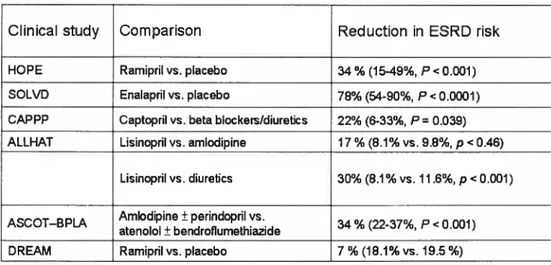
Clinical Trails ofACEi in Diabetic Patients
Six clinical studies in which ACEi were given to patients with
hypertension or other risk factors for cardiovascular disease (CVD) indeed
suggest that they decrease the risk cf ESRD (Table 3).
The Heart Outcomes Prevention Evaluation (HOPE) trial found a 34%
reduction (P
<0.001) in the relative risk of new-onset diabetes in patients
treated daily with up to 10 mg of the ACEi ramipril (146). Similarly, the
effect cf enalapril on the incidence of new-onset diabetes was assessed in
a subgroup of patients from studies cf left ventricular dysfunction (SOLVD).
A significant 78% reduction (P< 0.0001) in the incidence of new-onset
diabetes was noted in comparison to placebo (147). Together with the
small number of patients included in the sub-analysis, this limited wider
interpretation cf the findings. Nevertheless, the data from both HOPE and
SOLVD suggested that ACEi prevent the development cf diabetes in
subjects with CVD, meriting further investigation in prospective trials.
Table 3. Clin ical evidence for prevention cf diabetes with ACE inhibitors
Clinical study
Comparison
Reduction in ESRD risk
HOPE Ramipril vs. placebo 34% (15-49%, P< 0.001) SOLVD Enalapril vs. placebo 78% (54-90%, P<0.0001) CAPPP Captopril vs. beta blockers/diuretics 22% (6-33%, P=0.039) ALLHAT Lisinopril vs. amlodipine 17 % (8.1% vs. 9.8%, p<0.46)
Lisinopril vs. diuretics 30% (8.1% vs. 11.6%, p<0.001) AScOT—BPLA Amlodipine ± perindopril vs. 34% (22-37% P<0.001)
atenolol ± bendroflumethiazide
DREAM Ramipril vs. placebo 7 % (18.1% vs. 19.5%)