Université de Montréal
The biological importance of apurinic/apyrimidinic endonucleases in the maintenance of genetic stability
par
Ratsavarinh Vongsamphanh
C
Département de microbiologie et immunologie faculté de médecine
Thèse présentée à la Faculté des études supérieures en vuede l’obtention du grade de philosophiae doctor (Ph.D)
en microbiologie et immunologie
Octobre 2006
q
Université
(JI
de Montréal
Direction des biblïotlièques
AVIS
L’auteur a autorisé l’Université de Montréal à reproduire et diffuser, en totalité ou en partie, pat quelque moyen que ce soit et sur quelque support que ce soit, et exclusivement à des flns non lucratives d’enseignement et de recherche, des copies de ce mémoire ou de cette thèse.
L’auteur et les coauteurs le cas échéant conservent la propriété du droit d’auteur et des droits moraux qui protègent ce document. Ni la thèse ou le mémoire, ni des extraits substantiels de ce document, ne doivent être imprimés ou autrement reproduits sans l’autorisation de l’auteur.
Afin de se conformer à la Loi canadienne sur la protection des renseignements personnels, quelques formulaires secondaires, coordonnées ou signatures intégrées au texte ont pu être enlevés de ce document. Bien que cela ait pu affecter la pagination, il n’y a aucun contenu manquant. NOTICE
The author of this thesis or dissertation has granted a nonexclusive license aNowing Université de Montréal to reproduce and publish the document, in part or in whole, and in any format, solely for noncommercial educational and research purposes.
The author and co-authors if applicable retain copyright ownership and moral rights in this document. Neither the whole thesis or dissertation, nor substantial extracts from it, may be printed or otherwise reproduced without the author’s permission.
In compliance with the Canadian Privacy Act some supporting forms, contact
information or signatures may have been removed from the document. While this may affect the document page count, it does flot represent any loss of content from the document.
Cette thèse intitulée:
The biological importance of apurinic/apyrimidinic endonucteases in
the maintenance of genetic stability
présentée par:
RATSAVARINH VONGSAMPHANH
o
a été évaluée par un jury composé des personnes suivantes:
Dr. Elliot Drobetsky président-rapporteur Dr. Dindial Ramotar directeur de recherche Dr. Janos Filep membre du jury Dr. Daniel Durocher examinateur externe Dr. Jean-François Côté
représentant du doyen de la FES
111
RÉSUMÉ
Les dérivés réactifs de l’oxygène produisent de multiples lésions à l’ADN, notamment des sites abasiques (AP). Les dommages oxydatifs de l’ADN sont réparés principalement via la voie de réparation par excision de bases dans laquelle les AP endonucléases jouent un rôle crucial. Les cellules déficientes pour les AP endonucléases sont hypersensibles aux agents aikylants (e.g. MIVIS) et oxydants (e.g. H202). Nous sommes intéressés par l’étude moléculaire des AP endonucléases eucaryotiques afin de mieux comprendre leurs mécanismes enzymatiques. Notre première étude porte sur la structure et la fonction de la principale AP endonucléase de la levure $accharornyces cerevisiae, Apnl. Nos résultats démontrent clairement l’importance des acides aminés G1u15$ et Asp192 pour l’activité biologique d’Apnl, étant donné qu’une mutation d’un de ces résidus abolit significativement son activité enzymatique. Lors de notre deuxième étude, nous avons évalué le rôle de l’ADN glycosylase Oggi de la levure dans l’instabilité des séquences poly(GT) mitochondriales. Nous avons observé qu’une surproduction d’Apnl cause une
(
augmentation de l’instabilité des séquences poly(GT). Cet effet fût toutefois contrebalancé par la surexpression d’ Ogg 1. Nos résultats indiquent que la protéine Oggl compétiolme avec Apnl possiblement pour la liaison des sites AP afin d’empêcher la réparation incontrôlée par Apnl. Notre troisième étude avait comme but initial l’identification de l’homologue humain d’endo IV. Cependant, nous avons isolé l’enzyme glycolytique, GAPDH. Nos résultats démontrent que GAPDH interagit avec APEÏ afin de stimuler son activité AP endonucléase en convertissant la forme oxydée d’APEl en sa forme réduite. De plus, les résidus Cys 152 et Cys156 de GAPDH sont importants pour la stimulation de APE1. Le rôle que joue GAPDH dans la résistance cellulaire face aux agents génotoxiques, tels le MMS et H202, fût également démontré par des tests de viabilité cellulaire. Collectivement, nos études confirment l’importance biologique des AP endonucléases dans la réparation des sites AP. De plus, elles ont permis de comprendre davantage les mécanismes par lesquels ces enzymes contribuent à la stabilité génétique dans la celltile.Mots-clés : dommage oxyc1atf à 1 ‘ADÀ’ réparation par excision de bases, interaction
(
protéique, modUication posttransÏationneÏle.ABSTRACT
Living organisms are constantly exposed to reactive oxygen species which are known to cause a variety of DNA lesions, such as apurinic/apyrirnidinic (AP) sites. The repair of oxidative DNA damage is primarily mediated by the base excision repair (BER) pathway. AP endonucleases are key enzymes in this process. Celis deficient in AP endonucleases are hypersensitive to aikylating (e.g. MMS) and oxidizing (e.g. 11202) agents. We are interested in studying eukaryotic AP endonucleases at the molecular level to gain insights into their mechanisms of DNA binding and cleavage at AP sites. n our first study, we carried out the first structure/fttnction analysis of Apnl, the major AP endonuclease in yeast
Saccharornyces cerevisiae. Our data indicate that two conserved amino acids residues of Apnl, G1u15$ and Asp192, are critical for its biological function, since glycine substitutions at these positions abolished its DNA repair activities. In our second study, where we evaluated the role of yeast Oggi DNA glycosylase in the maintenance of a mitochondrial poly(GT) tract reporter system. we discovered that overproduction of Apnl causes instability of the poly(GT) tracts. Interestingly, this effect was counteracted by overexpression of Oggi. Our results clearly suggest that uncontrolled DNA cleavage by Apnl is prevented by Oggi which most likely competes with Apnl for binding to AP sites. In our final study, we set out to identify the endo IV homolog in human ceils, but we unexpectedly re-isolated GAPDH, a classical glycolytic enzyme. We demonstrated that GAPDH interacts with APE1 to stimulate the AP endonuclease activity by converting the oxidized forms of APE 1 to the reduced forms. Futhermore, we also showed that Cys 152 and Cysi 56 of GAPDH are important for APE1 reactivation. Consistent with this notion, we demonstrated that GAPDH plays an important role in the cellular resistance against genotoxic agents such as MMS and H202 by cell survival assays. Taken together, our studies reinforce the biological significance of eukaryotic AP endonucleases in the repair of AP sites, but more irnportantly, they provide valuable insights into the rnechanisms by which AP endonucleases maintain genomic stability in the cell.
Keywords: oxidative DNA damage, base excision repair, protein interaction, posttranslationaÏ modlification.
V
Table of Contents
RÉSUMÉ III
ABSTRACT IV
TABLE 0F CONTENTS V
LIST 0F FIGURES VIII
LIST 0F TABLES X
LIST 0F ABBREVIATIONS XI
ACKNOWLEDGEMENTS XIV
CONTRIBUTIONS 0F AUTHORS XV
CHAPTER 1: GENERAL iNTRODUCTON 1
OXIDATIVE DNA DAMAGE 2
GENERATI0N AND DETOXIFICATION 0F REACTIVE OXYGEN SPECIES 2 TYPES 0F OXIDATIVEDNALES1ONS 3
Base damage 3
Sugar dam age 3
REPAIR 0F OXIDATIVE DNA DAMAGE $
BASE EXCISION REPAIR $
AN ALTERNATIVEBERPATHWAY 12 POTENTIAL MECHANISMS 0F SWITCHING EETWEENBERSUBPATHWAYS 12 ASSOCIATION BETWEENBERDEFECTS AND HUMAN CANCER 13
Geneticpotymorphisms 13
Mouse models 14
AP ENDONUCLEASES 14
SEQUENCE AND STRUCTURAL CONSERVATION 0FAPENDONUCLEASES 16
E.COLIAPENDONUCLEASES 1$ Exo III 18 EndolU 19 S.CEREVISIAIAPENDONUCLEASES 19 ApnI 19 Apn2 20 HuMANA?ENDONUCLEASES 21 APEI 21 APE2 22
APE] more than a DNA repair enzyme 23
REGULATION 0FAPE1 ACTIVITIES 23 PROTEIN-PROTEIN INTERACTIONS INVOLVINGAPE1 24
HYPOTHESIS AND OBJECTIVES 27
CHAPTER 2: CHARACTERIZATION 0F TWO INDEPENDENT AMINO ACID SUBSTITUTIONS THAT DISRUPT THE DNA REPAIR FUNCTIONS
0F THE YEAST APN1 2$
ABSTRACT 30
INTRODUCTION 31
STRAIN5, MEDIA, GENETIC ANALYSIS, AND TRANSFORMATION.34
CoNsTRUcTIoN 0F THE PLASMIDSGFP-APNI ANDGST-APNI 34
SITE-DIRECTED MuTAGENEsIs 35
CONSTRUCTION 0FGFP-APN I (E 158G)ANDPGFP-APN I (D 192G) 35
GRADIENT PLATE AsSAY 35
IMMUNODETECTION 36
PuRIFICATION 0F APN1, G5T-APN1 AND THE MUTANT F0RMs 36
APENDONUCLEASE AND 3-DIE5TERASE ASSAYS 37
DNAGLYCOSYLASE-INDEPENDENT INCISION 0F OXIDATIVELY DAMAGEDDNA 3$ ELEcTR0PHORETIC MOBILITY SHIFT A5sAY 39
RESULTS 40
TUE CONSTRUCTGEP-APN I EXPRESSES A FUNCTIONALLY ACTIVE FUSION PROTEIN 40 THE GFP-APN1 (E 158G)AND GFP-APN1 (D 192G)MUTANT PLASMIDS CONFER NO DRUG RESISTANCE TO THEAPENDONUCLEASE-DEFICIENT STRA[NYW78 I 40
TUE APNI MUTANT PLASMIDS EXPRESS NORMAL LEVELS 0F TUE MUTANT PROTEINS AND ARE
LOCALIZED TO TUE NUCLEUS 43
EXPRESSION AND COMPLEMENTATION ANALYSES 0F GsT-APN1 AND TUE GST-APNI MUTANT FORMS IN TUEDNAREPAIR DEFICIENTE.COLI STRAINBW528 46
APN1 MUTANTS LACKDNAREPAIR ACTIVITIES 51 GsT-APN1 MUTANTS BIND TO DOUBLE STRANDED OLIGONUCLEOTIDE 57
DISCUSSION 61
ACKNOWLEDGMENT 63
CHAPTER 3: SACCHAROMYCES CEREVISIAEOGG1 PREVENTS
POLY(GT) TRACT INSTABILITY IN THE MITOCHONDRIAL GENOME 64
ABSTRACT 66
INTRODUCTION 67
MATERIALS AND METHODS 69
YEAST STRAINS 69
CONSTRUCTION 0F TUE PLASMIDPOGG I -GFP 69 PREPARATION 0F MITOCUONDRIAL FRACTIONS FROM YEAST 70
WESTERN BLOT 71
PREPARATION 0FDNASUBSTRATE AND ASSAY FOR OGG1 ACTIVITY 71 MEASUREMENT 0F MUTATIONS RATES 0F MITOCUONDRIALDNA 72 ANALYSIS 0F TUE LENGTU 0F MITOCUONDRIAL MICROSATELLITES 72 ANALYsIS 0F 8-oxo-DGU0 IN MTDNA BYHPLC 73
RESULTS 75
TUE MITOCUONDRIAL FORM 0F OGGI IS ACTIVE AT PROCESSING 8-0XO-DGUO LESIONS 75 OGGIA MUTANTS EXHIBIT ELEVATED POLY(GT) TRACT INSTABILITY 75
PCRANALYSIS 0FGTTRACT INSTABILITY IN TFIE PARENT AND TUE OGGIAMUTANT $1 ANAER0BIc CONDITION ELIMINATES TUE ELEVATED MUTATIONS IN TUE OGGIANULL 81 APNI OVEREXPRESSION IN TUE OGG1A MUTANT STIMULATES POLY(GT) TRACT INSTABtLITY 84 POLY(GT) TRACT INSTABILITY IS NOT AFFECTED BYNTG 1 DELETION 87
DISCUSSION $9
Vii
CHÂPTER 4: HUMAN GAPDH FUNCTIONS AS A REDOX FACTOR TO
Q
REACTIVATE THE OXIDIZED FORM 0F THE DNA REPAIR ENZYMEAPE1 93
ABSTRACT 95
INTRODUCTION 96
MATE RIALS AND METHODS 9$
BAcTERIA STRAINS 9$
CELLcuLTURE 9$
AP ENDONUCLEASE ASSAY 9$
PURIFICATION 0F A PROTEIN WITH ASSOCIATEDAPENDONUCLEASE ACTIVITY 9$
PROTEIN SEQUENCING 100
PLA5MIDs 100
PROTEIN5 100
GAPDHASSAY 101
BINDING 0FGST-GAPDHTOHIS-APEICOLUMN 101
GST PULL DOWN ASSAYS 102
IMMUNOPRECIPITATION 102
IMMUNODETECTION 102
OxIDATI0N 0FHIs-APEI PROTEIN WITHF1707 103
SITE-Dl RECTED MUTAGENESIS 103
CHEMICAL OXIDATION 0FGST-GAPDH 103
SIRNA-MEDIATEDGAPDHSILENC[NG 104
DRUG EXPOSURE AND CLONOGENIC ASSAY 104
UV IRRADIATION 105
WESTERN BLOT 105
RESULTS 107
PURIFICATION 0F A MG2-INDEPENDENTAPENDONUCLEASE ACTIVITY 107
IDENTIFICATION 0FGAPDHIN TUE PURIFIED PROTEIN FRACTION 110
PURIFIEDGAPDHEXHIBITS NOAPENDONUCLEASE ACTIVITY 110
INTERACTION 0FGAPDHWITHAPE 1 113
ACTIVATION 0FAPENDONUCLEASE ACTIVITY 0FAPEI BYGAPDH 116
GAPDH FUNCTIONS TO REDUCE THE OXIDIZED FORM 0FAPE 1 117
MUTATION 0f CYSTEINE 152AND 1560FGAPDH10 GLYCINE AFFECTS THE REACTIVATION THEAP
ENDONUCLEASE ACTIVITY 122
OvERExPREsSI0N 0FGAPDHENHANCES CELLULAR RESISTANCE TOMMSANDH202,BUT NOT
UVC 123
GAPDHf UNCTION IS NECESSAY FOR PROTECTION AGAINST APEI-REPAIRABLEDNADAMAGE... 126
DISCUSSION 131
CHAPTER 5: GENERAL DISCUSSION 135
DISCUSSION 136
C0NCLUDING REMARKS 144
REFERENCES 146
List of Figures
FIGURE1-l. CHEMIcAL STRUCTURES 0F THE MAJOR OXIDATIVE DNA BASE LESIONS 4 FIGURE 1-2. CHEMIcAL STRUCTURES 0F COMMONS FORMS 0f ABASIC(AP) DNA
DAMAGE 6
FIGURE l-3. SCHEMATIC REPRESENTATION 0F Tu BASE EXCISION REPA1R (BER)
PATHWAY 9
FIGURE2-1.DRUG RESISTANCE DETERIIINATI0N INS. CEREVISIAE STRAINS YW465
(PARENT) AND YW781 APN1AAPN2A) HARBORING PLASMIDS CARRYING EITHER THE NATIVE OR MUTANT ALLELES 0FTHEAPNJGENE fUSED 10
GFP 41
FIGURE2-2. C0MPARIs0N 0F THE EXPRESSION LEVELS 0F NATIVE APN1 AND ITS
MUTANT FORMS FUSED TO GFP 44
FIGuRE2-3.NUcLEAR LOCALIZATION 0F GFP-APN1 AND ITS MUTANT FORMS 47 FIGURE 2-4. ExPRESsIoN AND PURIFICATION 0F NATIVE AND MUTANT FORMS,E158G
AND D 192G, 0f APN1 USINGE. COL! 49
F1GuRE2-5.DRUG COMPLEMENTATION ANALYSIS 0F STRAIN BW528 CARRYING
PLASMIDS EXPRESSING THE NATIVE AND MUTANT FORMS 0F APNJ FUSED 10
GST 52
FIGuRE2-6. COMPARISON 0F THEDNAREPAIR ACTIVITIES 0F APN1 AND THE MUTANT
FORMS,E158GANDD 192G 54
FIGURE2-7.M0BILITY-SHIFT ANALYSIS 0F A NATURAL AND A MISMATCH-CONTAINING 42-MER-LABELLED DOUBLE STRANDED OLIGONUCLEOTIDE BY PURIfIED
GST-APN 1,GST-APN1 (E 158G), G5T-APN 1 (Dl 92G),AND GsT 59
FIGURE3-1.MITOcHONDRIAL OGG1 IS fUNCTIONALLY ACTIVE 76 FIGURE 3-2. OGGJ IS REQUIRED TO MAINTAIN POLY(GT) TRACT STABILITY 79 FIGuRE3-3.PCRANALYSIS FOR POLY(GT) TRACT INSTABILITY 82 FIGURE3-4.ANAEROBIC CONDITIONS REDUCE 111E P0LY(GT) TRACT INSTABILLTY 85 FIGURE4-1.PURIFICATIoN 0F A MG2-1NDEPENDENT AP ENDONUCLEASE ENZYME 10$ FIGuRE4-2.PURIFIED POLYPEPTIDE ISIHE GLYCOLYTIC ENZYME GAPDH 111
FIGURE4-3. GAPUIIINTERACTS DIRECTLY WITHAPE1 114
FIGuRE4-4. GAPDHSTIMULATESAPEI APENDONUCLEASE ACTIVITY 11$
FLGURE 4-5.GAPDHSTIMULATESAPE1 AP ENDONUCLEASE ACTIVITY VIA A REDOX
C
ix
FIGURE4-6.CYsTEINE152AND156 RESIDUES 0fGAPDHARE IMPORTANT TO
STIMULATEAPEJ AP ENDONUCLEASE ACTIVITY 124
FIGuRE4-7. GAPDHPLAYS AN IMPORTANT ROLE IN PROTECTING CELLS AGAINSTMMS
List of Tables
o
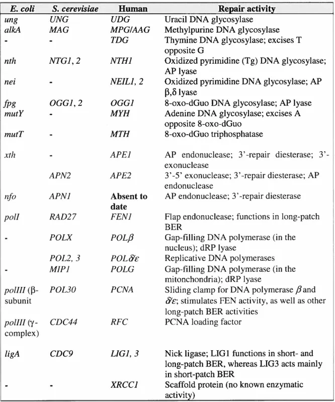
TABLEI-I. SUMMARY 0F 111E PRINCIPALBERENZYMES IN EscHERIcHIA COU, SACCHAROMYcEs CERF VJSME, AND HUMAN 11
TABLEI-II. SUMMARY 0FBERMOUSE MODELS 15
TABLEI-III. C0MPARIs0N 0FAPENDONUCLEASES IN BACTERIA, YEAST, AND
HUMAN 17
TABLE14V.EFFECTs 0F 111E VARIOUS PROTEINS INTERACTIONS INVOLVINGAPE 1
ON THEBERPATHWAY 26
TABLEIII-I. EFFECT 0FDNAREPAIR PROTE INS ON 111E RATE 0F POLY(GT) TRACT INSTABILITY IN 111E MITOCHONDRIAL GENOME 8$
xi
List of Abbreviations
AP apurinic/apyrimidinic
AP- 1 activator protein- 1
APE 1 apurinic/apyrimidinic endonuclease 1 (human) Apn 1 apurinic/apyrimidinic endonuclease 1 (yeast)
ATP adenosine triphosphate
BER base excision repair
bp base pair
BRCT BRCA1 C-terminus
CanR Canamycin resistance
cDNA complimentary deoxyribonucleic acid C. eÏegans Caenorhabditis elegans
CR0 Chinese hamster ovary
CKI/II casein kinase 1/11
DHT 5,6-dihydrothymine DRU 5,6-dihydrouracil dNTP deoxynucleotide triphosphate dRP 5’ -deoxyribose-5 -phosphate dRpase 5 ‘-deoxyribose-5-phosphodiesterase DTT dithiothreitol
E. cou Escherichia cou
EDIA ethylenediaminetetraacetic acid EMSA electrophoretic mobility shifi assay Endo III endonuclease III
Endo IV endonuclease IV
EryR erythromycin resistance Exo III exonuclease III
fapyA 4,6-diamino-5-formamidopyrimidine
FapyG 2,6-diamino-4-hydroxy-5 -formamidopyrimidine
Q
GAL1GAPDH galactose 1glyceraldehyde-3 -phosphate dehydrogenase Gfp green fluorescent proteinGst glutathione-S-transferase
H-Bond hydrogen bond
111F-la hypoxia-inducible factor- la
F1202 hydrogen peroxide
HPLC high performance liquid chromatography HSP7O heat-shock protein 70
IPTG isopropyl thio-fi-D-galactoside
kb kilobase
kDa kiloDalton
LB Luriabroth
MMR mistmatch repair
MMS methyl methane sulfonate
(
MSI microsatellite instabilitymtDNA mitochondrial DNA
MWCO molecular weight cut-off
NAC N-acetylcysteine
NAD+ nicotinamide adenine dinucleotide NER nucleotide excision repair
NF-KB nuclear factor-KB
NLS nuclear localization signal
Oggi 8-oxo-deoxyguanosine DNA glycosylase superoxide anion
0H hydroxyl radical
8-oxo-dGuo 8-oxo-deoxyguanosine/8-oxo-7, 8-dihydro-2 ‘-deoxyguanosine PARP Poly(ADP-ribose) polymerase 1
PCNA proliferating ceil nuclear antigen
PCR polymerase chain reaction
xlii
Pin Protein with internai repeats
PKC protein kinase C
Pol 13, , polymerase beta, delta, epsilon
Ref- 1 redox effector factor- 1 RF-C replication factor-C
RNAi RNAinterference
ROS reactive oxygen species
rRNA ribosomal ribonucleic acid Rrpl Recombination repair protein 1 S. cerevisiae Saccharornyces cerevisiae Siahi Seven in absentia homolog 1
$DS-PAGE sodium dodecyl sulfate polyacrylamide gel electrophoresis siRNA small interfering RNA
S. pombe Schizosaccharomyces pombe
ssb single-strand break
Tg thyrnine glycol
THF tetrahydrofuran
TRX thioredoxin
UDG uracil DNA glycosylase
UV ultraviolet
WRN Werner protein
YPD yeast peptone dextrose
XPG xeroderma pigmentosum complernentation group G XRCC1 x-ray cross-species complementing Ï
Acknowledgements
In 199$, I carried out my first research project as a Master student in microbiology and immunology at the University of Montreal. I chose as subject DNA Repair. I came in an amazing world of researchers, scientists, and congresses that fascinated me. Afier obtaining my Master degree in 2001, I had the possibility to pursue my graduate studies in the amazing field of DNA Repair as a Ph.D. student. Now, a littie more than five years later, that opportunity bas resulted in this thesis.
I would like to thank the Terry Fox Foundation of the National Cancer Institute of Canada (NCIC) for providing funding for my research studentship from July 2002 to July 2005.
Writing a thesis is flot something you just do by yourself at an unimpeded moment. During the period of five years, many people have been a great support. Colleagues at the Guy-Bernier Research Center, friends, and family have helped me in accomplishing this work. Therefore, I would like to take this opportunity to thank a number ofthem in particular.
First and foremost, my supervisor, Dr. Dindial Ramotar, whose enthusiastic cry of ‘Any Results!T wilI haunt me for the rest of my days. His unquenchable curiosity and love for the subject are probably the most valuable lessons I have leamed from this Ph.D., and bis continuaI support and encouragement have kept me going over the last eight or so years!!!
Furthermore, I would like to express my deepest gratitude and thanks to my colleagues at the Guy-Bemier Research Center, especially Géraldine M., Marie-Eve B, Julie R., and Isabelle L., and all past and present members ofDr. Ramotar’s group, particularly Xiaoming Y., Anick L., Arshad J., Andrea S., and Sonish A., for contributing to such an inspiring and pleasant atmosphere.
Obviously, the mentioned time period of five years was not spent twenty-four hours a day, seven days a week on research and writing. Fortunately, I am in the wealthy position of being surrounded by a lot of ftiends with whom I spent many holidays. Their presence gave me the very rnuch-needed distraction to my research.
Last but not least, I want to thank my family. The warm relationship I have with my brother, Prattana, is absolutely a pleasant environment in which to write a thesis.
I would like to thank my father, Bounpheng, for his encouragement and kind support which were feit even from being miles a part.
Finally, my heartily profound thanks, gratitude, and appreciation are addressed to my mother, Thongnhoune, who has undoubtedly been my strongest motivator. While raising two children by herself, she must have had her hands full and presumably bas gone through not always easy time. I hope this thesis shows that things tumed out fine.
xv
Contributions of Authors
The following describes the contribution of each co-author for these manuscripts:
(1) Jilani, A., Vongsamphanh, R., Leduc, A., Gros, L., Saparbaev, M., and Rarnotar, D. (2003) Characterization of Two Independent Amino Acid Substitutions that Disrupt the DNA Repair Functions of the Yeast Apnl. Biochernistry, Vol. 42, No. 21. pp. 6436-6445 (Chapter 2).
Ratsavarinh Vongsamphanh made the following constructs which were all used in this study: pGfP-APN1. pGfP-APN1(E158G), and pGFP-APN1(D192G). She deterrnined that the pGfP-APN1 plasmid expressed a functionally active Gfp Apnl fusion protein by drug complernentation assay in apn]Aapn2A (YW7$1) double mutant strain (figure 2-l). Miss Vongsamphanh also demonstrated that neither pGfP-APN 1 (E 15$ G) nor pGfP-APN 1 (Dl 92G) mutant plasmid conferred resistance to MMS (figure 2-l), suggesting that the mutant proteins are inactive.
Ratsavarinh Vongsamphanh compared the expression levels of native Apnl and its mutant forms fused to Gfp in yeast strains YW465 (parent) and YW7$1
(apnïAapn2A) by Western blot (figure 2-2) using monoclonal anti-Gfp antibodies to
exciude the possibility that the lack of drug complementation is due to a reduction in the expression level ofthe mutant proteins.
Anick Leduc examined the cellular location of Gfp-Apnl and its mutant forms by immunofluorescent microscopy (f igure 2-3). She showed that the native Gfp Apnl and the mutants Gfp-Apnl E15$G and Gfp-Apnl D192G fusion proteins are localized to the nucleus, thus exciuding the possibility that the mutant proteins harbor a defect in nuclear transiocation.
Arshad Jilani made the pGST-APNI construct. Anick Leduc then used this plasmid as the template for glycine substitution at positions E15$ and D192 which produce the plasmids pGST-APN1(E15$G) and pGST-APNI(D192), respectively. Dr. Jilani evaluated the expression of all three GST constructs in bacteria BW52$
(xthAnfoA) by Coomassie blue-stained SDS-PAGE gel (figure 2-4A) and purified the
j.— fusion proteins on a glutathione-8-transferase affinity co}urnn (figure 2-4B) followed
by ion-exchange chromatography on monoS (Figure 2-4C).
Miss Leduc tested if the native fusion protein, Gfp-Apnl, and the mutant fusion proteins Gst-Apnl E15$G and Gst-Apnl D192G complernented the drug sensitivity of bacteria 3W52$ (Figure 2-5). The Gst-Apnl fusion protein was able to restore parental resistance to both MMS (Figure 2-SA) and H202 (Figure 2-5B) but not the mutant proteins, suggesting that these mutant proteins are unable to act on the damaged DNA.
Arshad Jilani and Laurent Gros compared the ability of purified Gst-Apnl fusion protein and the mutant forms to process DNA damage in vitro. Mr. Jilani assessed the AP endonuclease and the 3’-diesterase activities (Figure 2-6A and B, respectively) and Mr. Gros analyzed the nucleotide incision repair activity (Figure 2-6C).
Dr. Jilani monitored the ability ofa fixed amount ofpurified Gst, Gst-Apnl, Gst-Apnl E15$G, and Gst-Apnl D192G to bind to DNA using an electrophoretic
(
mobility shift assay (Figure 2-7A). Ratsavarinh Vongsamphanh analyzed the binding of increasing amounts of the purified proteins to the 42-mer labeled (UG) oligonucleotide by mobility shift analysis (f igure 2-7B).(2) Vongsarnphanh, R., Wagner, J.R., and Ramotar, D. (2006) Saccharomyces cerevisiae Oggi prevents poly(GT) tract instability in the mitochondrial genome.
DNA Repair, Vol. 5, No. 2, pp. 235-247 (Chapter 3).
Ratsavarinh Vongamphanh generated the following mutants by one-step gene targeting approach: RVY6 (ogg]zl), RVY7 (apnlA), RVY8 (ntgli), RVY9 (oggiA
ntgM), and RVY1O (ogg]zI apnlzi). Ail ofthese strains were used in this study. Miss Vongsamphanh made the pOGGY-GFP construct which was
subsequently used to subclone the OGGJ-GfP fragment into the multicopy vector pYES2.0 to create the plasmid pYES-OGG1-GFP. This later construct produces a higher expression level ofthe Oggl-Gfp fusion protein.
Ratsavarinh Vongsamphanh prepared the mitochondrial extracts from the parent (CAB193) (kindly provided by Dr. E.A. Sia, University of Rochester, New
xvii
t
York, USA) and the oggiA mutant strain. She found that Oggi is functionally active in the mitochondria by an enzymatic assay for Oggi activity (Figure 3-1).Miss Vongsarnphanh determined the rate of mutation to Arg in the parent strain CAB193, the oggli nuli mutant, the parent with the plasmid pOGGÏ (kindly provided by Dr. S. Boiteux, CNR$-CEA, Fontenay aux Roses, france), and the
ogg]A nul! mutant with pOGGY under aerobic conditions (Figure 3-2) and under
anaerobic conditions (Figure 3-4).
Dr. Richard Wagner isolated mitochondrial DNA from both the parent and the
oggiA nuil mutant strains and compared the level of 8-oxo-Guo lesions present in the the mitochondrial poly(GT) tract using HPLC (data flot shown). However, this quantative approach failed to reveal any significant difference between the ogg]A
mutant and the parent strain.
Ratavarinh Vongsamphanh extracted mitochondrial DNA ftom the parent CAB193, the oggiA nuli mutant, fifieen Arg derivatives of the parent, and fifteen Arg revertants of the ogg]A nul! mutant strains. She examined the types of alterations present on the poly(GT) tract by PCR analysis (Figure 3-3).
Miss Vongsamphanh evaluated the effect of DNA repair proteins on the rate of the poly(GT) tract instability in the mitochondrial genome by calculating the rate of the Arg revertants in various strains containing certain vector or plasmid (Table III—I).
(3) Jilani, A., Vongsamphanh, R., Azam, S., and Ramotar, D. (2006) Human GAPDH functions as a redox factor to reactivate the oxidized form of the DNA repair enzyme APE1. In preparation (Chapter 4).
Arshad Jilani made the following constructs which were used in this study: pHIS-APE! and pGST-GAPDH. He then used the latter plasmid as the template for glycine substitution at positions C152, C156, and C247 by site-directed mutagenesis to produce the plasmids pGST-GAPDH C152G, pGST-GAPDH C156G, pGST GAPDH C247G, respectively.
Dr. Jilani cultured LF 1 human lung fibroblasts. He prepared the ccli extracts ç
Mg2-independent AP endonuclease activity. He analyzed the six eluted fractions from the final step of purification by SDS-PAGE gel stained with Coomassie blue (Figure 4-lA) and by AP endonuclease assay (Figure 4-13). Ratsavarinh Vongsamphanh measured the AP endonuclease activity of the purified polypeptide (Figure 4-ÏC). Protein sequencing by LC-MS. which was done by Harvard’s Microchemistry Facillty, revealed that the purified protein is GAPDH.
Both Mr. Jilani and Miss Vongsamphanh purified the following fusion proteins: GST-GAPDH, GST-GAPDH C152G, GST-GAPDH C156G, G$T-GAPDH C247G, and HIS-APE1. Ail the purified fusion proteins were used in our in vitro
studies. Ratsavarinh Vongsamphanh measured GAPDH glycolytic activity of the purified polypeptide, the various purified GST-GAPDH fusion proteins as well as the the commercial preparations of GAPDH (Figure 4-2A). Dr. Jilani analyzed the expression level ofthe purified GST-GAPDH by Coornassie blue stained SDS-PAGE gel (Figure 4-2B).
The interaction between GAPDH and APE1 was detected by HIS-APE1
(
affinity columns (done by Arshad Jilani; Figure 4-3A), and by GST pull-down assays and by immunoprecipitation experiments (both done by Ratsavarinh Vongsamphanh; Figure 4-33 and C, respectively). Arshad and Ratsavarinh performed an AP endonuclease assay on commercial preparations of GAPDH from various organisms (Figure 4-3D).Both Dr. Jilani and Miss Vongsamphanh measured the effect of increasing concentrations of purified GST-GAPDH on a fixed amount of purified HIS-APE1. They found that GAPDH stimulated APE 1 AP endonuclease activity in a dose dependent manner (Figure 4-4).
Mr. Jilani and Ratsavarinh analyzed the effect of GST-GAPDH on the redox status of the reduced and oxidized HIS-APE1 by SDS-PAGE gel stained with silver stain (Figure 4-5A and data not shown). Both Arshad and Ratsavarinh verify the effect of GAPDH on the AP endonuclease activity of oxidized APE1 (Figure 4-5B). Ratsavarinh tested if other reducing agents (e.g. DTT, glutathione, and NAC), magnesium, or DTT pre-soaked APE1 can also stimulate oxidized APEI (data not
xix
Dr. Jilani did the AP endonuclease assay with the various GAPDH mutants (Figure 4-6). The mutants GAPDH C152G and GAPDH C156G are unabic to stimulate APE1 AP endonuclease activity. Miss Vongsamphanh demonstrated by an AP endonuclease assay that only reduced GAPDH can stimulated oxidized APE1 (data not shown).
Miss Vongsamphanh cultured the DLD-1 celi une stab!y transfected with either pGFP or pGFP-GAPDH pÏasmid. She measured ce!! survival by clonogenic assay following MMS, F1202, and UVC treatment (Figure 4-7A).
Sonish Azam did the siRNA knockdown of GAPDH in HCT1 16 by tranfection. Dr Azam ana!yzed the effectiveness of the siRNA treatment by Western Mot (Figure 4-7B) and she performed the clonogenic assay afier MMS and UVC treatments (figure 4-7C).
CHAPTER 1
GENERAL INTRODUCTION
o
2
OXIDATIVE DNA DAMAGE
Living organisms are constantly exposed to potentially harmful reactive oxygen species (ROS). One of the cellular targets for oxidative damage by ROS is DNA [1]. failure to repair oxidative DNA lesions may lead to mutagenesis. carcinogenesis, and aging [2].
Generation and detoxificatïon of reactive oxygen species
ROS such as hydrogen peroxide (11202), superoxide anions (02) and
hydroxyl radicals (0H) are generated through both endogenous and exogenous sources including oxidative electron transport chain in the mitochondria, pathological processes such as infection and inflammation, rnetabolism of exogenous compounds (e.g. paraquat), cigarette smoke, ultraviolet (UV) light, and ionizing radiation [3-5]. Amongst ail the oxygen radicals, the 0H is the most reactive towards biological molecules [6].
To prevent the negative effects of ROS, aerobic organisrns have evolved numerous defense mechanisms which scavenge and inactivate ROS. The simplest defense mechanism consists of several low molecular weight nonenzyrnatic antioxidants that include vitamins C and E, glutathione, and thioredoxin [7]. The other, more complex, defense mechanism impiicates the activation of enzymes, such as glutathione peroxidase, superoxide dismutase, and catalase [8,9]. Low levels of ROS can escape these natural cellular defense systems, thus, resulting in a tolerable background level of damage. If the production of ROS however overwhelms the cellular detoxification mechanisms, oxidative stress is the resuit [10]. This condition causes an accumulation of oxidative damage to biomolecules such as lipids. proteins, and DNA [5]. Since proteins and lipids are readily degraded and resynthesized, the most significant consequence of oxidative stress is thought to be DNA damage, specifically, to its base and sugar moieties [11,12].
Types of oxidative DNA lesions
Base damage
More than 70 different types of base damage have been identified in DNA following exposure to oxidative stress, thus forming the major class of ROS-induced DNA lesions [7]. The chemical structures ofthe major oxidative DNA base damages are shown in Figure 1-1. The C5=C6 double bond of pyrimidines is particularly sensitive to an attack by 0H, generating a variety of oxidative pyrimidine lesions, including thymine glycol (Tg), uracil glycol, 5-hydroxy-dU, and 5-hydroxy-dC [13-15]. Similarly, the C4-, C5- and Cg-position of purines are highly susceptible to an addition of 0H, producing mainly 8-oxo-dGuo, 8-oxo-dA, and formamidopyrimidines [7]. 1g and 8-oxo-dGuo are representative of oxidized pyrimidines and purines, respectively (insets in Figure 1-1). The biological consequences of many oxidative base lesions have been studied extensively. for example, unrepaired 1g is cytotoxic as it block DNA replication, whereas 8-oxo-dGuo is highly mutagenic since it readily bypassed by DNA polymerases [1,16-18]. Unrepaired 8-oxo-dGuo frequently mispairs with adenine during DNA replication, thereby generating GC to 1A transversion mutations [17,18].
Sztgar damage
In addition to the base residue, 0H also targets the sugar moiety of DNA, resulting in the abstraction of hydrogen atoms from carbon atoms of the deoxyribose residue
[71.
The oxidized carbon can undergo additional reactions, depending on the availability of molecular 02, thus producing several sugar modifications, includingsingle-strand breaks (ssb) and a variety of apurinic/apyrimidinic (AP) sites (Figure I
-2) [7]. AP sites are also formed spontaneously at a substantial rate due to the lost of purine bases. [19] It lias been estimated that at Ieast 10 000 depurination events occur in a single mammalian ce!! per day, thus making AP sites one of the most frequent lesions in DNA [20]. Sugar modifications, such as ssb, may act as an
4
Figure 1-1. Cliemical structures ofthe major oxidative UNA base lesions.
The representative of oxidized pyrimidines and purines: thymine glycol (1g) and 8-oxo-deoxyguanosine (8-oxo-dGuo), respectively, are shown in the insets.
I
o
o
/ X 2 \ / t Xo
jO
ii
4 Iô
X Iz
l-Jz
z
o
/o
Q /\
zI ro
o
N/1
‘z z z-kX
z
X o I N X z±00
0
u.L
/z’
E
Ô
o,[1
Xz
ZI
(
t, / \0=:
z9
z N IG
o
‘I4
t,6
figure 1-2. Chemical structures of commons forms of abasic (AP) DNA damage. Structure of AP Ïesions: natural AP site (top), C4’-oxidized AP site (middle), and C1’-oxidized AP site, 2-deoxyribonolactone (bottom).
• — — • — © ©
P
© — •—r
o
o
o
X o oz 4 z o. o 4 z o,$
inhibitor of DNA replication, and are thus toxic to celis [7]. On the other hand, unrepaired AP sites can resuit in a block to DNA replication, cytotoxicity, and mutations [20].
Thus, to prevent such harmful outcomes of the numerous DNA base and sugar lesions, ail organisms have evoÏved several DNA repair mechanisms to ensure proper removal of oxidative DNA damages.
REPAIR 0f OXIDATIVE
DNADAMAGE
The highly conserved base excision repair (BER) pathway is believed to be the primary defense mechanism against oxidative DNA base damage induced by ROS or simple aikylating agents, such as methyl methane sulfonate (MM$) [21].
Base excision repair
The multi-step BER process involves the concerted action of several proteins that recognize, excise, and repair an oxidative DNA base lesion, ultimately replacing the damaged moiety with the correct nucleotide (figure 1-3 and Table I-I) [22,23]. BER is initiated by a DNA glycosylase that cleaves the N-glycosilic bond between the damaged base and DNA sugar phosphate backbone [24,25]. DNA glycosylases are divided into two groups: monofunctional and bifunctional DNA glycosylases [26,27]. Monoftmctional glycosylases possess only base excision activity (Figure 1-3, depicted to the left), whereas bifunctional glycosylases act both to excise a damaged base and to incise 3’ at the resulting AP site with their associated AP lyase activity, yielding a strand break with a 3’-blocking group and a normal 5’-phosphate residue (Figure 1-3, depicted to the right) [26,27]. for monofunctional glycosylases, the resulting AP site is cleaved at its 5’ by an AP endonuclease, leaving behind a strand break with a normal 3’-OH group and an abnormal 5’-deoxyribose-5-phosphate (dRP) fragment (Figure 1-3, middle). In either case, the abnormal 3’-blocking group and 5’-dRP residue, produced by an AP lyase and AP endonuclease activity, respectively, must be processed prior to repair completion. The 3’-blocking group is excised by an AP endonuclease-associated 3’-phosphodiesterase activity, while the
o
Figure 1-3. Schematic representation of the base excision repair (BER) pathway. Shown is a general model of the short-patch (right) and the long-patch (lefi) BER pathways, and the enzymes to be involved in the individual steps. $ee main text for additional details.
DNA contaïning damaged base àààà..à Monofunctional g1ycosy1ase%7.Z’ N%functionaI glycosylase ••à•à’ààeà àààà• —
.,.,...
AP endonuclease E. cou: exo III, endo IV Yeast: Apnl, Apn2 Mammals: APEI, APE2 3’ phosphodiesterase __________________ (APendonuclease) dRPase Mammals: Pol for Pol PCNA+RF-C E. coït: RecJ, Fpg O OH Eukaryotes: Polf
Long-patch
Short-patcli
Mammals: FEN1 E. cou: polI PCNA4,
Mammals: Pol fi/XRCCÏ P HOP àà.àâàI
Mammals: Ligase II1/XRCCI E. coïl: ligase or Ligase I Mammals: Ligase IIIJXRCCÏ or Ligase Ià..ààààààààa
à..à..ààâN_àààFIGURE
1-3
Table I-I. Summary of the principal BER enzymes in Escherichia cou, Saccharornyces cerevisiae, and human.
E. cou S. cerevisiae Human Repair activity
ung UNG UDG Uracil DNA glycosylase
aÏkA MAG MPGIAAG Methylpurine DNA glycosylase
- - TDG Thymine DNA glycosylase; excises T
opposite G
nth NTGJ, 2 NTH] Oxidized pyrimidine (Tg) DNA glycosylase; AP lyase
nei - NEIL], 2 Oxidized pyrimidine DNA glycosylase; AP
f3,1yase
fpg OGG], 2 OGGJ 8-oxo-dGuo DNA glycosylase; AP lyase
mutY - MYH Adenine DNA glycosylase; excises A
opposite $-oxo-dGuo
rnutT - MTH $-oxo-dGuo triphosphatase
xth - APE] AP endonuclease; repair diesterase;
3’-exonuclease
APN2 APE2 3’-5’ exonuclease; 3’-repair diesterase; AP endonuclease
nfo APNJ Absent to AP endonuclease; 3’-repair diesterase
Q
datepolI RAD27 FEN] flap endonuclease; functions in long-patch BER
POLX POL/3 Gap-filling DNA polymerase (in the nucleus); dRP lyase
POL2, 3 FOL Replicative DNA polymerases
MIP] POLG Gap-fihling DNA polymerase (in the
mitonchondria); dRP lyase
poli11
(f3-
POL3O PCNA Siiding clamp for DNA polymerase fiandsubunit c; stimulates FEN activity, as well as other
long-patch BER activities
poli11 (y- CDC44 RFC PCNA loading factor complex)
ligA CDC9 LIG], 3 Nick ligase; LIG1 functions in short- and long-patch BER, whereas LIG3 acts mainly in short-patch BER
- XRCC] Scaffold protein (noknown enzymatic
activity)
12
transient 5’-dRP residue is removed by DNA polymerase
fi
(Pol /J)-associated 5’-deoxyribose-5-phosphodiesterase (dRPase) activity. Then, Polfi
inserts the correct nucleotide, and a DNA ligase seals the nick, thereby, repairing the damage [2$]. The repair pathway described above is named short-patch BER (F igure 1-3, pathway on the right).An alternative BER pathway
In mammals, an alternative BER pathway exists, termed the long-patch pathway which involves the replacement of approximately 2 to 10 nucleotides, including the damaged base (Figure 1-3, pathway on the lefi) [29]. In addition to the protein described for the short-patch pathway, the long-patch BER involves the replicative enzymes Pol c5 and the accessory factors PCNA (proliferating ccli nuclear antigen) and RF-C (replication factor-C), as well as the endonuclease FEN1 (flap endonuclease 1) [30,31]. Either Pol
fi
or Pol &, with the assistance of PCNA and RF-C, carnes out strand-displacement synthesis to replace the missing nucleotides [30,31]. The resulting flap structure containing the 5’-dRP residue is subsequently removed by the flap endonuclease activity of FEN1, and the remaining nick is then sealed by DNA ligase I [30,31]. Studies have shown that both the fission yeast Schizosaccharomyces pombe (S. pombe) [32] and the budding yeast Saccharomyces cerevisiae (S. cerevisiae) have homologues to mammalian FEN 1, PCNA, and RF-C [26]. However, the existence of the long-patch repair pathway lias been confirmed only in S. pombe [32].Potential mechanisms of switching between BER subpathways
The short-patch BER predominates over the long-patch pathway in mammals, yet the precise mechanism that controls the switching between these two pathways remains undetermined [33,34]. Previous reports have postulated that the type ofDNA lesions and the nature of DNA glycosylases involved constituted the major determinants in the choice of the BER subpathway. Base lesions (e.g. $-oxo-dGuo and Tg) that are excised by bifunctional giysosylases (e.g. OGG1 and NTHÏ) were shown to trigger mainly the short-patch repair since the 5’-phosphate residue
produced afier AP lyase incision prevents strand dispiacement by Pol ,B (Figure 1-3, pathway on the right) [35]. In contrast, both the short- and long-patch BER subpathways are activated when monoftmctional glycosyalses (e.g. UDG) initiate repair of damaged bases, such as uracil [35J. Recently, Petermaim et aï. however provided strong evidence for a key role of the Ligase III/XRCC 1 complex in regulating the switching between the BER subpathways [36]. They demonstrated that the short-patch subpathway predominates when ATP-dependent ligation of the nick containing 5’-dRP residue takes place. Conversely, when ligation is inhibited due to ATP shortage and/or mutations in Lig III, XRCC 1 stirnulates Pol fl-dependent DNA synthesis of a multi-nucleotide patch, thus resulting in the activation of the long-patch BER subpathway. Consistent with this notion, the authors showed that the long-patch repair prevails over the short-patch subpathway in HeLa celis knockdowned for the Lig III!XRCC1 complex [36].
Association between BER defects and human cancer Genetic polyrnoiphisrns
Many highly prevalent single-nucleotide polymorphisms have been found in several BER genes, such as OGG1, APE1, Pol fland XRCCI [37]. However, to date, no human diseases have been directly connected to their defects. Various case-control studies have investigated the association between the genetic variants of OGG1 (Ser326Cys), APE1 (Aspl4$Glu), Pol /3 (Lys289Met and Ile26OMet), and XRCC1 (Argl94Trp, Arg28OHis, and Arg399Gln) and cancer risk [37]. However, these epidemiological investigations have yet to clearly demonstrate a direct effect of these polymorphisms on cancer susceptibility. For example, the Ser326Cys polymorphism ofOGGi lias inconsistently been associated with risk of lung cancer [37]. Out offive studies which analyzed the homozygous mutant alleles, two were statistically significant whereas two studies showed no interaction [37]. Similarly, resuits of frmnctional studies on the Ser326Cys OGGY variant are also inconclusive. Some studies suggest reduced repair activity with the Ser326Cys variant, whereas others did
flot find an association between the Ser326Cys polymorphism in cancer and DNA
14 clearly establish a reÏationship between cancer and these genetic variants of BER genes.
However, Al-Tassan and colleagues have demonstrated that inherited defects of the adenine DNA glycosylase MYH is associated with somatic GC to TA tranversion mutations in the adenornatous polyposis cou (APC) gene, which is normally linked to familial adenomatous polyposis, a type of hereditary colon cancer [38]. MYH is thus the flrst BER gene associated with a human cancer syndrome. Mouse models
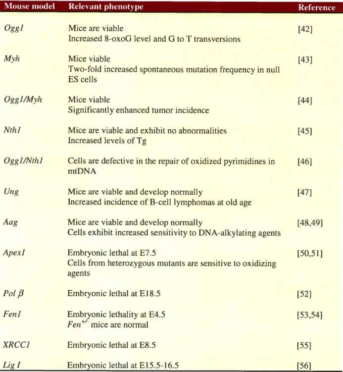
Over the iast decade, several BER-deficient mouse models have been generated to study the relationship between BER dysftinction and carcinogenesis [39]. A surnmary of the BER mouse models, and their related phenotypes, is delineated in Table I-II. At first glance, it is evident that the next generation ofmouse modeis should comprised of combined deficiencies of DNA glycosylases which initiate the BER pathway as weII as ofconditionai modeis ofthe intermediate and late steps of BER before a direct link can be established between BER deficiency and cancer.
AP ENDONUCLEASES
In addition to being central damage intermediates in BER, AP sites are considered to 5e one ofthe most frequent endogenous lesions in DNA [19,40]. Due to their mutagenic and cytotoxic properties, the repair of AP sites is essential to ensure genome stability and ccli survivai [41]. AP sites can 5e processed either by AP endonucleases or glycosylases/AP iyases (figure 1-3). StiIl, The 3’-blocking groups produced following incision by glycosylases/AP lyases must be further processed to produce an accessible 3’-OH termini required for repair synthesis by DNA polymerases [1]. The majority of the celiular 3’-phosphodiesterase activity is provided by AP endonucleases (figure 1-3, pathway on the right) [1]. Thus, this class of enzymes is considered to be far more efficient in repairing AP sites than glycosylases/AP lyases.
Table I-II. Summary of BER mouse models.
Mouse model Relevant phenotype Reference
Oggi Mice are viable [421
Jncreased 8-oxoG level and G to T transversions
Mvii Mice viable [43]
Two-fold increased spontaneous mutation frequency in null ES cells
Ogg]/M)’h Mice viable [44]
Significantly enhanced tumor incidence
Nth] Mice are viable and exhibit no abnormalities [45]
Increased levels of Tg
Ogg]/Nth] Celis are defective in the repair of oxidized pyrimidines in [46] mtDNA
Ung Mice are viable and develop normally [47]
Increased incidence of B-cell lymphomas at old age
Aag Mice are viable and develop normally [48,49]
Celis exhibit increased sensitivity to DNA-alkylating agents
Apex] Embryonic lethal at E7.5 [50,51]
Cells from heterozygous mutants are sensitive to oxidizing agents
Pot
fi
Embryonic lethal at E 18.5 [52]Fen] Embryonic lethality at E4.5 [53,54]
Fen mice are normal
XRCC] Embryonic lethal at E8.5 [55]
Lig J Embryonic lethal at E 15.5-16.5 [56]
16
(
Sequence and structural conservation of AP endonucleasesAP endonucleases are evolutionary highly conserved DNA repair enzymes which are grouped into two distinct families, the exonuclease (exo) III and the endonuclease (endo) IV families, named afier the Escherichia cou (E. cou) members [1,57-59]. The exo III family includes E. cou exo III, 8. cerevisiae Apn2, Caenorhabditis elegans (C. etegans) EXO-3, human APE1 and APE2. The exo III members are small, monomeric, divalent metal ion-dependent enzymes that are sensitive to metal-chelating agents, such as EDTA [60-62]. On the other hand, AP endonucleases of the endo IV family, comprising E. cou endo IV, £ cerevisiae Apnl, and C. elegans APN- 1, are resistant to inactivation by EDTA since they are metal ion-independent enzymes [60,63]. Although no homolog of the endo IV family has yet been identified in human celis, such activity is Iikely to be present in there as well (See Table I-III for a summaiy ofthe AP endonuclease enzymes).
In addition to their sequence sirnilarity, related AP endonucleases also share structural homology, as suggested by high-resolution crystal structures for their representative members. Both E. cou exo III and human APE1 possess a characteristic four-layered u43-sandwich fold which is also found in the digestive enzyme DNase I [64,65]. However, their DNA-binding domain loops adopts an extrahelical conformation that is flot observed for DNase I. These protruding loops are proposed to be involved in damage recognition and bond cleavage at AP sites [65]. Recently, Kaneda and colleagues demonstrated by site-directed mutagenesis that the amino acid residues, Trp-212 and Trp-2$0, near the catalytic site of exo III and APE 1, respectively, are critical for AP site recognition as well as AP endonuclease activity [66]. They propose a model whereby the tryptophan residue ofexo III-related enzymes acts as an AP site ‘recognizer’ by intercalating its aromatic side chain into the AP site pocket, followed by flipping out of the AP site from the duplex into the enzyme catalytic pocket with DNA kinking at the AP site [66]. for
©
Table I-III. Comparison of AP endonucleases in bacteria, yeast, and human. E. coti S. cerevisiae Human Gene name Protein name Family nameCofactors Inhibitors Length Molecular
weight AP endonuclease activity 3 ‘-phosphodiestease activity 3’-5’ exonuclease activity Nucleotide incision activitv Relevant mutant phenotpe xth exo III Exo UI Mg2 EDTA 268 aa 3 0.97 kDa 90% 90% Robust Specific for 3’ recessed ends
ofDNA Absent Hypersensitive to
oxidizing agents nfo endo IV Endo IV no no 285 aa 31.4$ kDa 10% *2Ofld induction by
paraquat 10% *2Ofld induction
by
paraquat High Preference
for
3’
recessed
substrate Present Hypersensitive to
oxidizing
agents
APNI Apnl Endo
1V no no 367 aa 41.44 kDa 97% . 97% Robust Preference for 3’ recessed
DNA Present Hypersensitive to
alkyÏating and oxidizing agents APN2 APEI APE2 Apn2 APE1 APE2 Exo III Exo III Exo III 2+ 2+ 2+ Mg Mg Mg EDTA EDTA EDTA 520aa 317aa 51$aa 59.45 kDa 3 5.42 kDa 57.4 kDa 3% 95% Weak 30-to 40-fold 100-to 200-Strong more efficient fold less than AP efficient than endonuclease P?AP endonuçlease 30-to 40-fold 100-to 200-Robust more efficient fo1d less than AP efficient than endonuclease ‘ AP endonuclease Absent Present Absent Sensitive to Ernbryonic Growth aikylating lethal in nuil retardation agents mice nul! mice
1$
the endo IV family, the high-resolution structure of E. cou endo IV suggests that this AP endonuclease and its homologues may use the classic agf3s TIM banel fold to promote flipping of both the AP site and its orphan nucleotide out of the duplex with
a 900 bend in the DNA, but allowing only the damaged nucleotide in the enzyme active site pocket [67]. The conformational changes induced in endo IV upon DNA binding would allow the AP site to directly make contact with the three Zn2 ions, which are essential for the catalytic activities of ail endo IV-related AP endonucleases [67]. Thus, solving the crystal structures of representative AP endonucleases has provided the structural basis for understanding the catalytic rnechanism of the exo III and endo IV family members [60].
E. cou AP endonucleases
Exo III
The exo III enzyme, encoded by the xth gene, was initialiy identified as a
(
phosphatase-exonuclease, but subsequently was discovered to be the major constitutive AP endonuclease in E. cou extracts. Exo III is a 26$ amino acid enzyme with a molecular mass of 31 kDa and accounts for approximatively 90% of the cellular AP endonuclease activity [59,68,69]. Exo III is also endowed with a strong 3’-phosphodiesterase activity, an efficient 3’-5’ exonuclease activity, and an endonuclease activity at urea residues in oxidized DNA [70]. E. cou mutants lacking exo III (xthA) are sensitive to oxidizing agents such as H202 and are hypersensitive to UVB light [7 1-74]. A recent study clearly suggests that exo III plays a crucial role in the repair of UVB-induced toxic lesions, as pre-treatment with dipyridyl (an iron chelator) completely restores wild-type resistance in the xthA strain [74]. Dipyridyl prevents the Fenton reaction (Fe2 + F1202 —> Fe3 + Q14 + 0W) to occur via the chelation of a fenous iron (Fe2), thus protecting ceils from ROS-induced DNA lesions [74]. In fact, several studies have demonstrated that xthA strains accurnulate large amounts of unrepaired ssb with blocked 3’ termini which are genotoxic damages [73,75].EndoIV
The xth mutants possess a residual (10%) AP endonuclease activity that displays no requirement for metal ions. A large-scale screening of clones overexpressing a Mg2tindependent AP endonuclease activity lcd to the isolation of the nfo gene which encodes the endo IV protein. The nfoA mutant is less sensitive to oxidative and aikylating agents (e.g. FT202 and MMS, respectively) as compared to the xthA strain, indicating that endo IV serves as an back-up enzyme in the repair of AP sites [75]. However, recently, endo IV was demonstrated to play a determinant role in the avoidance of UVB-induced mutagenic lesions [74]. Moreover, the protein expression levels of endo IV can be induced more than 20-fold by paraquat, a superoxide-producing agent. The up-regulation of endo IV may be important during oxidative stress or when certain DNA lesions are refractory to repair by exo III [76].
S. cerevisïaeAP endonucleases
C
Apn]In the budding yeast L cerevisiae, Apnl is the predominant AP endonuclease representing more than 90% of the total AP endonuclease activity [77]. Apnl was first described as a 3’-phosphodiesterase since the purified Apnl cleaved synthetic DNA substrates containing 3’-phosphoglycolaldehyde esters [77]. Both the AP endonuclease and 3’-phosphodiesterase activities of yeast Apnl showed no metal requirement similarly to bacteria endo IV [77-79]. Screening a yeast 2gt1 1 expression cDNA library in E. cou using a polyclonal antibody produced from the purified Apnl lead to the successful identification of the AFNJ gene [$0]. Comparison of the primary structure of Apn 1 with that of E. cou endo IV reveaÏed that the yeast protein is indeed homologous to endo IV and bears an extra $2 amino acid residues at the C-terminus [$0,81]. This additional C-teminal region of APN] contains a bipartite nuclear localization signal (NLS) which when deleted causes cytoplasmic accumulation of Apnl [$1]. Moreover, we have previously demonstrated that Pin, a cell wall constituent, interacts with Apnl bipartite NLS, causing Apnl to translocate
20
into the mitochondria [$2]. We observed in pir]A mutants a striking nuclear accumulation (‘—3-fold) of Apnl, which coincided with drastically reduced levels in the mitochondria [$2]. Apnl encompasses several enzymatic activities: (j) an AP endonuclease activity, that hydrolyses the phosphodiester backbone 5’ to an AP site [1], (ii) a 3’ -phosphodiesterase activity, that removes 3’ DNA-blocking groups such as 3’ -phosphates, 3’ -phosphoglycolates, and 3’ -ct, 13-unsaturated aldehydes [11, (iii) a 3’-tyrosyl-DNA phosphodiesterase activity, that hydrolyzes the tyrosine residue of topoisomerase I linked to a 3’ DNA end [$3], (iv) an endonuclease activity, that nicks 5’-side of oxidatively damaged DNA bases such as fapyA and FapyG [84], and (y) a
3’—>5’ exonucleaseactivity, that removes either a 3’-terminal nucleotide at a nick or a misincorporated $-oxo-dGuo [85,86].
Apnl-deficient strains of S. cereviasiae display hypersensitivity to H202 and MMS which produces ssb with blocked 3’-termini and abasic sites, respectively [40]. Interestingly, apn]A nuli mutants are not sensitive to bleomycin, suggesting the presence of another enzyme more proficient at removing bleomycin-induced
3’-C
blocking groups. Loss of Apnl resuits in a 10- to 15- fold increase in the nuclear spontaneous mutation rate [40]. The mutations correspond mainly to AT to CG transversions [$7]. The high frequency of spontaneous mutations in the apnli nuil mutants is primarlly due to unrepaired AP sites [8$]. Deletion of Apnl has no effect on the rates of spontaneous mitochondrial mutations. However, when Apnl-deficient cells are exposed to MMS, they dispïay approximately 6-fold increase in mitochondrial spontaneous mutation rates, suggesting an important role of Apnl in the repair of darnaged mitochondrial DNA [$2,89].Apn2
A residual (5%) AP endonuclease activity was partially purified initiaÏly by Sander and Ramotar [90] and termed Pde (3’-phosphodiesterase). Later, Johnson et aï. independently characterized the Pde protein and renamed it Apn2 [$9]. Apn2 shares 36% and 33% sequence homology with E. cou exo III and human APE1, respectively [89,91]. In addition to its AP endonuclease activity, Apn2 also contains 3’-phosphodiesterase and 3’—÷S’ exonuclease activities [92]. These two latter
activities of Apn2 were estimated to be 30- to 40-fold higher than its AP endonuclease activity [92]. Furthermore, a direct interaction of Apn2 with PCNA strongly stimulates its 3’-phosphodiesterase and the 3’—>5’ exonuclease activities [93].
Ceils deficient in Apn2 are less sensitivity to MM$ than the apn]A strains. However, apn]Aapn2A double mutants are 1 5-fold more sensitive to MM$ and 2- to 3-fold more sensitive to H202 and bleomycin than apn]zl single mutants [91,94]. The apn]Aapn2A mutant strains show both enhanced spontaneous and MM$-induced mutation rates in comparison to the wild type [$9,91]. The expression of Apn2 in apnMapn2A mutants restores some resistance to MMS and partially decreases the rate of spontaneous mutations [91]. Together, these observations indicate that Apn2 provides important back-up DNA repair activities for Apnl.
Human AP endonucleases AFE]
In humans, the exo III-related protein APE 1 (also called Ref- 1, HAP 1, and APEX1) is considered the major AP endonuclease, representing approximately 95% of the total cellular AP endonuclease activity [95-9$]. APE1 harbors its own bipartite NLS, located within its N-terminal 20 amino acid residues, which allows for its nuclear import but also localization to the mitochondria following cleavage by an unknown specific mitochondria-associated peptidase [99,100]. APE1 is also endowed with a 3’-phosphodiesterase as well as a 3’—+5’-exonuclease activity, which are much weaker than its AP endonuclease activity [1]. Moreover, Gros et aï. demonstrated that APE1 is also capable of incising directly 5’ of oxidative base lesions, sucli as 5,6-dihydro-2-deoxyuridine, in a DNA glycosylase-independent manner [101].
The biological importance of APE1 is underlined by the embryonic lethality in knock-out mice and the lack of stable APE 1 -deficient celI fines [102]. Recently, Fung and Demple addressed the cellular role of APE1 by using RNAi tecbnology [103]. They showed that APEY downregulation blocked cdl proliferation and
22
stimulated apoptosis, which was correlated with accumulation of AP sites [103]. These effects were reversed by expression of S. cerevisiae Apnl, a protein that is structurally unrelated to APE 1 but shares several enzymatic activities in the repair of AP sites [1031 [104]. Their resuits clearly establish that APE1 plays a vital role in the repair of endogenous DNA damage that, when lefi unrepaired triggers apoptotic celi death.
APE2
To date, no endo IV homologs have been identified in human celis. In their own search for nove! human AP endonuclease/3 ‘-phosphodiesterase enzymes, Hadi and Wilson have however identified a second exo Ili-like protein termed APE2 [105]. The ubiquitously expressed APE2 enzyme is composed of 51$ amino acids with a predicted molecular mass of 62 kDa and shares significant amino acid sequence similarity to the core nuclease domain of APE1 and exo III (29% and 27% sequence identity, respectively) [105]. Peptide sequence comparison revealed that APE2 lacks the extended N-terminal domain present in APE 1 but possesses a longer C-terminal region, which is absent in APE1 and exo III, thus indicating the existence of a second exolli-like subfamily. This new exo lil-like subgroup comprises also of S. cerevisiae
Apn2 and S. pombe Apn2 [106].
An initial insight into the biological significance of APE2 was provided by homozygous APE2-null mice which exhibited growth retardation and dyshematopoiesis accompanied by G2/M arrest [107]. To shed more light on the role of human APE2, Burkovic et al. expressed recombinant APE2 in yeast and showed that recombinant enzyme exhibits a weak AP endonuclease activity, but shows strong 3’—5’ exonuclease and 3’-phosphodiesterase activities that act preferentially on mistmatched 3’-nucleotides from DNA [10$]. Moreover, mutational analysis revealed that Asp177 is an active site residue ofAPE2 [10$]. This study strongly suggests that the intrinsic activities of APE2 are determinant in the removal of 3’-blocking termini, as well as in the proofreading oferrors incorporated by DNA polymerases [10$].
APE] more than a DNA repair enzyme
APE1 is also known as Ref-1 (redox effector factor-1) due to its redox activity that acts on redox-sensitive transcription factors [109]. APE 1 /Ref- 1 was first found to copurify with the transcription factor AP-1 during column chromatography [110]. Subsequently, APE1/Ref-1 was demonstrated to reduce the conserved cysteine residues in the DNA binding domains of c-Fos and c-Jun, the two subunits of AP-1, thus promoting the binding to their DNA cognate sequences [97]. Since then, APE1/Ref-1 has been shown to modulate the DNA binding activities, and thus the transcriptional regulatory potential of proteins such as NF-KB (nuclear factor-KB), Myb, HIf-YŒ (hypoxia inducible factor-1Œ) and p53 via a redox mechanism [111].
The redox function of APE1/Ref-1 is located in the N-terminal 127 amino acids, whereas its nuclease activity resides in the C-terminal 157 amino acids [112]. The nuclease region of APE 1 /REf- 1 shares a high degree of sequence homology with other functionally related repair proteins such as bacteria exo III and fruit fly Rrp 1, but the redox domain is present only in mammals [112]. The redox activity of APE1/Ref-1 may have evolved to sustain embryonic development, since deletion of an exo Ili-like protein does flot cause lethality in lower organisms [112]. However, at the cellular level, APE1/Ref-1 nuclease function is more important for the repair of spontaneous DNA damage that, when accumulated, can induce celi death [102,112,113]
Regulation of APE1 activities
Considering the key roles of APEY in DNA repair and gene regulation, the ccli has developed several strategies to regulate the APEY enzyme at both the transcriptional and posttranslational levels [111]. In terms of transcriptional regulation, it has been shown that ROS, such as H202, induce APEY mRNA, which correlates with an increase in APE1 protein levels and activity [114]. APE1 cellular functions are further modulated through two non-mutally exclusive posttranslational mechanisms, namely subcellular localizations and posttranslational modification [115]. Upon exposure to ROS, APE1 accumulates in the nucleus as a resuit ofboth an
24
increased in nuclear transiocation and a decreased in nuclear export via the binding of its bipartite NLS with nuclear importins cd and Œ2 and the inhibition of its nuclear export signal, respectively [99]. Several posttranslational modifications, such as acetylation, phosphorylation and oxidation/reduction have been reported to influence APEI functions [111,115]. Acetylation of APE1 by p300 was shown to affect the transcriptional regulatory function ofAPE1 [1 15]. However, the reports on the impact of phosphorylation by PKC (protein kinase C), CKI and CKII (casein kinase I and II, repectively) on APE1 activities remain inconsistent [1 15]. Some studies suggest that phosphorylation of APE1 might abolish its AP endonuclease activity [1151.
The redox status of APE1 is another important regulatory factor determining its cellular activities [111,115]. Hirota et al. demonstrated that thioredoxin (TRX), an endogenous dithiol-reducing molecule, acts as a hydrogen donor for APE 1. Reduced APEI, in tum. potentiates the DNA-binding and transactivation abilites of AP-l [116]. In a similar maîmer, TRX and APE1 cooperate in the control of basal p53 stability and activity [117]. While reduction of APE 1, on the one hand, appears to be a positive reguÏatory mechanism for its transcriptional functions, oxidation, on the other, lias a negative impact on APE1 AP endonuclease activity [118]. Treatment of APE1 with oxidizing agents, such as H202 and diamide, causes a significant decrease in AP endonuclease activity, which was neutralized by the addition of certain amounts of DTT, suggesting a direct effect of oxidation [11$]. Furthermore, site directed mutagenesis studies revealed that Cys3lO residue of APE1 is likely to be oxidized, whereas Cys63 and Cys95 are most susceptible to a reduction reaction [115] [112].
Protein-protein interactions involving APE1
APE1 has been demonstrated to be an important modulator of the multi-step BER pathway. APE1 accomplishes this regulatory role mainly through physical and/or functional protein-protein interactions with both up- and down-stream BER enzymes (Table I-IV) [115]. BER is initiated by DNA glycosylases which recognize and excise a damaged base leaving behind an AP site. The activity of several
Q
glycosylases, such as OGGI [119], UNG [120], TDG [121] and NIH1 [122], isstimulated by an association with APE 1. However, only TDG and MYH interact physically and form stable complexes with APE1 [121,123]. Since most of the glycosylases display a high affinity for base lesions and AP sites, APE1 is thought to stimulate the dispiacement of the glycosylases from their DNA products via protein interactions, thus facilitating glycosylase turnover. APE1 was shown to interact also with downstream BER proteins, sucli as fEN1 [124], Pol 3 [1251, PCNA t124], XRCCI [126] and LIG1 [127]. In these interactions, the AP endonuclease modulates its patner’s activity, with the exception of XRCC1, which alters the ftmctions of APE1 (see Table I-IV).Adding to its complex network of protein interactions with BER proteins, APE1 also interacts with proteins participating in other cellular processes, such as
transcription and replication. APE1 was demonstrated to interact with WRN (Werner
syndrome protein), PARP (Poly(ADP-ribose) polymerase 1), HSP7O (heat-shock protein 70), and the tumor-suppressor protein p53 [128-132]. The effects of these
26
G
Table I-IV. Effects of the various proteins interactions involving APE1 on theBER pathway.Interacting patner Conseguence of the interaction Reference BER proteins
UDG StimuÏates dispiacement ofUDG from AP sites [120]
TDG Stimulates the turnover o [121]
f TDG at A? sites
MYH Facilitates the release ofMYH from its DNA [123]
products
FEN1 Enhances both the exo- and endonuclease [124,127]
activities ofFEN1 Stimulates LP-BER
Pol
fi
Stimulates the 5’-dRP lyase activity of Polfi
[125]PCNA Coordinates LP-BER [124]
XRCC1 Increases APE1 A? endonuclease and 3’- [126]
phosphodiesterase activity Promotes SP-BER
LIG1 Stimulates DNA ligase 1 activity [127]
Other prote ins
WRN Inhibits WRNhelicase activity on BER [128]
intermediates
PARP Blocks APE1 stimulation ofstrand-displacement [129]
DNA synthesis by Pol
f3
Inhibits the exonuclease activity ofAPE1
HSP7O Stimulates APE I AP endonuclease activity [1301
p53 Stimulates p53 DNA binding by promoting its [132]
tetramerization
Induces p53 transactivation and pro-apoptotic activities
HYPOTHESIS
ANDOBJECTIVES
AP endonuclease enzymes are crucial players in the BER pathway. They are grouped into two distinct familles, the exo III famiÏy and endo IV family. Celis lacking AP endonuclease activity are hypersensitive to oxidizing and alkylating agents, such as H202 and MMS, which induce ssb with blocked 3’-termini and AP sites, respectively. We were particularly interested in understanding the molecular mechanisms by which eukaryotic AP endonucleases maintain genomic integrity in the nucleus as well as in the mitochondria. We set out to study the structure/function andlor protein interactions of AP endonucleases, which could provide us with valuable insights into their mechanisms of binding and cleavage of oxidative DNA lesions as well as their regulation inside the celi. In our first study (Chapter 2) which aimed at identifying the critical arnino acid residues of S. cerevisiae Apnl for its biological function, we showed by mutational analysis that G1u15$ and Asp192 are crucial for the DNA repair functions of Apnl. In our second study (Chapter 3), we set out to evaluate the role of yeast Oggl DNA glycosylase in the maintenance of a mitochondrial poly(GT) tract reporter system. Interestingly, we discovered that overproduction of Apnl triggers instability of the poly(GT) tract, but which was counteracted by the simultaneous overexpression of Oggi. In our third study (Chapter 4), we initially aimed at purifying an endo IV homolog in human celis. However, the goal of this project changed when we unexpectedly re-isolated GAPDH, a well established glycolytic enzyme, during our purification of a Mg2tindependent AP endonuclease. We demonstrated that GAPDH physically interacts with APE1 to reduce the oxidized forms of APE1, which resulted in the reactivation of the AP endonuclease activity. In addition, we demonstrated the importance of Cys 152 and Cys 156 residues of GAPDH in the reactivation of APE 1 by site-directed mutagenesis. The biological importance of GAPDH in the cellular protection against genotoxic agents, which induce APE1-repairable lesions, was further supported by clonogenic assays using human colon carcinoma celis overexpressing GAPDH or knockdowned for GAPDH.
o
CHAPTER 2
Cliaracterization of two independent amino acid
substitntions that disrupt the DNA repair
functions of the yeast Apnl
o
Characterization of
two
independent amino acid substitutions that
disrupt the DNA repair functions of the yeast Apn 1”
ARSHAD JILANI’, RATSAVARINH VONGSAMPHANH1, ANICK LEDUC’, LAURENT GROS2, MURAT SAPARBAEV2, AND DINDIAL RAMOTAR1
Running titie: Two variants ofApnl lacking DNA repair functions
Corresponding author:
Research Centre, University of Montreal, 5415 de l’Assomption, Montreal, Quebec, Canada HiT 2M4. Telephone: (514) 252-3400 ext 4684. Fax: (514) 252-3430.
E-mail:
2Groupe Réparation de l’ADN, Unité Mixte de Recherche $532 CNRS, Laboratoire de Biotechnologies et Pharmacologie Génétique Appliquée-École Normale Supérieure Cachan, Institut Gustave-Roussy, 94805 Villejuif Cedex, France. Telephone: 33 1 42115404. Fax: 33 1 42115276.
E-mail:
WThis work was partially funded by the National Cancer Institute of Canada (NCIC) with funds from the Canadian Cancer Society and the Natural Sciences and Engineering Research Council of Canada (D.R.), and by European Community Grant QLK4-2000-00286 and Association pour la Recherche sur le Cancer (M.S.). D.R. is supported by a senior fellowship from the Fonds de la Recherche en Santé du Québec, and L.G. is supported by a postdoctoral fellowship from European Community.