UNCORRECTED PR
OOF
1
Surfactins modulate the lateral organization of
fluorescent membrane polar lipids:
2
A new tool to study drug:membrane interaction and assessment of the role of
3
cholesterol and drug acyl chain length
4
L.
Q1
D'Auria
a,b, M. Deleu
c, S. Dufour
c, M.-P. Mingeot-Leclercq
d, D. Tyteca
a,b,⁎
5 aCELL Unit, de Duve Institute, UCL B1.75.02, Avenue Hippocrate, 75, B-1200 Brussels, Belgium6 b
Université Catholique de Louvain, UCL B1.75.02, Avenue Hippocrate, 75, B-1200 Brussels, Belgium
Q2
7 c
Centre de Biophysique Moléculaire Numérique, Gembloux Agro-Bio Tech, Université de Liège, Gembloux, Belgium
8 d
Cellular and Molecular Pharmacology, Louvain Drug Research Institute, Université Catholique de Louvain, Brussels, Belgium
9 10
a b s t r a c t
a r t i c l e i n f o
11 Article history: 12 Received 27 February 201313 Received in revised form 16 April 2013
14 Accepted 8 May 2013 15 Available online xxxx 16 17 18 19 Keywords: 20 Surfactin:membrane interaction
21 Micrometric lipid domain
22 Living erythrocyte
23 Vital confocal imaging
24 Cholesterol
25 BODIPY-lipid
26 The lipopeptide surfactin exhibits promising antimicrobial activities which are hampered by haemolytic toxicity.
27 Rational design of new surfactin molecules, based on a better understanding of membrane:surfactin interaction,
28 is thus crucial. We here performed bioimaging of lateral membrane lipid heterogeneity in adherent living
29 human red blood cells (RBCs), as a new relevant bioassay, and explored its potential to better understand
30 membrane:surfactin interactions. RBCs show (sub)micrometric membrane domains upon insertion of BODIPY
31 (*) analogs of glucosylceramide (GlcCer*), sphingomyelin (SM*) and phosphatidylcholine (PC*). These domains
32 exhibit increasing sensitivity to cholesterol depletion by methyl-β-cyclodextrin. At concentrations well below
33 critical micellar concentration, natural cyclic surfactin increased the formation of PC* and SM*, but not GlcCer*,
34 domains, suggesting preferential interaction with lipid* assemblies with the highest vulnerability to
methyl-35 β-cyclodextrin. Surfactin not only reversed disappearance of SM* domains upon cholesterol depletion but further
36 increased PC* domain abundance over control RBCs, indicating that surfactin can substitute cholesterol to promote
37 micrometric domains. Surfactin sensitized excimer formation from PC* and SM* domains, suggesting increased
38 lipid* recruitment and/or diffusion within domains. Comparison of surfactin congeners differing by geometry,
39 charge and acyl chain length indicated a strong dependence on acyl chain length. Thus, bioimaging of micrometric
40 lipid* domains is a visual powerful tool, revealing that intrinsic lipid* domain organization, cholesterol abundance
41 and drug acyl chain length are key parameters for membrane:surfactin interaction. Implications for surfactin
42 preferential location in domains or at their boundaries are discussed and may be useful for rational design of better
43 surfactin molecules. 44 © 2013 Published by Elsevier B.V. 45 46 47 48 49 1. Introduction
50 Decades of world-wide antibiotic use have led to an increased bac-51 terial resistance which urges tofind new agents. Biological properties 52 of surfactin, a lipopeptide produced by Bacillus subtilis, indicate to be a 53 potential antibacterial agent
Q3 . Natural surfactin (hereafter referred to
54 as“surfactin”) is composed of an heptapeptide cycle closed by a C13
55 to C15hydroxy fatty acid forming a lactone ring, with strong
amphi-56 philic character explaining bioactivity as surfactant. Surfactin exhibits
57 additional biological properties, including antibacterial, antiviral and
58 hemolytic activities [for a review, see1]. Surfactin's biological activity
59 is determined by an interaction with membranes, including insertion
60 into lipid bilayers, modification of permeability and membrane
solu-61 bilization by a detergent-like mechanism[2]. This interaction is
high-62 ly dependent on surfactin concentration, as demonstrated in model
63 membranes with coexistingfluid disordered and gel phases[3]. To
64 prevent hemolysis, a major limitation to medical applications, Dufour
65 and collaborators have synthesized various linear surfactin analogs
66 differing by charge and hydrophobicity (for structures, see Suppl.
67 Fig. 1). In comparison to cyclic congeners, linear surfactins showed
68 reduced surface activity and hemolysis[4].
69 So far, membrane:surfactin interactions have been mainly studied
70 in elementary artificial model systems made of one or two
phospho-71 lipids, thus ignoring major membrane components such as
cholester-72 ol and sphingolipids (SLs), as well as membrane lateral heterogeneity.
Biochimica et Biophysica Acta xxx (2013) xxx–xxx
Abbreviations: BODIPY, boron dipyromethene, referred here as *; DF-BSA, defatted bovine serum albumin; FRAP, fluorescence recovery after photobleaching; GlcCer, glucosylceramide; GSL, glycosphingolipid; Ld, liquid-disordered; Lo, liquid-ordered;
mβCD, methyl-β-cyclodextrin; PC, phosphatidylcholine; PM, plasma membrane; So,
solid-ordered; SAL14, Surfactin Acylated Linear with 14C; SL, sphingolipid; SM, sphingomyelin; SNC14, Surfactin Natural Cyclic with 14C-acyl chain length; SSL10, Surfactin Synthetic Linear with 10C; SSL14, Surfactin Synthetic Linear with 14C; SSL18, Surfactin Synthetic Linear with 18C; surfactin-C13–C15, natural cyclic surfactin,
referred here as surfactin
⁎ Corresponding author. Tel.: +32 2 764 75 91; fax: +32 2 764 75 43. E-mail address:[email protected](D. Tyteca). 0005-2736/$– see front matter © 2013 Published by Elsevier B.V. http://dx.doi.org/10.1016/j.bbamem.2013.05.006
Contents lists available atSciVerse ScienceDirect
Biochimica et Biophysica Acta
j o u r n a l h o m e p a g e : w w w . e l s e v i e r . c o m / l o c a t e / b b a m e mUNCORRECTED PR
OOF
73 SLs include the zwitterionic sphingomyelin (SM), which bears the74 same phosphocholine headgroup as phosphatidylcholine (PC), and 75 glycosphingolipids (GSLs), a heterogeneous family comprising mono 76 (e.g. glucosylceramide; GlcCer), di (e.g. lactosylceramide) and more 77 complex GSLs such as ganglioside GM1 [for a review, see5]. Membrane 78 lipid bilayers, long viewed as homogenous solvent for membrane pro-79 teins[6], actually show lateral heterogeneity at two different scales: 80 transient nanometric rafts[7–11] vs more stable (sub)micrometric/ 81 mesoscale domains. Such domains have not only been evidenced 82 on artificial vesicles [8,12–17] but also documented on living cells. 83 They were initially predicted by FRAP[18]; further implied by single-84 molecule tracking based on discrete jumps between “mesoscale” 85 domains[19–21]; and directly visualized by confocal imaging after in-86 sertion offluorescent analogs at trace levels in various cells, including 87 red blood cells (RBCs)[22–27]. Yeast plasma membrane (PM) proteins 88 also show a patchwork of distinct micrometric domains[28–31]. 89 Since antibacterial potential of surfactin is hampered by intrinsic 90 hemolytic properties which are influenced by drug interaction with 91 the bilayer, we here performed bioimaging of lateral membrane lipid 92 heterogeneity in adherent human red blood cells (RBCs), as a new 93 relevant bioassay, and explored its potential to better understand mem-94 brane:surfactin interactions. RBCs show micrometric domains readily 95 evidenced by confocal microscopy upon insertion of trace levels of 96 fluorescent analogs (BODIPY, *) of major polar lipids[22,26,27]. We 97 thus probed the effect of various natural and synthetic surfactins on 98 membrane organization using lipid* domains as read-out: (i) cyclic 99 natural surfactin (referred to as surfactin-C13–C15or surfactin), bearing
100 13, 14 and 15C acyl chains; (ii) the purified natural SNC14 (Surfactin 101 Natural Cyclic with 14C-acyl chain length); as well as (iii) synthetic lin-102 ear analogs differing by charge and hydrophobicity, SAL14 (Surfactin 103 Acylated Linear with 14C-acyl chain length) and three SSLs with 104 increased acyl chain lengths (SSL10, 14 and 18, Surfactins Synthetic 105 Linear with 10, 14 or 18C).
106 RBCs offer several advantages as experimental system. First, they 107 allow studying lipid lateral organization without artifacts: (i) they 108 are featureless at the micrometric level; (ii) they do not perform 109 endocytosis nor lipid metabolism; (iii) their membrane asymmetry 110 is well-characterized[32], including the occurrence of rafts[33–35]. 111 Second, RBCs have a uniquely high content of cholesterol (~ 40 mol% 112 vs ~ 30 mol% infibroblasts vs ~15 mol% in blood platelets), which is 113 a key regulator of both membrane fluidity via lipid packing and 114 membrane deformability via modulation of PM protein interactions 115 at the cortical cytoskeleton interface[36]. RBCs also exhibit a strong 116 membrane:cytoskeleton anchorage, thanks to two non-redundant 117 4.1R and ankyrin-based complexes[37]. Third, RBCs have been used 118 to evaluate surfactin toxicity [4]. Fourth, we recently reported in 119 details by vital confocal imaging segregation of BODIPY analogs of 120 GSLs* (e.g. GlcCer*), SM* and PC* into structurally distinct micrometric 121 domains in RBCs[22,26,27]. We observed that all GSLs*, SM* and PC* 122 domains disappear upon RBC stretching, indicating a control by mem-123 brane tension. However, domains are differentially modulated by: (i) 124 temperature (peaking at 20 °C for SM* and PC* while steadily
Q4 increasing
125 up to 37 °C for GlcCer*); (ii) cholesterol (suppression of SM* and PC* 126 domains by minor cholesterol depletion but preservation of GlcCer* 127 domains); and (iii) the two membrane:cytoskeleton anchorage com-128 plexes (differential association with 4.1R complexes upon antibody 129 patching and differential response to uncoupling at 4.1R and ankyrin 130 complexes). The relevance of BODIPY-lipid micrometric domains for en-131 dogenous lipids despite BODIPY substitution of acyl chain is supported 132 by three observations: (i) co-localization of exogenous GM1* with 133 endogenous GM1 labeled by cholera toxin in RBCs; (ii) identity of PM 134 domains in CHO cells upon insertion of SM* vs metabolic conversion of 135 ceramide* into SM*; and (iii) selective disappearance of SM* domains 136 upon depletion of endogenous SM[22,26,27].
137 We found that interaction of surfactins with RBC membrane, as 138 reflected by impact on fluorescent micrometric lipid domains, is dictated
139 by endogenous cholesterol content, lipid* domain organization and
140 surfactin acyl chain length. Implications for preferential surfactin
interac-141 tion with membranes of specific lipid composition and lateral
heteroge-142 neity are discussed. This straightforward confocal imaging assay may
143 help understanding surfactin surface activity and designing less toxic
144 surfactin derivatives.
145 2. Materials and methods
146 2.1. RBC isolation and immobilization
147 This study was approved by the Medical Ethics Institutional
148 Committee and the blood donors gave written informed consent.
149 RBCs were isolated from healthy volunteers. Blood was collected by
150 venopuncture into dry EDTA (K+salt)-coated tubes, diluted 1:10 in
151 medium (DMEM containing 25 mM glucose and 25 mM HEPES) and
152 washed twice by centrifugation at 133 g for 2 min and resuspension.
153 For spreading onto poly-L-lysine-coated coverslips, RBCs were plated
154 at ~ 20.106cells/ml onto 2-cm2coverslips precoated with 0.1 mg/ml
155 70–150 kDa poly-L-lysine (PLK; Sigma) at 20 °C for exactly 4 min
156 after which the suspension was removed and replaced by fresh
medi-157 um, in which RBCs were allowed to spread for another 4 min.
158 2.2. RBC treatments
159 Surfactin-C13–C15(a natural mixture of 13, 14 and 15C-acyl chain
160 lengths, in proportion 3:42:52) and SNC14 (for Surfactin Natural
161 Cyclic with 14C) were extracted from a B. subtilis S499 culture
super-162 natant. Synthetic surfactins, SAL14 (Surfactin Acylated Linear with
163 14C) as well as SSL10, SSL14 and SSL18 (Surfactin Synthetic Linear
164 with 10, 14 and 18C in the acyl chain respectively), were prepared
165 as described[4]. Unless otherwise stated, RBCs were preincubated
166 in suspension with 0–1 μM surfactins at 37 °C for 30 min, before
167 spreading onto poly-L-lysine-coated coverslips. For cholesterol
deple-168 tion, cells were preincubated with 0.25 mM methyl-β-cyclodextrin
169 (mβCD; Sigma) at 37 °C for 1 h. For combined treatments, RBCs
170 werefirst treated with mβCD for 30 min at 37 °C then with surfactin
171 in the continued presence of mβCD for another 30 min. These RBCs
172 were pelleted at 133 g for 2 min and gently resuspended in DMEM
173 for adhesion to poly-L-lysine-coated coverslips. Alternatively, RBCs
174 labeled with BODIPY-lipids (lipids*) were imaged during exposition
175 to surfactins. To measure residual cholesterol, lipid was extracted
176 and cholesterol was determined by Amplex Red Cholesterol kit
177 (Invitrogen) in the absence of cholesterol esterase[22]. Q5
178 2.3. RBC labeling and vital imaging
179 RBCs were labeled with BODIPY-lipids (lipids*; Invitrogen) after
180 spreading onto poly-L-lysine-coverslips. Briefly, cells were rinsed in
181 DMEM and labeled at 20 °C for 15 min with 0.75μM SM* or 1 μM
182 PC* or 1μM GlcCer* (except otherwise stated) in DMEM containing
183 equimolar defatted bovine serum albumin (DF-BSA; Sigma)[26]. For
184 confocal imaging, coverslips were placed bottom-up into Lab-Tek
185 chambers and examined in the green channel with a Zeiss LSM510
con-186 focal microscope using a plan-Apochromat 63× NA 1.4 oil immersion
187 objective in a thermostated cabinet set at 37 ± 1 °C (XL/LSM incubator,
188 Zeiss; Tempcontrol 37-2, PeCon)[26]. For excimer studies, RBCs were
189 excited at 488 nm and images were simultaneously acquired in the
190 green (λem520 nm) and red channels (λem605 nm)[27].
191 2.4. Hemolysis
192 Hemolysis was evaluated at 0.5μM surfactins by hemoglobin
193 release[22,38]. 0.2% Triton X-100 induced complete hemolysis, yielding
194 the 100% control value.
UNCORRECTED PR
OOF
195 2.5. Thin layer chromatography196 Lipids* were inserted in the RBC membrane at 0.75 or 1μM. After 197 washing, all lipids (endogenous and inserted*) were extracted, separated 198 by thin layer chromatography (TLC) in chloroform:methanol:15 mM 199 CaCl2(65:35:8; v/v/v)[39]and revealed by charring densitometry after
200 staining with 10% cupric sulfate in 8% O-phosphoric acid[40]. Band in-201 tensity of inserted lipid* was quantified and expressed by reference to 202 the sum of major lipids (cholesterol, PC, phosphatidylethanolamine, cer-203 amide and SM) from the same sample, after correction for band intensity 204 of corresponding endogenous lipid.
205 2.6. Statistical analyses
206 Values are means ± SEM. Statistical significance of comparisons 207 was tested by Student's t test. NS, not significant; *, p b 0.05; **, 208 pb 0.01; and ***, p b 0.001.
209 3. Results
210 3.1. Micrometric lipid* domains in control RBCs are restricted by RBC 211 stretching and membrane:cytoskeleton anchorage but favored by cholesterol 212 Using vital confocal imaging, we recently reported that fluores-213 cent lipid analogs of glycosphingolipids (e.g. BODIPY-GlcCer 214 [GlcCer*]), sphingomyelin (BODIPY-SM [SM*]) and phosphatidyl-215 choline (BODIPY-PC [PC*]) spontaneously form (sub)micrometric 216 domains at the plasma membrane (PM) of living RBCs adherent 217 onto poly-L-lysine (PLK)-coated coverslips and of CHO cells (for
218 lipid analog structures, see[26,27]). These domains are (i) readily 219 visible on RBCs partially spread onto PLK-coated-coverslips, (ii) 220 structurally and kinetically distinct, (iii) of decreasing packing: 221 GlcCer* > SM* > PC* and (iv) preferentially found at the outer PM 222 leaflet, as revealed by their complete disappearance upon surface 223 back-exchange by BSA (data not shown). Domains are probe 224 concentration-independent, since increasing SM* concentration 225 from 0.5 to 3μM changed neither the number nor the size of 226 domains (data not shown;[22]). The relation with endogenous 227 lipid compartmentation has been discussed elsewhere[22,26,27]. 228 All micrometric lipid* domains are strongly dependent on membrane 229 tension since they can be seen on control RBCs partially spread on the 230 coverslip but not in most spread cells. Moreover, domains are numerous 231 when RBCs are barely attached, decline to a stable low number at partial 232 spreading, and vanish upon maximal stretching [22]. Interestingly, 233 they (re-)appear or increase in size upon incubation into mildly hypo-234 tonic medium (data not shown;[26]). In addition, domains are differen-235 tially restricted by membrane:cytoskeleton anchorage[22], presumably 236 preventing domain increase in size and number. Accordingly, we ob-237 served that the combination of membrane:cytoskeleton uncoupling at 238 4.1R complexes upon PKC activation and membrane relaxation by incu-239 bation in mildly hypotonic medium led to a strong increase of GlcCer* 240 domain abundance in comparison to control RBCs (data not shown). In 241 contrast, cholesterol appears as a stabilization factor for PC* and SM* 242 domains[22].
243 3.2. Cyclic natural surfactin-C13–C15 promotes PC* and SM*, but not
244 GSLs*, micrometric domains
245 Because membrane:surfactin interaction was so far mainly studied 246 on model membranes containing mixtures of phosphatidylcholines 247 with various acyl chain lengths and saturation levels [3,41,42], we 248 first examined if surfactin, a natural mixture of 13, 14 and 15C-acyl 249 chain lengths (surfactin-C13–C15), could affect the less-packed PC*
250 micrometric domains when applied at concentrationsb1 μM, i.e. well 251 below the critical micellar concentration ([3,43]; see Suppl. Table 1). 252 As previously reported [22], PC* analogs labeled (sub)micrometric
253 domains on control RBCs partially spread on the coverslip (typically
254 b8 μm in diameter; arrowheads atFig. 1A, a ), but not in most spread
255 cells (arrows atFig. 1A, a), presumably due to high membrane tension.
256 When RBCs were preincubated for 30 min with 0.5μM surfactin, PC*
257 domain abundance was increased by ~2-fold (Fig. 1A, b; quantification
258 atFig. 2B). The effect of surfactin can be attributed neither to drug toxicity
259 (no hemolysis was observed; data not shown) nor to an increased
inser-260 tion of PC* in the RBC membrane, as evaluated by thin layer
chromatog-261 raphy (data not shown). The kinetics of domain induction was monitored
262 by time-lapse imaging on stage in cells prelabeled with PC*. To minimize
263 photobleaching, a slightly higher surfactin concentration was used
264 (0.75μM instead of 0.5 μM). Induction of new PC* micrometric domains
265 by surfactin was obvious but slow. Thus, like incubation in mildly
hypo-266 tonic medium or membrane:cytoskeleton uncoupling (seeSection 3.1), Q6
267 surfactin increases the abundance of PC* domains.
268 To further address if the effect of low (up to 1μM) concentrations of Q7
269 surfactin on micrometric domains depended on lipid* domain packing,
270 we next looked at the more packed SM* and GlcCer* domains[27]. As
271 shown inFig. 2, surfactin also increased the abundance of SM* domains
272 (Fig. 2A, i, j), but not that of GlcCer* domains (Fig. 2A, k–o). The effect
273 was concentration-dependent, peaking at 0.5μM for PC* (Fig. 2A, c) vs
274 0.75μM for SM* (Fig. 2A, i). Thus, surfactin best promoted domains
275 for the less packed lipid analogs (PC* > SM*), without detectable effect
276 on most packed GSLs*.
277 3.3. Surfactin-C13–C15reverses the attrition of PC* and SM* domains
278 induced by cholesterol depletion
279 Because of the high cholesterol concentration of RBCs and the
280 highest vulnerability of less packed domains to marginal cholesterol
A
B
0min
10min
20min
control -> surf-C
13
-C
15
->
-> surfactin-C
13
-C
15
PC*
PC*
PC*
a
b
n
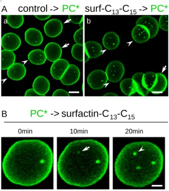
Fig. 1. In adherent RBCs, natural cyclic surfactin-C13–C15increases the abundance of
BODIPY-PC (PC*) micrometric domains. (A) Vital imaging of RBCs preincubated with surfactin. Freshly isolated RBCs were either preincubated in suspension with 0.5μM surfactin for 30 min (b) or kept untreated (a), then attached onto poly-L-lysine (PLK)-coated coverslips for 4 min and allowed to spread for additional 4 min, labeled with PC*, washed and immediately imaged at 37 °C. Notice at left that PC* labels several micrometric domains on partially spread cells (b8 μm; arrowheads) but not on more spread cells (arrows). At right, the number of domains is increased by surfactin, including on highly spread cells. Scale bars, 5μm. (B) Time-lapse vital imaging of RBCs incubated on stage with surfactin. RBCs were attached–spread on PLK-coverslips as above, labeled with PC*, washed and imaged at 37 °C following the addition of 0.75μM surfactin. Notice progressive domain appearance (arrow) and enlargement (arrowhead). Scale bar, 2μm.
UNCORRECTED PR
OOF
281 depletion[22], we thus asked whether surfactin can overcome the 282 effect of cholesterol depletion. To this aim, we induced a moderate cho-283 lesterol depletion (~25%) by 0.25 mM methyl-β-cyclodextrin (mβCD), 284 which causes complete attrition of PC* and SM*, but largely preserves 285 GlcCer*, domains ([22];Fig. 3A, c, g and quantification atFig. 3B). Expo-286 sure of mβCD-treated RBCs to surfactin not only prevented disappear-287 ance of PC* and SM* domains but further increased the abundance 288 of PC* domains by >3-fold as compared to untreated control RBCs 289 (Fig. 3A; quantification atFig. 3B;film atFig. 3C). These results indicated
290 that surfactin can substitute cholesterol to favor PC* and SM* micrometric
291 domains or that both act in concert.
292 3.4. Surfactin-C13–C15increases excimer formation at PC* and SM* domains
293 Based on the reversion by surfactin of the attrition of PC* and SM*
294 domains induced by mβCD, we then ask more directly whether the
295 lipopeptide could, like cholesterol, affect lipid* domain organization.
296 To this aim, we looked at clustering-dependent shift of BODIPY spectral
297 properties, known as excimer formation[27]. This phenomenon results
298 from a partial conversion of the primary emission peak atλem520 nm
299 (green) to a secondary emission at 605 nm (red). We therefore looked
300 at green and redfluorescence emission from PC* and SM* domains,
301 either at usual concentrations (Fig. 4a–d) or at higher SM* concentration
302 to sensitize excimer formation ([23,26,27];Fig. 4e). In control RBCs, no
303 significant excimer formation was detected by line scans at the usual
304 PC* and SM* concentration (Fig. 4a, c), but the phenomenon was obvious
305 upon image inspection at 3μM SM* (Fig. 4e, right, arrowheads) and can
306 be quantified by line scan (up to ~25% red/green emission ratio;Fig. 4e′).
307 Upon treatment with surfactin-C13–C15, excimer formation of cells
308 exposed to 1μM PC* or 0.75 μM SM* became detectable (arrowheads
309 at panels b,d, right; compare with panels a, c), reaching emission ratios
310 of ~15% (Fig. 4b′, d′).
311 3.5. Synthetic surfactins increase PC* domain abundance in an acyl chain
312 length-dependent manner
313 Having shown that the mixture known as surfactin-C13–C15affected
314 PC* and SM* domains in a cholesterol-sensitive manner, we next aimed
315 at identifying the structural features of the surfactin molecule
responsi-316 ble for this effect. To this aim, several surfactins were compared: (i)
317 purified natural cyclic surfactin with uniform acyl chain length of 14C
318 (referred as SNC14); (ii) linear analogs with the same 14C-acyl chain
319 and further differing in charge (2 vs 3 acid groups), referred as SAL14
320 and SSL14; and (iii) linear analogs differing in acyl chain length (10 vs
321 14 vs 18 carbons), referred as SSL10, SSL14 and SSL18 (for structures
322 and characteristics, see Suppl. Fig. 1 and Suppl. Table 1, respectively).
323 All congeners were used at the same concentrations as natural cyclic
324 surfactin-C13–C15and none caused any hemolysis (data not shown).
325 Irrespective of their geometry (cyclic vs linear) and charge (2 vs 3), all
326 tested surfactins with 14C (purified natural cyclic SNC14 as well as the
327 linear SAL14 and SSL14 with respectively 2 and 3 negative charges)
328 increased by ~2-fold the number of PC* domains (Fig. 5A, b, c, h), like
329 natural surfactin mixture with 13 to 15C. This indicated that surfactin
330 overall geometry and charge density were not determinant factors for
331 drug effect on PC* micrometric domains. In contrast, increasing surfactin
332 acyl chain length from 10 (SSL10) to 14 (SSL14) to 18C (SSL18)
differen-333 tially increased PC* domain abundance, from ~1.5-fold to ~3-fold as
334 compared to control cells (panels g, h, i atFig. 5A and quantification at
335 Fig. 5B, upper panel), indicating that surfactin acyl chain length is instead
336 a key determinant for the increase of PC* micrometric domains.
337 3.6. Relation between synthetic surfactin acyl chain length and the increase
338 of PC* domain abundance is inverted upon cholesterol depletion
339Q8 Next, to evaluate if all tested surfactins can overcome the attrition of
340 PC* domains induced by cholesterol depletion, a similar experiment was
341 performed after the removal of ~25% cholesterol by 0.25 mM mβCD
342 [22]. Like natural cyclic surfactin-C13–C15, purified cyclic surfactin
343 SNC14 and synthetic linear compounds (SAL14, SSL10-18), whatever
344 their geometry, charge and acyl chain length, suppressed the effect of
345 cholesterol depletion on PC* domains (Fig. 5A, +mβCD). However,
346 while surfactin with the longest acyl chain (SSL18) induced the
stron-347 gest increase of PC* domain abundance in RBCs with normal cholesterol
348 content (seeFig. 5,−mβCD), the opposite was observed in RBCs treated
349 with mβCD (Fig. 5B, lower panel). Altogether, these results indicate that
0.00 0.25 0.50 0.75 1.00 0 50 100 150 200
A
B
number of domains (% of control RBCs)
CTL
0.25µM
0.5µM
0.75µM
1µM
***
***
**
***
***
, PC*; , SM*; , GlcCer*
surfactin (µM)k
f
a
l
g
b
m
h
c
n
i
d
o
j
e
SM* GlcCer*
PC*
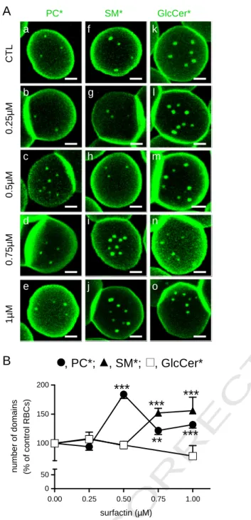
Fig. 2. Surfactin-C13–C15 favors PC* and SM*, but not GlcCer*, domains in a
concentration-dependent manner. (A) Representative confocal images. Freshly isolated RBCs were preincubated (b–e; g–j; l–o) or not (a, f, k) in suspension with the indicated con-centration of surfactin-C13–C15for 30 min, attached–spread onto PLK-coverslips, labeled
with PC* (a–e), SM* (f–j), or GlcCer* (k–o), washed and immediately imaged at 37 °C, as inFig. 1. Notice the selective increase of PC* (c–e) and SM* domains upon surfactin (i, j), with different peak concentrations (0.5μM surfactin for PC* and 0.75 μM for SM*); the abundance of GlcCer* domains is unchanged. All scale bars, 2μm. (B) Morphometry. Micrometric domains are means ± SEM of (i) 44–664 RBCs for PC*; (ii) 23–322 RBCs for SM*; and (iii) 99–316 for GlcCer*, pooled from 4 independent experiments and normalized to untreated RBCs taken as 100%.
UNCORRECTED PR
OOF
350 surfactin analogs increased PC* domain abundance in an acyl chain 351 length-dependent manner and that cholesterol depletion inverted this 352 tendency, suggesting that membrane:surfactin analog interaction de-353 pends on endogenous cholesterol level.
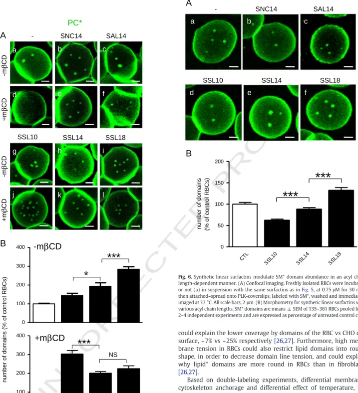
354 3.7. Synthetic surfactins also affect SM* domain abundance in an acyl 355 chain length-dependent manner
356 Because synthetic surfactins can substitute cholesterol to support 357 PC* micrometric domains, we then asked whether and how synthetic 358 surfactins could also affect the more packed SM* domains [27].
359 Whereas no effect was observed in RBCs incubated with SNC14 and
360 SAL14 (Fig. 6b, c ), acyl chain length differentially influenced SM*
361 domain abundance, from a ~ 1.5-fold decrease for the short SSL10 to
362 a 1.5-fold increase for the long SSL18 (Fig. 6A, d–f; quantification at
363 Fig. 6B). Moreover, removing cholesterol inverted this tendency, as
364 observed on PC* domains: the longest acyl chain length, the lowest
365 SM* domain abundance (data not shown). Thus, like for PC* domains,
366 SM* domain abundance can be modulated by surfactin acyl chain
367 length. However, in contrast to PC* domains, the effect on SM*
368 domain abundance was differential: decreased by short surfactins
369 (SSL10) but promoted by long surfactins (SSL18).
+
mβCD
-0 100 200 300 400 0 50 100 150 200+
-surf-C
13-C
15-
+
-
+
number of domains (% of control RBCs)
A
B
m
β CD ->
-> surfactin-C
13-C
15C
SM*
PC*
+
m
βCD
-
-
+
surf-C
13-C
15--
+
+
+
-
-
+
--
+
+
e
a
f
b
g
c
h
d
0’
14’
23’
30’
SM*
PC*
PC*
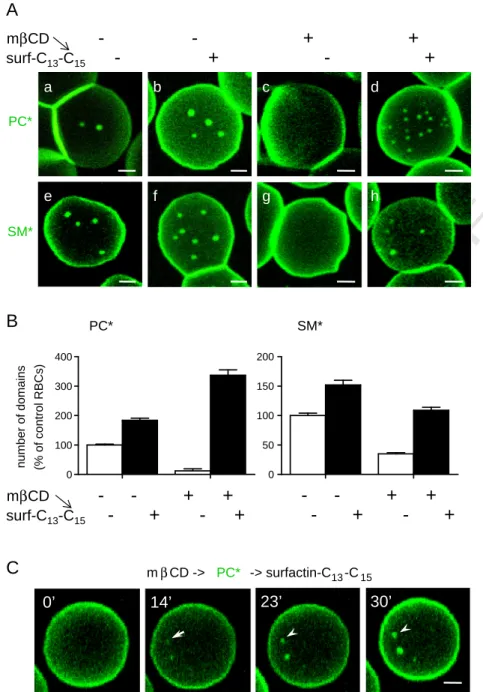
Fig. 3. Surfactin-C13–C15reverses the attrition of PC* and SM* domains induced by cholesterol depletion. (A) Representative confocal images. Freshly isolated RBCs were preincubated
(c, d, g, h) or not (a, b, e, f) in suspension with 0.25 mM methyl-β-cyclodextrin (mβCD) for 1 h to decrease endogenous cholesterol by ~25%. During the last 30 min, RBCs were further exposed to 0.5μM (b, d) or 0.75 μM (f, h) surfactin, in the continued presence of mβCD if appropriate. RBCs were then attached–spread onto PLK-coverslips, labeled as above with PC* (a–d) or SM* (e–h), washed and immediately imaged at 37 °C. Note that the disappearance of PC* and SM* domains by moderate cholesterol depletion (c, g) is reversed by surfactin. For PC* domains, number is even increased by ~3-fold in comparison to control cells. All scale bars, 2μm. (B) Morphometry. PC* (left panel) and SM* (right panel) domains upon combined treatment with mβCD and surfactin are means ± SEM of 288 and 258 RBCs pooled from 3 independent experiments, as percentage of untreated control cells. (C) Time-lapse vital imaging of mβCD-treated RBC incubated on stage with surfactin. Freshly isolated RBCs were preincubated for 30 min with 0.25 mM mβCD, then attached–spread onto PLK-coverslips, labeled with PC*, washed and continuously imaged by confocal microscopy at 37 °C upon treatment with 0.75μM surfactin for the indicated times. Notice progressive domain appearance (arrow) and enlargement (arrowheads). Scale bar, 2μm.
UNCORRECTED PR
OOF
370 4. Discussion
371 4.1. Current model for micrometric BODIPY-lipid domain biogenesis and 372 (co-)existence in control RBCs
373 Before discussing how surfactin affects lipid* domain organization 374 and abundance, let us summarize our current view, based on this 375 paper and our previous studies[22,26,27], for micrometric lipid* do-376 main (i) biogenesis; (ii) low cell surface coverage and round shape; 377 (iii) coexistence; and (iv) relevance for endogenous lipids.
378 Whereas micrometric lipid domains are observed with all the 379 three classes of polar lipids* used, i.e. GSLs*, SM* and PC*, their 380 abundance is differentially controlled by stabilization and restriction 381 machineries. Thus, GlcCer* domains are favored by high temperature 382 and ankyrin complexes, whereas PC* and SM* domains are promoted 383 by cholesterol and regulated linkage to the 4.1R complex[22,26,27]. 384 Moreover, our studies point to three differences between PC* and
385 SM* domains: (i) their intrinsic propensity to form excimers (SM*
386 but not PC*); (ii) their interaction with 4.1R complexes, providing
387 either internal stabilization (SM*) or peripheral retention (PC*); and
388 (iii) their control by cholesterol, as regulator of membranefluidity
389 (SM*) or membrane:cytoskeleton anchorage (SM* and PC*)[22,26,27].
390 In contrast, membrane stretching and membrane:cytoskeleton
anchor-391 age constitute restriction factors for domains, thereby preventing
392 domain expansion[22,27]. Biophysical studies should address the
me-393 chanical parameters governing the relation between membrane tension
394 and lipid* domain packing and size in RBCs. Nevertheless, phase
coexis-395 tence at the rabbit RBC membrane studied by multiphoton microscopy
396 after labeling with LAURDAN allows evidencing tightly packed domains,
397 with different lipid packing and sizes, moving in a morefluid
back-398 ground phase[35].
399 High membrane stretching and strong membrane:cytoskeleton
400 anchorage in RBCs, which constitute restriction factors for lipid*
401 micrometric domains thereby preventing domain expansion [22],
0 1 2 3 4 5 6 7 8 0 50 100 150 200 250 0 1 2 3 4 5 6 7 8 0 50 100 150 200 250
surf-C
13-C
15CTL
0 1 2 3 4 5 6 7 8 0 50 100 150 200 250 0 1 2 3 4 5 6 7 8 0 50 100 150 200 250 y 0 1 2 3 4 5 6 7 8 0 50 100 150 200 250 yc
1 3 2 intensity distance (µm)e
`
`
`
`
`
d
a
b
1 2 3 1 2 3 1 2 1 2 3C-f
r
u
s
L
T
C
13-C
15CTL
λ
em, 520
0.75µM SM*
1µM PC*
3µM SM*
λ
em, 605
1 2 3c
e
d
a
b
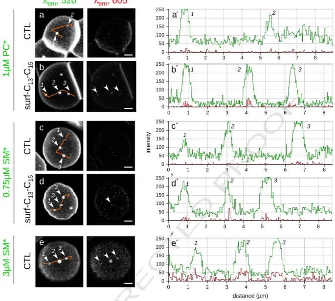
1 2 3 1 3 2 1 2 1 2 3Fig. 4. Surfactin-C13–C15induces a spectral shift (“excimers”) of PC* and SM* domains. Left, confocal imaging. Freshly isolated RBCs were incubated (b, d) or not (a, c, e) in suspension with
surfactin-C13–C15at 0.5μM (b) or 0.75 μM (d) for 30 min, then attached onto PLK-coverslips, labeled with PC* at 1 μM (usual concentration; a, b) or with SM* at either 0.75 μM (usual
concentration; c, d) or 3μM to sensitize excimer formation (e), washed and immediately examined by confocal microscopy. All images were generated with λexc488 nm, with
simulta-neous recording in the green (left;λem520 nm) and red channels (right;λem605 nm). Signal in red images (right) is indicative of packed clustering (excimers). Notice that excimers are
totally absent in control RBCs incubated with 1μM PC* (a) and 0.75 μM SM* (c) but induced by surfactin-C13–C15(b, d) and best seen with 3μM SM* in control RBCs (e). All scale bars,
2μm. Right, quantitation of conventional (green) and excimer emission (red). Intensity profiles were recorded along the paths indicated by continuous orange lines at left. Numbers 1–3 refer to the indicated patches. Average red/green emission ratio for PC* and SM* in control RBCs isb5% at usual lipid* concentrations (a′, c′), reaches ~15% upon incubation with surfactin-C13–C15(b, d), and up to ~25% at 3μM SM* without surfactin (e′).
UNCORRECTED PR
OOF
402 could explain the lower coverage by domains of the RBC vs CHO cell
403 surface, ~ 7% vs ~ 25% respectively[26,27]. Furthermore, high
mem-404 brane tension in RBCs could also restrict lipid domains into round
405 shape, in order to decrease domain line tension, and could explain
406 why lipid* domains are more round in RBCs than in fibroblasts
407 [26,27].
408 Based on double-labeling experiments, differential membrane:
409 cytoskeleton anchorage and differential effect of temperature, we
410 have previously suggested that the RBC PM is organized in at least
411 three segregated lipid* domains. However, only a fraction of the
412 lipids* at the PM is present in the round micrometric lipid* domains
413 and three lines of evidence support the existence of a surrounding
414 phase. We could infer this assumption by the % of PM covered by
415 lipid* domains, the effect of temperature on domain number and
416 the very fast lipid* recovery by FRAP. First, considering that SM*
417 domains cover ~ 7% of the PM, their ~ 8-fold enrichment indicates
418 that about half of the SM* is present in the domains and the other
419 half outside[26]. Second, the three classes of lipids* show a distinct
420 number of domains according to the temperature: whereas GlcCer*
421 shows an increasing domain number when temperature is increased
422 from 20 °C to 37 °C, SM* and PC* show a peak of domains at 20 °C
423 and a strong decrease thereafter; accordingly, a weak and
homoge-424 nous labeling with PC* can also be detected at 37 °C [27]. Third,
CTL SSL10 SSL14 SSL18 0 100 200 300 400 0 100 200 300 400
*
***
number of domains (% of control RBCs)
B
-m
βCD
+m
βCD
***
NSA
-
SNC14
SAL14
SSL14
SSL10
SSL18
+m
βCD -m
βCD
+m
βCD -m
βCD
a
b
h
g
c
i
d
e
f
k
j
l
PC*
Fig. 5. Synthetic linear surfactins increase PC* domain abundance in an acyl chain length- and cholesterol-dependent manner. (A) Confocal imaging. Freshly isolated RBCs were preincubated in suspension with 0.25 mM mβCD for 1 h (d–f; j–l) or not (a–c; g–i). During the last 30 min, cells were further exposed to the indicated surfactins (SNC14, Surfactin Natural Cyclic with 14C-acyl chain length; SAL14, Surfactin Acylated Linear with 14C; SSL10, SSL14 and SSL18, Surfactin Synthetic Linear with 10, 14 and 18C, respectively) at 0.5μM for 30 min in the continued presence of mβCD if appropriate. RBCs were attached-spread onto PLK-coverslips, labeled with PC*, washed and immediately imaged by confocal microscopy at 37 °C. All scale bars, 2μm. (B) Morphometry for synthetic linear surfactins with various acyl chain lengths. Data are means ± SEM of 53–421 RBCs pooled from 2–3 experiments and are expressed as percentage of untreated control cells.
number of domains (% of control RBCs)
B
CTL SSL10 SSL14 SSL18 0 50 100 150 200***
***
a
b
c
e
d
f
-
SNC14
SAL14
A
SSL14
SSL10
SSL18
SM*
Fig. 6. Synthetic linear surfactins modulate SM* domain abundance in an acyl chain length-dependent manner. (A) Confocal imaging. Freshly isolated RBCs were incubated or not (a) in suspension with the same surfactins as inFig. 5, at 0.75μM for 30 min, then attached–spread onto PLK-coverslips, labeled with SM*, washed and immediately imaged at 37 °C. All scale bars, 2μm. (B) Morphometry for synthetic linear surfactins with various acyl chain lengths. SM* domains are means ± SEM of 135–361 RBCs pooled from 2–4 independent experiments and are expressed as percentage of untreated control cells.
UNCORRECTED PR
OOF
425 although domains are immobile, they show a very fast recovery after 426 photobleaching indicating that domains* are large-scale immobile 427 assemblies of highly dynamic individual or small clusters of lipids*. 428 Relevance offluorescent lipid* domains for endogenous lipids is 429 supported by three observations: (i) co-localization of exogenous GM1* 430 with endogenous GM1 labeled with subunit B cholera toxin in RBCs; 431 (ii) identity of PM domains in CHO cells upon insertion of SM* vs meta-432 bolic conversion of ceramide* into SM* at physiological temperature; 433 and (iii) selective disappearance of SM* upon endogenous SM depletion 434 [22,26,27].
435 4.2. Preferential formation by surfactin of less-packed PC* and SM* domains 436 As shown by live cell imaging of RBCs, low concentrations of 437 surfactin induced the formation of new PC* and SM* micrometric 438 domains, without obvious effect on GlcCer* domains. Based on the com-439 plete abolishment of RBC PC* and SM* labeling upon back-exchange 440 with BSA (data not shown) and on the very limitedflip-flop of surfactin 441 from outer to inner leaflet[42], the increased abundance of domains 442 induced by surfactin should be due to PC* and SM* clustering from the 443 surrounding lipid* pool in the outer PM leaflet. By FRAP experiments 444 in RBCs, we indeed observed very fast (t1/2~ 10 s) and high recovery
445 of PC* and SM* domain constituents[22]. Assuming a representative 446 behavior of the surfaces we analyzed, area estimations indicate that 447 SM* domains account from ~7% of the RBC surface at 20 °C. The respec-448 tive∼8-fold enrichment in these micrometric domains indicates that 449 about half of total SM* would be clustered in these domains and half 450 outside[26].
451 Although PC* and SM* differ from GlcCer* by the same small and 452 zwitterionic phosphocholine headgroup, this explanation for a differen-453 tial effect of surfactin is not satisfactory because: (i) whereas surfactin 454 reversed the attrition of SM* domains induced by moderate cholesterol 455 depletion, it increased by >3-fold PC* domain abundance in compari-456 son to control RBCs; and (ii) changing the charge number of synthetic 457 surfactins (SAL14 with 2 charges vs SSL14 with 3 charges) had no effect 458 on the increase of PC* domain abundance.
459 Besides differences in polar headgroup size and charge, PC* and 460 SM* domains show a lower propensity to form excimers[23,26,27]. 461 Because surfactin preferentially increased the abundance of PC* and 462 SM* domains, we favor the view that the drug preferentially interacts 463 with less-packed lipid* domains. This proposal in living cellsfits with 464 the observation that surfactin shows a stronger insertion in mixed 465 monolayers containing phospholipids with short chain length and/or 466 in afluid-like organization[42]. In another study, binding affinity of 467 surfactin to LUVs was higher for So- than Ld- than Lo-phases [44],
468 again in agreement with our data on RBCs in which Ld/Sophases are
469 not expected to coexist due to their very high cholesterol content[45]. 470 The third difference between lipid* micrometric domains is unequal 471 sensitivity to cholesterol depletion, higher for PC* and SM* than for 472 GlcCer* domains. We will now discuss how surfactin could interact 473 with membranes, by systematic comparison with the well-known 474 effects of cholesterol on biological membranes.
475 4.3. Cholesterol like-effects of surfactin
476 Two lines of evidence indicate that surfactin and cholesterol similar-477 ly impact on micrometric domains: (i) cholesterol depletion by mβCD 478 and surfactin addition oppositely affected both PC* and SM* domain 479 abundance; and (ii) disappearance of PC* and SM* domains by mβCD 480 was completely abrogated by surfactin. The hypothesis of cholesterol-481 like effect of surfactin will guide a further discussion on how surfactin 482 could affect membrane lateral organization in micrometric domains. 483 Cholesterol not only regulates membrane fluidity at a global level 484 but also favors biogenesis of micrometric lipid domains at discrete 485 predefined spots by promoting intrinsic polar lipid packing [22,27]. 486 Cholesterol apparently concentrates at the boundaries between liquid
487 and gel-like phases, thereby reducing line tension[46]. Cholesterol
488 was also reported to modulate membrane:cytoskeleton uncoupling
489 [22,36,47], but this is poorly relevant for surfactins for whichflip-flop
490 from the outer to the inner leaflet is very limited [42,48]. Arguing
491 against a modulation by surfactin of global membranefluidity, the
492 three classes of polar lipids* were differentially affected by surfactin,
493 in agreement with the recent classification of surfactin into the group
494 of heterogeneously-perturbing surfactants which disrupt membrane
495 locally[49]. We thus favor the view that surfactin promotes biogenesis
496 specifically at PC* and SM* domains. We indeed observed a specific
497 increased abundance and excimer formation from these two domains.
498 Increased excimer formation might reflect that (i) lipid* domains got
499 fewer or smaller; (ii) lipids* showed a stronger preference for domains;
500 and/or (iii) lipid* diffusion and molecular motions within the domains
501 were enhanced. Based on increased domain abundance and size, the
502 first hypothesis can be ruled out. The effect of surfactin on domain
503 abundance and excimer formation would thus probably be due to a
504 combination of the two latter hypotheses. This can be explained by a
505 strengthening of hydrophobic interactions between acyl chains of lipids
506 and surfactins, reminiscent to the wedge-like shapes of SLs and
choles-507 terol that allow them to come in very close apposition via van der Waals
508 forces[50]. Similarly, sphingosine, which also behaves as a
surface-509 active amphiphile, rigidifies pre-existing gel domains in mixed bilayers
510 [15,51,52]. We also noticed that small changes of surfactin
concentra-511 tions lead to contrasting effects on lipid* domains, with a peak at
512 0.5μM and a subsequent decrease for PC* domains, with concomitant
513 increase of SM* domains. This concentration effect could be explained
514 by a shift of surfactin interaction,first with PC*domains, then with
515 SM* domains and/or at domain boundaries, thereby reducing line
516 tension at interface and eroding domains.
517 If this view is correct, then high local cholesterol concentrations
518 would prevent any effect of surfactin. This prediction is consistent
519 with the higher increase of PC* domains abundance by surfactin
520 in RBCs with lower cholesterol level vs normal RBCs. Accordingly, it
521 has been shown that the presence of cholesterol in the phospholipid
522 membrane attenuates the destabilizing effect of surfactins[53]and
523 that surfactin preferentially lyses cholesterol-free liposomes [54].
524 However, it seems atfirst glance inconsistent with the absence of
525 effect of addition of stigmasterol on surfactin interaction with LUVs
526 [44]. This apparent discrepancy might be explained by the very low
527 level of stigmasterol used in the latter study and/or by the ability of
528 cholesterol, but not stigmasterol, to form domains in DOPC/SM
529 bilayers[55]. The higher impact of surfactin on RBCs when cholesterol
530 content was decreased markedly contrasts to the behavior of other
531 lipopeptides produced by Bacillus species, such as fengycin, iturins
532 and mycosubtilin, which show high affinity for cholesterol[56–58]
533 via a tyrosyl residue[56]. Thus, whereas surfactin could substitute
534 cholesterol, the latter three drugs depend on it.
535 4.4. Critical surfactin structural features involved in micrometric lipid*
536 domain modulation
537 To prevent hemolysis, Dufour and collaborators have synthesized
538 various linear surfactin analogs differing by charge and hydrophobicity
539 (for structures and characteristics, see Suppl. Fig. 1 and Suppl. Table 1,
540 respectively). Whereas surfactin geometry and charge density did not
541 impact onfluorescent lipid lateral compartmentation in domains, the
542 acyl chain length was an important feature: the longest the chain
543 (SSL18), the highest the increase of PC* and SM* domain abundance.
544 This observation perfectly agrees with the highest insertion into DPPC
545 monolayer of surfactins bearing the longest acyl chain (Suppl. Table 1).
546 However, an opposite effect was observed in RBCs partially depleted in
547 cholesterol: the shortest the acyl chain, the highest the increase of PC*
548 and SM* domain abundance. This raises the possibility that cholesterol
549 removal could leave room for the small SSL10. These observations
550 underline that, besides surfactin structural features, host membrane
UNCORRECTED PR
OOF
551 composition, e.g. cholesterol abundance, is a key parameter for 552 membrane:surfactin interaction, and must be kept in mind for designing 553 new surfactins.
554 4.5. Model for surfactin:membrane interaction and surface activity, based on 555 surfactin structural features and host membrane composition/organization 556 An inverse relation can be established between critical micellar 557 concentration of linear surfactin analogs (CMC, 1114 vs 302 vs 8 for 558 SSL10, SSL14 and SSL18 respectively; see Suppl. Table 1) [41]and 559 micrometric lipid* domain abundance in normal RBCs: the lowest 560 the CMC, the highest the domain abundance; moreover, this relation 561 was inverted upon cholesterol depletion. We thus propose a new 562 model for surfactin:membrane interaction based on lipid domain or-563 ganization and cholesterol abundance/distribution. In RBCs with nor-564 mal cholesterol level, long surfactins (e.g. SSL18) would preferentially 565 insert inside domains, because of deep insertion into the hydrophobic 566 core of the membrane, while short surfactins (e.g. SSL10) could only 567 find their place at domain boundary, as already proposed[42], reduc-568 ing line tension and domain size. Accordingly, SSL10 did not increase 569 but decreased SM* domain abundance, in contrast to natural cyclic 570 surfactin. However, when cholesterol was removed, the short chain 571 SSL10 showed a stronger increase of PC* domains, which would fur-572 ther gain access inside domains and substitute cholesterol, thereby 573 favoring domain coalescence.
574 4.6. Conclusion
575 Taken together, our data imply that, in addition to surfactin structure 576 and concentration, the modulation of the lateral organization of PM 577 fluorescent lipids by surfactin appears dictated by lipid* domain packing 578 and sterol content. The preference for membranes with a lower global 579 cholesterol content and for domains with low packing could explain 580 why surfactin preferentially disrupts bacterial membranes since pro-581 karyotic membranes almost universally lack sterols and SLs[59]. This 582 contrasts with the poor toxicity of surfactin to fungi and plant mem-583 branes[44]that contain high sterol, inositolphosphoryglycolipids and 584 glycosphingolipids and show lateral compartmentation in micrometric 585 domains[59,60]. In conclusion, the cholesterol content of the host 586 membrane and its organization in domains must be taken into account 587 to evaluate surfactin surface activity and toxicity and for designing new 588 surfactin analogs.
589 Supplementary data to this article can be found online athttp:// 590 dx.doi.org/10.1016/j.bbamem.2013.05.006.
591
Q9 Acknowledgments
592 This work was supported by grants from the UCL (FSR), the 593 F.R.S.-FNRS, the Région Wallonne, the Région Bruxelloise, the Salus 594 Sanguinis Foundation, the Loterie Nationale, the ARC and the IUAP 595 (Belgium). MD thanks the FNRS for her position as Research Associate. 596 References
597 [1] F. Peypoux, J.M. Bonmatin, J. Wallach, Recent trends in the biochemistry of
598 surfactin, Appl. Microbiol. Biotechnol. 51 (1999) 553–563.
599 [2] R. Maget-Dana, M. Ptak, Interactions of surfactin with membrane models,
600 Biophys. J. 68 (1995) 1937–1943.
601 [3] M. Deleu, J. Lorent, L. Lins, R. Brasseur, N. Braun, K. El Kirat, T. Nylander, Y.F.
602 Dufrene, M.P. Mingeot-Leclercq, Effects of surfactin on membrane models
603 displaying lipid phase separation, Biochim. Biophys. Acta 1828 (2012) 801–815.
604 [4] S. Dufour, M. Deleu, K. Nott, B. Wathelet, P. Thonart, M. Paquot, Hemolytic activity
605 of new linear surfactin analogs in relation to their physico-chemical properties,
606 Biochim. Biophys. Acta 1726 (2005) 87–95.
607 [5] G. van Meer, D.R. Voelker, G.W. Feigenson, Membrane lipids: where they are and
608 how they behave, Nat. Rev. Mol. Cell Biol. 9 (2008) 112–124.
609 [6] S.J. Singer, G.L. Nicolson, Thefluid mosaic model of the structure of cell membranes,
610 Science 175 (1972) 720–731.
611
[7] D. Lingwood, K. Simons, Lipid rafts as a membrane-organizing principle, Science
612
327 (2010) 46–50.
613
[8] D. Lingwood, J. Ries, P. Schwille, K. Simons, Plasma membranes are poised for
614
activation of raft phase coalescence at physiological temperature, Proc. Natl.
615
Acad. Sci. U.S.A. 105 (2008) 10005–10010.
616
[9] L.J. Pike, Rafts defined: a report on the Keystone symposium on lipid rafts and cell
617
function, J. Lipid Res. 47 (2006) 1597–1598.
618
[10] A. Fujita, J. Cheng, M. Hirakawa, K. Furukawa, S. Kusunoki, T. Fujimoto, Gangliosides
619
GM1 and GM3 in the living cell membrane form clusters susceptible to cholesterol
620
depletion and chilling, Mol. Biol. Cell 18 (2007) 2112–2122.
621
[11]H. Mizuno, M. Abe, P. Dedecker, A. Makino, S. Rocha, Y. Ohno-Iwashita, J. Hofkens,
622
T. Kobayashi, A. Miyawaki, Fluorescent probes for superresolution imaging of
623
lipid domains on the plasma membrane, Chem. Sci. 2 (2011) 1548–1553.
624
[12] L.A. Bagatolli, J.H. Ipsen, A.C. Simonsen, O.G. Mouritsen, An outlook on organization
625
of lipids in membranes: searching for a realistic connection with the organization of
626
biological membranes, Prog. Lipid Res. 49 (2010) 378–389.
627
[13]N. Kahya, D. Scherfeld, K. Bacia, B. Poolman, P. Schwille, Probing lipid mobility
628
of raft-exhibiting model membranes byfluorescence correlation spectroscopy,
629
J. Biol. Chem. 278 (2003) 28109–28115.
630
[14]T. Baumgart, S.T. Hess, W.W. Webb, Imaging coexisting fluid domains in
631
biomembrane models coupling curvature and line tension, Nature 425 (2003)
632
821–824.
633
[15]F.M. Goni, A. Alonso, Effects of ceramide and other simple sphingolipids on
634
membrane lateral structure, Biochim. Biophys. Acta 1788 (2009) 169–177.
635
[16]J.V. Busto, J. Sot, J. Requejo-Isidro, F.M. Goni, A. Alonso, Cholesterol displaces
636
palmitoylceramide from its tight packing with palmitoylsphingomyelin in the
637
absence of a liquid-disordered phase, Biophys. J. 99 (2010) 1119–1128.
638
[17]L.A. Bagatolli, To see or not to see: lateral organization of biological membranes
639
andfluorescence microscopy, Biochim. Biophys. Acta 1758 (2006) 1541–1556.
640
[18]M. Edidin, The state of lipid rafts: from model membranes to cells, Annu. Rev.
641
Biophys. Biomol. Struct. 32 (2003) 257–283.
642
[19]A. Kusumi, K.G. Suzuki, R.S. Kasai, K. Ritchie, T.K. Fujiwara, Hierarchical mesoscale
643
domain organization of the plasma membrane, Trends Biochem. Sci. 36 (2011)
644
604–615.
645
[20] A. Kusumi, T.K. Fujiwara, R. Chadda, M. Xie, T.A. Tsunoyama, Z. Kalay, R.S.
646
Kasai, K.G. Suzuki, Dynamic organizing principles of the plasma membrane
647
that regulate signal transduction: commemorating the fortieth anniversary
648
of singer and Nicolson'sfluid-mosaic model, Annu. Rev. Cell Dev. Biol. 28
649
(2012) 215–250.
650
[21]T. Fujiwara, K. Ritchie, H. Murakoshi, K. Jacobson, A. Kusumi, Phospholipids
651
undergo hop diffusion in compartmentalized cell membrane, J. Cell Biol. 157 (2002)
652
1071–1081.
653
[22]L. D'Auria, M. Fenaux, P. Aleksandrowicz, P. Van Der Smissen, C. Chantrain, C.
654
Vermylen, M. Vikkula, P.J. Courtoy, D. Tyteca, Micrometric segregation offluorescent
655
membrane lipids: relevance for endogenous lipids and biogenesis in erythrocytes,
656
J. Lipid Res. 54 (2013) 1066–1076.
657
[23]R.D. Singh, Y. Liu, C.L. Wheatley, E.L. Holicky, A. Makino, D.L. Marks, T. Kobayashi,
658
G. Subramaniam, R. Bittman, R.E. Pagano, Caveolar endocytosis and microdomain
659
association of a glycosphingolipid analog is dependent on its sphingosine
stereo-660
chemistry, J. Biol. Chem. 281 (2006) 30660–30668.
661
[24]K. Gousset, W.F. Wolkers, N.M. Tsvetkova, A.E. Oliver, C.L. Field, N.J. Walker, J.H.
662
Crowe, F. Tablin, Evidence for a physiological role for membrane rafts in human
663
platelets, J. Cell. Physiol. 190 (2002) 117–128.
664
[25]M. Hao, S. Mukherjee, F.R. Maxfield, Cholesterol depletion induces large scale
665
domain segregation in living cell membranes, Proc. Natl. Acad. Sci. U.S.A. 98 (2001)
666
13072–13077.
667
[26]D. Tyteca, L. D'Auria, P.V. Der Smissen, T. Medts, S. Carpentier, J.C. Monbaliu, P. de
668
Diesbach, P.J. Courtoy, Three unrelated sphingomyelin analogs spontaneously
669
cluster into plasma membrane micrometric domains, Biochim. Biophys. Acta
670
1798 (2010) 909–927.
671
[27]L. D'Auria, P. Van der Smissen, F. Bruyneel, P.J. Courtoy, D. Tyteca, Segregation of
672
fluorescent membrane lipids into distinct micrometric domains: evidence for
673
phase compartmentation of natural lipids? PLoS One 6 (2011) e17021.
674
[28]G. Grossmann, J. Malinsky, W. Stahlschmidt, M. Loibl, I. Weig-Meckl, W.B.
675
Frommer, M. Opekarova, W. Tanner, Plasma membrane microdomains
regu-676
late turnover of transport proteins in yeast, J. Cell Biol. 183 (2008)
677
1075–1088.
678
[29]K. Malinska, J. Malinsky, M. Opekarova, W. Tanner, Visualization of protein
679
compartmentation within the plasma membrane of living yeast cells, Mol. Biol.
680
Cell 14 (2003) 4427–4436.
681
[30]J. Malinsky, M. Opekarova, W. Tanner, The lateral compartmentation of the yeast
682
plasma membrane, Yeast 27 (2010) 473–478.
683
[31] F. Spira, N.S. Mueller, G. Beck, P. von Olshausen, J. Beig, R. Wedlich-Soldner,
Patch-684
work organization of the yeast plasma membrane into numerous coexisting domains,
685
Nat. Cell Biol. 14 (2012) 640–648.
686
[32] P.F. Devaux, R. Morris, Transmembrane asymmetry and lateral domains in biological
687
membranes, Traffic 5 (2004) 241–246.
688
[33]A. Ciana, C. Achilli, C. Balduini, G. Minetti, On the association of lipid rafts to the
689
spectrin skeleton in human erythrocytes, Biochim. Biophys. Acta 1808 (2011)
690
183–190.
691
[34] A. Ciana, C. Balduini, G. Minetti, Detergent-resistant membranes in human
692
erythrocytes and their connection to the membrane–skeleton, J. Biosci. 30
693
(2005) 317–328.
694
[35] S.A. Sanchez, M.A. Tricerri, E. Gratton, Laurdan generalized polarizationfluctuations
695
measures membrane packing micro-heterogeneity in vivo, Proc. Natl. Acad. Sci.
696
UNCORRECTED PR
OOF
697 [36] M. Sun, N. Northup, F. Marga, T. Huber, F.J. Byfield, I. Levitan, G. Forgacs, The effect of
698 cellular cholesterol on membrane–cytoskeleton adhesion, J. Cell Sci. 120 (2007)
699 2223–2231.
700 [37]M. Salomao, X. Zhang, Y. Yang, S. Lee, J.H. Hartwig, J.A. Chasis, N. Mohandas, X. An,
701 Protein 4.1R-dependent multiprotein complex: new insights into the structural
702 organization of the red blood cell membrane, Proc. Natl. Acad. Sci. U.S.A. 105
703 (2008) 8026–8031.
704 [38]M.J. Parnham, H. Wetzig, Toxicity screening of liposomes, Chem. Phys. Lipids 64
705 (1993) 263–274.
706 [39]Z.J. Cheng, R.D. Singh, D.K. Sharma, E.L. Holicky, K. Hanada, D.L. Marks, R.E.
707 Pagano, Distinct mechanisms of clathrin-independent endocytosis have unique
708 sphingolipid requirements, Mol. Biol. Cell 17 (2006) 3197–3210.
709 [40]J.B. Marsh, D.B. Weinstein, Simple charring method for determination of lipids,
710 J. Lipid Res. 7 (1966) 574–576.
711 [41]G. Francius, S. Dufour, M. Deleu, M. Paquot, M.P. Mingeot-Leclercq, Y.F. Dufrene,
712 Nanoscale membrane activity of surfactins: influence of geometry, charge and
713 hydrophobicity, Biochim. Biophys. Acta 1778 (2008) 2058–2068.
714 [42]O. Bouffioux, A. Berquand, M. Eeman, M. Paquot, Y.F. Dufrene, R. Brasseur, M. Deleu,
715 Molecular organization of surfactin-phospholipid monolayers: effect of
phospholip-716 id chain length and polar head, Biochim. Biophys. Acta 1768 (2007) 1758–1768.
717 [43]H. Heerklotz, J. Seelig, Detergent-like action of the antibiotic peptide surfactin on
718 lipid membranes, Biophys. J. 81 (2001) 1547–1554.
719 [44]G. Henry, M. Deleu, E. Jourdan, P. Thonart, M. Ongena, The bacterial lipopeptide
720 surfactin targets the lipid fraction of the plant plasma membrane to trigger
721 immune-related defence responses, Cell. Microbiol. 13 (2011) 1824–1837.
722 [45] W.F. Wolkers, L.M. Crowe, N.M. Tsvetkova, F. Tablin, J.H. Crowe, In situ assessment
723 of erythrocyte membrane properties during cold storage, Mol. Membr. Biol. 19
724 (2002) 59–65.
725 [46]S. Mukherjee, F.R. Maxfield, Membrane domains, Annu. Rev. Cell Dev. Biol. 20
726 (2004) 839–866.
727 [47]F.J. Byfield, H. Aranda-Espinoza, V.G. Romanenko, G.H. Rothblat, I. Levitan, Cholesterol
728 depletion increases membrane stiffness of aortic endothelial cells, Biophys. J. 87
729 (2004) 3336–3343.
730
[48]M. Deleu, O. Bouffioux, H. Razafindralambo, M. Paquot, C. Hbid, P. Thonart, P.
731
Jacques, R. Brasseur, Interaction of surfactin with membranes: a computational
732
approach, Langmuir 19 (2003) 3377–3385.
733
[49]M. Nazari, M. Kurdi, H. Heerklotz, Classifying surfactants with respect to their
734
effect on lipid membrane order, Biophys. J. 102 (2012) 498–506.
735
[50]B. Ramstedt, J.P. Slotte, Membrane properties of sphingomyelins, FEBS Lett. 531
736
(2002) 33–37.
737
[51]F.X. Contreras, J. Sot, A. Alonso, F.M. Goni, Sphingosine increases the permeability
738
of model and cell membranes, Biophys. J. 90 (2006) 4085–4092.
739
[52]F.M. Goni, A. Alonso, Biophysics of sphingolipids I. Membrane properties of
sphin-740
gosine, ceramides and other simple sphingolipids, Biochim. Biophys. Acta 1758
741
(2006) 1902–1921.
742
[53]C. Carrillo, J.A. Teruel, F.J. Aranda, A. Ortiz, Molecular mechanism of membrane
743
permeabilization by the peptide antibiotic surfactin, Biochim. Biophys. Acta 1611
744
(2003) 91–97.
745
[54]L. Oftedal, L. Myhren, J. Jokela, G. Gausdal, K. Sivonen, S.O. Doskeland, L.
746
Herfindal, The lipopeptide toxins anabaenolysin A and B target biological
mem-747
branes in a cholesterol-dependent manner, Biochim. Biophys. Acta 1818 (2012)
748
3000–3009.
749
[55] V. Shahedi, G. Oradd, G. Lindblom, Domain-formation in DOPC/SM bilayers studied
750
by pfg-NMR: effect of sterol structure, Biophys. J. 91 (2006) 2501–2507.
751
[56]M.N. Nasir, F. Besson, Specific interactions of mycosubtilin with
cholesterol-752
containing artificial membranes, Langmuir 27 (2011) 10785–10792.
753
[57] M. Eeman, G. Francius, Y.F. Dufrene, K. Nott, M. Paquot, M. Deleu, Effect of cholesterol
754
and fatty acids on the molecular interactions of fengycin with Stratum corneum
755
mimicking lipid monolayers, Langmuir 25 (2009) 3029–3039.
756
[58]M.J. Quentin, F. Besson, F. Peypoux, G. Michel, Action of peptidolipidic antibiotics
757
of the iturin group on erythrocytes. Effect of some lipids on hemolysis, Biochim.
758
Biophys. Acta 684 (1982) 207–211.
759
[59]J.T. Hannich, K. Umebayashi, H. Riezman, Distribution and functions of sterols and
760
sphingolipids, Cold Spring Harb Perspect Biol 3 (2011) 1.
761
[60] M. Opekarova, J. Malinsky, W. Tanner, Plants and fungi in the era of heterogeneous
762
plasma membranes, Plant Biol. (Stuttg.) 12 (Suppl. 1) (2010) 94–98.
763 764