REVIEW
Breakthrough in cardiac arrest: reports
from the 4th Paris International Conference
Peter J. Kudenchuk
1, Claudio Sandroni
2, Hendrik R. Drinhaus
3, Bernd W. Böttiger
3, Alain Cariou
4,5, Kjetil Sunde
6,
Martin Dworschak
7, Fabio Silvio Taccone
8, Nicolas Deye
9, Hans Friberg
10, Steven Laureys
11, Didier Ledoux
12,
Mauro Oddo
13, Stéphane Legriel
14, Philippe Hantson
15, Jean‑Luc Diehl
16*and Pierre‑Francois Laterre
17Abstract
Jean-Luc Diehl The French Intensive Care Society organized on 5th and 6th June 2014 its 4th “Paris International Con‑
ference in Intensive Care”, whose principle is to bring together the best international experts on a hot topic in critical care medicine. The 2014 theme was “Breakthrough in cardiac arrest”, with many high‑quality updates on epidemiol‑ ogy, public health data, pre‑hospital and in‑ICU cares. The present review includes short summaries of the major presentations, classified into six main chapters:
• Epidemiology of CA • Pre‑hospital management
• Post‑resuscitation management: targeted temperature management
• Post‑resuscitation management: optimizing organ perfusion and metabolic parameters • Neurological assessment of brain damages
• Public healthcare
Keywords: Cardiac arrest, Cardio‑pulmonary resuscitation, Targeted temperature management, Therapeutic
hypothermia, Persistent vegetative state, Minimally conscious state, Organ donation
© 2015 Kudenchuk et al. This article is distributed under the terms of the Creative Commons Attribution 4.0 International License (http://creativecommons.org/licenses/by/4.0/), which permits unrestricted use, distribution, and reproduction in any medium, provided you give appropriate credit to the original author(s) and the source, provide a link to the Creative Commons license, and indicate if changes were made.
Review
Epidemiology of CA
Out‑of‑hospital cardiac arrest in 2014: incidence, outcome and disparities
Peter Kudenchuk Out-hospital-cardiac arrest (OHCA) remains a common public health problem. Each year, car-diac arrest claims more than 424,000 lives in the United States, 300,000 lives in Europe, and upwards of 3.7 million lives worldwide. Resuscitation is typically conducted in accordance with a uniform algorithmic approach embod-ied in the “chain of survival” which emphasizes the impor-tance of early activation of emergency medical services, prompt CPR, rapid defibrillation, advanced cardiac life support and post-cardiac arrest care. This, in fact might be
said to be the only uniform aspect of cardiac arrest, which is otherwise riddled with disparities. The first of these disparities lies in how few patients who are successfully resuscitated from cardiac arrest ultimately survive to hos-pital discharge. In Seattle, approximately 60 % of patients in whom cardiac arrest presents as ventricular fibrillation (VF) are successfully resuscitated and admitted to hospital; yet only about half of these (30 %) typically survive to hos-pital discharge. When cardiac arrest presents as asystole or pulseless electrical activity (PEA), outcomes are strikingly worse: only 20–30 % of such patients are successfully resuscitated, and an even smaller proportion of these, as few as 2 in 10—ranging from 2 to 5 % of patients, survive to hospital discharge [1].
A second disparity lies in the changing incidence of rhythms that precipitate cardiac arrest. In Seattle dur-ing the decade of the 1970s, VF accounted for approxi-mately 60 % of all out-of-hospital cardiac arrests treated by emergency medical services (EMS), whereas the
Open Access
*Correspondence: jldiehl@invivo.edu
16 Medical Intensive Care Unit, AP‑HP, European Georges Pompidou Hospital, Paris Descartes University and Sorbonne Paris Cité‑Medical School, Paris, France
remainder of acute rhythm presentations were equally divided between asystole and PEA. In the ensuing years, the proportion of cardiac arrests caused by VF has declined to 25–30 % of cases, such that now asystole and PEA represent the most common presenting rhythms. Extrapolating these incidence data from Seattle to the United States census, the annual rate of cardiac arrest due to ventricular fibrillation declined from about 85 persons per 100,000 in 1980 to 38/100,000 in 2000 [1]. Data from the Resuscitation Outcomes Consortium estimated a fur-ther decline in the incidence of VF cardiac arrest to 17.4 adults/100,000 in 2011 [2]. These statistics, coupled with the known worse survival prognosis of patients in whom cardiac presents as a non-shockable rhythm, pose a new and major challenge for the present and future treatment of this emerging “new wave” of cardiac arrest victims.
A third disparity lies in the marked differences in out-come from cardiac arrest between communities across Europe and the United States. For cardiac arrest due to VF, survival can vary significantly between major cities by many-fold (Fig. 1) [3–5]. To some extent, such differences in outcome are explained by inaccuracies in record keep-ing. For example, few communities actually report their incidence of cardiac arrest and outcome [6]. And even among those that do, complete capture of all cases of car-diac arrest and the reliability of their survival data can be questionable, accounting for some of the variability in outcomes that are reported. This said, record keeping alone does not entirely explain these discrepancies. Iden-tifying and targeting remediable causes of differences in survival between communities is imperative if we are to assure citizens that they are comparably “safe” from death by cardiac arrest in whatever locale they call home. Clinical factors that are known to account for differences
in survival outcome from cardiac arrest include patient characteristics—such as age, gender, and co-morbidities; and by the circumstances of the arrest—such as whether witnessed by bystanders, the location of its occurrence, and by the presenting arrest rhythm. Most would regard these as “factors of fate” and as such not alterable. Con-versely, the components of prehospital emergency medi-cal care, including rapid dispatch, dispatcher-assisted CPR, EMS training and time-to-treatment, are all poten-tially correctable factors. Targeting these aspects of pre-hospital care affords an opportunity to change outcome for the better after cardiac arrest. Among these, perhaps the one that can be most readily and immediately imple-mented is high-performance CPR.
High-performance CPR consists of training with meticulous attention to the details of performing CPR to the best known prescribed parameters of chest compres-sion rate, depth, full chest recoil, and minimized inter-ruptions, and applying strict compliance standards (e.g. a compression fraction of no less than 85 %) for their performance during resuscitation. In addition to striv-ing for “letter perfect” CPR performance, it also involves a system of accountability, whereby feed-back of CPR performance derived from a review of recordings of field resuscitations is consistently conveyed back to providers for further possible improvement. Deploying such a high-performance CPR protocol in King County, Washington, starting in 2005 resulted in a significant improvement in survival from both cardiac arrest due to ventricular fibril-lation (Fig. 2) [7], as well as asystole/PEA [8] that has been sustained in the ensuing years. The attractiveness of high-performance CPR is that it is relatively inexpensive to deploy (can be easily added to existing EMS training programs), does not require special equipment (hands
Fig. 1 Survival to hospital discharge from out‑of‑hospital ventricular
fibrillation in various communities outside the United States
Fig. 2 The impact of instituting high‑performance CPR in King
County, Washington, in 2005 on survival from witnessed out‑of‑ hospital cardiac arrest due to ventricular fibrillation
only), and has demonstrated that it can improve outcome for virtually all presentations of cardiac arrest.
While many of the identified disparities in cardiac arrest will continue to pose challenges for its manage-ment, a focus on improving systems-of-care, particularly deployment of high-performance letter-perfect CPR, offers the promise of a practical intervention that can be implemented immediately and an effort that promises to improve survival outcomes in any community.
Effectiveness of rapid response systems for prevention of cardiac arrest
Claudio Sandroni Despite the immediate availability of qualified life support, the outcome of in-hospital cardiac arrest (IHCA) remains poor. Survival to discharge after IHCA rarely exceeds 20 % [9] and it has remained stable in the last 25 years (Fig. 3). Rapid response systems (RRS) have been established to prevent IHCA in non-critical care areas of the hospital [10]. Those systems are based on timely detection of deteriorating patients by the ward personnel (the afferent limb of the system), who will there-fore summon a medical emergency team (MET; the effer-ent limb of the system), whose roles are to stabilize the patient in the ward or escalate the level of care. Although the theory underlying RRS is compelling, there is no defi-nite evidence that their implementation improves patient outcome. The major problem in evaluating the effective-ness of RRS is the choice of the outcome measure.
The first endpoint for a study addressing the effective-ness of RRS could be the rates of unexpected cardiac arrests occurring outside intensive care units (ICUs), that is, the rates of cardiac arrest occurring in ward patients for whom there is no do-not-attempt-resusci-tation (DNAR) order. This endpoint, however, is poten-tially biased by the fact that one of the tasks of METs is to identify ward patients for whom a resuscitation would be
inappropriate. Therefore, part of the observed reduction in the rate of unexpected cardiac arrest after the imple-mentation of a RRS is because the fraction of expected cardiac arrests is increased by placement of a DNAR order.
Another endpoint for measuring RRS effectiveness is the reduction of unplanned ICU admissions. The ration-ale is that the introduction of RRS should increase the number of ICU admissions that are planned early, before further deterioration occurs, and decrease those occur-ring as emergency admissions after resuscitation from cardiac arrest. This model has been indirectly demon-strated for ICU admissions from Emergency Depart-ment [11] where an earlier transfer to ICU has been demonstrated to decrease both ICU and hospital mor-tality. However, this is not always the case with RRS. In one large American before-and-after study [12] in which almost half of the MET interventions resulted in an ICU admission, the implementation of the RRS was followed by a reduction of non-ICU codes but it did not translate in a reduction of hospital mortality. In that study, mor-tality in patient transferred from ward to ICU was very high, and problems of patient selection, appropriateness and timeliness of ICU transfer have been advocated to explain these results.
The third, and most comprehensive endpoint for RRS effectiveness is hospital mortality. Unfortunately, meta-analyses of available evidence on this endpoint showed conflicting results, with some studies showing benefit and others showing no or only non-significant reduc-tion of hospital mortality after RRS implementareduc-tion [13]. Moreover, the quality of evidence is relatively low, with almost all studies having a before-and-after design, which make them prone to bias due to secular trends unrelated to the study intervention or to changes in hospital case mix, a variable which is difficult to adjust for.
The ultimate strategy for assessing RSS effectiveness would be a randomized trial with concurrent cohorts, which would allow the investigators to control for most possible confounders. However, this solution is ham-pered by both ethical and implementation issues. Ran-domization at individual patient level for interventions which are commonly believed to be beneficial would in fact be ethically questionable. Cluster randomization is ethically acceptable, but difficult to implement because RRS intervention cannot be blinded, and contamination between the two study arms would be unavoidable, as did actually occur in the MERIT trial, the only randomized study conducted since now on RRS [14]. Another major implementation issue in that trial, as in general for RRS, was an afferent limb failure [15], due to an incomplete compliance of the ward personnel with the MET calling criteria.
Fig. 3 The 25‑year trend of rates of survival to discharge after resus‑
citation from in‑hospital cardiac arrest in 100 observational studies, 1985–2010
A final issue is reproducibility. The vast majority of studies have been made in UK or Australian–New Zea-land systems, a minority of studies have been conducted in US and only very few studies have been conducted in other World areas as Continental Europe or Asia. The effectiveness of an RRS depends on the nature and the quantity of the urgent, unmet patients’ needs in general wards. This model may not work in places where the severity of patients in general wards, the education of the ward personnel or the resource availability is different from that of places where this model was developed.
In summary, there are different ways of measuring the effectiveness of RRSs. The major include the rate of unexpected CA outside ICU, the rate of unplanned ICU admissions, and hospital mortality. All these outcome measures have limitations and are prone to bias. The level of evidence supporting the effectiveness of RRSs is rela-tively low and almost all studies have a before-and-after design. Despite the ethical and implementation difficul-ties, high-quality randomized trials are warranted to reli-ably assess the effectiveness of RRS.
Pre‑hospital management
CPR quality has a deep impact
Hendrik Drinhaus and Bernd Böttiger Despite enormous efforts in recent years to improve quality of cardiopulmo-nary resuscitation (CPR) by the development of new CPR-guidelines and to enhance post-resuscitation care particu-larly by the introduction of mild therapeutic hypothermia and early coronary intervention, survival rates after OHCA remain unsatisfyingly low in many countries. As a prereq-uisite for successful post-resuscitation care on the Intensive Care Unit, high-quality CPR must be started as early as pos-sible. As advanced life support (ALS) by emergency medi-cal services (EMS) can only be expected to be commenced several minutes after OHCA, timely initiation of basic life support (BLS) by lay bystanders is crucial to ensure a timely perfusion of the brain and other vital organs. Sufficient per-fusion can only be obtained by high-quality CPR. Hence, it is vital to increase the percentage of bystander-CPR and to improve the quality of both BLS and ALS. According to the German Resuscitation Registry, bystander-CPR is per-formed in less than 20 % of OHCA cases [16], as opposed to 50 % or more in Scandinavian countries. Large-scale programmes raising public awareness of cardiac arrest and CPR as well as providing hands-on training in BLS are a promising tool to improve survival after OHCA. In a national initiative that included CPR-training as early as in primary school, telephone guidance to CPR by EMS-dispatchers, and distribution of automated external defibril-lators (AED) in Denmark [17], the rate of bystander-CPR could be increased from 21 to 45 % and 1-year survival
raised from 3 to 10 % during the intervention period. It is worth noting that the percentage of AED-use increased only from 1 to 2 %, which implies that the impressive improve-ment of outcomes of OHCA patients is rather due to prompt basic life support (as well as improved post-resuscitation care) than to AEDs. Similar initiatives are now being under-taken in Germany. Once CPR is started, its high quality is decisive for outcome. Compression depth must be sufficient (5–6 cm), frequency appropriate (100–120/min.), the chest must be released between compressions and interruptions of chest compressions must be kept as short as possible. Several studies have shown an association between these factors and survival [18–20]. During ALS by EMS-person-nel, further variables are part of CPR quality: in the OHCA-setting, automated chest compression devices have failed to prove superiority to manual CPR. There is still much debate on airway management and staffing of ambulance cars. Retrospective studies have shown higher survival rates in patients who received endotracheal intubation (ETI) as compared with extraglottic airways [21, 22]. In retrospec-tive studies, though, successful ETI might also be indica-tive of a generally higher skill-level of the EMS-personnel performing CPR. When using ETI during CPR, one needs to keep in mind that a high level of training is indispensa-ble and that prolonged ETI-attempts, which go along with long interruptions of chest compressions, must be avoided. In a recent prospective, non-randomized trial, presence of a physician during CPR of OHCA-patients was associated with an impressively higher rate of survival than in patients being resuscitated by paramedics only [23], which confirms results of previous trials that observed higher survival rates in physician-staffed EMS [24]. Taken together, we can save thousands of lives each year if we manage to increase the likelihood of bystander-CPR and to optimize quality of ALS.
What to perform prior to ICU admission? Percutaneous coronary intervention before hypothermia
Hendrik Drinhaus and Bernd Böttiger Mild therapeutic hypothermia (MTH) and percutaneous coronary interven-tion (PCI) are both established components of post-resus-citation care after cardiac arrest, as recommended by the 2008 Statement of the International Liaison Committee on Resuscitation (ILCOR) [25]. Already then, it was suggested that indication for early PCI be extended beyond obvious myocardial infarction (STEMI), given the high probability of significant coronary artery disease in OHCA patients [26]. Since then, several trials have underlined the benefi-cial effects of early (<6 h after the event) PCI after OHCA also without STEMI [4, 27, 28]. Combination of PCI and MTH is feasible and does not necessarily lead to longer
door-to-balloon times [29]. Its effects on survival and good neurological outcome appear to be synergistic [30, 31]. Hence, modern post-resuscitation care includes hypother-mia and early coronary intervention (unless a non-cardiac origin of cardiac arrest is assumed or confirmed). Induction of MTH is recommended to be started as soon as possible by the ERC guidelines. Meta-analyses of the existing data on very early and prehospital induction of MTH have not shown an improvement in survival [32]. In a recent large randomized controlled trial using large volumes of ice-cold saline to induce MTH before arrival at the hospital, no difference in survival or neurological outcome could be found, but patients who received cold saline had a slightly higher risk of renewed cardiac arrest during transport to the hospital and of transient pulmonary oedema [33]. In the light of the findings presented hitherto, we deem it impor-tant to stress the survival benefit of early PCI after cardiac arrest not only due to definite STEMI and to raise aware-ness among EMS personnel to transport OHCA-patients to hospitals in which early PCI, as well as other components of post-resuscitation care, can and will be performed at any time. Prehospital cooling (at least by infusion of cold saline) on the other hand appears not to convey a benefit, maybe even a risk, and hence, not too much time and effort should be spent on aggressively lowering temperature dur-ing transport usdur-ing ice-cold saline solution. In any case, induction of MTH must not delay arrival at an appropriate hospital. It appears worth considering to include into the guidelines on resuscitation and post-cardiac arrest care a recommendation to treat patients after cardiopulmonary in specialized centres that can provide early PCI, MTH and targeted temperature management as well as expert ICU-treatment wherever and whenever possible [34].
What to perform prior to ICU admission: is CT‑scan useful?
Alain Cariou Early identification of causes and conse-quences of cardiac arrest is generally considered of impor-tance, in order to prevent recurrence and subsequent clinical deterioration. Apart from coronary angiography, a brain and chest CT-scan can also be performed at admission, when an extra-cardiac cause is suspected and in the absence of an obvious pre-hospital etiology. In a recent study, this strategy was performed in 355 patients and provided a diagnosis in 72 patients (20 %), mainly stroke and pulmonary embolism (PE) [35]. Early identification of brain damages can lead to major changes in therapy such as the use of anticoagulants. In addition, since the rate of subsequent brain death is very high in this subgroup, organ donation can be considered rapidly if an early diagnosis is achieved. Regarding PE, cur-rent guidelines underline the potential benefit of identifying this curable cause of arrest [36]. Based on prodromes and clinical evidence, an algorithm can be proposed in order to manage early imaging after cardiac arrest (Fig. 4).
Thrombolysis in the treatment of cardiac arrest
Hendrik Drinhaus and Bernd Böttiger In non-traumatic OHCA, acute myocardial infarction (AMI) and pulmo-nary artery embolism (PAE) account for approximately 70–80 % of all cases [37]. Common feature of these aeti-ologies is the obstruction of vital arteries by a blood clot. Dissolving these blood clots by administering fibrinolytic substances to re-establish circulation in the respective ves-sel beds therefore appears to be a logical approach from a pathophysiological point of view. Several case-reports and case-series have shown impressive results in patients with presumed or documented PAE [38, 39]. Improvement of return of spontaneous circulation (ROSC) and survival
rates in patients who received thrombolysis during CPR has also been observed in non-randomised observational trials [40, 41]. A large randomised, double-blind, placebo-controlled trial—the TROICA-study—was therefore initi-ated to systematically evaluate thrombolysis during OHCA of presumed cardiac origin, not limited to suspected pul-monary artery embolism. In this trial, no difference with regard to ROSC or survival rates in an unselected OHCA-population could be detected. Intracranial haemorrhage occurred more frequently in the tenecteplase-group [37]. Hence, no recommendation to administer fibrinolytics as a standard of care in all OHCA-patients could be gener-ated from this trial. Accordingly, the current guidelines of the European Resuscitation Council recommend not to use fibrinolytics routinely during CPR. However, fibrinolytics should be considered if pulmonary embolism is the proven or suspected cause of cardiac arrest. In this case, cardio-pulmonary resuscitation should be maintained for at least 60–90 min after injection of the fibrinolytic, if necessary [42]. Taken together: if a coronary cause of cardiac arrest is assumed, fibrinolytic drugs should not be used and prompt coronary revascularisation, usually by percutaneous coro-nary intervention, is the treatment of choice—even during ongoing CPR. Fibrinolytics must be considered if pulmo-nary artery embolism is the suspected or proven cause of cardiac arrest.
CPR is better without epinephrine in cardiac arrest!
Kjetil Sunde Whereas there is clear evidence for improved survival with CPR and defibrillation during cardiac arrest management, there is today lacking evidence that any of the recommended and used drugs leads to any long-term benefit for the patients. Thus, until we have better drugs or combination of drugs, ALS can be performed without the use of drugs, and instead gains all focus on improving the tasks we know improve survival. Good-quality CPR, early defibrillation together with goal-directed post-resuscita-tion care is way more important than giving drugs with no proven benefit [43]. More drug studies are indeed required, and future research needs to incorporate better diagnos-tic tools to test more specific and tailored therapies that account for underlying aetiologies and individual respon-siveness. We should expand our diagnostic capabilities exploring the feasibility of utilizing technologies such as capnography, near-infrared spectrophotometry (NIRS), VF analysis, and ultrasound assessment to allow targeted ther-apy (while maintaining adequate CPR). When good quality of care and improved diagnostics have been ensured, more tailored drug approaches could eventually be tested based on underlying etiologies.
ECMO for cardiac arrest: ECMO is futile
Martin Dworschak Extended time periods of no and low flow after CA are generally associated with poor out-come. Although return of spontaneous circulation can be achieved after more than 20 min of CPR, only few patients will survive with good functional outcome [44, 45]. By rapid deployment of ECMO in patients with refractory conventional CPR (CCPR) systemic blood flow can be maintained to prevent irreversible organ damage. The best results with extracorporeal CPR (ECPR) have been obtained so far in neonates and children when ECPR was instituted during in-hospital cardiac arrests that had short response times. The benefit of ECPR in adults being resuscitated for in- or OHCA is less clear. One major con-founder is the fact that ECPR is frequently considered for a highly selected patient population (young age, ventricular fibrillation as the initial cardiac rhythm, witnessed arrests with immediate bystander CPR) only [46]. Nevertheless, quoted survival rates after ECPR [47] are either compa-rable with those in CCPR patients [48] or slightly better, but this does not seem to impact on neurological outcome [46, 49]. Accordingly, in 2010 the American Heart Asso-ciation did not recommend the routine use of ECPR [50]. Yet, it could be taken into consideration when it is readily available and the no-flow duration is brief. Furthermore, the conditions that led to the arrest should either be revers-ible or amenable. Particularly in OHCA, however, most patients are neither young nor hypothermic or intoxicated and they do not present with a shockable rhythm; fewer CAs are witnessed and the quality of BLS/ALS is usually unknown. Another handicap in OHCA appears to be fast deployment of ECMO with quick institution of therapeu-tic hypothermia [51]. Although ECPR seems to improve survival, especially after long-duration CPR, the rate of neurologically intact survivors still remains low. Future research should define criteria for the optimal indication of ECPR and criteria for ECPR as a bridge to LVAD, HTX, or organ procurement to guarantee efficient use of precious and scarce resources. Randomized controlled trials, as well as crucial analysis of uniformly reported CA data from reg-istries would greatly facilitate decision-making.
Post‑resuscitation management: targeted temperature management
How to cool?
Fabio Taccone Several cooling methods, both invasive and non-invasive, are currently used to achieve target tem-perature after post-anoxic brain injury. Invasive methods include the administration of intravenous cold fluids or the use of endovascular cooling catheters [52]. The use of
cold (4 °C) fluid infusion is cheap and easy-to-use, even in the pre-hospital setting. This method is generally rec-ommended to initiate cooling in comatose survivors after CA, either alone or in conjunction with other cooling sys-tems [53]. A 2-l bolus given over 30–60 min immediately after ROSC was associated with a mean decrease in core temperature of 1.3 °C [54]. This method is largely used also to induce hypothermia in the fields [33]. However, cold fluids are not effective to maintain target temperature [55] and, because of potential excessive volume-loading and reduced coronary perfusion pressure [56], have been recently associated with an increased risk of re-arrest and pulmonary oedema when given before hospital admission [33]. The endovascular cooling catheter contains a circu-lating cold solution, which is maintained at a controlled temperature; this method can easily achieve a cooling rate of 1.5–4.5 °C/h [52]. Moreover, the use of an endovascular system reduced the variability of body temperature around the target value and increased the proportion of time that patients spent within the therapeutic temperature targets during the maintenance phase [57]. Main limitations are related to the time to insertion into a large vein, the need of a bedside heat exchanger with energy supply (i.e. lim-ited used outside the ICU) and the potential risk of venous thrombosis or infection [58].
Non-invasive methods include external surface and ice packs or pads. Modern cooling blankets or fluid pads usually operate with a continuous temperature feedback mechanism, which reduced the risk of temperature vari-ability and overcooling, and present a cooling rate around 1.2–2.5 °C/h in CA patients [52, 59]. Unfortunately, these devices also depend on an external energy sup-ply and it remains difficult to use them outside the ICU. Ice packs can be easily applied to different areas of the body, are inexpensive and are not dependent on an exter-nal energy source; however, the cooling rate is extremely poor (<1 °C/h) and could expose patients to overcooling, as there is no feedback temperature control. Ice pads can provide a faster cooling rate (up to 3 °C/h) after CA, but they have been associated with thermal skin damage on the sites of application [60].
An attractive alternative to these methods is to induce brain hypothermia, especially because of the decreased risk of side effects associated with whole-body cooling. Cranial cooling cap devices placed around the head and neck can easily decrease tympanic temperature; how-ever, they are mostly effective in children because of their favourable ratio of head-to-body surface area [61]. Nasopharyngeal cooling devices, which use volatile cool-ant fluids via a specific nasal catheter, can rapidly reduce brain temperature with a concomitant reduction of core temperature around 1.0–1.5 °C/h [62]. These systems
are quite safe, are not dependent on an external energy source and can be initiated even during cardiopulmonary resuscitation [63]. Alternatives methods for cooling could be continuous renal replacement therapy, peritoneal lav-age and, in case of severe cardiogenic shock, the use of extracorporeal membrane oxygenation (ECMO) devices; however, these methods are particularly invasive and their use has been limited to very selected cases [64–66].
Finally, no prospective study has shown a clinical superiority of one method to another, while some retro-spective reports suggest a better survival rate for endo-vascular cooling when compared to surface cooling methods [67, 68]. Importantly, the endovascular catheter and modern surface devices have significant higher costs than intravenous fluids and ice packs [69]; however, their use was associated with an important reduction of nurse workload and improvement in patients’ care [70].
For how long?
Kjetil Sunde The precise description of patients that will benefit from therapeutic hypothermia (TH), or tar-geted temperature management (TTM), the ideal induction technique (alone or in combination), target temperature, optimal maintenance and rewarming times have yet to be established. Independently of the cooling method chosen, TH is easy to perform, and without severe side effects or complications associated with mortality. As for the dura-tion of TH/TTM, the majority of centers use 24 h as their treatment strategy; however, we are lacking of clinical data comparing different durations. Newborn asphyxial arrests are treated with TH for 72 h, with several RCTs proving its benefit compared to normothermia/fever. In a recent ani-mal study, post-cardiac-arrest TH resulted in comparable improvement of survival and survival with good neurologic function when initiated within 4 h after return of sponta-neous circulation. Interestingly, histological assessment of neuronal survival revealed a potentially broader thera-peutic window and greater neuroprotection when TH was maintained for 48 versus 24 h [71]. However, this benefit was not seen in the neurological outcome evaluation. In a clinical registry study among approximately 1000 cooled comatose cardiac arrest patients, factors related to the tim-ing of TH had no apparent association to outcome (Fig. 5). Any evidence for non‑shockable rhythm patients?
Nicolas Deye According to international guidelines, therapeutic hypothermia (TH) might also benefit coma-tose adult CA patients with spontaneous circulation after resuscitation from a non-shockable rhythm [72]. However, there is no large randomized controlled trial evaluating the clinical impact of TH in this situation. Using TH for non-shockable CA patients is supported by only low level of
scientific evidence and extrapolation of data resulting from shockable rhythms. Consequently, the potential impact of TTM including TH-implementation remains controver-sial for CA patients presenting with initial non-shockable rhythm [73, 74]. Treating such patients becomes a major health issue, as the proportion of these patients increases over decades while the proportion of OHCA patients resuscitated from shockable rhythms declines and now represents the minority of OHCA patients [75]. Prognosis of patients experiencing CA still remains poor. However, large registries reported recently for non-shockable CA patients hospitalized in ICU survival up to 26 %, favour-able neurological outcome at 6 months of 22 %, and sur-vival rate at 10 years after hospital discharge alive reaching 43 % [76, 77].
The pro-con debate on the use of TH in non-shockable patients has been previously described [74]. Briefly, some argue in favour of the use of TH in non-shockable CA patients: (1) the pathophysiological processes responsi-ble of post-anoxic (brain) damages seem independent of the initial rhythm (i.e. shockable versus non-shockable); (2) most of experimental and animals studies strongly support the use of TH in asphyxia and CA models irre-spective of the initial rhythm; (3) with more than 10 concordant large randomized controlled trials and meta-analyses, TH initiated within 6 h and targeted to 32.5– 35.5 °C for 72 h is unequivocally beneficial on survival and neurological outcome in newborns who sustained asphyxia (with or without cardiopulmonary resuscita-tion: CPR) and exhibit acidosis and/or neonatal hypoxic-ischaemic encephalopathy [73, 74, 78]; (4) TTM with TH is potentially the unique available treatment to date prone to minimize brain damages and long-term dis-abilities, and to increase survival without major sequelae in non-shockable CA patients [74]; (5) some observa-tional, retrospective or prospective, but non-randomized
clinical studies observed a benefit of TH in non-shocka-ble CA adults patients, with one meta-analysis suggesting a reduced in-hospital mortality without improvement of the neurological outcome in these patients.
Conversely, the potential increased TH-related side effects are not in favour of the use of TH in non-shock-able CA patients. Adverse events include the increased delay in time to recovery of consciousness leading to a prolonged delay to evaluate the neurological recovery. This delay seems mainly induced by the use of sedatives and neuromuscular blockers often required during the TH use, leading to a possible prolongation of mechani-cal ventilation and hospitalization durations, and conse-quently of ICU costs. The increased incidence of other main side effects, essentially pneumonia and sepsis, could be another explanation altering the risk/benefit ratio of TH, although neither firmly established nor related to the initial CA rhythm [74, 76, 79].
Additionally, several studies describing non-shockable CA patients have not shown significant prognostic effect of TH [74]. Most of the studies included in the first meta-analysis evaluating this issue presented substantial risks of bias and a very low quality of evidence. Thus the ben-eficial effect observed for in-hospital mortality was no longer significant when analysis was restricted to the two small RCTs available. Another meta-analysis similarly concluded that the group sizes for patients with asys-tole or non-cardiac causes of CA were too small to draw conclusions. At least, several recent non-randomized controlled studies—after adjustment in some studies— reported negative or even harmful TH-effects on out-come in this population [74, 80–82]. Similarly, with a TH implementation in nearly 50 % of patients and a global hospital survival of 2 % in the non-shockable rhythm group, TH was not significantly associated with hospital survival after adjusting for other prognostic factors [75].
Beyond ethical considerations regarding the TH-related futility/benefit ratio, some major issues remain unsolved regarding the ideal TTM in this specific population.
The correct selection of patients that could benefit or not from TTM or TH-implementations seems critical in this heterogeneous group, as well as the underlying co-morbidities and severity of the post-resuscitation syn-drome [74]. Indeed an initial non-shockable rhythm may represent: (1) either the first documented CA rhythm resulting from a severe non-cardiac aetiology with its own prognosis per se; (2) or the consequence of an initial shockable CA with a prolonged time to first CPR leading to prolonged ischaemic times, severe brain damages and/ or multiple organ failures. A non-prolonged time from collapse to return of spontaneous circulation (i.e. sum of the time to first CPR plus the duration of CPR) could be
a relevant prognosis factor able to identify patients who may mostly benefit from cooling independently of the initial rhythm. Conversely in witnessed-OHCA patients, the more prolonged is the time from collapse to first CPR the better could be the TH-benefit after adjustment on other factors including the initial rhythm. The target pop-ulation in non-shockable patient probably results from a complex balance between patients that can benefit from TH (i.e., those with TH-accessible brain damages with adequate durations of time to first CPR and CPR dura-tions) versus those who do not (i.e., those with multiple organ failure or too severe prognosis that could lead to early death or early treatments withdrawal). Addition-ally, the TH-implementation in non-shockable patients is often left at the bedside physician’s discretion in most studies evaluating this issue, and important bias should be introduced regarding the correct selection of patients [74, 83].
At last, the aetiology and/or mechanisms respon-sible for fever are possibly a key issue to correctly evaluate patients able to receive a TTM or a TH imple-mentation, especially after CA occurrence in non-shock-able patients. In a large population of unselected ICU patients, fever was not always associated with a poor prognosis and control of fever was not always associated with better prognosis, with important differences exist-ing between septic and non-septic patients [84]. Post-CA syndrome often mimics a sepsis-like syndrome. The correct discrimination of patients who may benefit for a strict normothermic control, those who may benefit for a TH implementation targeted to 32–35 °C, versus those who may—or not—benefit for a fever control within the 48–72 h after CA to avoid rebound hyperpyrexia, is the next step to optimize the temperature control after CA in the non-shockable population [74, 85].
The choice of the correct target temperature as the precise achievement of goal temperature seems also crucial in these patients. The recommended treatment for all post-CA patients is at present an early TH-imple-mentation of 12–24 h targeted to 32–34 °C [72]. An adapted scheme of TTM could be of critical importance in non-shockable patients, regarding the optimal dura-tion, speed cooling, level of temperature, therapeutic window, and rewarming rate of TTM, considering that the cerebral damages may be more severe in this popula-tion [72, 74]. Interestingly, in the large recent TTM-trial including 20 % of patients presenting with an initial non-shockable rhythm out of a total 939 enrolled patients, no difference were found between the 2 studied arm, i.e. 33 versus 36 °C applied for 28 h. The TTM-scheme included in both groups a gradual rewarming <0.5 °C/h to reach 37 °C, with tapering or discontinuation of the mandatory sedation at 36 h, and a maintenance of body
temperature for unconscious patients <37.5 °C until 72 h after CA [86]. Similar results were herein observed in all pre-planned subgroups including analyses performed according to initial rhythm (i.e. non-shockable versus shockable), suggesting that “strict normothermia” tar-geted to 36 °C should be an alternative in non-shockable rhythm CA patients.
This ongoing debate clearly underlines the need for a large multicentre study evaluating the effects of different scheme of TTM and TH in non-shockable CA patients after careful patients’ selection, including insights in sub-groups according to the pathophysiology of the CA. To date, the indication for TH in non-shockable patients should be based using a case-by-case approach, whereas a TTM approach targeting strict normothermia remains reasonable for these patients.
Hypothermia‑associated complications
Alain Cariou Besides infections, several other adverse events such as arrhythmias, seizures, bleeding or thrombo-sis, electrolyte and metabolic disorders, occur commonly in comatose patients treated in critical care units after out-of-hospital cardiac arrest. These events may be related to the cause precipitating the cardiac arrest, the post-cardiac arrest syndrome or the critical care treatment. As it may affect many physiologic processes and responses, hypo-thermia itself is commonly suspected to promote these events (Fig. 2) [87]. However, most of these events are probably not related to TH, as suggested by the results of a recent Cochrane systematic review that revealed no significant difference in reported adverse events between hypothermic and control patients [88]. Regarding meta-bolic disorders, rapid changes in glycemia are possibly the most clinically relevant event that could be worsened by hypothermia [89, 90]. As it may worsen brain damages, it suggests minimizing glycaemic variations during the post-resuscitation period. In addition, TH most often requires sedation, ventilation, and neuromuscular blockade, which may delay the possibility of neurological evaluation. What is a real targeted temperature management: TTM at 36 °C
Hans Friberg In 2002, two landmark trials were pub-lished showing that lowering body temperature to 33 °C improved neurological outcome and saved lives in coma-tose survivors of out-of-hospital cardiac arrest [52, 91]. This therapy was rapidly adopted by international guide-lines and not questioned until a systematic review using the GRADE methodology and trial sequential analysis showed that the quality of the evidence for treating cardiac arrest patients at 33 °C was low [86]. Criticism included the low number of enrolled patients and the fact that both trials had
weaknesses, including quasi-randomization, lack of power analysis and early stopping which was not adjusted for. Furthermore, patients in the control groups were treated according to standard care of that time which allowed fever [92].
This led to the design of a new trial, randomizing patients to two controlled temperatures, 33 and 36 °C [93]. The Target Temperature Management after Out-of-hospital Cardiac Arrest trial (TTM-trial) included and randomized 950 patients in 27 months, almost three times the number of the two previous trials combined. Thirty-two sites enrolled patients in nine countries in Europe plus Australia. The TTM-trial differs from pre-vious trials in several aspects, the most important one being that all patients received controlled temperature management avoiding fever in both intervention arms (Fig. 6). Other differences include a contemporary setting with improved pre-hospital care with a high bystander CPR rate, and improved hospital care in patients with ROSC. For example, two of three patients in the TTM-trial received an early angiography (<24 h). Other qual-ity criteria that were acknowledged in the TTM-trial, as opposed to previous trials, were that the design and the statistical analysis plan were published in advance. [93,
94] In addition, prognostication of comatose patients was performed according to a strict and predefined protocol and the rules for decisions on withdrawal of life-sustain-ing therapy (WLST) were transparent and published in advance [90]. The primary outcome was survival until the end of trial. A neurological assessment, using both the Cerebral Performance Category scale (CPC) and the modified Rankin Scale (mRS), was performed at 6 months and was blinded.
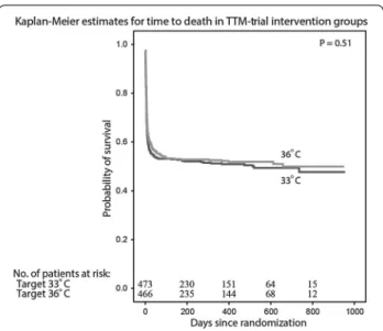
In summary, the TTM-trial has sent a clear message to the medical community that no difference in survival (Fig. 7) or neurological outcome could be detected when
comatose patients after cardiac arrest were treated at either 33 or 36 °C [86]. We can therefore conclude that a real target temperature management after cardiac arrest is a controlled temperature at either of the two tempera-tures, both avoiding fever. This should be part of a bun-dle of care, including treatment of acute coronary disease and contemporary and active intensive care.
Our rationale for changing to 36 °C is that this tem-perature is closer to normal and less invasive, reducing known and unknown risks. The number and severity of adverse events in the two intervention arms did not dif-fer in the TTM-trial, although there was a tendency towards less events in the 36°-arm (p = 0.086). Treat-ing the patients closer to normal temperatures allows for predictable drug effects, including those of sedatives and antithrombotic therapy. In a recently published post hoc analysis of the TTM-trial of the subgroup of patients in circulatory shock at admission, a higher mortality at ICU discharge was suggested in the 33 °C-arm (66 ver-sus 44 %, adjusted p value 0.03) [95]. We cannot exclude that other subgroups may benefit from treatment at 36 °C or for that matter, from treatment at 33 °C. The optimal temperature, duration of temperature management and target population are yet to be defined.
Post‑resuscitation management: optimizing organ perfusion and metabolic parameters
Oxygen and carbon dioxide after cardiac arrest: friend or foe?
Nicolas Deye In CA patients, neurological injury is a major cause of mortality [72]. A first hypoxo-anoxia phe-Fig. 6 Mean bladder temperature in the 33 and 36 °C intervention
groups of the Target Temperature Management after Out‑of‑hospital Cardiac Arrest Trial (TTM‑trial), during the 36 h of temperature intervention. Temperature values are presented with 95 % confidence intervals. In the original publication, temperature curves displayed the means ± 2SD. From Wise MP et al. [179]. Reprinted with permis‑ sion
Fig. 7 The TTM‑trial included and randomized 950 comatose
patients to either 33 or 36 °C. Probability of survival through the end of the trial and the number of patients at risk at each time point is presented. From Nielsen et al. [86]. Copyright © (2013) Massachusetts Medical Society. Reprinted with permission
nomenon occurs initially during CA before and during CPR maneuvers. The ischemia–reperfusion syndrome occurring in the post-resuscitation phase after obtaining an ROSC can lead to secondary insults including oxidative stress with free radical formation or mitochondrial dys-function [72, 96]. Oxygen (O2) and carbon dioxide (CO2) abnormalities can promote these insults. Targeted tempera-ture management (TTM), including therapeutic hypother-mia (TH) targeted to 32–34 °C, is one of the therapeutic measure improving prognosis and neurological outcome after CA. Induced hypothermia could minimize the CA-related injuries by decreasing O2 free-radical production,
mitochondrial dysfunction, brain O2 consumption [96]. Thus TH could minimize neuronal death and improve neu-rological outcome and survival. However, hypothermia is associated with several arterial blood gas (ABG) modifica-tions, mainly induced by the leftward shift of the hemo-globin dissociation curve, increased O2 and CO2 solubility, modification of the pH regulation, with hypoventilation and hypometabolism, leading per se to hypoxia and hypocap-nia. Iatrogenic dyscarbia incidence (hypo- or hyper-carbia) has been reported up to 69 % [97], hypoxia up to 63 % [98], and hyperoxia up to 41 % [99]. International guide-lines mainly focused on potential hypocapnia and hypoxia/ hyperoxia harmful effects for CA patients [72, 100]. They advocate initial resuscitation with 100 % O2 ventilation to avoid hypoxia followed by a titration of O2 therapy target-ing arterial O2 saturation levels between 94 and 96(−98) %
especially during the initial post-CA period to avoid hyper-oxia.
Beyond general detrimental effects such as decreased stroke volume, cardiac output and coronary blood flow and increased systemic vascular resistances, poten-tial neurological harm of O2 therapy have been largely described [100, 101]. Hyperoxia can exacerbate cellular oxidative stress injury and mitochondrial dysfunction to key mitochondrial enzymes or mitochondrial lipids, leads to cerebral O2-related vasoconstriction with a decreased cerebral blood flow (CBF) and cerebral energy metabolism impairment. It can increase the neuronal lipid peroxida-tion and protein oxidaperoxida-tion, enhance O2 free radical for-mation and reactive O2 species, or react with nitrite oxide to produce toxic metabolites (peroxynitrite, superoxide ion, hydrogen peroxide). All of this will finally participate to cell death. Despite several limitations or controversial results in specific experimental models [100–102], most animal studies have suggested that hyperoxia after CA could worsen neurological outcome [103].
In a retrospective cohort including 145 adult OHCA patients with available arterial O2 pressure (PaO2) on ABG sample during CPR attempts, an increased PaO2 was associated with improved rate of hospital admission
alive in multivariate analysis [104]. Incidence of hyper-oxia defined as PaO2 >300 mmHg reached 14 %, whereas hypoxia was defined as PaO2 <60 mmHg. The potential deleterious effect of hypoxia during the resuscitative efforts has been also found in another recent retrospec-tive study [100].
Most clinical studies depicting the O2 effects after ROSC from CA are methodologically of low level quality. The only small randomized clinical trial evaluating this issue found a significant decreased value of the Neuron Specific Enolase biomarker in favor of a normal level of PaO2 versus a high level PaO2 in the subgroup of patients without TH [105]. However, this result was not observed for the S100B Protein biomarker and in the whole cohort including the 28 patients regardless TH implementa-tion. Hypoxia and hyperoxia harmful effects regarding in-hospital mortality and functional status were initially described in a large American registry in 2010 [98]. Sev-eral issues have been pointed out for these studies: (1) the statistical methods (registries and databases versus scarce randomized studies, retrospective versus prospective studies, single center versus multicentre studies, adjust-ment not usually performed to control for other poten-tial confounders); (2) the definitions regarding hyperoxia thresholds (i.e. what precise level of PaO2 to choose?); (3) the different time-point measurements and the period of data collection (i.e. what value to consider between the first ABG, the ABG on admission, the ABG within the first hours or the first day after ROSC or CA, between the mean or median versus the maximal or minimal PaO2 values or the worst PaO2 using worst (A-a) DO2 or the PaO2/FiO2 ratio?); (4) the associated treatments (i.e. TH versus the absence of TTM); (6) the best endpoint (i.e. in-hospital mortality versus neurological outcome).
A recent meta-analysis concluded that hyperoxia in the post-resuscitation phase after ROSC was significantly associated with an increased in-hospital mortality [99]. Conversely, the poor neurologic outcome at hospital dis-charge did not reach significance suggesting a possible lack of association or a review underpowered. However, these results need further confirmation because of its sig-nificant heterogeneity (results were inconsistent in sub-group and sensitivity analyses) and the limited number of studies analyzed (3 abstracts out of the 10 pooled studies were finally included). Since this review, two other stud-ies describing a low hyperoxia incidence rate (3 and 6 %) have been published [106, 107]. The potential harmful effect of hyperoxia regarding mortality was not signifi-cant. In only one of these 2 studies [106], hyperoxia was significantly associated with poor neurological outcome in the multivariate analysis, depicting a “V-shaped” rela-tionship between probability of unfavorable outcome and the mean PaO2 value obtained from ROSC to rewarming.
Cerebral autoregulation physiologically maintain a con-stant CBF within a large range of mean arterial pressure [100, 108]. This relationship is modified by dyscarbia as cerebral perfusion depends on CO2. Hypercapnia leads to cerebral vasodilation and potentially increased intracra-nial pressure, whereas hypocapnia leads to cerebral vaso-constriction and potentially ischemia (decreasing PaCO2 of 1 mmHg can decrease CBF up to 3 %). Ensuring physi-ological CO2 tension in CA patients seems important to prevent worsening of the neurological status. Further-more, impaired autoregulation has been described in some brain-injured areas, in some TH-treated patients, and inconstantly in CA patients. Recommendations in resuscitated CA patients suggest a PaCO2 target of 40–45 mmHg during the post-ROSC period in this popu-lation of brain-injured patients regardless of TH use [72,
100]. In a retrospective single-center study implementing TH in 41 %, hypocapnia (defined by PaCO2 ≤30 mmHg), hypercapnia (PaCO2 ≥50 mmHg), and association of both dyscarbia occurring within the first 24 h after ROSC were all independently associated with poor neu-rologic outcome at hospital discharge [97]. An observa-tional multicenter registry recently enrolled 16.542 adult CA patients TH-treated in 39 %. Increased in-hospital mortality and rate of poor outcome were observed in the hypocapnia group compared to normocapnia after adjustment for illness severity and propensity score [109]. Conversely, hypercapnia (PaCO2 ≥45 mmHg) within the first 24 h after admission was independently associated with similar in-hospital mortality and a higher rate of dis-charge home among survivors. In a smaller study of TH-treated patients, hypocapnia but not hypercapnia was independently associated with an increased risk of in-hospital death [106]. A “U-shaped” relationship between the mean PaCO2 and the in-hospital mortality was found with the best survival observed for the normocapnia group (35–45 mmHg). No association between hypo- and hyper-capnia with poor neurologic outcome were observed. Another recent multicenter and prospective study applying TH in 71 % defined PaCO2 as “low” when <30 mmHg, “middle” when 30–37.5 mmHg, “intermedi-ate” when 37.5–45 mmHg, and “high” when >45 mmHg [107]. Patients with poor versus good outcome had simi-lar highest, mean and lowest PaCO2. The mean 24-h PaCO2 and the time spent in PaCO2 >45 mmHg regard-less TH-implementation were independently associated with better 1-year good outcome. However, same criti-cisms can be made for all these studies than those made above regarding statistics, threshold, data collection, or treatments. To date mainly because of paucity of data on this issue, no clear thresholds have been found regard-ing the harmful impact of hypo- or hyper-capnia after CA. Whereas hypocapnia seems consistently associated
with worse outcome, hypercapnia is not [100]. The man-agement of PaCO2 after CA by using specific mechanical ventilation strategy could influence the outcome of these patients especially during TH. In a recent retrospective study focusing on initial post-CPR mechanical ventila-tion settings minute ventilaventila-tion was weakly correlated with the initial PaCO2 [110]. Normocapnia alone (31– 49 mmHg) was again associated with a better favorable neurological outcome.
ABG measurements performed at 37 °C are sec-ondarily expressed either as temperature-corrected or uncorrected according to biochemical centers [108]. Substantial discrepancies can be related to these meth-ods: PaO2 = 100 mmHg at 37 °C becomes 79 mmHg if corrected at 33 °C, when PaCO2 = 36 mmHg becomes 30 mmHg at 33 °C. There are no clear recommendations regarding the ventilation strategy to be used in resusci-tated CA patients despite such differences. Normocap-nia in hypothermic patients can be achieved according to two different mechanical ventilation strategies: α-stat versus pH-stat. In the α-stat strategy ventilation is set to achieve physiological arterial CO2 tension measured at 37°, unadjusted to the patient’s temperature, whereas in the pH-stat strategy ventilation is set to achieve physi-ological arterial CO2 tension measured at the patient’s actual temperature. The latter strategy leads to a relative hypoventilation compared to the α-stat strategy. Using either α-stat or pH-stat strategy to guide normocapnia after CA remain open to discussion. In the recent mul-ticenter Finnish study focusing on ABG abnormalities after CA, 13 ICUs used temperature-correction; whereas, eight did not [107]. Two recent exploratory studies in TH-treated CA patients compared either a “lower versus upper threshold normocapnia” (32 versus 45 mmHg) or the α-stat versus the pH-stat strategies while maintaining a PaCO2 target level between 36 and 42 mmHg. The first study showed that the “lower threshold normocapnia” induced decreased internal jugular vein O2 saturation and CBF mean velocity suggesting an increased risk of cerebral ischemia. The second study found that the alpha-stat strategy increased jugular vein desaturation and cer-ebral O2 extraction and decreased transcranial Doppler cerebral velocities in survivors but not in non-survivors.
In summary, dyscarbia especially hypocapnia should be associated with an increased harm after CA leading to increased unfavorable outcome or in-hospital mortality. Conversely O2 seems a two halves phenomenon with: (1) a former deleterious effect of hypoxia during resuscita-tion efforts, the potential benefit of hyperoxia implying more evaluations; (2) followed by a deleterious effect of both hypoxia and hyperoxia after ROSC in the post-CA phase. To date because O2 and CO2 derangements have no obvious benefit, aiming at normoxia and normocapnia
after CA are potentially two very easy targets to achieve in post-resuscitation care bundles. Meanwhile all stud-ies describing ABG parameters in TTM- or TH-treated CA patients should actually emphasize the strategy used, either a temperature-corrected or a non-corrected strat-egy to more precisely evaluate potential thresholds.
Other means of cardio‑ and neuro‑protection
Fabio Taccone Heart and brain protection strategies aim to prevent or attenuate disease progression and secondary injuries by halting or at least slowing the loss of cardio-myocytes and/or neurons [111]. Although many pathologi-cal mechanisms, including endothelial damage and tissue hypoperfusion, inflammation, impaired mitochondrial res-piration with induction of reactive oxygen species, calcium overload or excitotoxicity, are common in both cardiac and neurological injury following CA [112], some drugs may present a more specific organ protection or have a lower brain penetration, thus acting especially on the cardiac tis-sue. Many therapeutic agents have been shown to be effec-tive in protecting the heart and/or the brain in animal mod-els of global or local ischemia [113]; nevertheless, these findings were flawed because of the differences between the experimental setting and the clinical scenario (i.e. absence of co-morbid diseases and need for anesthetics in animal studies), the low mortality rates or the administra-tion of the specific drug before the development of injury (i.e. pre-treatment approach).
A promising approach to reduce brain excitotoxic-ity (i.e. excessive extracellular glutamate levels) could be the administration of intravenous magnesium or inhaled noble gases (i.e. xenon or argon). In two studies, magne-sium administration did not result in a better survival or neurological outcome for CA patients [114, 115]. How-ever, hypothermia was not used in these studies, while the combination of magnesium with cooling procedures has been shown to result in the highest neuroprotective effects [116]. Importantly, many questions remain unan-swered on the optimal timing to initiate magnesium per-fusion, optimal dosing and circulating levels and potential side-effects, which may explain the negative results of magnesium therapy after subarachnoid hemorrhage and traumatic brain injury [117, 118]. Noble gases have been proved to reduce the extent of neurological damage after ischemia in animal models of CA [119, 120]. The admin-istration of Xenon was also feasible in the human setting, reporting both no adverse events and an improvement of cardiovascular function after CA [121]. Unfortunately, we still do not have any human data describing the potential neurological benefits of such treatment.
Mitochondrial dysfunction could be attenuated by the administration of erythropoietin (EPO), the principal
hematopoietic hormone regulating erythropoiesis, which shows also anti-apoptotic, anti-inflammatory and anti-oxi-dant properties [122]. In a swine model of VF, high-dose EPO administration during CPR reduced post-resuscita-tion myocardial dysfuncpost-resuscita-tion and improved cardiac func-tion [123]. However, post-ischemic EPO administration in rats exposed to CA exerted no protective effect on hip-pocampal neurons [124]. One human study has compared the effects of 90.000 UI of EPO given during CPR to an historical matched control group [125]. The EPO group had higher rates of ROSC (92 versus 53 %, p = 0.006) and hospital survival (54 versus 20 %, p = 0.011) when com-pared to the control group. In another study [126], EPO therapy was associated with a trend towards higher full neurological recovery (55 versus 38 %) when compared to an historical cohort. Unfortunately, a recent unpublished randomized clinical trial (RCT) found no benefits of EPO administration on comatose survivors after CA when compared to placebo (Cariou et al.—presented at the 27th ESICM congress—Barcelona, October 2014).
Reperfusion injury leads to mitochondrial dysfunc-tion also through the opening of a nonspecific pore in the inner mitochondrial membrane, known as the mito-chondrial permeability transition pore (MPTP) [127]. This phenomenon causes the loss of ionic homeostasis and ultimately cell swelling and death. The inhibition of the MPTP opening may provide some protection against reperfusion injury; importantly, this may be mediated by a direct interaction of cyclosporine A (CsA) with a pro-tein located on the MPTP, called cyclophilin-D [127]. In a murine model of CA, CsA was effective in reducing myocardial dysfunction when given at the onset of resus-citation but not after ROSC [128]. In one human study conducted in patients suffering from acute myocardial infarction, the administration of a 2.5 mg/kg bolus of CsA before percutaneous coronary intervention was associ-ated with a significant reduction in biomarkers of myo-cardial injury (i.e. troponin I) and the extent of ischemic areas on cardiac magnetic resonance imaging [129]. Nowadays, no clinical studies have evaluated the effects of CsA on the neurological recovery of CA survivors yet.
Also, mitochondrial dysfunction can be modulated through the nitric oxide (NO)-related pathways; NO inhibits ROS-producing enzymes and directly scavenges ROS production [130]. Other potential beneficial effects are the direct vasodilation of coronary arteries, which could improve cardiac function in this setting. Experi-mental models have suggested a protective role of intra-venous NO-donors or inhaled NO both on cardiac and neurological function after CA [131, 132]. Unfortunately, only one pilot study showed the feasibility and safety of low-dose nitrite infusion in cardiac arrest survivors, but did not report any improvement in outcome [133].
Finally, recent data suggest that the early administration of corticosteroids after in-hospital CA was associated with an increased survival rate and reduced occurrence of extra-cerebral organ failures [134]; whether these effects were secondary to an anti-inflammatory effect or the treatment of a relative adrenal insufficiency [135], it remains to be further evaluated. Similarly, abnormalities of peripheral microcirculation, similar to those found in septic patients, have been described after CA [136]; the pathophysiology of such alterations as well as their role on patients’ outcome and the various therapeutic options to manipulate the microvascular flow have to be better characterized in future studies.
Neurological assessment of brain damages
Minimal consciousness: classification and prognosis in CA patients
Steven Laureys and Didier Ledoux Successful resuscita-tion after cardiac arrest or cerebral hypoxia can have an extremely wide variety of functional and cognitive conse-quences, ranging from transient cognitive or motor dys-function lasting several weeks or months; to awakening after coma in a state of unawareness or inability to com-municate lasting months to years or decades (e.g. as seen in vegetative or minimally conscious states) [137]. At pre-sent, the medical and economic impacts of differences in clinical management (including therapeutic hypothermia), care pathways and disparate health policies within Europe remain unclear.
Figure 8 illustrates the timeline of events that may occur after anoxic coma. When cerebral hypoxia leads to a prolonged loss of consciousness (lasting >2 h to differ-entiate from syncope), the patient is considered to be in coma. Patients in coma will never open the eyes, even if stimulated by a loud noise or intense noxious stimulus, and will only show reflex movements. Coma will not last longer than a couple of days to weeks. Most patients who show good recovery will rapidly show signs of conscious-ness and functional communication. Some patients, however, will evolve to brain death (i.e., irreversible coma with absent brainstem reflexes) and organ dona-tion should be discussed [138]. Many patients will remain with some brainstem function (i.e., will not be brain death), but clinical and complimentary testing shows that there is no reasonable chance for a meaningful recovery and the decision is made to withhold or withdraw treat-ment. In these cases, organ donation after cardiac death can be discussed.
Patients who survive their coma may awaken (i.e., open the eyes) without any behavioural sign of consciousness (i.e., only show reflex or automatic movements), a condi-tion coined “persistent vegetative state” or “unresponsive
wakefulness syndrome” (PVS/UWS) [139] or without recovery of functional communication or functional object use, a condition referred to as “minimally con-scious state” (MCS) [140]. In contrast to coma, such chronic disorders of consciousness can last for many months to years and at present there are no reliable epi-demiological data regarding these challenging patients. The heterogeneous MCS group is subcategorized in MCS—when patients only show non-reflexive behav-iour such as eye tracking, orientation to pain or contin-gent behaviour to specific stimuli (e.g. smiling exclusively in the presence of a family member) and MCS+ when a reproducible (albeit often inconsistent) response to com-mand can be observed [141].
When patients remain in PVS/UWS for over 3 months after the cardiac arrest, the condition is considered irre-versible and life-sustaining treatment (i.e., artificial hydration and nutrition) may be considered as futile and hence be withdrawn. At present, the chances of recovery after post-anoxic MCS are considered to be better than for PVS/UWS but remain ill defined. Many patients who recover from coma or related disorders of consciousness will show cognitive dysfunction and may remain insti-tutionalized or dependent of others for many activities of daily living, but few reliable data exist regarding their remaining quality of life.
Recent advances in automated clinical EEG analysis [142], combined EEG transcranial magnetic stimulation studies [143] and structural and functional neuroimaging have permitted to better document the clinical diagnosis [144] and levels of consciousness in patients with severe post-anoxic encephalopathy [145]. Several studies also show their value in predicting the chances of recovery after cardiac arrest (e.g., the assessment of EEG reactiv-ity [142]; the quantification of white matter damage using


![Fig. 10 Assessment of health condition in the long‑term follow‑up after cardiac arrest (adapted from World Health Organization [171])](https://thumb-eu.123doks.com/thumbv2/123doknet/6001624.149573/17.892.88.434.767.1019/assessment-health-condition-follow-cardiac-adapted-health-organization.webp)
